
Article

Immunohistochemistry and Ultrastructural Evaluation of the Interaction Between Nano-Hydroxyapatite/β-Tricalcium Phosphate Composite Spheroids and Bone Marrow-Derived Mesenchymal Stem Cells in a 3D Cell Culture Model

by
Igor Da Silva Brum
1,*,
Carlos Nelson Elias
2,
Lucio Frigo
3,
Bianca Torres Ciambarella
4,
Debora Ornelas
5,
Simone Carvalho
4,
Erika Cortez
5,
Alessandra Thole
5,
Ana Lúcia Rosa Nascimento
4,
Karina Ribeiro Silva
5,
Ivonete Sena Dos Santos
4 and
Jorge José De Carvalho
4
1
Faculty of Dentistry, State University of Rio de Janeiro, Blvd. 28 De Setembro, 157-Vila Isabel, Rio De Janeiro 20551-030, Brazil
2
Materials Science Department, Instituto Militar De Engenharia, Praça Gen. Tibúrcio, 80-Urca, Rio De Janeiro 22290-270, Brazil
3
Basic Sciences Department, Faculdade de Odontologia da APCD, R. Voluntários da Pátria, 547-Santana, São Paulo 02011-000, Brazil
4
Department of Histology and Embryology, State University of Rio De Janeiro, Rua Prof. Manoel De Abreu, 444, Rio De Janeiro 20550-170, Brazil
5
Laboratory of Stem Cell Research, Histology and Embryology Department, Biology Institute, State University of Rio De Janeiro, Rua Prof. Manoel De Abreu, 444, Rio De Janeiro 20550-170, Brazil
*
Author to whom correspondence should be addressed.
J. Compos. Sci. 2026, 10(8), 406; https://doi.org/10.3390/jcs10080406
Submission received: 26 June 2026 / Revised: 27 July 2026 / Accepted: 28 July 2026 / Published: 31 July 2026
(This article belongs to the Section Biocomposites)

Abstract

The nano-hydroxyapatite/β-tricalcium phosphate composite (nano-HA/β-TCP) is widely used in various medical and dental procedures. The absence of in vivo toxicity of nano-HA/β-TCP has been extensively studied, and it is considered one of the most effective synthetic biomaterials for promoting cell differentiation in bone regeneration. Bone marrow-derived mesenchymal stem cells (BM-MSCs) are the primary cell type involved in the osteoinductive process of guided bone regeneration following injury. In the present study, rat BM-MSCs were cultured with nano-HA/β-TCP (80/20%) composite spheroids, and the interaction between the cells and the composite was analyzed using transmission electron microscopy (TEM). Ultrathin sections examined by TEM showed extensive interaction between nano-HA/β-TCP and BM-MSCs. Semi-thin sections stained with toluidine blue revealed the incorporation of the biomaterial into the cell cytoplasm. For the immunohistochemistry analysis, eight adult male Wistar rats weighing approximately 300 g were used in each group. Two bilateral, non-critical-sized 3 mm defects were created in the parietal bones of the calvaria: Control, Bio-Oss®, and Blue Bone® (n = 24) during a 12-week experimental period. Bone formation was evaluated through osteonectin and osteopontin expression. At the ultrastructural level, internalization of the biomaterial and close association with the endoplasmic reticulum (ER) and mitochondria were observed. TEM analysis also revealed no harmful effects on the cells, such as apoptotic or necrotic bodies or cell lysis. These findings indicate that the nano-HA/β-TCP composite demonstrates in vitro biocompatibility and interacts appropriately with BM-MSCs, including incorporation into the cell cytoplasm. In vivo, the Blue Bone® group exhibited superior bone formation when compared with the other groups.

1. Introduction

Osteoconduction and osteoinduction are key processes that support bone healing and represent the primary goals of graft biomaterials. Osteoconduction refers to the property of biomaterials that allows new bones to grow along their surface. In contrast, osteoinduction refers to the ability to induce stem cells to commit to the osteoblastic lineage and synthesize the bone matrix [1].
Osteoinduction is considered a multifactorial process involving chemical, physical, and biological elements. The architecture of the biomaterial scaffold plays a critical role, particularly surface characteristics and microporosity. While the importance of calcium and phosphate in osteoinduction is well established, calcium ions and their interaction with calcium receptors have been increasingly emphasized in the literature. Additionally, several cell types participate in this process, including macrophages, osteoclasts, and stem cells, along with a myriad of signaling molecules and modulators [2].
The central event in the osteoinductive process is the commitment of stem cells and their differentiation into osteoblasts. It is believed that pre-osteoblasts originate from mesenchymal progenitor cells—collectively referred to as skeletal stem/progenitor cells (SSPCs)—which then mature into osteoblasts, bone lining cells, and ultimately osteocytes. Each cellular stage is characterized by specific gene expression profiles and the production of particular proteins and enzymes [3,4].
The commitment of mesenchymal stem cells (MSCs) to the osteogenic lineage is regulated by various hormonal and paracrine factors, distinct cell signaling pathways, and physical characteristics of the extracellular matrix (ECM) [5].
Hydroxyapatite is one of the ECM components that contribute to MSC differentiation. The potential of nano-hydroxyapatite (nano-HA) and biphasic calcium phosphate (BCP) compounds to promote proper cell differentiation in bone remodeling has been investigated [6]. Nano-HA and BCP have not shown cytotoxic effects or induced cell lysis [7]. Moreover, their scaffolding properties help prevent soft tissue ingrowth into the healing site [8,9,10].
Nano-HA demonstrates osteoinductive potential and has been shown to support various stages of the bone remodeling process. Nonetheless, autogenous bone remains the gold standard in terms of osteoinductive properties (Figure 1) [7,11].
The ability of nano-HA to induce MSC differentiation has been studied in in vitro models. Human Wharton’s Jelly-derived MSCs (hWJ-MSCs), when co-cultured with human umbilical vein endothelial cells (HUVECs) and incubated in nano-HA-containing media, exhibited a dose-dependent relationship between nano-HA concentration and cytotoxicity, along with upregulation of key osteogenic signaling genes and extracellular matrix production [12].
Additional studies using hWJ-MSC monocultures were conducted to analyze the influence of HA nanoparticles internalized by the cells on osteogenic gene activation and extracellular matrix (ECM) synthesis.
Initially, the particles adhered to the cell membrane, followed by their enclosure through cytoplasmic projections, ultimately resulting in full internalization. The engulfed particles remained within the cytoplasm and were not observed in the nucleus [13].
Nano-HA has been combined with tricalcium phosphate (TCP) in various ratios to harness the complementary osteoinductive and osteoconductive properties of both materials. These biphasic calcium phosphate (BCP) compounds have been widely studied in both animal models and clinical trials, with evidence suggesting their osteoinductive potential [14,15,16,17]. However, it remains unclear whether BCPs elicit the same ultrastructural mechanisms in MSC osteogenic commitment as pure nano-HA.
Immunohistochemistry is considered in the literature to be the most accurate method for evaluating bone interaction with the cells involved in the remodeling process. Immunomarkers such as osteopontin (an osteoblast marker) and osteonectin (an osteoclast marker) are essential for confirming the presence of these cells and, consequently, for accurately quantifying bone formation [18].
Biomaterials used in scaffolds exhibit different characteristics from those used solely as bone grafts. Scaffold materials tend to possess properties suitable for printing, in addition to biocompatibility. Scaffolds activate biophysical and biochemical processes to accelerate tissue repair. One approach involves adding piezoelectric materials to generate electrical activity upon deformation. These biomaterials create electrochemical microenvironments that induce cellular signaling, thereby facilitating tissue regeneration [19]. Zhang et al. [20] analyzed the possibility of using 3D printing of hydroxyapatite to obtain scaffolds similar to the structure of trabecular bone. For printing, they incorporated methacrylated gelatin hydrogels to obtain structures similar to the extracellular matrix. The resulting scaffold presented a suitable environment for biomimetic 3D culture for osteoblasts and vascular progenitor cells. The results obtained showed that the printed scaffold presents a suitable environment for developing biomaterials for the regeneration and repair of bone tissues.
In this context, the present study aims to evaluate, at the ultrastructural level, the interaction and potential internalization of BCPs (nano-HA/β-TCP composite) by bone marrow-derived mesenchymal stem cells (BM-MSCs).

2. Materials and Methods

The present work compared the performance of two biomaterials for bone graft: Blue Bone® (experimental group), an alloplastic biomaterial, and Bio-Oss® (control group), a xenogeneic biomaterial. Blue Bone® is a synthetic mixture of nano-HA (80%) and β-TCP (20%) supplied by Regener Biomaterials Co. (Curitiba, Brazil). Bio-Oss® (Geistlich Pharma AG, Wolhusen, Switzerland) has an organic origin and is produced with deproteinized bovine bone. Blue Bone® biomaterial in granule form has a particle size from 130 to 300 nm [21] and a porosity of 63.84% [22].

2.1. Bm-Msc Isolation and 3D Cell Culture

Bone marrow-derived MSCs (BM-MSCs, at passage 3 (p-3)) were obtained from 3-month-old male Wistar rats (three months old) euthanized in a CO2 chamber. After collecting tibias and femurs, bone medullary cavities were exposed and harvested by centrifugation at 350 g for 10 min.
The bone marrow harvested after centrifugation was resuspended in 0.01 M phosphate-buffered saline (PBS) and mechanically dissociated by gentle pipetting. The cell suspension was centrifuged at 300 g for 5 min, and the pellet was then re-suspended in Dulbecco’s modified Eagle’s medium F12 (DMEM-F12, Sigma-Aldrich, St. Louis, MO, USA) supplemented with 20% fetal bovine serum (FBS; Gibco, Thermo Fisher Scientific, Grand Island, NY, USA) and antibiotics. Cells were plated in 25 cm2 culture flasks. The samples were maintained at 37 °C in a humid atmosphere with 5% CO2 for cell expansion.
The culture medium was changed every three days until the cell monolayer of MSCs reached 80% confluence. Adherent cells were harvested from culture flasks with trypsin-EDTA 0.25% (Sigma-Aldrich, St. Louis, MO, USA) and cultured for further expansion up to the third passage.
The Ethics Committee in Animal Experimentation of the State University of Rio de Janeiro has approved all procedures (registered under CEUA/001/2019).
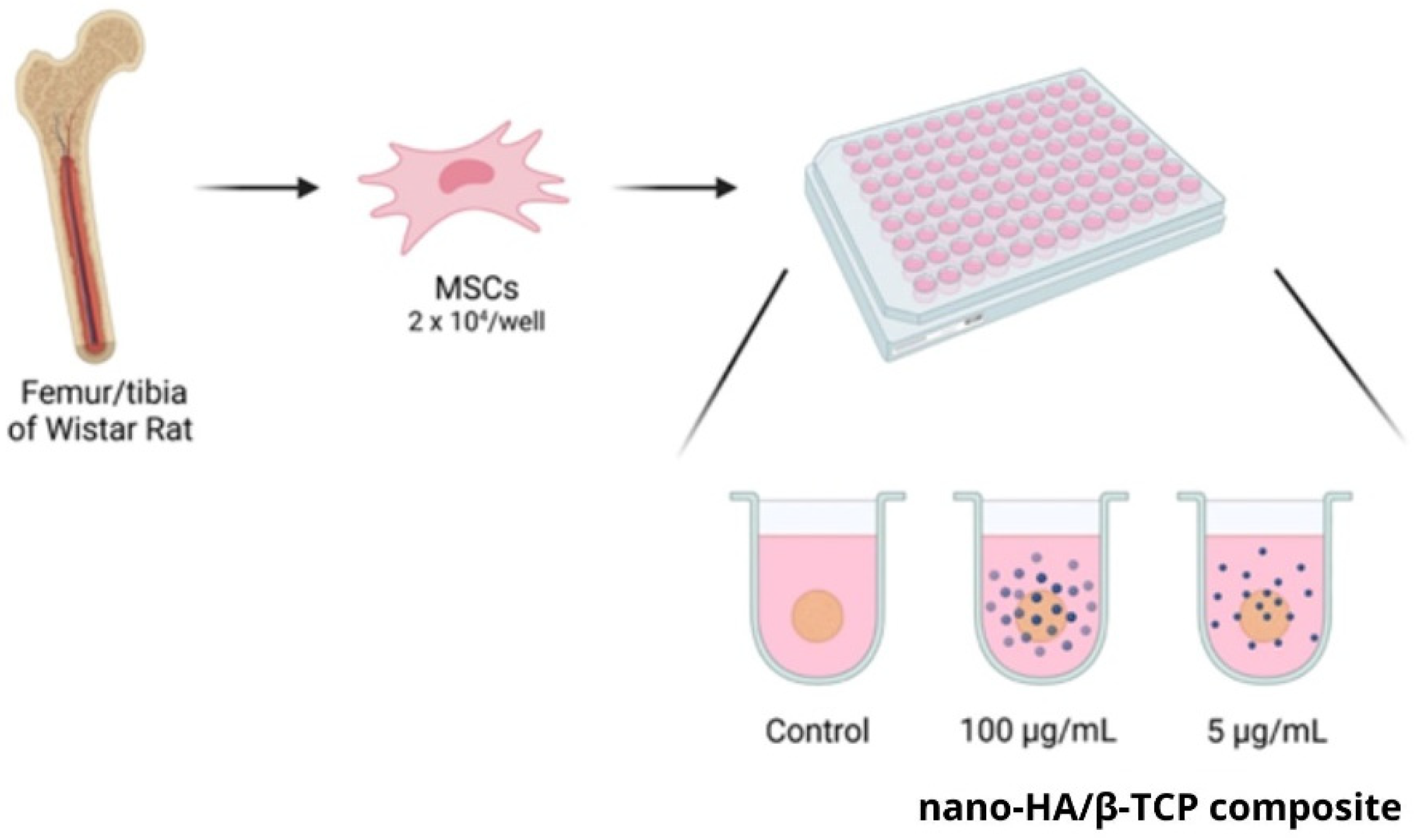
The BM-MSCs (2 × 10⁴/well) were seeded in 96-well U-bottom plates previously coated with 1% ultrapure agarose. Cells were incubated for 24 h at 37 °C in a humid atmosphere with 5% CO2. Three-dimensional spheroid cell cultures were exposed to 5 or 100 μg/mL nano-HA/ß-TCP composite for three days. Cells without nanoparticles were used as controls.
For nanoparticle interaction evaluation, 2 × 10⁴ BM-MSCs were seeded per well of 96-well U-bottom plates (Corning Incorporated, Cornig City, NY, USA) previously coated with a thin layer of 1% ultrapure agarose (Thermo Fisher Scientific, Invitrogen, Waltham, MA, USA—in 0.9% NaCl). Cells were plated in 200 μL/well of DMEM-F12 medium supplemented with 10% FBS and antibiotics (3D cell culture medium). The mixture was incubated for 24 h at 37 °C in a humid atmosphere with 5% CO2. After this procedure, BM-MSC spheroids were formed. The 3D cell cultures were exposed to 5 and 100 μg/mL nanoparticles for three days. Cells immersed in a mixture without nanoparticles were used as controls (Figure 1).
After this period, spheroids were washed with 0.01 M PBS and fixed using 2.5% glutaraldehyde.

2.2. Transmission Electron Microscopy

The ultrastructure of the 3D spheroids containing the nano-HA/β-TCP composite was analyzed using transmission electron microscopy (TEM). The fixed samples were washed in 3 baths of cacodylate buffer solution (0.1 M; pH 7.4) for 15 min, followed post-fixation with osmium tetroxide 1% and potassium ferrocyanide 0.8% for 5 min, dehydration with a crescent grade of acetone (30 up 100%; 15 min each), and infiltration with increased concentration of Epon-812 diluted in acetone P.A. (2:1, 1:1, and 1:2; 2 h each).
The samples were incubated overnight with Epon-812, included in resin, and polymerized between 48 and 72 h at 60 °C.
The blocks were cut to a thickness of 1 µm, and the sections were stained with toluidine blue 0.5% for morphological analysis by optical microscopy and field selection. Then, the block was cut with an ultramicrotome to obtain a 70 nm slide thickness. The slides were collected on 300 mesh copper grids.
The contrast was enhanced with uranyl acetate for 20–30 min. The images were analyzed using a JEOL JEM-1011 transmission electron microscope (JEOL, Ltd., Akishima, Tokyo, Japan), operating at 60 kV. Digital micrographs were captured using an ORIUS CCD digital camera (Gatan, Inc., Pleasanton, CA, USA) at 8000×, 10,000×, and 25,000× magnification.

3. Immunolabeling Protocol

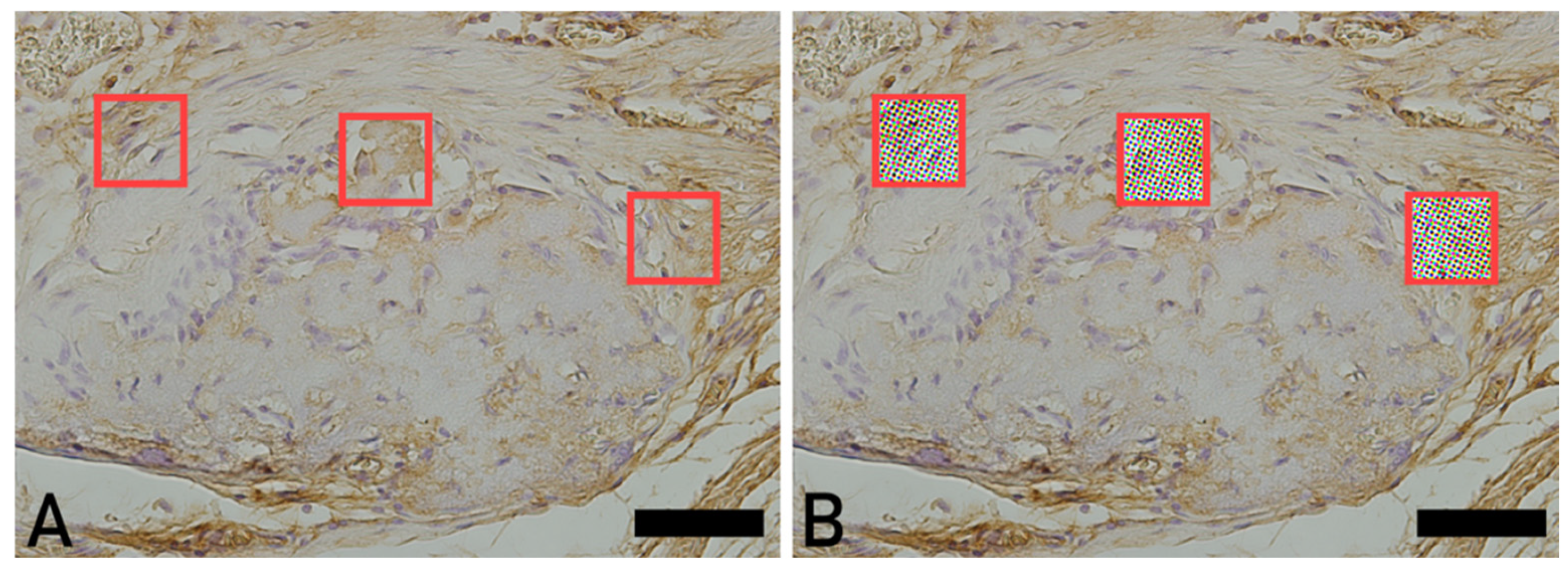
Immunohistochemical analyses were performed using markers for osteopontin (OPN) and osteocalcin (OCN). The staining in the tissue sections exhibited brown coloration due to development with diaminobenzidine (DAB). For the immunolabeling procedures, sections were initially deparaffinized in three xylene baths (5 min each), followed by rehydration in a graded ethanol series (100%, 90%, 70%, and distilled water; 5 min each). Endogenous peroxidase activity was blocked by incubating the sections in 3% hydrogen peroxide diluted in distilled water for 15 min in the dark. The sections were then washed in three baths of phosphate-buffered saline (PBS; pH 7.2) for 5 min each. The subsequent steps for each immunolabeling procedure are described in the respective sections for each antibody, as shown in Figure 2.

3.1. Osteopontin

This immunolabeling allows for the visualization of OPN-positive areas. Osteopontin is a non-collagenous protein closely associated with hydroxyapatite, acting as a mediator between hydroxyapatite crystals and osteoprogenitor cells. Antigen retrieval was performed in citrate buffer (pH 6.0) at 60 °C for 20 min. The sections were allowed to cool, washed in three PBS baths (pH 7.2; 5 min each), and nonspecific binding sites were blocked with 3% PBS/BSA for 20 min.
The sections were then incubated with the primary anti-osteopontin antibody (Thermo Fisher Scientific, Santa Cruz, MA, USA, sc-21742), diluted 1:200 in 1% PBS/BSA, overnight at 4 °C in a humid chamber. After washing in three PBS baths (pH 7.2; 5 min each), the sections were incubated at room temperature with a biotinylated secondary antibody (Vector Laboratories, VECTASTAIN® Universal Quick HRP Kit, Newark, CA, USA) for 1 h, followed by washing in PBS and incubation with streptavidin (VECTASTAIN® Universal Quick HRP Kit) for 30 min.

3.2. Osteocalcin

To assess the mineralized tissue matrix, immunohistochemical staining for osteocalcin (OCN) was performed as a continuation of the previously described immunolabeling protocol. Antigen retrieval was performed using citrate buffer (pH 6.0) at 60 °C for 20 min. The sections were allowed to cool to room temperature, rinsed in three changes of PBS buffer (pH 7.2) for 5 min each, and incubated with 3% bovine serum albumin (BSA) in PBS for 20 min to block nonspecific binding sites. Subsequently, the sections were incubated with the primary anti-osteocalcin antibody (Thermo Fisher Scientific, Santa Cruz Biotechnology, sc-365797, Waltham, MA, USA), diluted in 1% PBS/BSA (1:200), overnight at 4 °C in a humidified chamber. After incubation, the sections were washed in three changes of PBS buffer (pH 7.2) for 5 min each and incubated at room temperature with a biotinylated secondary antibody (VECTASTAIN® Universal Quick HRP Kit) for 1 h. The sections were then washed three times with PBS buffer (pH 7.2) for 5 min each and incubated with streptavidin–HRP (VECTASTAIN® Universal Quick HRP Kit) for 30 min.

3.3. Animal Groups and Statistical Analysis

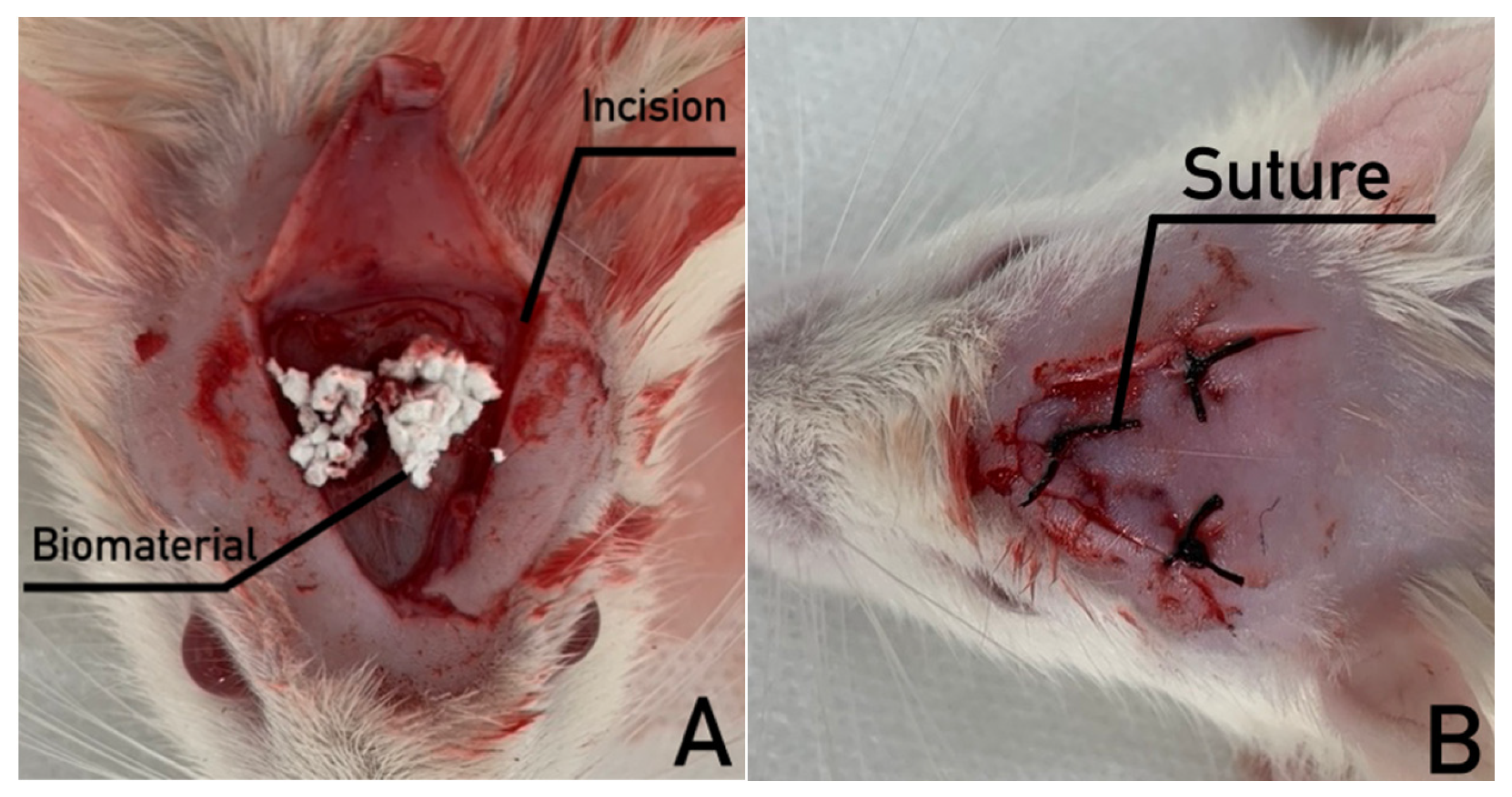
The animals were randomly allocated into 3 experimental groups (n = 8 per group), Control (sham), Blue Bone® and Bio-Oss® during a 12-week experimental period. In the sham group, bone defects were surgically created without the placement of a grafting material, and the defects were allowed to heal with the formation of a blood clot only. The defects were filled with bone substitute materials. The defects were then closed with three-point sutures (Figure 3A,B).
The experimental protocol was approved by the local Animal Ethics Committee (#001/2019). A total of 24 adult male Wistar rats, weighing 300 g, were included in the study. The animals were obtained from the Institute of Biology of Rio de Janeiro State University.
Statistical analyses were performed using the nonparametric Kruskal–Wallis test, adopting a significance threshold of p < 0.05. All analyses were conducted using GraphPad Prism (version 8.0) and BioEstat (version 5.0) software.

4. Results

4.1. Control Group

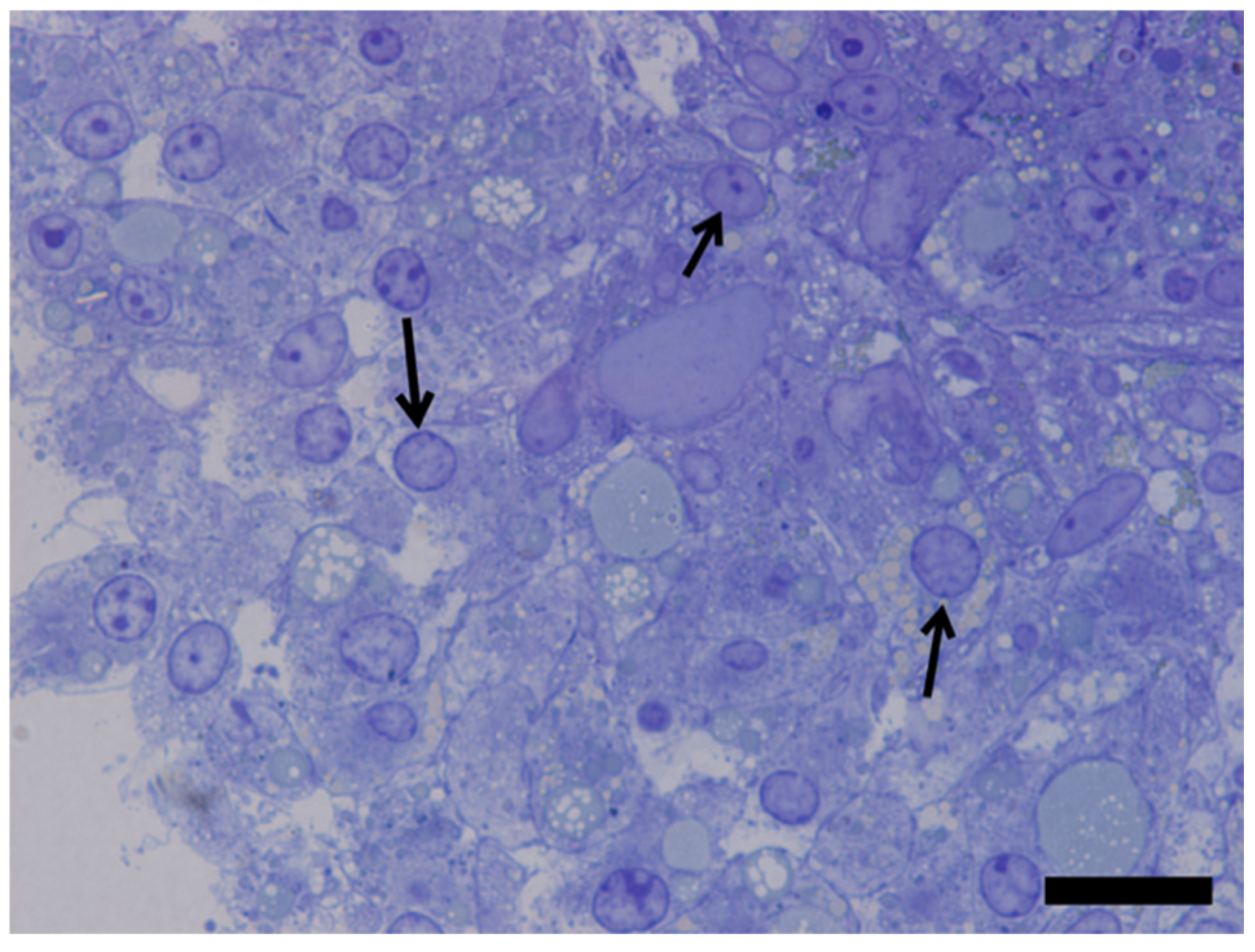
In the control group, it was possible to confirm, through the semi-thin section, that cells interacted with each other through cell contacts; regular round-shaped nuclei and one or more nucleoli were present and virtually no degenerating cells or apoptotic bodies profiles were observed (Figure 4).

4.2. Biomaterial-Containing Groups

The first finding worth mentioning is that there was no evident morphological difference between the 100 µg/mL and 5 µg/mL biomaterial groups, nor morphological images of degenerating cells, suggesting that the toxic threshold was not reached. In fact, the only way to know which group had been observed was by consulting specimen (slides, grids) labels.
In this respect, a single description of both experimental groups will be provided.
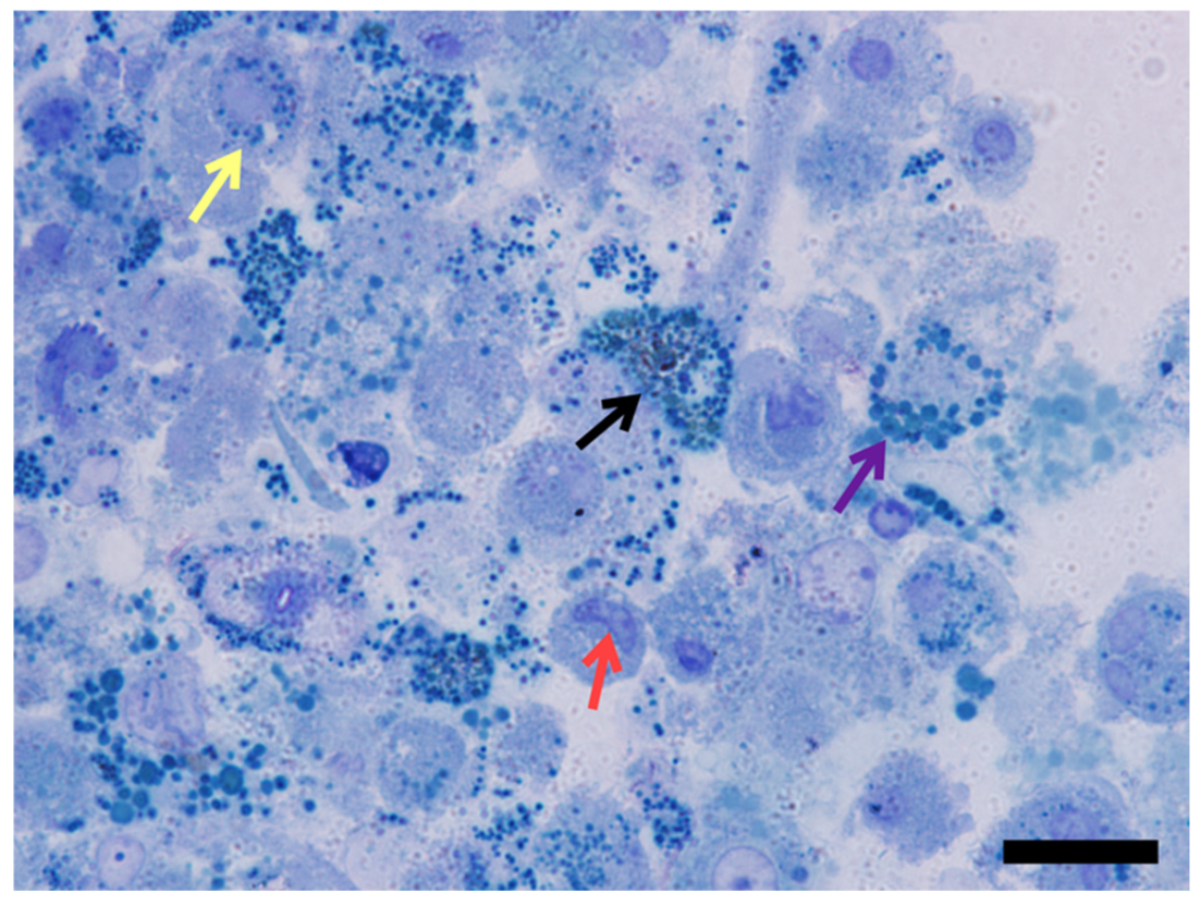
The toluidine blue-stained slides evidenced the biomaterial spheroids as bluish dots of different shades from cells. The biomaterial rounded blue dots were observed in the culture medium and inside cells. Still, in some of them, it was heavily concentrated in the cytoplasm and dispersed around the nucleus (Figure 5).
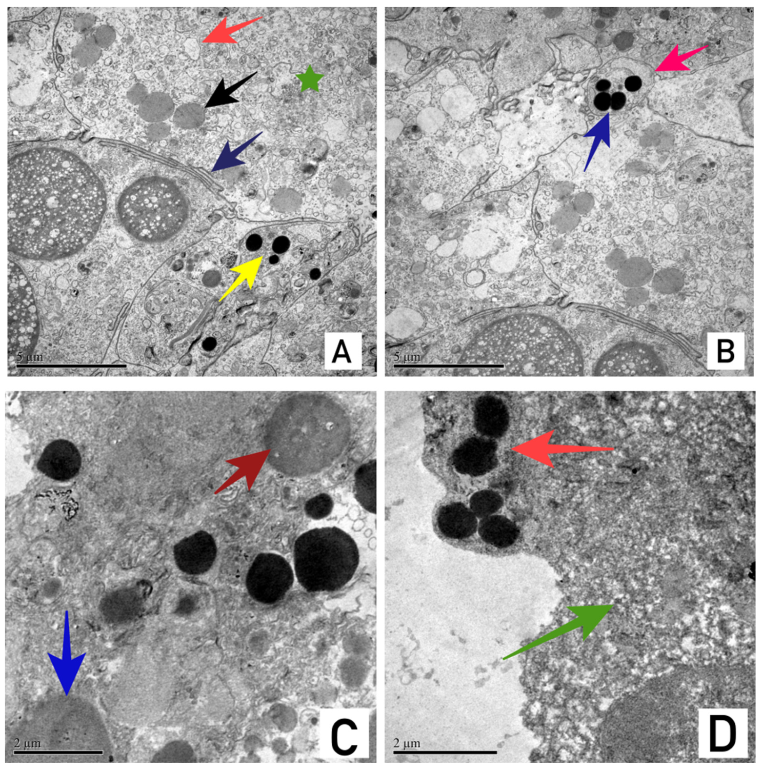
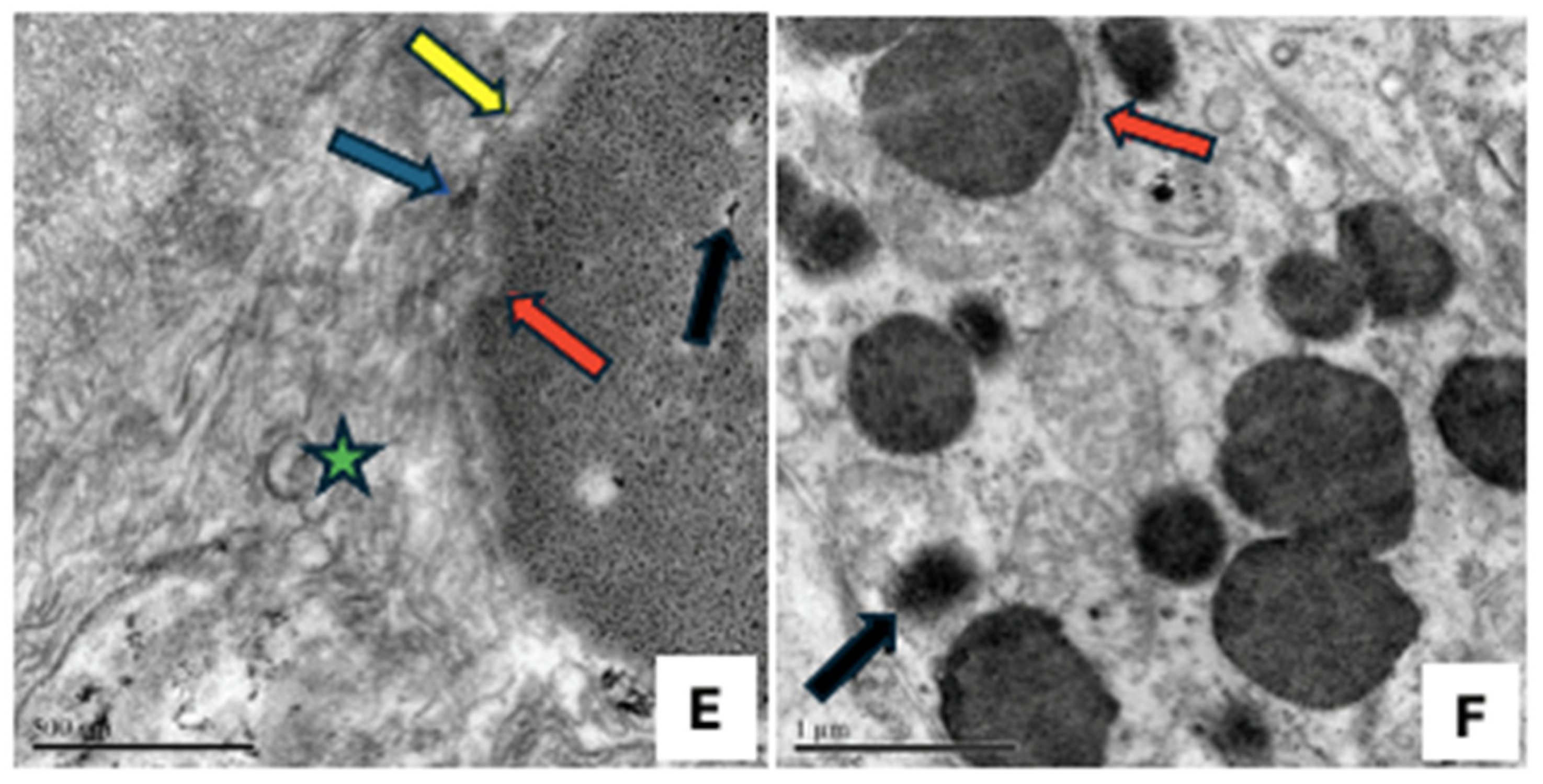
The cytoplasmatic engulfed biomaterial could be observed in different grades of electron density (different shades from black to light gray), suggesting some sort of dilution/degradation. (Figure 6C,D) The light gray-shaded engulfed biomaterial appeared as a granulated substance at higher magnifications (Figure 6E). In addition, engulfed biomaterials were frequently observed closely related to cisternal membrane profiles and mitochondria (Figure 6D–F).
Ultrastructural image analysis via transmission electron microscopy (TEM) demonstrated the stages of contact, internalization, and intracellular degradation of the nano-HA/β-TCP compound spheroids by BM-MSCs. Initially, biomaterial spheroids were observed in direct contact with the cell surface, wrapped in an electron-dense fibrillar material suggestive of glycocalyx (Figure 6A).
Biomaterial internalization really occurred, as spheroids could be observed in the cell cytoplasm. (Figure 6B). Once it gained access to the cell cytoplasm, the biomaterial spread over the cytoplasm and surrounded the nucleus but did not gain access to it. (Figure 6C).
The internalized biomaterial exhibited varying degrees of electron density, ranging from deep black to light gray shades, indicating an active process of dilution or degradation (Figure 6C,D).
Under higher magnification, the degraded light gray-shaded biomaterial displayed a distinct granulated appearance (Figure 6E). Furthermore, the engulfed spheroids were frequently found in close relationship with host cell organelle networks, specifically associated with cisternal membrane profiles, mitochondria (Figure 6D,E), and the endoplasmic reticulum (Figure 6F).

4.2.1. Osteopontin

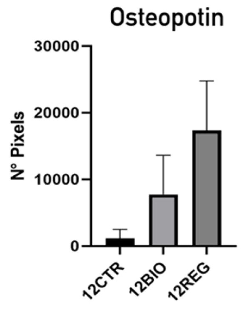
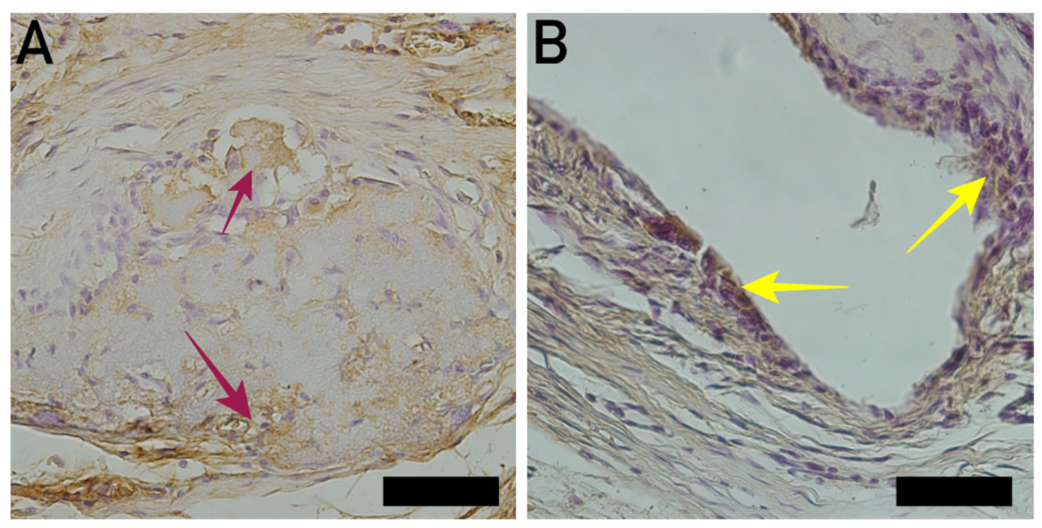
In the osteopontin group, it was possible to observe a predominance of osteoblasts in the Regener group, suggesting that the bone regeneration process in this group demonstrated superior performance compared with the other groups (Figure 7 and Table 1).
Figure 7 and Figure 8 shows the osteopontin express after 12 weeks. The Kruskal–Wallis statistical analysis showed a significant difference (p < 0.05) between the control and Bio-Oss® groups. The same significant difference (p < 0.05) was observed between the Regener® and the control and Bio-Oss® groups. Table 1 shows the statistical data analysis.

4.2.2. Osteocalcin

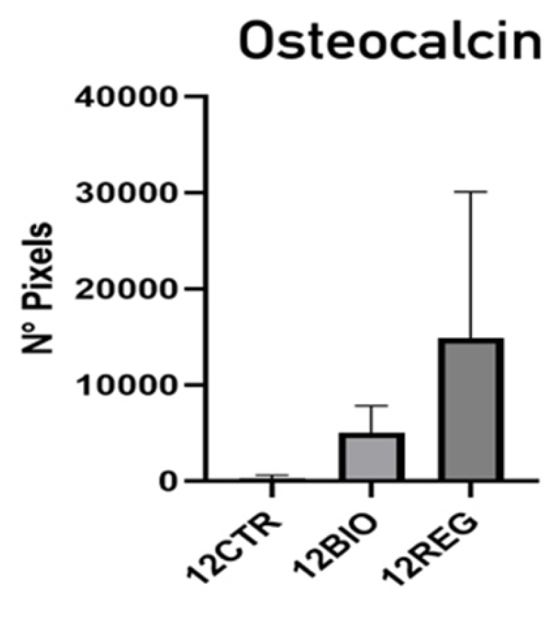
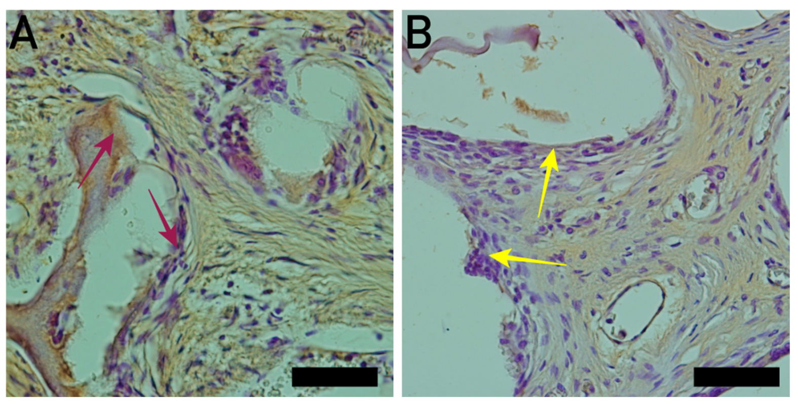
In the osteocalcin group, it was possible to observe a predominance of osteoclasts in the Regener group, suggesting that the bone replacement process occurs more rapidly in this group (Figure 9 and Figure 10; Table 1).

5. Discussion

The ultrastructural findings suggest that BM-MSCs actively internalize nano-HA/β-TCP compound spheroids through energy-dependent endocytic pathways, rather than passive penetration. The presence of a fibrillar, glycocalyx-like coat on the cell surface capturing the biomaterial points toward initial receptor-mediated adherence or nonspecific adsorption. The reaction is localized in the cytoplasm, where it is frequently associated with cisternal profiles and mitochondria, and undergoes some degree of degradation. Once internalized, the spheroids remain strictly compartmentalized within membrane-bound endolysosomal structures, preventing direct exposure or damage to the cell nucleus.
The observed spectrum of electron densities, shifting from solid black to a light gray, granular appearance, strongly demonstrates progressive intracellular degradation and biomaterial dissolution. This active breakdown occurs in proximity to metabolic hubs like mitochondria and the endoplasmic reticulum, suggesting a coordinated intracellular response. The close positioning to these organelles implies that the cellular machinery is actively engaged in processing the breakdown products, which could potentially trigger downstream osteogenic signaling pathways in the progenitor cells.
Nano-HA has applications in several fields, including bone regeneration and drug delivery. Some studies have investigated the impact of nanoparticles on cell cytotoxicity. Results showed that high concentrations of nano-HA can induce the formation of multivesicular bodies, which act as calcium reservoirs—an attribute that may be useful for treating cancer cells [23]. In the present study, the observation of multivesicular bodies indicative of apoptosis was rare, suggesting that the nano-HA/β-TCP composite has low cytotoxic potential in the context of bone regeneration. However, [24] reported a 15–20% reduction in hWJ-MSC viability after 72 h of exposure to 100 µg/mL nano-HA.
The osteoinductive potential of nano-HA and β-TCP has been studied individually, both in pure forms and in combination with various scaffold materials. Studies on nano-HA-containing hydrogels and other biological polymers have demonstrated its critical role in inducing the differentiation of MSCs and MC3T3-E1 cells into odontoblasts [25,26,27,28,29,30,31].
Strontium-substituted hydroxyapatite has also attracted interest due to the chemical similarity between Sr2+ and Ca2+ ions, which may enhance MSC osteogenic differentiation [32,33,34]. Collectively, these findings provide robust scientific evidence supporting the osteoinductive potential of nano-HA in MSCs.
The behavior of mouse MC3T3-E1 pre-osteoblasts and hFOB1.19 osteoblasts derived from human embryos was evaluated in the presence of porous HA or β-TCP spheres. The results indicated that HA and β-TCP have complementary roles in promoting cell adhesion and differentiation. When used in combination, they enhanced osteoblast differentiation more effectively than when used individually [35].
Porous HA has also been shown to induce osteogenic differentiation in human placenta-derived mesenchymal stem cells (hPMSCs). Similarly, HA scaffolds have demonstrated osteogenic potential in human bone marrow- and umbilical cord-derived MSCs [36].
Our findings on the nano-HA/β-TCP composite in BM-MSC cultures at the ultrastructural level are consistent with literature results [37]. Who studied nano-HA alone in hWJ-MSC cultures. In both cases, the biomaterial adhered to the cell surface and was internalized through a wrapping mechanism. Once internalized, it remained confined to vesicles and lysosomes within the cytoplasm, without entering the nucleus.
Nano-HA uptake has also been observed in hMSC cultures, where the material was found distributed in cytoplasmic vesicles of varying sizes and aggregated irregularly [38]. In MC3T3-E1 cells, nano-HA internalization was associated with a lysosome-based autophagic process, which plays a critical role in maintaining cellular homeostasis [39].
The molecular mechanisms underlying the effects of calcium phosphate-containing biomaterials have been explored in the literature. The uptake of extracellular phosphate (PO43−) via the SLC20a1 solute carrier promotes osteogenic differentiation in hMSCs through adenosine signaling, which activates the A2b adenosine receptor [40]. Conversely, extracellular calcium (Ca2+) is internalized through voltage-gated and receptor-operated ion channels. Intracellular Ca2+ is largely buffered by calcium-binding proteins—some with enzymatic activity—and is also sequestered into the endoplasmic reticulum (ER) and mitochondria via distinct membrane transporters. This calcium influx activates the calmodulin (CaM) and calmodulin-dependent kinase II (CaMKII) pathway, which regulates key transcription factors involved in osteogenic lineage commitment [41]. In this context, our finding that engulfed nano-HA/β-TCP spheroids are frequently associated with the ER and mitochondria is particularly noteworthy.
Another technological advancement that enables greater control and standardization of hydroxyapatite particle porosity is Digital Light Processing (DLP). This fabrication technique generates microparticles capable of releasing biologically active ions, such as Mg2+ and Ca2+, which have been shown to enhance cellular differentiation [42]. In the present study, the novel aspect is the application of nano-HA/β-TCP, which demonstrated superior performance in bone regeneration, resulting in increased new bone formation and enhanced extracellular bone matrix deposition.
In another study investigating cell differentiation and proliferation in relation to the degradation of biomaterial-based scaffolds, materials exhibiting controlled degradation were shown to promote the gradual release of calcium ions, thereby enhancing surface hydrophilicity and biomineralization [43]. The study demonstrated that a composition containing 80% nano-hydroxyapatite and 20% nano-β-tricalcium phosphate significantly improved bone regeneration. This effect was mainly attributed to the rapid resorption of nano-β-tricalcium phosphate, which releases calcium ions into the local microenvironment and generates additional porous spaces, facilitating cellular infiltration, vascularization, and cell–scaffold interactions. At the same time, the nano-HA/β-TCP phase provides structural stability and sustained osteoconductive support for new bone formation.
Based on Figure 6, progressive intracellular degradation and dissolution of the nano-HA/β-TCP spheroids may occur during the experiment. Transmission electron microscopy showed a light gray granular matrix, which is hypothesized to play a pivotal role in driving the osteogenic differentiation of BM-MSCs. Upon endolysosomal internalization, the acidic microenvironment triggers the sustained breakdown of the biomaterial, leading to a localized influx of calcium (Ca2+) and phosphate (PO43) ions into the cytoplasm. This intracellular ion enrichment can modulate key osteogenic signaling cascades. Specifically, elevated cytosolic Ca2+ acts as a secondary messenger that activates the calmodulin/calcineurin pathway, subsequently inducing the nuclear translocation of nuclear factor of activated T-cells (NFAT) and upregulating runt-related transcription factor 2 (Runx2) expression. Concurrently, the released inorganic phosphate can stimulate the mitogen-activated protein kinase (MAPK/ERK) pathway via specific phosphate transporters. This synergistic ion-mediated signaling cascade ultimately enhances the transcription of crucial bone-related extracellular matrix proteins, such as osteocalcin and osteopontin, reinforcing the intrinsic osteoinductive potential of the nano-HA/β-TCP compound.
A study has demonstrated that osteocalcin can be used as a gene expression level marker of osteogenesis in early bone marrow-derived mesenchymal stem cells (BMSCs) [44]. In our study, the use of osteocalcin was essential for confirming the presence of osteoclasts throughout the entire bone remodeling process.
Osteopontin can be used as a marker for osteoblast expression control, as reported in a study that employed osteopontin to quantify osteoblast expression. The study concluded that immunohistochemistry is a highly efficient technique, as it enabled the precise differentiation of osteoblastic expression levels across groups [45]. Based on the findings regarding osteocalcin and osteopontin expression, it may be suggested that the Blue Bone® group demonstrated a more balanced bone remodeling process, with a more favorable relationship between bone formation and degradation events associated with the inflammatory response, compared with the other groups (Figure 7 and Figure 9).
The Blue Bone® and Bio-Oss® have demonstrated excellent biocompatibility and osteoconductive capabilities; however, differences in composition, morphology, particle size, and surface characteristics can influence the bone healing kinetics.
The Bio-Oss®, composed of highly crystalline natural hydroxyapatite, serves as a slowly resorbing scaffold that maintains graft volume and provides long-term structural support for bone healing. On the other hand, the Blue Bone® is composed of biphasic calcium phosphate containing 80% nano-hydroxyapatite and 20% β-tricalcium phosphate and is degraded more rapidly due to dissolution of the β-TCP phase. The accelerated degradation promotes faster extracellular matrix remodeling and earlier collagen deposition. The Blue Bone® nano-hydroxyapatite composition enhances cell contact area, promotes cell membrane interaction, and facilitates vesicle internalization for further lysosomal processing, as observed in the ME experiment.
The naïve micro- and macroporous trabecular structure of Bio-Oss® is more related to supporting volume maintenance in bone healing. In contrast, the nanoporous architecture and nanometric particle size of Blue Bone® increase the surface area-to-volume ratio, which enhances protein adsorption, cell adhesion, and vascular infiltration during the early stages of healing.
The Blue Bone® is composed of a highly hydrophilic and negatively charged surface that can promote faster blood wetting and enhance cell migration compared with the slightly negative Bio-Oss® surface, potentially supporting accelerated bone healing [7].
Some authors have suggested that, compared with direct cell seeding onto bulk scaffolds, multicellular spheroids function as pre-organized microtissues in which cell–cell interactions and endogenous extracellular matrix are established before contact with the biomaterial [46,47]. Consequently, the biological response is less dependent on the initial attachment, distribution, and colonization of isolated cells within the scaffold—processes that are strongly influenced by scaffold architecture and seeding efficiency—and instead reflects the interaction between the biomaterial and an already organized three-dimensional cellular construct [46]. This approach more closely resembles the early in vivo scenario, where implanted biomaterials interact with surrounding tissues rather than isolated cells. Therefore, spheroid-based models represent a valuable complementary approach for investigating the interaction between osteoconductive nanobiomaterials and a pre-established cellular microenvironment. Consistent with this concept, recent studies employing hydroxyapatite- and titanium dioxide-based nanomaterials further support spheroid-based models as physiologically relevant platforms for investigating cell–biomaterial interactions and osteogenic responses to bone-regenerative nanomaterials [48,49].
It is important to note that transmission electron microscopy (TEM) is inherently limited in its ability to provide quantitative analysis. Future studies could explore cell mediator expression and changes in extracellular culture medium composition to complement these findings.
The authors acknowledge the limitations of the present study, as the current in vitro 3D spheroid model represents a static or semi-dynamic environment that differs substantially from the complex biological microenvironment of bone defects in vivo. Therefore, further in vitro studies will be conducted to understand the mechanisms by which cellular interactions with the nanobiomaterial enhance osteogenesis and promote improved bone tissue formation.
The utilization of a 3D cell culture spheroid model in this study offers distinct pathophysiological advantages over both conventional 2D monolayer cultures and traditional cell seeding on bulk solid scaffolds like rigid, solid scaffolds, which often suffer from non-uniform cell attachment, limited core cellular infiltration, and altered mechanical signaling. The 3D spheroid configuration closely recapitulates the native, highly cellular mesenchymal condensations during embryonic skeletogenesis. Within these dense spheroids, the enhanced cell-to-cell and cell-to-matrix interactions establish a more biomimetic physiological tissue microenvironment. This spatial arrangement creates localized, realistic nutrient, oxygen, and metabolic gradients, while also enabling rich autocrine and paracrine signaling networks. Consequently, the combination of this high-density 3D microenvironment with the close proximity of internalizing nano-HA/β-TCP particles create a highly synergistic niche that accelerates and synchronizes the osteogenic commitment of the progenitor cells.

6. Conclusions

Based on the experimental results, it is possible to conclude the following:
(a)
The nano-HA/β-TCP particles were internalized by BM-MSCs without causing apparent cellular damage, remaining confined to the cytoplasm and associated with organelles involved in cellular metabolism.
(b)
The experimental synthetic biomaterial made with the mixture of nano-HA (80%) and β-TCP (20%) exhibited greater osteogenic potential in vivo than an organic biomaterial produced with deproteinized bovine bone.
(c)
The nanostructured biomaterial enhances both cell material interactions and bone regeneration.

Author Contributions

I.D.S.B.: Conceptualization, surgery, and co-wrote the manuscript. C.N.E.: Analysis of results, writing—review, and editing. L.F., B.T.C., D.O., S.C., E.C., A.T., A.L.R.N., K.R.S. and I.S.D.S.: Co-wrote the manuscript. J.J.D.C.: Analyzed the experimental results of the materials. All authors have read and agreed to the published version of the manuscript.

Funding

This research was supported by the Brazilian agencies CNPq, FAPERJ, CAPES, and UERJ (Fisclinex).

Institutional Review Board Statement

The study was conducted following the Declaration of the Ethics Committee in Animal Experimentation of the State University of Rio de Janeiro, which has approved all procedures (registered under CEUA/001/2019).

Informed Consent Statement

Not applicable.

Data Availability Statement

The original contributions presented in this study are included in the article. Further inquiries can be directed to the corresponding author(s).

Acknowledgments

The authors are grateful to the Brazilian Agencies CAPES, CNPq, Fisclinex, FAPERJ, and FINEP.

Conflicts of Interest

The authors declare no conflicts of interest.

References

  1. Agrawal, S.; Srivastava, R. Osteoinductive and osteoconductive biomaterials. In Racing for the Surface; Springer: Berlin/Heidelberg, Germany, 2020; pp. 355–395. [Google Scholar] [CrossRef]
  2. Bohner, M.; Miron, R.J. A proposed mechanism for material-induced heterotopic ossification. Mater. Today 2019, 22, 132–141. [Google Scholar] [CrossRef]
  3. Huang, W.; Yang, S.; Shao, J.; Li, Y.P. Signaling and transcriptional regulation in osteoblast commitment and differentiation. Front. Biosci. 2007, 12, 3068–3092. [Google Scholar] [CrossRef]
  4. Zhu, S.; Chen, W.; Masson, A.; Li, Y.P. Cell signaling and transcriptional regulation of osteoblast lineage commitment, differentiation, bone formation, and homeostasis. Cell Discov. 2024, 10, 71. [Google Scholar] [CrossRef] [PubMed]
  5. Thomas, S.; Jaganathan, B.G. Signaling network regulating osteogenesis in mesenchymal stem cells. J. Cell Commun. Signal. 2022, 16, 47–61. [Google Scholar] [PubMed]
  6. Salam, N.; Gibson, I.R. Lithium-ion doped carbonated hydroxyapatite compositions: Synthesis, physicochemical characterization and effect on osteogenic response in vitro. Biomater. Adv. 2022, 140, 213068. [Google Scholar] [CrossRef] [PubMed]
  7. Da Silva Brum, I.; Frigo, L.; Lana Devita, R.; da Silva Pires, J.L.; Hugo Vieira de Oliveira, V.; Rosa Nascimento, A.L.; de Carvalho, J.J. Histomorphometric, Immunohistochemical, Ultrastructural Characterization of a Nano-Hydroxyapatite/Beta-Tricalcium Phosphate Composite and a Bone Xenograft in Sub-Critical Size Bone Defect in Rat Calvaria. Materials 2020, 13, 4598. [Google Scholar] [CrossRef] [PubMed] [PubMed Central]
  8. Liang, W.; Ding, P.; Li, G.; Lu, E.; Zhao, Z. Hydroxyapatite Nanoparticles Facilitate Osteoblast Differentiation and Bone Formation Within Sagittal Suture During Expansion in Rats. Drug Des. Dev. Ther. 2021, 15, 905–917. [Google Scholar] [CrossRef] [PubMed]
  9. Demirel, M.; Aslan, N.; Aksakal, B.; Arslan, M.E. Fabrication of hydroxyapatite-based nano-gold and nano-silver-doped bioceramic bone grafts: Enhanced mechanostructure, cell viability, and nuclear abnormality properties. J. Biomed. Mater. Res. B Appl. Biomater. 2023, 111, 1386–1397. [Google Scholar] [CrossRef] [PubMed]
  10. Sivakumar, P.M.; Yetisgin, A.A.; Sahin, S.B.; Demir, E.; Cetinel, S. Enhanced properties of nickel-silver codoped hydroxyapatite for bone tissue engineering: Synthesis, characterization, and biocompatibility evaluation. Environ. Res. 2023, 238, 117131. [Google Scholar] [CrossRef] [PubMed]
  11. Kontogianni, G.-I.; Coelho, C.; Gauthier, R.; Fiorilli, S.; Quadros, P.; Vitale-Brovarone, C.; Chatzinikolaidou, M. Osteogenic Potential of Nano-Hydroxyapatite and Strontium-Substituted Nano-Hydroxyapatite. Nanomaterials 2023, 13, 1881. [Google Scholar] [CrossRef] [PubMed]
  12. Wang, Z.; Han, T.; Zhu, H.; Tang, J.; Guo, Y.; Jin, Y.; Wang, Y.; Chen, G.; Gu, N.; Wang, C. Potential Osteoinductive Effects of Hydroxyapatite Nanoparticles on Mesenchymal Stem Cells by Endothelial Cell Interaction. Nanoscale Res. Lett. 2021, 16, 67. [Google Scholar] [CrossRef] [PubMed]
  13. Shi, X.; Zhou, K.; Huang, F.; Zhang, J.; Wang, C. Endocytic mechanisms and osteoinductive profile of hydroxyapatite nanoparticles in human umbilical cord Wharton’s jelly-derived mesenchymal stem cells. Int. J. Nanomed. 2018, 13, 1457–1470. [Google Scholar] [CrossRef] [PubMed]
  14. Brum, I.D.S.; Frigo, L.; Ribeiro da Silva, J.F.; Ciambarella, B.T.; Nascimento, A.L.R.; Pereira, M.J.D.S.; Elias, C.N.; de Carvalho, J.J. Comparison Between Nano-Hydroxyapatite/Beta-Tricalcium Phosphate Composite and Autogenous Bone Graft in Bone Regeneration Applications: Biochemical Mechanisms and Morphological Analysis. Int. J. Mol. Sci. 2024, 26, 52. [Google Scholar] [CrossRef] [PubMed]
  15. Pires, J.L.D.S.; de Carvalho, J.J.; Pereira, M.J.D.S.; Brum, I.D.S.; Nascimento, A.L.R.; Dos Santos, P.G.P.; Frigo, L.; Fischer, R.G. Repair of Critical Size Bone Defects Using Synthetic Hydroxyapatite or Xenograft with or without the Bone Marrow Mononuclear Fraction: A Histomorphometric and Immunohistochemical Study in Rat Calvaria. Materials 2021, 14, 2854. [Google Scholar] [CrossRef] [PubMed]
  16. Ebrahimi, M.; Pripatnanont, P.; Monmaturapoj, N.; Suttapreyasri, S. Fabrication and characterization of novel nano hydroxyapatite/β-tricalcium phosphate scaffolds in three different composition ratios. J. Biomed. Mater. Res. A 2012, 100, 2260–2268. [Google Scholar] [CrossRef] [PubMed]
  17. Jia, J.; Shen, J.; Yang, F.; Zhang, J.; Shuai, C.; Feng, P. Piezoelectric scaffold for tissue engineering: Material, structure, fabrication and function. Int. J. Extrem. Manuf. 2026, 8, 022013. [Google Scholar] [CrossRef]
  18. Zhang, S.; Zhang, Y.; Wang, X.; Zhao, W.; Song, L.; Li, X.; Yan, J.; Chen, G.; Ji, Y. A biomimetic physical-biological coupled bone organoid 3D culture system for bone repair. Mater. Today Adv. 2026, 29, 100739. [Google Scholar] [CrossRef]
  19. Wang, R.; Hu, H.; Guo, J.; Wang, Q.; Cao, J.; Wang, H.; Li, G.; Mao, J.; Zou, X.; Chen, D.; et al. Nano-Hydroxyapatite Modulates Osteoblast Differentiation Through Autophagy Induction via mTOR Signaling Pathway. J. Biomed. Nanotechnol. 2019, 15, 405–415. [Google Scholar] [CrossRef] [PubMed]
  20. Zhang, G.; Lu, Y.; Song, J.; Huang, D.; An, M.; Chen, W.; Han, P.; Yao, X.; Zhang, X. A multifunctional nano-hydroxyapatite/MXene scaffold for the photothermal/dynamic treatment of bone tumors and simultaneous tissue regeneration. J. Colloid Interface Sci. 2023, 652, 1673–1684. [Google Scholar] [CrossRef] [PubMed]
  21. da Silva Brum, I.; Frigo, L.; Goncalo Pinto dos Santos, P.; Nelson Elias, C.; da Fonseca, G.A.M.D.; Jose de Carvalho, J. Performance of Nano-Hydroxyapatite/Beta-Tricalcium Phosphate and Xenogenic Hydroxyapatite on Bone Regeneration in Rat Calvarial Defects: Histomorphometric, Immunohistochemical and Ultrastructural Analysis. Int. J. Nanomed. 2021, 16, 3473–3485. [Google Scholar] [CrossRef] [PubMed]
  22. da Silva Brum, I.; de Carvalho, J.J.; da Silva Pires, J.L.; de Carvalho, M.A.A.; Dos Santos, L.B.F.; Elias, C.N. Nanosized hydroxyapatite and β-tricalcium phosphate composite: Physico-chemical, cytotoxicity, morphological properties and in vivo trial. Sci. Rep. 2019, 9, 19602. [Google Scholar] [CrossRef] [PubMed] [PubMed Central]
  23. Fu, M.; Li, J.; Liu, M.; Yang, C.; Wang, Q.; Wang, H.; Chen, B.; Fu, Q.; Sun, G. Sericin/Nano-Hydroxyapatite Hydrogels Based on Graphene Oxide for Effective Bone Regeneration via Immunomodulation and Osteoinduction. Int. J. Nanomed. 2023, 18, 1875–1895. [Google Scholar] [CrossRef] [PubMed]
  24. Klimek, K.; Palka, K.; Truszkiewicz, W.; Douglas, T.E.L.; Nurzynska, A.; Ginalska, G. Could Curdlan/Whey Protein Isolate/Hydroxyapatite Biomaterials Be Considered as Promising Bone Scaffolds? Fabrication, Characterization, and Evaluation of Cytocompatibility towards Osteoblast Cells In Vitro. Cells 2022, 11, 3251. [Google Scholar] [CrossRef] [PubMed]
  25. Almasi, D.; Lau, W.J.; Rasaee, S.; Abbasi, K. Fabrication and in vitro study of 3D novel porous hydroxyapatite/polyether ether ketone surface nanocomposite. J. Biomed. Mater. Res. B Appl. Biomater. 2022, 110, 838–847. [Google Scholar] [CrossRef] [PubMed]
  26. Xu, Y.; Wu, X.; Wang, S.; Yang, C.; Li, Y.; Cao, Y. Hydroxyapatite Nanoparticle-Crosslinked Peptide Hydrogels for Three-Dimensional Culture and Differentiation of MC3T3-E1 Osteoblasts. J. Biomed. Nanotechnol. 2019, 15, 2351–2362. [Google Scholar] [CrossRef] [PubMed]
  27. Carles-Carner, M.; Saleh, L.S.; Bryant, S.J. The effects of hydroxyapatite nanoparticles embedded in a MMP-sensitive photoclickable PEG hydrogel on encapsulated MC3T3-E1 pre-osteoblasts. Biomed. Mater. 2018, 13, 045009. [Google Scholar] [CrossRef] [PubMed]
  28. Vitale, M.; Ligorio, C.; McAvan, B.; Hodson, N.W.; Allan, C.; Richardson, S.M.; Hoyland, J.A.; Bella, J. Hydroxyapatite-decorated Fmoc-hydrogel as a bone-mimicking substrate for osteoclast differentiation and culture. Acta Biomater. 2022, 138, 144–154. [Google Scholar] [CrossRef] [PubMed]
  29. Arkharova, N.A.; Suvorova, E.I.; Severin, A.V.; Khripunov, A.K.; Krasheninnikov, S.V.; Klechkovskaya, V.V. SEM and TEM for structure and properties characterization of bacterial cellulose/hydroxyapatite composites. Scanning 2016, 38, 757–765. [Google Scholar] [CrossRef] [PubMed]
  30. Suzuki, K.; Fukasawa, J.; Miura, M.; Lim, P.N.; Honda, M.; Matsuura, T.; Aizawa, M. Influence of Culture Period on Osteoblast Differentiation of Tissue-Engineered Bone Constructed by Apatite-Fiber Scaffolds Using Radial-Flow Bioreactor. Int. J. Mol. Sci. 2021, 22, 13080. [Google Scholar] [CrossRef] [PubMed]
  31. Chang, H.; Xiang, H.; Yao, Z.; Yang, S.; Tu, M.; Zhang, X.; Yu, B. Strontium-substituted calcium sulfate hemihydrate/hydroxyapatite scaffold enhances bone regeneration by recruiting bone mesenchymal stromal cells. J. Biomater. Appl. 2020, 35, 97–107. [Google Scholar] [CrossRef] [PubMed]
  32. Ran, L.; Liu, L.; Gao, J.; Pan, Y.; Ramalingam, M.; Du, X.; Liu, Y.; Cheng, L.; Shi, Z. Strontium-doped hydroxyapatite and its role in osteogenesis and angiogenesis. Int. J. Dev. Biol. 2023, 67, 137–146. [Google Scholar] [CrossRef] [PubMed]
  33. Akshata, C.R.; Murugan, E.; Harichandran, G. Alginate templated synthesis, characterization and in vitro osteogenic evaluation of strontium-substituted hydroxyapatite. Int. J. Biol. Macromol. 2023, 252, 126478. [Google Scholar] [CrossRef] [PubMed]
  34. Liu, X.; Huang, H.; Zhang, J.; Sun, T.; Zhang, W.; Li, Z. Recent Advances of Strontium Functionalized in Biomaterials for Bone Regeneration. Bioengineering 2023, 10, 414. [Google Scholar] [CrossRef] [PubMed]
  35. Nomoto, H.; Maehashi, H.; Shirai, M.; Nakamura, M.; Masaki, T.; Mezaki, Y.; Park, J.; Aizawa, M.; Ohkawa, K.; Yoshida, K.; et al. Bio-artificial bone formation model with a radial-flow bioreactor for implant therapy-comparison between two cell culture carriers: Porous hydroxyapatite and β-tricalcium phosphate beads. Hum. Cell 2019, 32, 1–11. [Google Scholar] [CrossRef] [PubMed]
  36. Ren, X.; Wang, Q.; Liu, C.; Zhao, Q.; Zheng, J.; Tian, K.; Xu, H.; Mu, Y. Osteogenic ability using porous hydroxyapatite scaffold-based delivery of human placenta-derived mesenchymal stem cells. Exp. Ther. Med. 2021, 22, 1091. [Google Scholar] [CrossRef] [PubMed]
  37. Meesuk, L.; Suwanprateeb, J.; Thammarakcharoen, F.; Tantrawatpan, C.; Kheolamai, P.; Palang, I.; Tantikanlayaporn, D.; Manochantr, S. Osteogenic differentiation and proliferation potentials of human bone marrow and umbilical cord-derived mesenchymal stem cells on the 3D-printed hydroxyapatite scaffolds. Sci. Rep. 2022, 12, 19509. [Google Scholar] [CrossRef] [PubMed]
  38. Shi, H.; Zhou, K.; Wang, M.; Wang, N.; Song, Y.; Xiong, W.; Guo, S.; Yi, Z.; Wang, Q.; Yang, S. Integrating physicomechanical and biological strategies for BTE: Biomaterials-induced osteogenic differentiation of MSCs. Theranostics 2023, 13, 3245–3275. [Google Scholar] [CrossRef] [PubMed]
  39. Yang, X.; Li, Y.; Liu, X.; Zhang, R.; Feng, Q. In Vitro Uptake of Hydroxyapatite Nanoparticles and Their Effect on Osteogenic Differentiation of Human Mesenchymal Stem Cells. Stem Cells Int. 2018, 2018, 2036176. [Google Scholar] [CrossRef] [PubMed]
  40. Shih, Y.R.; Hwang, Y.; Phadke, A.; Kang, H.; Hwang, N.S.; Caro, E.J.; Nguyen, S.; Siu, M.; Theodorakis, E.A.; Gianneschi, N.C.; et al. Calcium phosphate-bearing matrices induce osteogenic differentiation of stem cells through adenosine signaling. Proc. Natl. Acad. Sci. USA 2014, 111, 990–995. [Google Scholar] [CrossRef] [PubMed]
  41. Lu, H.; Zhou, Y.; Ma, Y.; Xiao, L.; Ji, W.; Zhang, Y.; Wang, X. Current Application of Beta-Tricalcium Phosphate in Bone Repair and Its Mechanism to Regulate Osteogenesis. Front. Mater. 2021, 8, 698915. [Google Scholar] [CrossRef]
  42. Guo, W.; Li, P.; Wei, Y.; Zhao, L.; Pang, Y.; Huang, Y.; Ye, X.; Wang, S.; Liu, B.; You, H.; et al. Substituição iônica através de doping bredigite para microestrutura e ajuste de desempenho em andaimes ósseos POROS HA TPMS impressos em DLP 3D. Prototipagem Virtual Fís. 2024, 19, e2423840. [Google Scholar] [CrossRef]
  43. Guo, W.; Wei, Y.; Xu, C.; Li, B.; Wu, Y.; Gong, Y.; Mai, H.; Wang, S.; Zhang, Y.; Long, Y. Functionalized degradable soybean oil-based biomimetic porous scaffolds for complex bone defects: Vat photopolymerization additive manufacturing, photothermal-mediated shape memory and tumor thermotherapy. Compos. Sci. Technol. 2026, 273, 111412. [Google Scholar] [CrossRef]
  44. Hsieh, C.W.; Chang, L.H.; Wang, Y.H.; Li, W.T.; Chang, J.K.; Chen, C.H.; Ho, M.L. O Sulfato Indoxílico Inibe a Osteogênese em Células-Tronco Mesenquimais da Medula Óssea através da Via AhR/Hes1. Int. J. Mol. Sci. 2024, 25, 8770. [Google Scholar] [CrossRef] [PubMed]
  45. Bargowo, L.; Kusumawardhani, B.; Perdana, S.; Wijaksana, I.K.E.; Saskianti, T.; Ridwan, R.D.; Setijanto, D.; Prahasanti, C.; Saquib Abullais, S. Expressão de osteopontina e osteocalcina em células de osteoblasto expostas a uma combinação de polimetilmetacrilato (PMMA) e hidroxiapatita (HAp): Um estudo observacional prospectivo. Medicina 2024, 103, e40088. [Google Scholar] [CrossRef] [PubMed] [PubMed Central]
  46. Di Caprio, N.; Burdick, J.A. Engineered biomaterials to guide spheroid formation, function, and fabrication into 3D tissue constructs. Acta Biomater. 2023, 165, 4–18. [Google Scholar] [CrossRef] [PubMed]
  47. Cesarz, Z.; Tamama, K. Spheroid culture of mesenchymal stem cells. Stem Cells Int. 2016, 2016, 9176357. [Google Scholar] [CrossRef] [PubMed]
  48. de Souza Castro, G.; de Souza, W.; Lima, T.S.M.; Bonfim, D.C.; Werckmann, J.; Archanjo, B.S.; Granjeiro, J.M.; Ribeiro, A.R.; Gemini-Piperni, S. The Effects of Titanium Dioxide Nanoparticles on Osteoblasts Mineralization: A Comparison between 2D and 3D Cell Culture Models. Nanomaterials 2023, 13, 425. [Google Scholar] [CrossRef] [PubMed]
  49. Liu, Y.; Ye, J.; Hu, H.; Song, Y.; Qiang, H.; Wang, J.; Zhou, L.; Wang, X.; Fei, X.; Zhu, M. 3D stem cell spheroids with urchin-like hydroxyapatite microparticles enhance osteogenesis of stem cells. J. Mater. Chem. B 2024, 12, 1344–1358. [Google Scholar] [CrossRef] [PubMed]
Figure 1. Diagram of experimental procedures and group division.
Jcs 10 00406 g001
Figure 2. Histomorphometric analysis was performed on sections stained with osteopontin. The photomicrographs present representative images of each experimental group evaluated. (A) The delineated region selected for analysis. (B) Brown staining denotes osteoblastic activity, Scale bar = 100 µm, ×100.
Jcs 10 00406 g002
Figure 3. (A) The creation of the triangular incision: the preparation of two defects using a 3 mm diameter trephine at 800 rpm with a depth of 2 mm. Irrigation was performed with sterile saline solution, followed by placement of the biomaterial into the calvarial defects. (B) Closure of the triangular incision after biomaterial implantation.
Jcs 10 00406 g003
Figure 4. A photomicrograph of the control group (no biomaterial added in the culture medium); well-defined round cell nuclei (black arrow) were observed. Toluidine blue. Scale bar = 100 µm, ×100.
Jcs 10 00406 g004
Figure 5. The nuclei (red arrow) and the biomaterial close to the cell surface (black arrow) can be observed. Inside the cells, biomaterial images can be seen (yellow arrow). Accumulations of intracellular biomaterials close to the cell nucleus (purple arrow) can be seen. Toluidine blue. Scale bar = 100 µm, ×100.
Jcs 10 00406 g005
Figure 6. TEM electromicrographs of nano-HA/β-TCP compound spheroids and BM-MSCs. (A) An electron micrograph showing biomaterial spheroids (yellow arrow) in contact with the surface of BM-MSCs. Fibrillar electron-dense structures, suggestive of the glycocalyx, together with plasma membrane interdigitations (blue arrow), are observed at the cell–biomaterial interface. The cytoplasm (green star), rough endoplasmic reticulum (red arrow), and mitochondria (black arrow) are also evident. Magnification: ×6000. (B) Engulfed spheroids (blue arrow) enclosed by a thin cytoplasmic layer (red arrow) [×5000]. (C) Biomaterial surrounding the cell nucleus (blue arrow) without entering it, displaying varied electron density indicative of degradation (red arrow) [×2000]. (D) Internalized biomaterial (red arrow) in close relation with mitochondria (green arrow) [×3000]. (E) Spheroid surrounded by a membrane-like structure (yellow arrow), associated dense body (blue arrow), granular degradation matrix (red arrow), internal dense core (black arrow), and surrounding cisternal profile (green star) [×12,000]. (F) The endoplasmic reticulum profile (red arrow) in proximity to the biomaterial sphere [×6000].
Jcs 10 00406 g006aJcs 10 00406 g006b
Figure 7. The variation in osteopontin expression in the Control (CTR), Bio-Oss® (BIO) and Regener® (REG) groups after 12 weeks. The Kruskal–Wallis statistical analyses showed a statistically significant difference (p < 0.05) in the expression of osteopontin (OPN) among the Control, Bio-Oss®, and Regener® groups.
Jcs 10 00406 g007
Figure 8. In (A), a more advanced stage of ossification can be observed compared with (B). The pink arrows indicate a newly formed bone matrix containing entrapped cells (osteoblasts), which subsequently differentiate into osteocytes involved in mature bone formation. In contrast, the yellow arrows indicate the presence of several multinucleated cells (osteoclasts) located at the periphery of the regenerated area, Scale bar = 100 µm, ×100.
Jcs 10 00406 g008
Figure 9. The variation in osteocalcin expression in the Control (CTR), Bio-Oss® (BIO) and Regener® (REG) groups after 12 weeks. The Kruskal–Wallis statistical analyses showed a statistically significant difference (p < 0.05) in the expression of osteocalcin (OCN) among the Control, Bio-Oss®, and Regener® groups.
Jcs 10 00406 g009
Figure 10. In (A), a more advanced stage of ossification can be observed compared with (B), together with cells surrounding the margins of the nanobiomaterial (pink arrows). In contrast, the yellow arrows indicate several cells bordering the biomaterial without significant bone formation, Scale bar = 100 µm, ×100.
Jcs 10 00406 g010
Table 1. Data from statistical analyses of osteocalcin (OCN) and osteopontin (OPN) expression in Control, Bio-Oss®, and Regener® groups. Kruskal–Wallis test.
ResultsOCN_12 Weeks OPN_12 Weeks
p value0.0050.0033
Exact or approximate p value?ExactExact
Do the medians vary signif. (p < 0.05)?YesYes
Number of groups33
Kruskal–Wallis statistic8.789.26
Disclaimer/Publisher’s Note: The statements, opinions and data contained in all publications are solely those of the individual author(s) and contributor(s) and not of MDPI and/or the editor(s). MDPI and/or the editor(s) disclaim responsibility for any injury to people or property resulting from any ideas, methods, instructions or products referred to in the content.