Proceeding Paper

In Vivo Recognition of Vascular Structures by Near-Infrared Transillumination †

Department of Electrical, Computer and Biomedical Engineering, University of Pavia, 27100 Pavia, Italy
*
Author to whom correspondence should be addressed.
Presented at the 6th International Electronic Conference on Sensors and Applications, 15–30 November 2019; Available online: https://ecsa-6.sciforum.net/.
Proceedings 2020, 42(1), 24; https://doi.org/10.3390/ecsa-6-06639
Published: 15 November 2019

Abstract

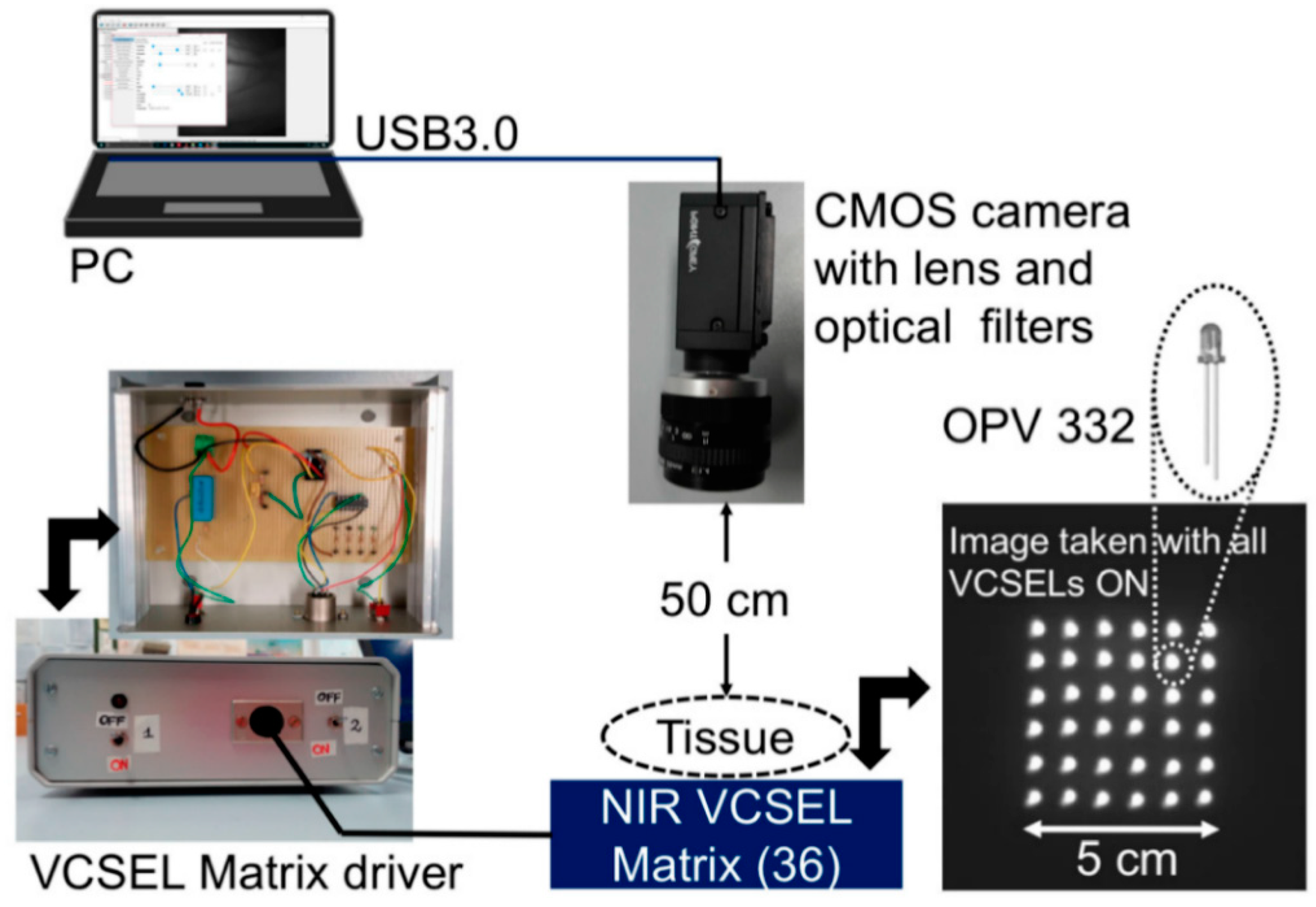
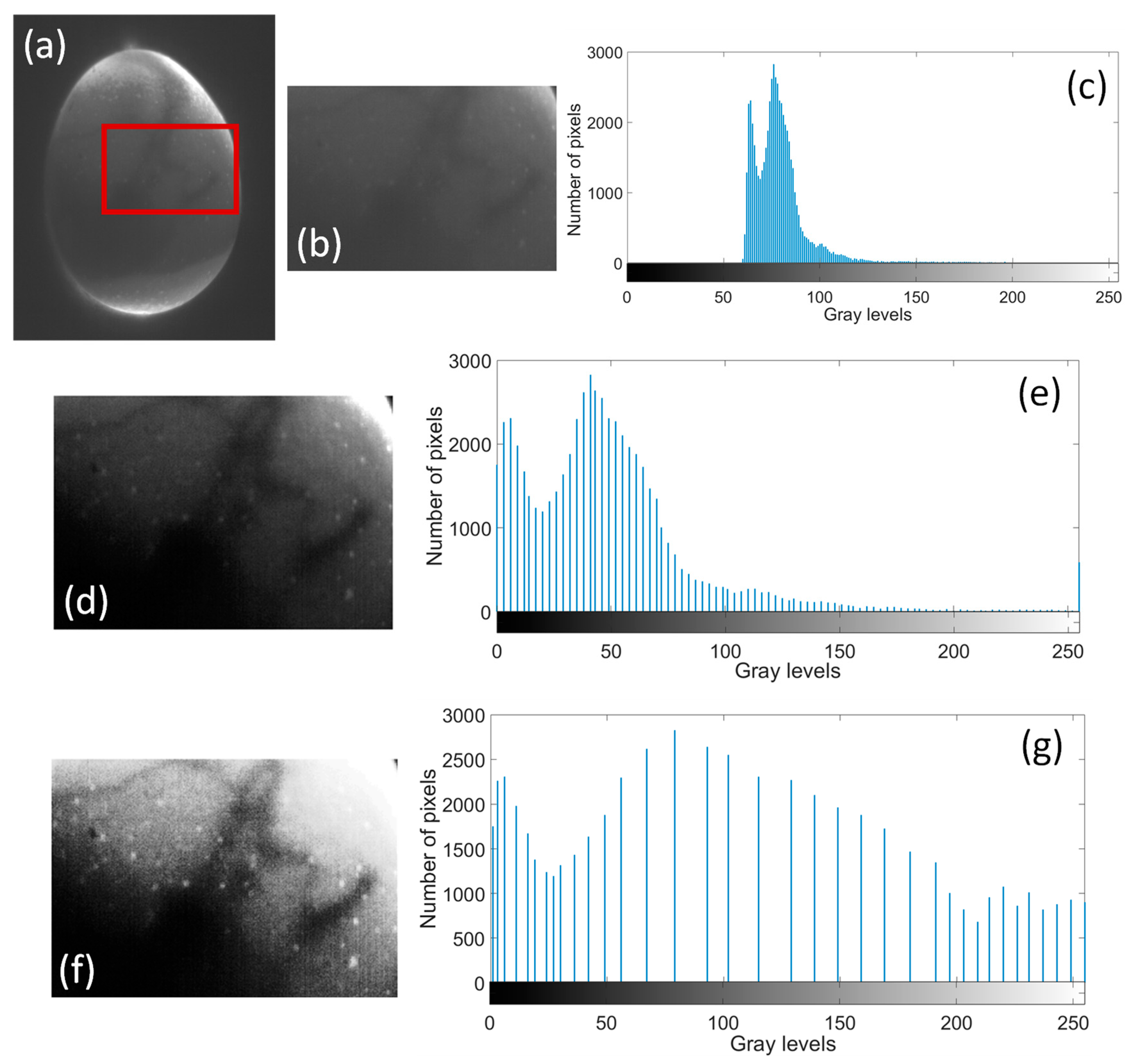
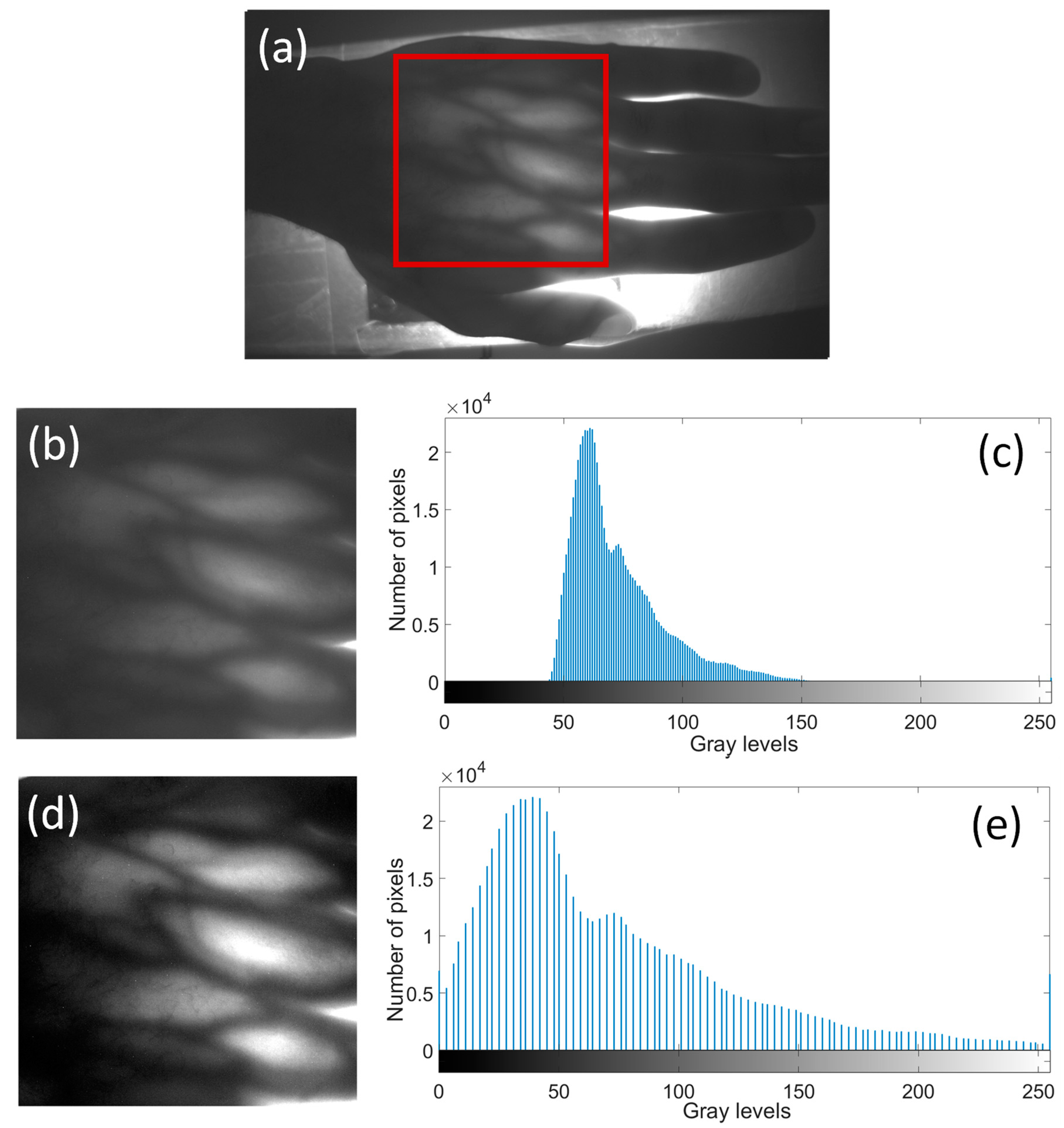
Transillumination is a very well-known non-invasive optical technique that relies on the use of non-ionizing radiation to obtain information about the internal morphology of biological tissues. In a previous work, we implemented a laser-based illuminator operating at a wavelength of 850 nm, combined with a CMOS digital camera and narrow-band optical detection that showed great potential for in vivo imaging. A great advantage is the use of low-cost semiconductor lasers, driven by a very low current (about 11 mA, spatially distributed as a 6-by-6 matrix covering a 25 cm2 area). Thanks to the strong absorption of hemoglobin at this wavelength, we have collected raw data of vascular structures that have been further processed to achieve images with much better quality. In particular, here we show that a higher contrast can be attained by the expansion of gray level histograms to exploit the full range, 0–255. This elaboration can be, for instance, exploited for the recognition of vascular structures with better resolution. Examples are reported relative to hand dorsal vein patterns and live chick embryos’ blood vessels. Analyses can be successfully performed without applying any thermal or mechanical stress to the human tissue under test and without damaging or puncturing any parts of the eggshell.
Keywords: transillumination; in vivo imaging; chick embryo; hand dorsal vein pattern; VCSEL transillumination; in vivo imaging; chick embryo; hand dorsal vein pattern; VCSEL