Automatic Characterization of Epiretinal Membrane in OCT Images with Supervised Training †


 ,
,  ,
, 
Abstract
:1. Introduction
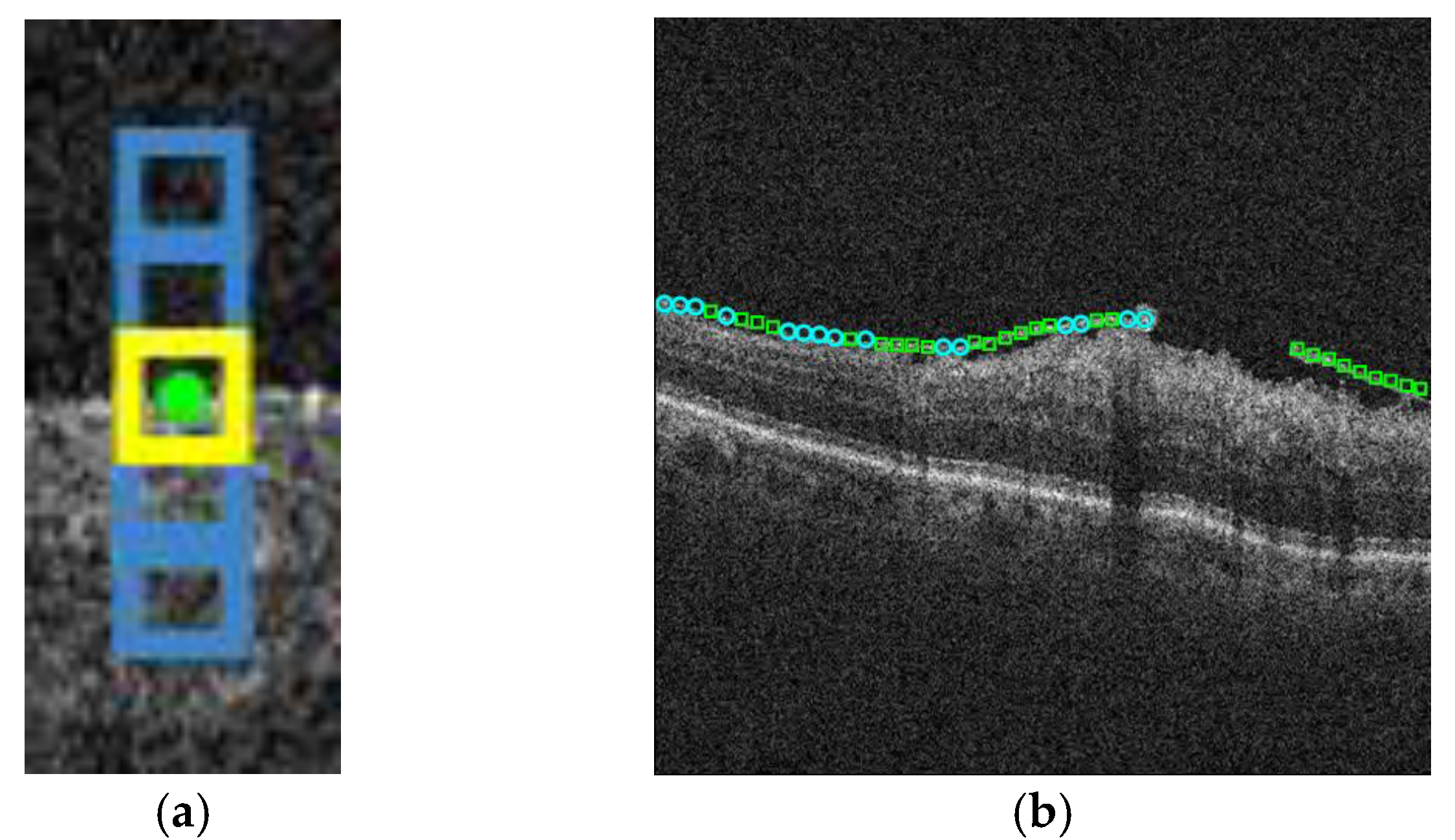
2. Methodology
3. Experimental Results
Acknowledgments
Conflicts of Interest
References
- Baamonde, S.; de Moura, J.; Novo, J.; Ortega, M. Automatic Detection of Epiretinal Membrane in OCT Images by Means of Local Luminosity Patterns. Adv. Comput. Intell. 2017, 10305, 222–235. [Google Scholar]
Publisher’s Note: MDPI stays neutral with regard to jurisdictional claims in published maps and institutional affiliations. |
© 2022 by the authors. Licensee MDPI, Basel, Switzerland. This article is an open access article distributed under the terms and conditions of the Creative Commons Attribution (CC BY) license (https://creativecommons.org/licenses/by/4.0/).
Share and Cite
Baamonde, S.; Moura, J.d.; Novo, J.; Barreira, N.; Ortega, M. Automatic Characterization of Epiretinal Membrane in OCT Images with Supervised Training. Proceedings 2018, 2, 1161. https://doi.org/10.3390/proceedings2181161
Baamonde S, Moura Jd, Novo J, Barreira N, Ortega M. Automatic Characterization of Epiretinal Membrane in OCT Images with Supervised Training. Proceedings. 2018; 2(18):1161. https://doi.org/10.3390/proceedings2181161
Chicago/Turabian StyleBaamonde, Sergio, Joaquim de Moura, Jorge Novo, Noelia Barreira, and Marcos Ortega. 2018. "Automatic Characterization of Epiretinal Membrane in OCT Images with Supervised Training" Proceedings 2, no. 18: 1161. https://doi.org/10.3390/proceedings2181161
APA StyleBaamonde, S., Moura, J. d., Novo, J., Barreira, N., & Ortega, M. (2018). Automatic Characterization of Epiretinal Membrane in OCT Images with Supervised Training. Proceedings, 2(18), 1161. https://doi.org/10.3390/proceedings2181161