1. Introduction
The high-throughput and cost-effective
in vitro assay of skin permeation is crucial for several applications as important as pharmaceutics, toxicology, nanomedicine and cosmetics [
1,
2,
3]. Traditional
in vitro methods for cutaneous permeation analysis are based on the permeability of a test substance across human or animal skin via static diffusion cells (Franz cells) [
4]. In this set-up, a skin membrane is placed between two glass compartments, namely the donor and the receptor, and the concentration profile of the compound under test is obtained by sampling the receptor solution at predetermined time-steps [
5,
6]. Though widely used, the large volume of the Franz cells (5–10 mL) and the time-consuming procedures necessary to obtain transport parameters (e.g., diffusivity) limit the chances for the high-throughput and cost-effective skin permeation assay.
To overcome these issues, we present a 3D-printed, pocket-size diffusion cell (3D-PDC) as an
in vitro tool for skin permeation analysis. As depicted in
Figure 1A, the skin is magnetically clamped between two elements: the top cover presents a donor chamber (350 µL), while the bottom receptor chamber (500 µL) integrates two microchannels and a glass cover for the coupling of the 3D-PDC with optical detection systems. Drugs and nanotherapeutics can be directly administrated from the top aperture, while the microchannels allow for nutrient delivery and retrieval of metabolites or diffused substances (
Figure 1B). Remarkably, the pocket-size of the 3D-PDC, not only reduce reagents consumption and waste production, but also support the coupling with external instruments for the control of the environment (e.g., humidity, CO2) and the real-time quantification of the cumulative amount vs. time of the compound of interest. Here we exploited this versatility by connecting our device with an inverted fluorescence microscope to study the permeation of a model molecule (FITC-dextran, 4 kDa) through two membrane models (healthy and microneedle-treated mice skins).
2. Materials and Methods
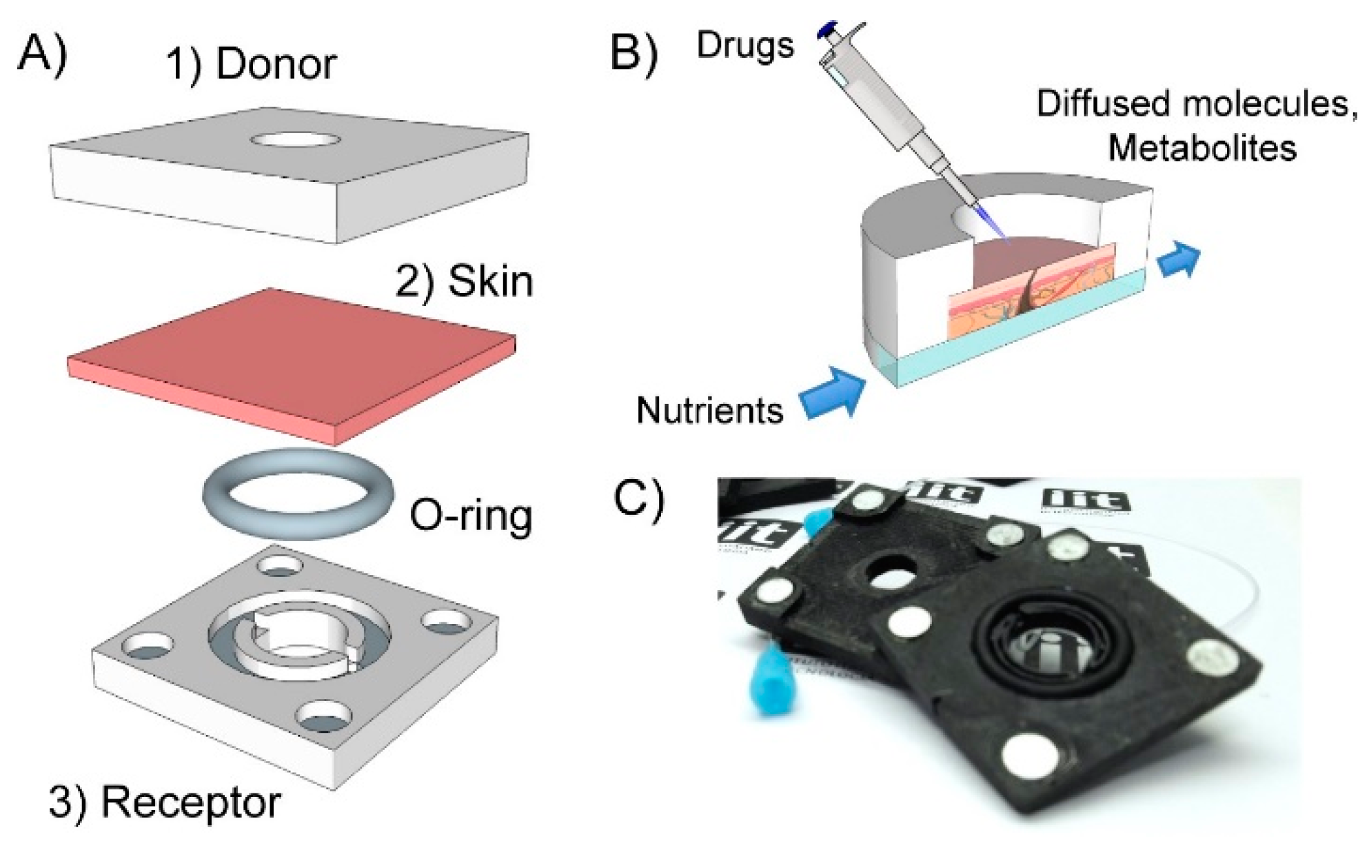
2.1. Fabrication of the 3D-PDC
Diffusion cells were fabricated by fused deposition molding (FDM) of poly-lactic acid (PLA). A commercial 3D-printer (CubePro, 3D Systems with a resolution of 100 µm) was used to extrude PLA filament through a heated nozzle (T = 220 °C). The 3D-PDC was built layer-by-layer by moving the nozzle in the XYZ-plane according to a G-code. Finally, an optically transparent cover and 4 magnets completed the device.
2.2. Skin Tissue Preparation
All experiments with animals were carried out in accordance with the guidelines of the European Communities Council (Directive 2010/63/EU) and the Italian Ministry of Health. 8–12 weeks old male CD1 mice (Charles River, Italy) were sacrificed, the back of the animal was shaved, and the full thickness skin was isolated. For in vitro viability test we employed fresh skin, whereas for the diffusion test, the explanted skin was frozen at −20 °C. Before the experiment, the skin was thawed at 37 °C for 30 min and then rinsed in phosphate buffer saline (PBS, 1x) for 30 min. For the microneedle treatment, a commercial roller (250 µm stainless steel needle, Pearl Enterprises LLC) was rolled on the epidermis 6 times in an asterisk-like direction.
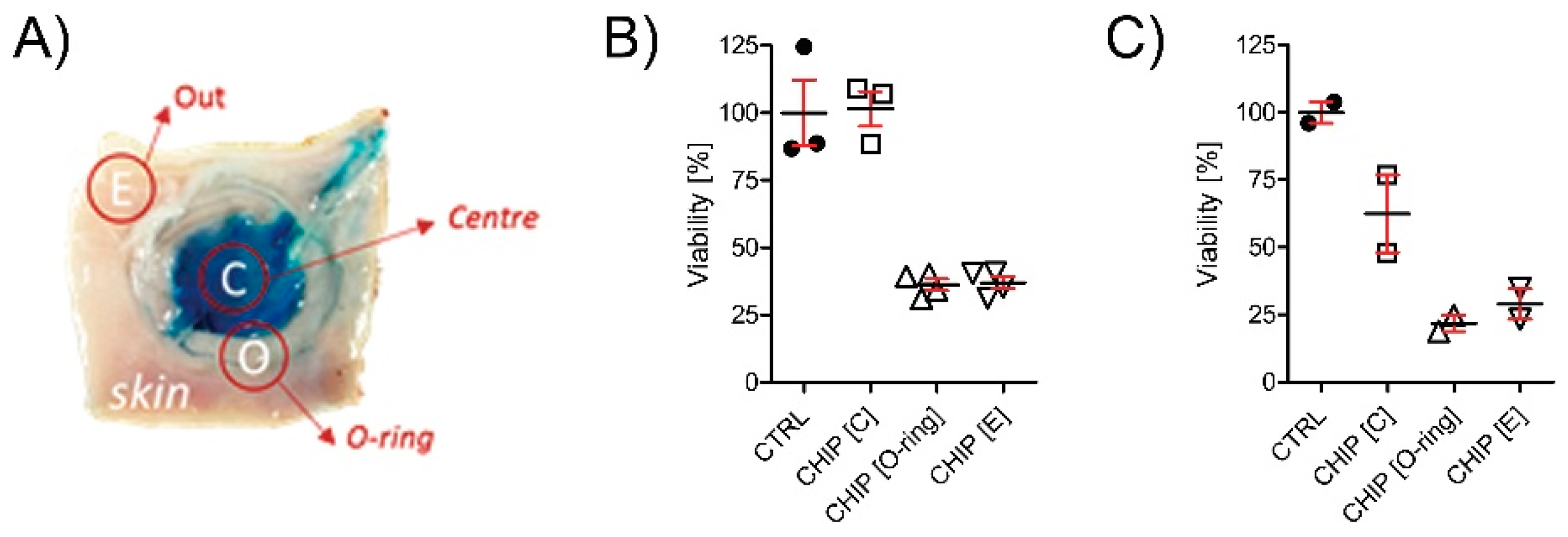
2.3. In Vitro Viability Test
Skin survival within the 3D-PDC was evaluated with Thiazolyl blue staining (MTT). Briefly, the skin was clamped between the donor and the receptor chamber, previously filled with culture media +20% Fetal Bovine Serum, and the 3D-PDC was transferred in a sterile incubator (37 °C, 5% CO2). After 12 and 24 h of incubation, the skin was cut with a 6-mm skin biopsy punch and transferred in 24 well plates with 0.5 mL of serum free media and 5% MTT reagent. After 3.5 h of incubation at 37 °C, each skin piece was fragmented, and formazan was extracted from the tissue by immersion in absolute ethanol. After 2 h of incubation under gentle shaking (dark, room temperature) the absorbance was measured at 570 and 670 nm.
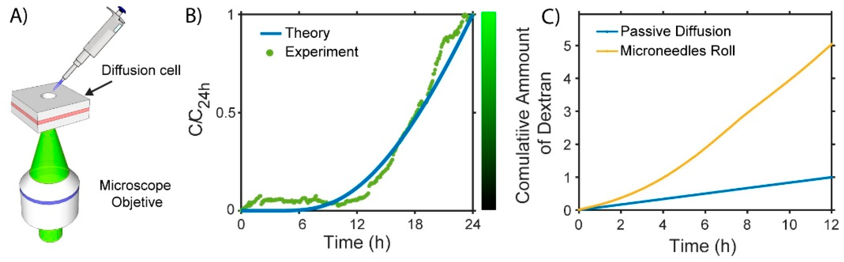
2.4. Diffusivity Analysis
Skin permeation of dextran solutions was studied though time-lapse fluorescence microscopy. A 2-mm-thick mice skin (2.5 cm × 2.5 cm) was inserted in the 3D-PDC with PBS filling the receptor. Afterwards, the 3D-PDC was positioned on a heated chamber (T = 35 °C) and fixed to the XY stage of an inverted microscope (Ti-E, Nikon, Tokyo, Japan). An external control unit (Okolab, Naples, Italy), connected to a CO2 tank and a heated water bath, provided the correct gas mixture and humidity. Finally, the donor chamber was loaded with 350 µL of a FITC-dextran solution (4 kDa Sigma Aldrich, Milan, Italy, λex = 490 nm, λem = 520 nm) at the concentration of 0.5 mg/mL. A glass cover was applied on top of the donor chamber to avoid evaporation. Time-lapse images, 1 frame every 5 min over 24 h, were collected with a microscope objective (Nikon Plan Apo 10×–0.45 NA) and a CMOS camera (Andor). A FITC filter (λcutoff = 505 nm) was used to cut off the excitation light and to capture only the fluorescence. The focal plane of the objective was 2.5 mm below the dermis.
3. Discussion
We designed, fabricated and tested a 3D-PDC for in vitro skin permeation assays. Specifically, we used FDM to fabricate the donor and receptor compartments of the device. The direct additive-manufacturing nature of this method allowed to rapidly prototype (printing time ~ 1 h) the entire device with minimal cost and, more importantly, to proper tailor the design to operate with skin membranes. As a matter of fact, once assembled, the 3D-PDC maintained the needed pressure to keep the skin stretched and avoid liquid spillage or uncontrolled perfusion during both the static and the flow-through operations. Furthermore, a simple visual inspection of the skin, up to 24 h incubation in the diffusion cell, reveled the absence of possible physical damages.
To gain a deep insight into the integrity and conditions of the skin during its incubation in the 3D-PDC, we performed viability assays. In this experiment, we studied the viability of various regions of the skin in contact with the 3D-PDC parts (
Figure 2A). Importantly, we found that the skin in the central region of the receptor chamber remained viable, presenting up to 100% and 70% viability for incubation times of 12 and 24 h, respectively (
Figure 2B,C). This result further demonstrates that the skin preserves its integrity and viability over moderately long incubation times in the 3D-PDC, thus ensuring that the data obtained during the diffusion experiments are valid and relevant. Notably, this encouraging result might extend the use of 3D-PDCs as tools for other skin-on-chip applications, such as wound healing, tissue engineering and organ-on-chip.
The 3D-PDC is compatible with a standard fluorescence microscope, as schematically depicted in
Figure 3A. This result, which is due to the pocket-size of our design, is key for the real-time detection of the concentration profile of the compound under test.
Figure 3B shows the results of the diffusion of the dextran molecules through the healthy skin membrane, as predicted by our theoretical model and investigated by time-lapse microscopy. The concentration profile is non-linear with an initial plateau up until 12 h. At this time-step the concentration starts to increase indicating that the dextran is accumulating in the receptor chamber. Remarkably, we found that the experimental profile is in really good agreement with the theoretical one that we calculated by solving the Fick’s second law for the donor/skin/receptor system. Specifically, we found that the diffusivity of the dextran in the tested skin is about 2·10−10 m2/s, in agreement with the literature on this subject.
Finally, we extended our investigation to active transdermal drug-delivery (TDD) systems. As a case study, we assessed the functionality of a microneedles-roll as vehicle to enhance the skin diffusivity of FITC-dextran.
Figure 3C shows the cumulative amount over time of dextran diffusing in the receptor for both passive and microneedle-assisted diffusion. A significant enhancement (~5 folds) in the dextran diffusion through the treated skin with respect to the untreated one is evident. This is not surprising, considering that microneedles help to cross the epidermis and accelerate the molecular diffusion. Importantly, this result clearly demonstrates that our method can be used to study and quantify the efficiency of a transdermal drug-delivery system.
4. Conclusions and Future Perspectives
3D-PDCs are suitable for in vitro skin permeation assays and quantitative assessment of TDD systems. Furthermore, the good viability of the incubated skin makes the device promising for applications where the viability of the tissue is an issue. In this regard, optimization of the current design and setup would also allow prolonged and advanced studies, such as tissue barrier diseases and wound healing.
Our compact design also offers the possibility of integrating the 3D-PDC with various detection instruments (e.g., photo-spectrometer), thus widening the potential applications of our method to other emerging fields, such as nanotoxicology, nanotherapeutics and regenerative medicine.