Proceeding Paper

Continuous Live-Cell Culture Monitoring by Compact Lensless LED Microscopes †

1
Institute of Semiconductor Technology (IHT), TU Braunschweig, 38106 Braunschweig, Germany
2
Laboratory for Emerging Nanometrology (LENA), TU Braunschweig, 38106 Braunschweig, Germany
3
Research Center for Electronics and Telecommunication, Indonesian Institute of Sciences (LIPI), 40135 Bandung, Indonesia
4
Faculty of Information Technology, Universitas Tarumanagara, 11440 Jakarta, Indonesia
5
Institute of Pharmacology, Toxicology and Clinical Pharmacy (IPT), TU Braunschweig, 38106 Braunschweig, Germany
6
Institute of Microtechnology (IMT), TU Braunschweig, 38124 Braunschweig, Germany
7
Institute for Biochemistry, Biotechnology and Bioinformatics, TU Braunschweig, 38106 Braunschweig, Germany
8
MIND-IN2UB, Department of Electronic and Biomedical Engineering, University of Barcelona, 08028 Barcelona, Spain
*
Authors to whom correspondence should be addressed.
Presented at the Eurosensors 2018 Conference, Graz, Austria, 9–12 September 2018.
Proceedings 2018, 2(13), 877; https://doi.org/10.3390/proceedings2130877
Published: 5 December 2018
(This article belongs to the Proceedings of EUROSENSORS 2018)

Abstract

:
A compact lensless microscope comprising a custom-made LED engine and a CMOS imaging sensor has been developed for live-cell culture imaging inside a cell incubator environment. The imaging technique is based on digital inline-holographic microscopy, while the image reconstruction is carried out by angular spectrum approach with a custom written software. The system was tested with various biological samples including immortalized mouse astrocyte cells inside a petri dish. Besides the imaging possibility, the capability of automated cell counting and tracking could be demonstrated. By using image sensors capable of video frame rate, time series of cell movement can be captured.

1. Introduction

Growth and maintenance of cell and tissue cultures are predominantly determined by end-point investigation. However, determining proliferation and differentiation kinetics would enable monitoring of processes, which contribute to the outcome of end-point investigations. Applying live-cell imaging to monitor cell dynamics requires highly equipped microscopes, which are extremely expensive [1]. Nevertheless, real-time in situ analysis of cell cultures is highly demanded by both research academia and industry in order to gain more insights into cell growth, including the cell dynamic behavior and morphology [2]. Therefore, miniaturized microscopes, which enable lensless real-time imaging of cells inside a conventional CO2 cell culture incubator, are a promising inexpensive alternative.
In this work, a compact and inexpensive imaging device based on digital inline-holography was built and applied to living cell cultures both outside and inside a cell incubator environment. By means of computational image analysis, key parameters (e.g., cell count, size and movement) could be extracted from the imaging data. The setup was also tested and optimized for long-term measurement scenarios and continuous online cell observation.

2. Materials and Methods

2.1. Microscopy Setup

For observation of cell cultures, a digital inline-holographic microscope [3] was built by utilizing a multi-color LED light source, a pinhole, and a CMOS sensor (Figure 1). The multi-color light source, having illumination peaks at 466 nm, 517 nm, and 629 nm, respectively, was spatially filtered by a 90 μm pinhole and positioned at a distance of 60 mm over the image sensor. The biological sample inside a petri dish was placed directly on top of the image sensor, which determines the effective sample-to-sensor distance by the thickness of the bottom of the petri dish and its refractive index. For the image sensor, an ON Semiconductor MT9P031 with a resolution of 2592 × 1944 pixels and a pixel pitch of 2.2 μm was interfaced with a controller board and connected to a PC via USB 3.0. The controlling of the light source was performed by a microcontroller, which was also connected to the measurement PC. An appropriate housing for fixing the components and allowing both inside cell incubator and stand-alone operations has been fabricated out of non-transparently dyed poly(methyl methacrylate) (PMMA) by 3D printing technique.
The image acquisition was performed by a custom written software, which is controlling the switching of the desired colors of the light source, and is saving the image automatically after brightness control of the camera has been stabilized. The data was saved locally and streamed to a cloud storage for a remote observation.

2.2. Image Reconstruction and Examination

The obtained raw image data containing a diffraction image of the illuminated sample was reconstructed by an in-house written software using the angular spectrum approach [4] allowing the extraction of both amplitude and phase information images from the microscopic data. Afterwards, cell counting based on blob detection or artificial neural network approach respectively was performed.

2.3. Samples

For testing and calibration of the developed microscope, polystyrene particles with various diameters (i.e., 1.3 μm, 5 μm, and 10 μm) as well as a USAF1951 resolution test chart were used. Meanwhile, in terms of the online measurements, immortalized mouse astrocyte cells (IMA, [5]) in agarose nutrition medium and a MIN6 cell line were investigated.

3. Results

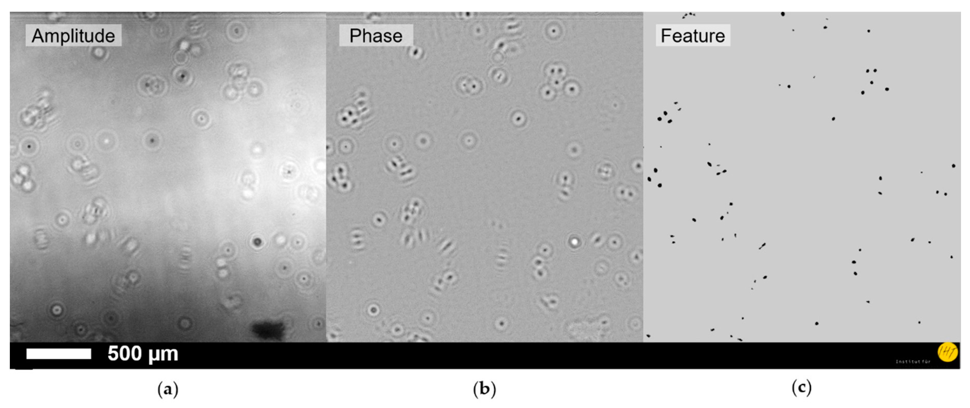
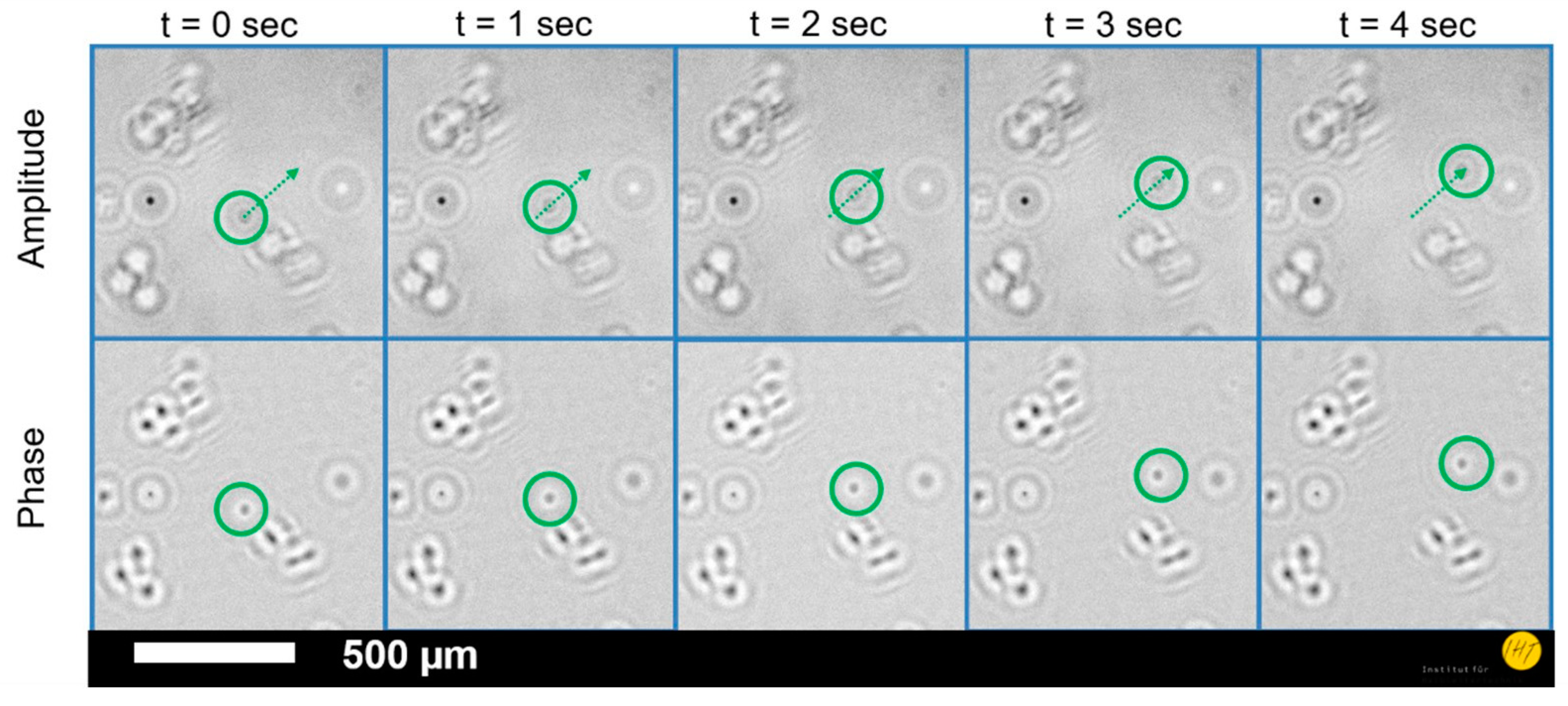
The preliminary examinations of the microscopic setup with the resolution and image quality test samples reveal a resolution down to 2.46 μm and a field-of-view of 24.4 mm². The combination of an inline-holographic microscope with living cell cultures for cell counting could be successfully performed revealing both amplitude (Figure 2a) and phase images (Figure 2b) of the culture. The images could be used for cell identification (Figure 2c) by appropriate algorithms. In case of the IMA cells, the phase image shows a better contrast than the amplitude image. Cells that appear to be blurred in the amplitude image show up clearly in the phase image. Due to the automated timed acquisition of the measurements, time series of the cell cultures could be made, revealing cell movements (Figure 3).
In order to validate the long-term operation, the microscope was placed inside a cell incubator (100% relative humidity by 37 °C and 5% CO2) for 3 days and adjusted to take a measurement automatically every 10 min with all 3 wavelengths of the light source. The data was both stored locally and streamed to a cloud storage device. Regardless of the required integration of a cooling device into the system due to the heated image sensor causing damage on the observed MIN6 cells, the developed microscope has been able to be operated without software or hardware malfunction over the whole time.

4. Discussion

The wide field-of-view, in combination with the sufficiently high resolution, can be used for the visualization and observation of cell culture growth. The difference in the amplitude and phase images allows an enhancement of the usable information content, while it also becomes apparent, that the used method has to be chosen carefully in order to avoid misleading results. The utilization of a timed acquisition software with cloud storage integration opens a path to study both cell dynamics and long-term cell observation, while the experimental setup has proven its capability to carry out such operations.

5. Conclusions

It has been demonstrated that the utilization of inline-holographic microscopy for cell culture investigation proves to be a promising way of revealing cell growth and dynamics during incubation. A system, capable of performing the task of long-term observation in combination with the acquisition of quantitative information like cell size, cell count and mobility has been presented. Nevertheless, further optimization of the system in both hardware and software is still required to completely unlock the full potential of this approach. An advantageous use of this compact microscope in combination with transparent microfluidic cell cultivation is foreseen. This can give a new insight into cell cultures, which finally can lead to new potential cure methods in medical research and therapy.

Author Contributions

Conceptualization, G.S., S.M., I.S., A.B.D. and H.S.W.; Methodology, G.S.; Software, A.B.D., I.S. and G.S.; Validation, J.H., P.H. and T.S.; Formal Analysis, A.B.D.; Investigation, I.S.; Resources, T.S., K.M., P.H., A.D., I.R., and K.H.; Data Curation, G.S.; Writing-Original Draft Preparation, G.S.; Writing-Review & Editing, J.H., J.D.P., H.S.W.; Visualization, G.S.; Supervision, H.S.W. and A.W.; Project Administration, H.S.W. and A.W.; Funding Acquisition, A.W.

Funding

This work is performed within projects of LENA-OptoSense and QUANOMET funded by the Lower Saxony Ministry for Science and Culture, Germany, as well as European project of ChipScope funded by the European Union’s Horizon 2020 research and innovation program under grant agreement No. 737089.

Acknowledgments

The authors thank K.-H. Lachmund for the technical support. S.M. thanks the Georg-Christoph-Lichtenberg PhD scholarship (Tailored Light). A.B.D. thanks Kemenristekdikti-LPDP for the Ph.D. scholarship. I.S. acknowledges RISTEKDIKTI for the Ph.D. scholarship of RISET-Pro. J.D.P. acknowledges the support of the European Unionʹs Seventh Framework Program (FP/2007-2013)/ERC Grant Agreement n. 336917, the Serra Húnter Program and the DFG Project GrK NanoMet.

Conflicts of Interest

The authors declare no conflict of interest.

References

  1. Kim, J.; Henley, B.M.; Kim, C.H.; Lester, H.A.; Yang, C. Incubator embedded cell culture imaging system (EmSight) based on Fourier ptychographic microscopy. Biomed. Opt. Express 2016, 7, 3097. [Google Scholar] [CrossRef] [PubMed]
  2. Dosch, J.; Hadley, E.; Wiese, C.; Soderberg, M.; Houwman, T.; Ding, K.; Kharazova, A.; Collins, J.L.; van Knippenberg, B.; Gregory, C.; et al. Time-lapse microscopic observation of non-dividing cells in cultured human osteosarcoma MG-63 cell line. Cell Cycle 2018, 17, 174–181. [Google Scholar] [CrossRef] [PubMed]
  3. Scholz, G.; Xu, Q.; Schulze, T.; Boht, H.; Mattern, K.; Hartmann, J.; Dietzel, A.; Scherneck, S.; Rustenbeck, I.; Prades, J.; et al. LED-Based Tomographic Imaging for Live-Cell Monitoring of Pancreatic Islets in Microfluidic Channels. Proceedings 2017, 1, 552. [Google Scholar] [CrossRef]
  4. Latychevskaia, T.; Fink, H.-W. Practical algorithms for simulation and reconstruction of digital in-line holograms. Appl. Opt. 2014, 54, 2424–34. [Google Scholar] [CrossRef] [PubMed]
  5. Schildknecht, S. Characterization of mouse cell line IMA 2.1 as a potential model system to study astrocyte functions. ALTEX 2012, 29, 261–274. [Google Scholar] [CrossRef] [PubMed]
Figure 1. Portable lensless digital-holographic imaging device for real-time cell culture monitoring: (a) schematic drawing of the measurement principle and (b) picture of the imaging setup with 3D printed mechanical housing.
Proceedings 02 00877 g001
Figure 2. Microscopic images of immortalized mouse astrocytes during cell culture process: (a) amplitude image, (b) phase contrast image, and (c) processed data for cell counting with marks indicating the detected cell positions.
Proceedings 02 00877 g002
Figure 3. Time-series (1 s step) of cell culture images, in which both amplitude and phase contrast figures show the movement of a mouse astrocyte cell detached from the culture container base.
Proceedings 02 00877 g003
Publisher’s Note: MDPI stays neutral with regard to jurisdictional claims in published maps and institutional affiliations.