Proceeding Paper

LED-Based Tomographic Imaging for Live-Cell Monitoring of Pancreatic Islets in Microfluidic Channels †

1
Institute of Semiconductor Technology (IHT), TU Braunschweig, D-38106 Braunschweig, Germany
2
Laboratory for Emerging Nanometrology (LENA), TU Braunschweig, D-38106 Braunschweig, Germany
3
Institute of Pharmacology, Toxicology and Clinical Pharmacy (IPT), TU Braunschweig, D-38106 Braunschweig, Germany
4
Institute of Microtechnology (IMT), TU Braunschweig, D-38124 Braunschweig, Germany
5
MIND-IN2UB, Department of Engineering: Electronics, University of Barcelona, E-08028 Barcelona, Spain
*
Author to whom correspondence should be addressed.
Presented at the Eurosensors 2017 Conference, Paris, France, 3–6 September 2017.
Proceedings 2017, 1(4), 552; https://doi.org/10.3390/proceedings1040552
Published: 22 August 2017
(This article belongs to the Proceedings of Proceedings of Eurosensors 2017, Paris, France, 3–6 September 2017)

Abstract

:
A portable lensless imaging device combining light-emitting diodes (LEDs) and a CMOS image sensor was developed and its suitability for non-invasive live-cell in vitro monitoring of pancreatic islets was demonstrated. A microfluidic lab-on-a-chip platform containing micro wells with various depths was also fabricated and integrated into the optical sensor system, which allows for immobilization of the single islets and continuous recording of their behavior. This promising technique may provide further insight into the structure and function of pancreatic islets and their deficiencies in type 2 diabetes.

1. Introduction

Type 2 diabetes mellitus is a strongly increasing health risk in developed and developing countries [1] and has been therefore intensively investigated by basic and clinical research. Most recent studies have been focused on the role of pancreatic islets in this disease by studying e.g., differentiation, proliferation, morphology, migration, regeneration and cell death [2]. However, although sophisticated cell imaging tools are available, they are of high cost and large dimension, which limits their usage to tabletop operation, making e.g., data collection inside a cell incubator setup infeasible. Therefore, a highly increasing demand by both research institutes and industries aims at a compact, cost-effective, non-invasive, and straightforward sensing technique, which can enable real-time cell culture monitoring, possibly even in-vivo [3]. Meanwhile, LED-technology gains enormous interest for sensing and imaging applications because of offering high brightness, wide wavelength range, low power consumption and chances for miniaturization into highly compact optical sensing systems.

2. Materials and Methods

2.1. Digital Holographic Microscope

To observe pancreatic cell islets, we realized a fully integrated, compact and portable digital holographic microscope (Figure 1) by combining an LED light engine with a CMOS image sensor and an in-house developed microfluidic perfusion system (MPS) [4]. The distance between the LED-light source and the image sensor is 59.8 mm, and the MPS is placed in direct contact to the image sensor. The MPS has a thickness of 2.2 mm, and due to the micro-channel geometry inside the MPS, the sensor-to-islet distance can be estimated to 1.2 mm. An RGB-LED with peak wavelengths of 466 nm, 517 nm, and 629 nm, respectively, was used as light source, whereas the image acquisition was performed by a Sony IMX219 CMOS-sensor with a pixel pitch of 1.12 µm and a resolution of 3280 × 2465 pixels. Control and data acquisition of the system were done using an embedded system with a Broadcom BCM2837 processor running Raspbian Linux, whereas the image processing was carried out in a regular PC. The raw image data was normalized using a reference image measured without the MPS to minimize the inhomogeneity of the optical system. The holographic images of the MPS and the islets inside were then reconstructed in the Fourier space, applying the angular spectrum method [5] to the normalized data. Afterwards, the twin image and out of focus artifacts were suppressed via twin image elimination method by Latychevskaja and Fink [6].

2.2. Optical Tomographic 3D Microscope

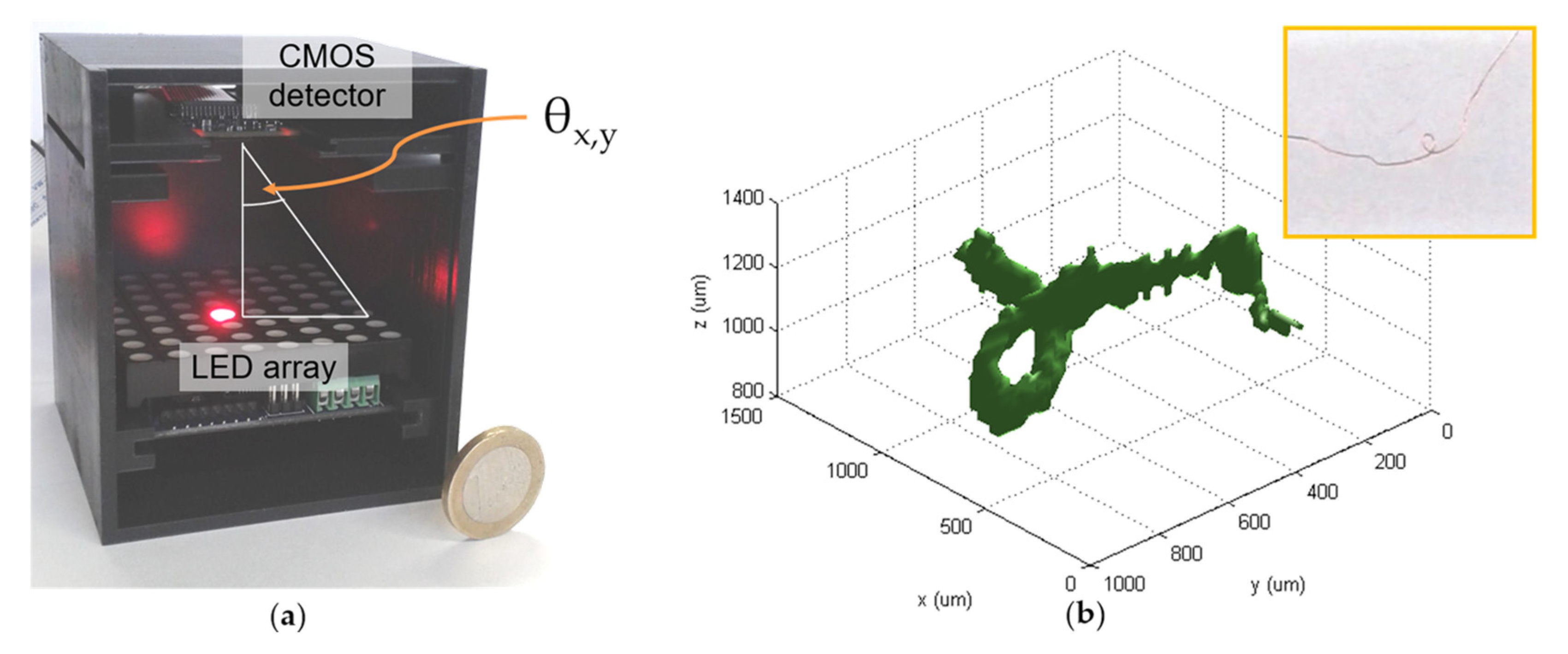
To measure the 3-dimensional (3D) shape of the pancreatic islets in relation to the micro-wells inside the MPS, the holographic setup was enhanced by utilizing an 8 × 8 pixel RGB-LED matrix (Seed Studio GTM2088RGB, controlled by a colorduino v1.4) instead of a single RGB-LED light source. The matrix was placed in 41.14 mm distance to the detector which leads to observation angles θ of up to ~29.59° in both x- and y-directions. To reconstruct the 3D object shape, a threshold function was first applied to the data, which could reduce the influence of noise and image artifacts on the result. Afterwards, the 2D images were extruded into the 3D space with respect to their angles and combined through multiplication [7].

3. Results

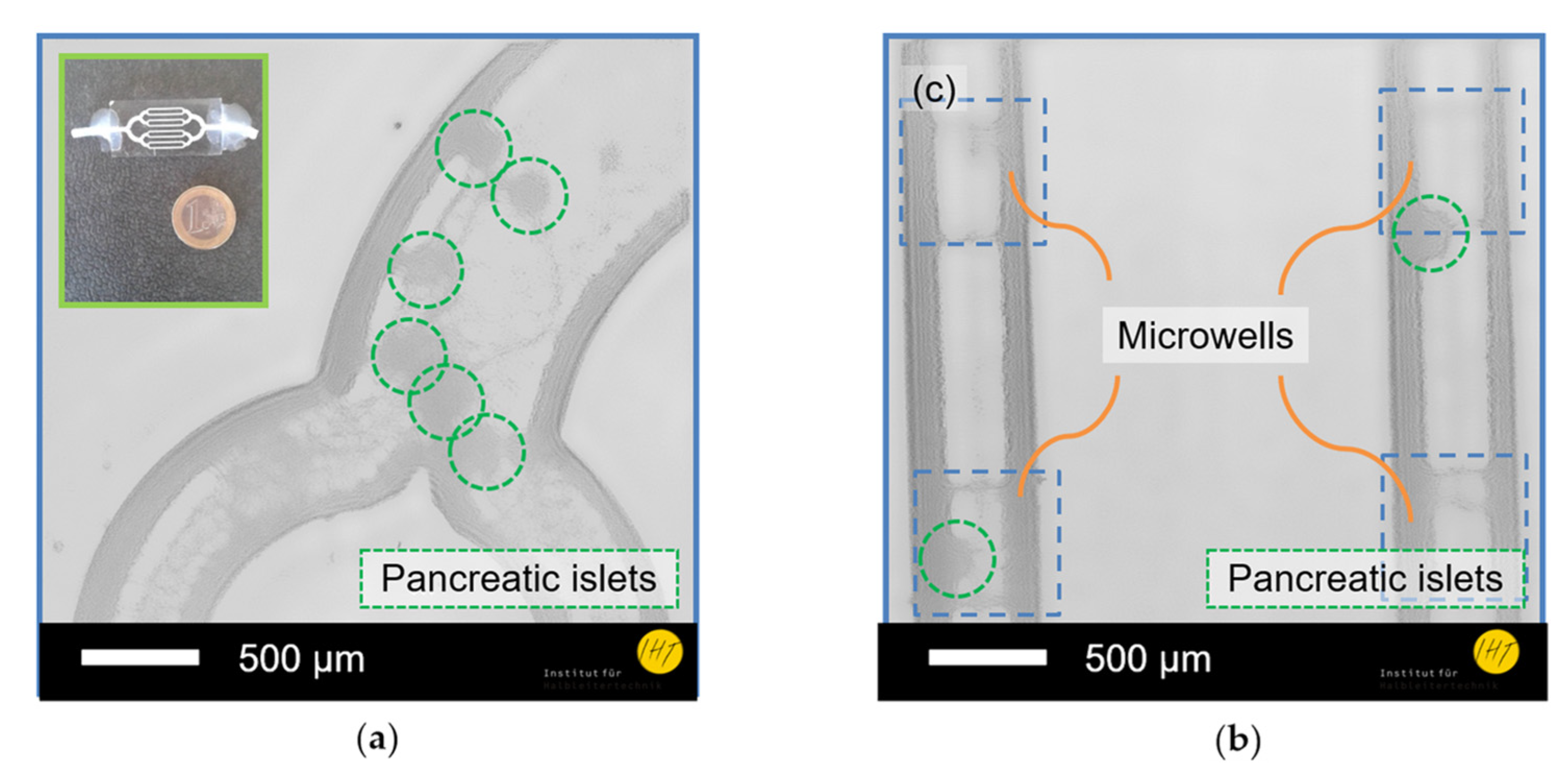
In order to test the abilities of the digital-holographic microscope, pancreatic islets, being collected for diabetes research purpose from NMRI mice through collagenase digestion technique, were inserted in Krebs-Ringer medium into a MPS [8]. Pancreatic islets of an average size of 160 ± 10 µm were monitored inside channel bifurcations (Figure 2a) and micro-wells within the channels (Figure 2b). The measured field of view was 3.67 mm × 2.76 mm, the maximum reachable resolution measured with a USAF-1951 pattern was below 2.46 µm and the position of the pancreatic islets could be imaged in relation to four micro-wells of the MPS simultaneously.
The extended setup for 3D tomographic imaging (Figure 3a) was tested by reconstructing a twisted copper wire (Figure 3b inset) mounted on glass substrate. The 3D image of the wire (Figure 3b) is capable of clearly resolving the wire and its loop inside a 3D space of 1500 µm × 1000 µm × 1400 µm.

4. Discussion

The digital inline-holographic microscope shown in this work offers a compact, lightweight, and inexpensive platform for pancreatic islet monitoring inside microfluidic devices. Both the islets themselves and the inner structure of the MPS could be distinguished in the reconstructed image, allowing for position checking of the islets. Moreover, the device is capable of imaging multiple wells in the MPS at once, offering a respectably large field of view of 10.1 mm2, which is beneficial for online measurement of multiple islets and leads to a feasible generation of large statistic data. Because of the small and lightweight setup, the system can be easily integrated into a long-term measurement setup, making interruptions of the experiment for measurement unnecessary due to online measurement. The enhanced tomographic imaging setup will allow for deeper insight into the islet morphology and into the mechanical interaction between channel and islet. On top of that, information of the islet morphology during the experiment could reveal interesting details about the process of insulin secretion as well as over the islets general condition.

5. Conclusions

Lensless imaging combined with a microfluidic perfusion device offers new opportunities for the in-situ investigation of living cells, here demonstrated with pancreatic islets. This could lead to a better understanding of the function and lifecycle of the tissues of the pancreatic system, and possibly to new ways in diagnostics and treatment of diabetes. Especially, the feasibility of directly integrating the imaging system into an incubator setup makes long-term studies and serial examination of cell cultures more feasible. The possibility of having a lightweight and accessible method for tomographic imaging provides the opportunity of an extensive study of islet morphology under different experimental conditions.

Author Contributions

G.S., Q.X., H.B., J.H., and H.S.W. developed the microscope and performed the experiments; K.M. and A.D. developed the microfluidic perfusion system (MPS); T.S., S.S., I.R. prepared for pancreatic islets; G.S. and Q.X. analyzed the data; G.S. wrote the paper; J.H., J.D.P., S.F., H.S.W., and A.W. revised the paper and provided significant inputs on LED-based imaging; H.S.W. and A.W. led the development of the optical system.

Acknowledgments

The authors thank K.-H. Lachmund for the technical support. This work is performed within projects of LENA-OptoSense and QUANOMET funded by the Lower Saxony Ministry for Science and Culture, Germany. G.S., H.S.W., and S.F. acknowledge the support from Photonik Inkubator GmbH (i.e., Superlight Photonics). H.B. thanks for the Georg-Christoph-Lichtenberg PhD scholarship (Tailored Light).

Conflicts of Interest

The authors declare no conflict of interest.

References

  1. Shaw, J.E.; Sicree, R.A.; Zimmet, P.Z. Global estimates of the prevalence of diabetes for 2010 and 2030. Diabetes Res. Clin. Pract. 2010, 87, 4–14. [Google Scholar] [CrossRef]
  2. Ashcroft, F.M.; Rorsman, P. Diabetes Mellitus and the β Cell: The Last Ten Years. Cell 2012, 148, 1160–1171. [Google Scholar] [CrossRef]
  3. Marciniak, A.; Cohrs, C.M.; Tsata, V.; Chouinard, J.A.; Selck, C.; Stertmann, J.; Reichelt, S.; Rose, T.; Ehehalt, F.; Weitz, J.; et al. Using pancreas tissue slices for in situ studies of islet of Langerhans and acinar cell biology. Nat. Protoc. 2014, 9, 2809–2822. [Google Scholar] [CrossRef]
  4. Schulze, T.; Mattern, K.; Früh, E.; Hecht, L.; Rustenbeck, I.; Dietzel, A. A 3D microfluidic perfusion system made from glass for multiparametric analysis of stimulus-secretioncoupling in pancreatic islets. Biomed. Microdevices 2017, 19, 47. [Google Scholar] [CrossRef] [PubMed]
  5. Latychevskaia, T.; Fink, H.-W. Practical algorithms for simulation and reconstruction of digital in-line holograms. Appl. Opt. 2014, 54, 2424–2434. [Google Scholar] [CrossRef] [PubMed]
  6. Latychevskaia, T.; Fink, H.-W. Solution to the Twin Image Problem in Holography. Phys. Rev. Lett. 2007, 98, 233901. [Google Scholar] [CrossRef] [PubMed]
  7. Williams, L.; Nehmetallah, G.; Banerjee, P.P. Digital tomographic compressive holographic reconstruction of three-dimensional objects in transmissive and reflective geometries. Appl. Opt. 2013, 52, 1702–1710. [Google Scholar] [CrossRef] [PubMed]
  8. Schulze, T.; Morsi, M.; Reckers, K.; Brüning, D.; Seemann, N.; Panten, U.; Rustenbeck, I. Metabolic amplification of insulin secretion is differentially desensitized by depolarization in the absence of exogenous fuels. Metabolism 2017, 67, 1–13. [Google Scholar] [CrossRef] [PubMed]
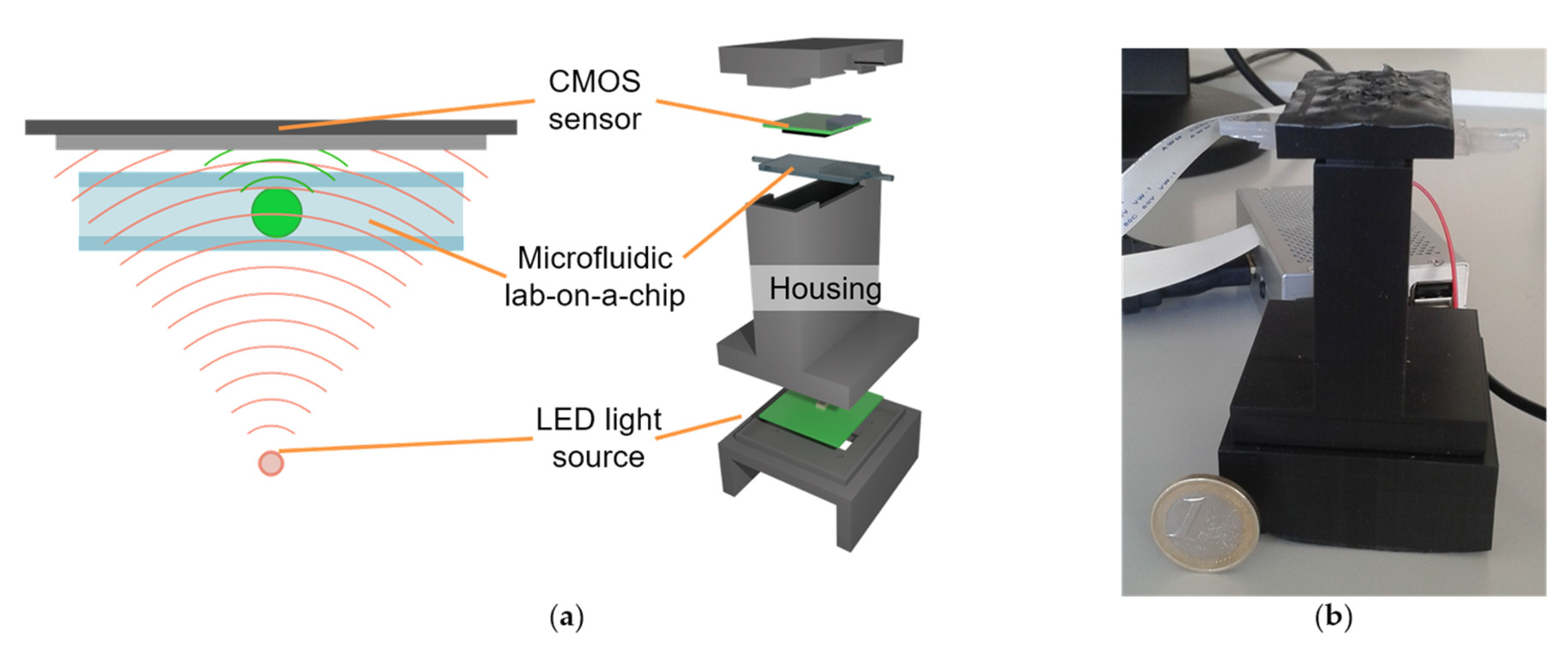
Figure 1. (a) Scheme of the digital holographic microscope showing its crucial elements; (b) Image of the described holographic microscope (with 1 € coin as a size reference).
Proceedings 01 00552 g001
Figure 2. Digital-holographic images of multiple pancreatic islets inside a microfluidic perfusion system (MPS). (a) Islets in a channel bifurcation and an image of the MPS (inset); (b) Islets inside four micro wells that are engraved into two channels.
Proceedings 01 00552 g002
Figure 3. (a) Tomographic setup consisting of RGB-LED matrix, CMOS-sensor, and custom-made 3D-printed housing; (b) Tomographic image of a twisted copper wire (photo in inset).
Proceedings 01 00552 g003
Publisher’s Note: MDPI stays neutral with regard to jurisdictional claims in published maps and institutional affiliations.