Proceeding Paper

Detection of Adrenaline Based on Bioelectrocatalytical System to Support Tumor Diagnostic Technology †

1
Institute of Nano- and Biotechnologies (INB), FH Aachen, Campus Jülich, Heinrich-Mußmann-Street 1, 52428 Jülich, Germany
2
Department of Pharmaceutical Chemistry, Philipps-University Marburg, Marbacher Weg 6-10, 35037 Marburg, Germany
3
Division of Endocrinology and Metabolism, Rostock University Medical Center, Ernst-Heydemann-Street 6, 18057 Rostock, Germany
4
Peter Grünberg Institute (PGI-8, Bioelectronics), Forschungszentrum Jülich GmbH, Wilhelm-Johnen-Street 6, 52425 Jülich, Germany
*
Author to whom correspondence should be addressed.
Presented at the Eurosensors 2017 Conference, Paris, France, 3–6 September 2017.
Proceedings 2017, 1(4), 506; https://doi.org/10.3390/proceedings1040506
Published: 7 August 2017
(This article belongs to the Proceedings of Proceedings of Eurosensors 2017, Paris, France, 3–6 September 2017)

Abstract

:
An amperometric biosensor based on the bioelectrocatalytic measurement principle for the detection of adrenaline has been developed. The adrenaline sensor has been prepared by modification of a platinum thin-film electrode with a pyrroloquinoline quinone-dependent glucose dehydrogenase. The enzyme was immobilized via cross-linking method. Lower detection limit of 1 nM of adrenaline has been achieved by measuring at physiological level at pH 7.4.

1. Introduction

Adrenal vein sampling (AVS) is the method of choice for characterizing aldosterone-producing adenoma of the adrenal gland and to distinguish unilateral from bilateral diseases [1,2]. While patients with bilateral disease are treated with life-long medication, patients with unilateral disease are offered adrenalectomy which leads to significant improvement and has the potential of cure [3,4]. However, AVS is an invasive and laborious technique because both adrenal veins must be sampled for meaningful comparison. Furthermore, placing the catheter tip within the right adrenal vein is quite challenging. Therefore, measuring a specific biomarker during catheterization could reduce X-ray exposure time and enhance result quality of the medical examination. In this case, adrenal veins’ adrenaline (epinephrine) concentrations can be measured to confirm successful catheterization of the adrenal veins: there is a gradient of the adrenaline concentration between adrenal blood (>100 nM) and peripheral blood (~1 nM) that can be employed as an indicator for correct catheter tip position.
Several sensors have been proposed for the detection of adrenaline or other catecholamines by using recycling processes for achieving signal amplification and to improve lower detection limit [5,6,7,8,9]. However, the optimum working pH of the described sensors does not comply with the pH value of blood that limits their application for the adrenaline detection in real blood samples.
In this contribution, a high-sensitive adrenaline thin-film biosensor based on the bioelectrocatalytic measurement principle has been realized by using a pyrroloquinoline quinone (PQQ)-dependent glucose dehydrogenase (GDH), which is stable in a broad pH range. Figure 1 shows the bioelectrocatalytical amplification principle. The recycling system implies an amplification of the sensor signal. The results of investigation of adrenaline detection in buffer at physiological level of pH 7.4 are presented.

2. Materials and Method

2.1. Chemicals

The enzyme GDH was bought from Sorachim SA (Switzerland, 757 U/mg) [10] and PQQ was purchased from Wako (Japan). Glutaraldehyde, bovine serum albumin (BSA), glycerol, CaCl2 and the phosphate buffer (PBS) components (monosodium phosphate and disodium phosphate) were obtained from Sigma-Aldrich. Adrenaline solution (1 mg/mL) was purchased from Infectopharm (Germany). Adrenaline stock solution of 1 µM and 100 µM were stored at 4 °C in the dark.

2.2. Preparation of the Adrenaline Biosensor

The adrenaline biosensor was realized by modification of a platinum thin-film electrode with an enzyme membrane (see Figure 2). The sensor chip was fabricated by means of conventional silicon and thin-film technologies. For the preparation of the enzyme membrane, 4 µL of GDH solution solved in PBS, together with 20 µM PQQ, 1 mM CaCl2 [11], 8 µL of BSA (10 vol %) and 8 µL of a glutaraldehyde (2 vol %)/glycerol (10 vol %) solution were mixed. The membrane cocktail was then dropped onto the platinum electrode. After drying, the chip was stored at 4 °C until required.

2.3. Electrochemical Sensor Characterization

For the electrochemical characterization, the adrenaline biosensor was connected to a potentiostat (PalmSens, Palm Instruments BV, The Nertherlands). A three-electrode arrangement was used, where a conventional liquid-junction Ag/AgCl electrode (Metrohm, Herisau, Switzerland) was utilized as a reference electrode and a platinum wire as a counter electrode. For the oxidation reaction of adrenaline, a constant potential of +450 mV vs. Ag/AgCl was applied to the platinum working electrode and the resulting current was recorded over time.

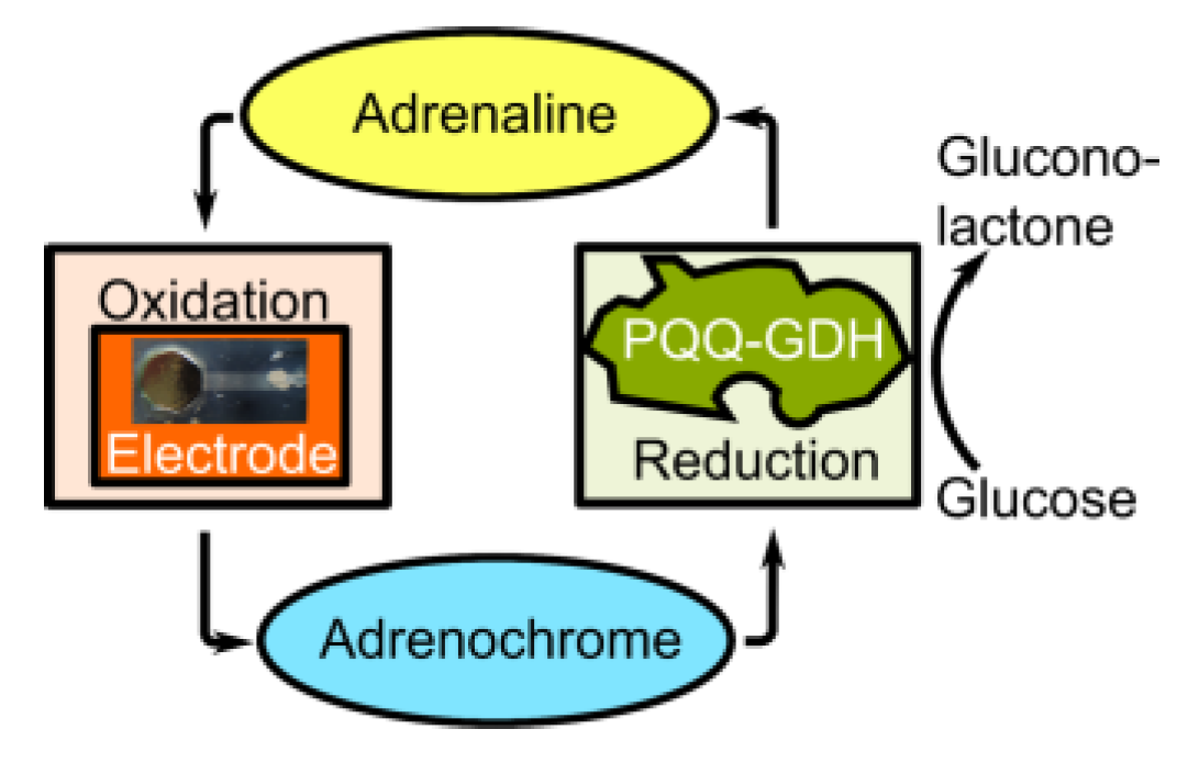
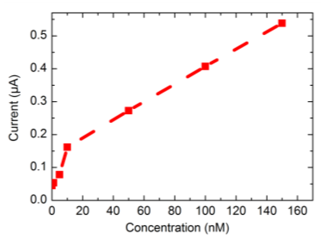
3. Results and Discussion

Figure 3 depicts the calibration curve of the developed adrenaline biosensor. Different adrenaline concentrations in the range of 1 nM to 150 nM were measured in buffer solution of pH 7.4 in the presence of 20 mM glucose. A lower detection limit of ~1 nM adrenaline could be reached.

4. Conclusions and Outlook

In this work, an amperometric thin-film biosensor for the detection of adrenaline has been presented. Using the bioelectrocatalysis measurement principle for the signal amplification, a lower detection limit of 1 nM in buffer solution at physiological level of pH 7.4 has been achieved. Ongoing experiments focus on the investigation of this biosensor in Ringers’ solution as well as real blood samples. We see potential for application in adrenal venous sampling for adrenal gland disease, for catheter-based detection of paragangliomas and also in stress-research.

Acknowledgments

This research is supported by a Ph.D. scholarship from FH Aachen.

Conflicts of Interest

The authors declare no conflict of interest. The founding sponsors had no role in the design of the study; in the collection, analyses, or interpretation of data; in the writing of the manuscript, and in the decision to publish the results.

References

  1. Rossi, G.P.; Auchus, R.J.; Brown, M.; Lenders, J.W.M.; Naruse, M.; Plouin, P.F.; Satoh, F.; Young, W.F. An expert consensus statement on use of adrenal vein sampling for the subtyping of primary aldosteronism. Hypertension 2013, 63, 151–160. [Google Scholar] [CrossRef] [PubMed]
  2. Funder, J.W.; Carey, R.M.; Mantero, F.; Murad, M.H.; Reincke, M.; Shibata, H.; Stowasser, M.; Young, W.F., Jr. The management of primary aldosteronism: Case detection, diagnosis, and treatment: An endocrine society clinical practice guideline. J. Clin. Endocrinol. Metab. 2016, 101, 1889–1916. [Google Scholar] [PubMed]
  3. Catena, C.; Colussi, G.; Nadalini, E.; Chiuch, A.; Baroselli, S.; Lapenna, R.; Sechi, L.A. Cardiovascular outcomes in patients with primary aldosteronism after treatment. Arch. Intern. Med. 2008, 168, 80–85. [Google Scholar] [CrossRef] [PubMed]
  4. Stowasser, M.; Gordon, R.D. Primary aldosteronism—Careful investigation is essential and rewarding. Mol. Cell. Endocrinol. 2004, 217, 33–39. [Google Scholar] [CrossRef] [PubMed]
  5. Scheller, F.; Siegbahn, N.; Danielsson, B.; Mosbach, K. High-sensitivity enzyme thermistor determination of L-lactate by substrate recycling. Anal. Chem. 1985, 57, 1740–1743. [Google Scholar] [CrossRef]
  6. Cybulski, D.; Male, K.B.; Scharer, J.M.; Moo-Young, M.; Luong, J.H.T. Substrate recycling scheme for tetrachloro-p-benzoquinone using bilirubin oxidase and NADH: Application for pentachlorophenol assay. Environ. Sci. Technol. 1999, 33, 796–800. [Google Scholar] [CrossRef]
  7. Scheller, F.W.; Bauer, C.G.; Makower, A.; Wollenberger, U.; Warsinke, A.; Bier, F.F. Coupling of immunoassays with enzymatic recycling electrodes. Anal. Lett. 2001, 34, 1233–1245. [Google Scholar] [CrossRef]
  8. Mizutani, F.; Yabuki, S.; Asai, M. Highly-sensitive measurement of hydroquinone with an enzyme electrode. Biosens. Bioelectron. 1991, 6, 305–310. [Google Scholar] [CrossRef]
  9. Eremenko, A.; Makower, A.; Jin, W.; Rüger, P.; Scheller, F. Biosensor based on an enzyme modified electrode for highly-sensitive measurement of polyphenols. Biosens. Bioelectron. 1995, 10, 717–722. [Google Scholar] [CrossRef]
  10. Glucose-Dehydrogenase (PQQ-Dependent). Available online: http://www.sorachim.com/enzymes/glucose-dehydrogenase-pqq-dependent.html (accessed on 2 August 2017).
  11. Laurinavicius, V.; Razumiene, J.; Ramanavicius, A.; Ryabov, A.D. Wiring of PQQ-dehydrogenases. Biosens. Bioelectron. 2004, 20, 1217–1222. [Google Scholar] [CrossRef] [PubMed]
Figure 1. Scheme of bioelectrocatalytical measurement principle for amplification of the adrenaline sensor signal. In a first reaction step, adrenaline is oxidized to adrenochrome. Then, reduction of adrenochrome was catalyzed by the enzyme PQQ-GDH.
Proceedings 01 00506 g001
Figure 2. Adrenaline thin-film biosensor arrangement with enzyme membrane.
Proceedings 01 00506 g002
Figure 3. Calibration curve of adrenaline biosensor in the concentration range of 1 nM–150 nM.
Proceedings 01 00506 g003
Publisher’s Note: MDPI stays neutral with regard to jurisdictional claims in published maps and institutional affiliations.

