Advancing Dental Diagnostics: A Review of Artificial Intelligence Applications and Challenges in Dentistry
Abstract
1. Introduction
- Providing a schema for the current X-ray imaging literature;
- Reviewing the state-of-art of AI models used in dental practice;
- Analyzing the possibilities, limitations, and future trends in using artificial intelligence in dentistry.
2. Materials and Methods
2.1. Protocol
2.2. Electronic Search Strategy
- Peer-reviewed journal articles;
- Conference papers;
- Review articles;
- Clinical studies;
- Case studies;
- Technical reports;
- Theses and dissertations.
2.3. Eligibility Criteria
2.3.1. Inclusion Criteria
- I.
- Timeline: Publications over the past 14 years (2010–2023) focused on the application of artificial intelligence, deep learning, and neural networks in dentistry;
- II.
- Language: All English-language publications were incorporated, regardless of the location of publication;
- III.
- Data and Outcome: Studies that adequately explain the datasets exploited, supported by explicit reporting of predictive and measurable outcomes to measure the effectiveness of the suggested model.
2.3.2. Exclusion Criteria
- I.
- Type of data used: research that does not provide precise details on the kinds of data that were used;
- II.
- Methodology: inadequately documented research on deep learning, machine learning, and computer vision techniques;
- III.
- Outcome: Studies that failed to document quantifiable results.
2.4. Study Selection and Items Collected
3. Challenges in Automated Dental Disease Diagnosis
- The significant quantity of data needed for training, validation, and testing is a significant obstacle in deploying artificial intelligence (AI) for dental caries detection. This involves extremely sensitive patient data, such as dental pictures and medical histories. Data sensitivity is a critical issue due to the personal and private nature of health information, which includes identifiable patient records and medical histories. The misuse of or unauthorized access to this data can lead to severe ethical and privacy violations. Ensuring data privacy and security is paramount to maintaining the trust of patients and upholding ethical standards in AI applications for dentistry diagnostics. Strict adherence to data protection regulations, such as GDPR, and implementing robust encryption and anonymization techniques are essential to safeguarding patient information [5].
- One major concern revolves around the transparency of artificial intelligence algorithms and data. The reliability of AI systems’ predictions greatly depends on the accuracy of annotations and labeling in the training dataset. Inaccurately labeled data can lead to subpar outcomes. This issue is especially pronounced in clinic-labeled datasets, where the lack of consistent quality further limits the transparency and effectiveness of AI systems [6].
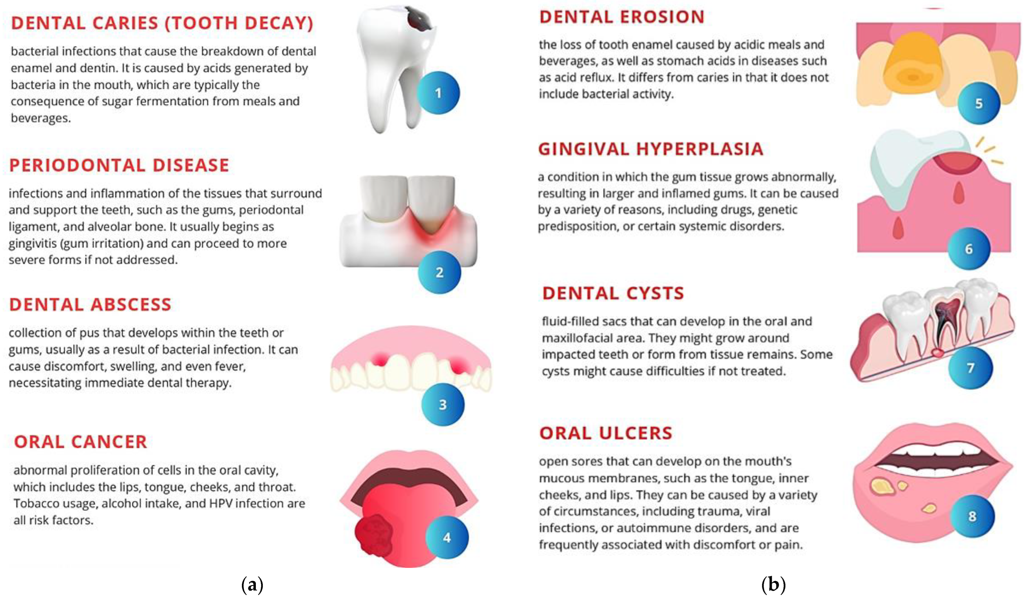
4. Types of Dental Diseases
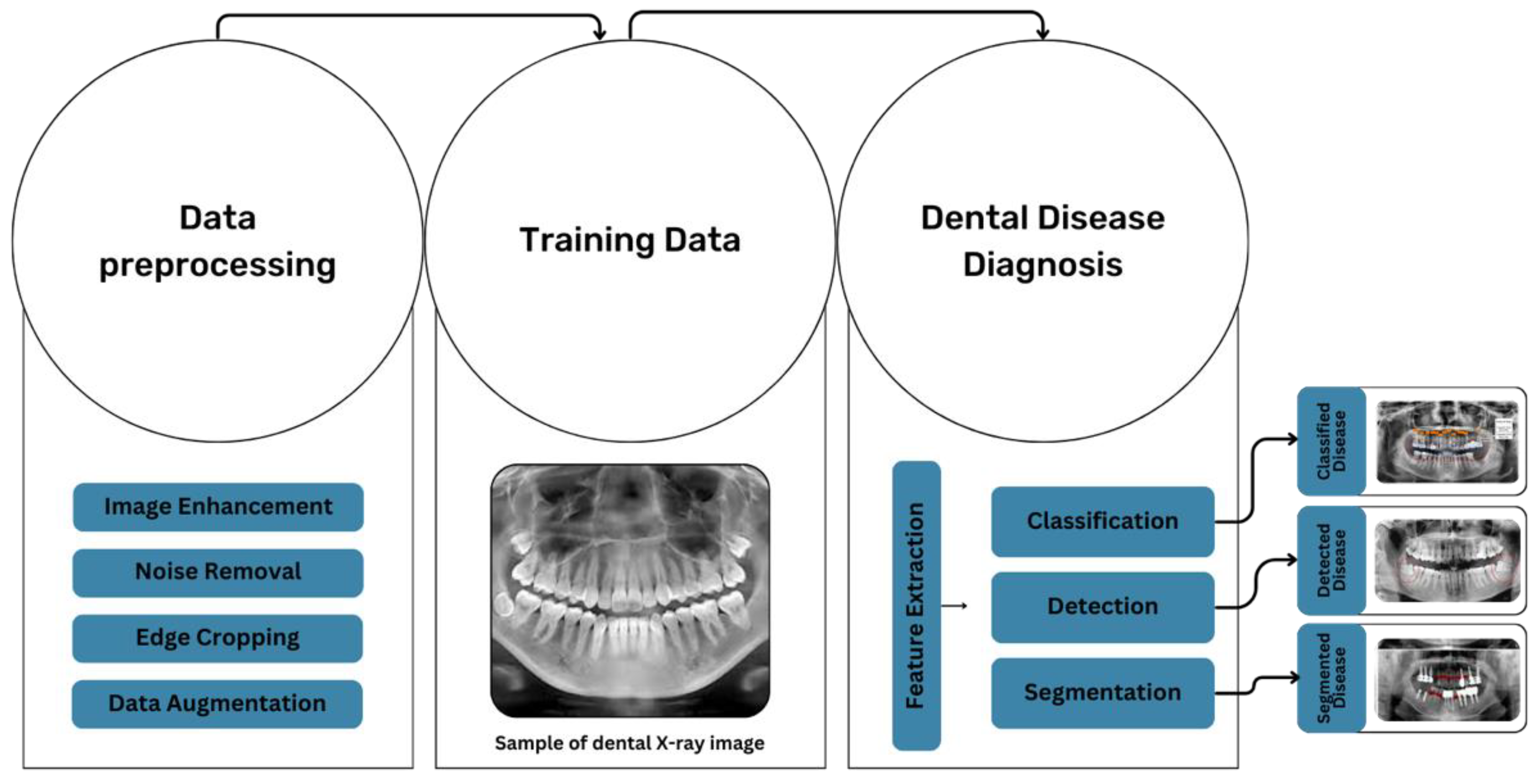
5. Approaches to Diagnosing Dental Diseases through X-ray Imaging
6. Preprocessing Techniques for Dental X-ray Pictures
7. Datasets Used for AI-Based Systems for Dentistry E-Health
8. Evolution of AI-Diagnostic Tools in Dentistry
8.1. Convolutional Neural Networks (CNNs)
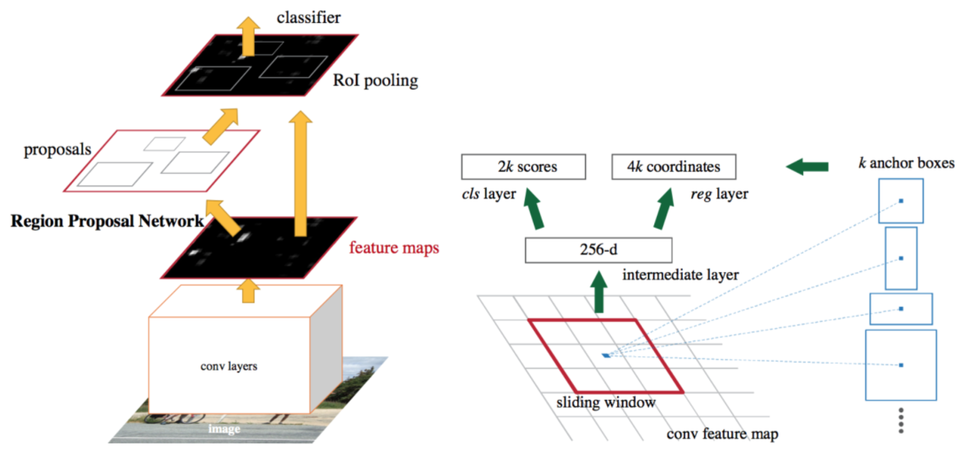
8.1.1. Faster R-CNN
8.1.2. ResNet
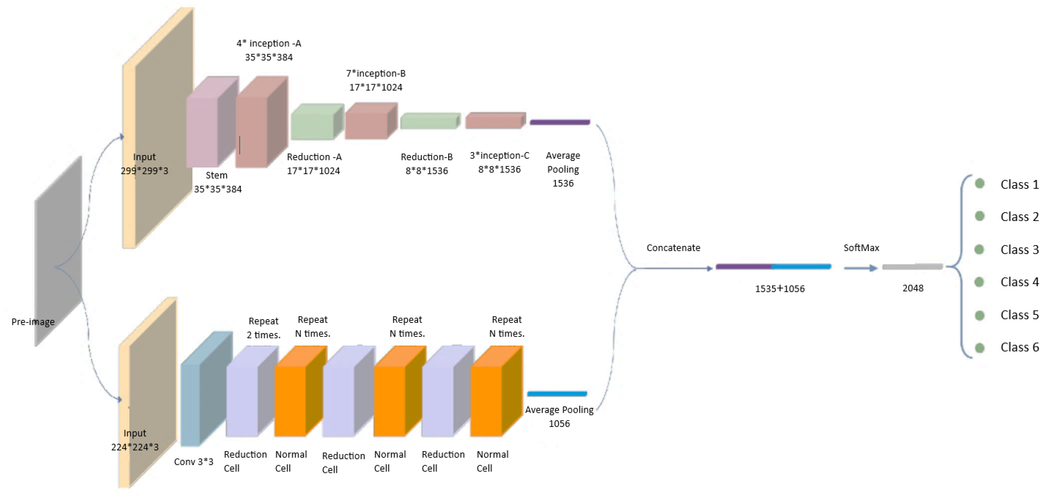
8.2. NASNetMobile
8.3. YOLOv3
9. Relevant Work Experiences
10. Role of X-ray Imaging in Dental Diagnostics
11. Impact of AI in Dental Healthcare
12. Knowledge Gaps and Future Research Directions
13. Conclusions
Author Contributions
Funding
Data Availability Statement
Conflicts of Interest
References
- Shan, T.; Tay, F.R.; Gu, L. Application of Artificial Intelligence in Dentistry. J. Dent. Res. 2021, 100, 232–244. [Google Scholar] [CrossRef] [PubMed]
- Martins, M.V.; Baptista, L.; Luís, H.; Assunção, V.; Araújo, M.-R.; Realinho, V. Machine Learning in X-ray Diagnosis for Oral Health: A Review of Recent Progress. Computation 2023, 11, 115. [Google Scholar] [CrossRef]
- Mahdi, S.S.; Battineni, G.; Khawaja, M.; Allana, R.; Siddiqui, M.K.; Agha, D. How Does Artificial Intelligence Impact Digital Healthcare Initiatives? A Review of AI Applications in Dental Healthcare. Int. J. Inf. Manag. Data Insights 2023, 3, 100144. [Google Scholar] [CrossRef]
- Shafi, I.; Fatima, A.; Afzal, H.; Díez, I.d.l.T.; Lipari, V.; Breñosa, J.; Ashraf, I. A Comprehensive Review of Recent Advances in Artificial Intelligence for Dentistry E-Health. Diagnostics 2023, 13, 2196. [Google Scholar] [CrossRef] [PubMed]
- Anil, S.; Porwal, P.; Porwal, A. Transforming Dental Caries Diagnosis Through Artificial Intelligence-Based Techniques. Cureus 2023, 15, 7. [Google Scholar] [CrossRef] [PubMed]
- Lee, J.H.; Kim, D.H.; Jeong, S.N.; Choi, S.H. Use of artificial intelligence in dentistry: Current clinical trends and research advances. J. Can. Dent. Assoc. 2021, 87, 1488–2159. [Google Scholar]
- Tuan, T.M.; Duc, N.T.; Van Hai, P. Dental Diagnosis from X-Ray Images using Fuzzy Rule-Based Systems. Int. J. Fuzzy Syst. Appl. 2017, 6, 1–16. [Google Scholar]
- Lee, J.H.; Kim, D.H.; Jeong, S.N.; Choi, S.H. Detection and diagnosis of dental caries using a deep learning-based convolutional neural network algorithm. J. Dent. 2018, 77, 106–111. [Google Scholar] [CrossRef] [PubMed]
- Jader, G.; Fontineli, J.; Ruiz, M.; Abdalla, K.; Pithon, M.; Oliveira, L. Deep Instance Segmentation of Teeth in Panoramic X-Ray Images. In Proceedings of the 2018 31st SIBGRAPI Conference on Graphics, Patterns and Images (SIBGRAPI), Parana, Brazil, 29 October 2018–1 November 2018; pp. 400–407. [Google Scholar]
- Tuzoff, D.; Tuzova, L.; Bornstein, M.; Krasnov, A.; Kharchenko, M.; Nikolenko, S.; Sveshnikov, M.; Bednenko, G. Tooth detection and numbering in panoramic radiographs using convolutional neural networks. Dentomaxillofacial Radiol. 2019, 48, 20180051. [Google Scholar] [CrossRef] [PubMed]
- Chen, H.; Zhang, K.; Lyu, P.; Li, H.; Zhang, L.; Wu, J.; Lee, C.H. A deep learning approach to automatic teeth detection and numbering based on object detection in dental periapical films. Sci. Rep. 2019, 9, 3840. [Google Scholar] [CrossRef]
- Geetha, V.; Aprameya, K.S. Dental Caries Diagnosis in X-ray Images using KNN Classifier. Indian J. Sci. Technol. 2019, 12, 5. [Google Scholar] [CrossRef]
- Vinayahalingam, S.; Xi, T.; Bergé, S.; Maal, T.; De Jong, G. Automated detection of third molars and mandibular nerve by deep learning. Sci. Rep. 2019, 9, 9007. [Google Scholar] [CrossRef] [PubMed]
- Wang, Y.; Sun, L.; Zhang, Y.; Lv, D.; Li, Z.; Qi, W. An Adaptive Enhancement Based Hybrid CNN Model for Digital Dental X-ray Positions Classification. arXiv 2020, arXiv:2005.01509. [Google Scholar]
- You, W.; Hao, A.; Li, S.; Wang, Y.; Xia, B. Deep learning-based dental plaque detection on primary teeth: A comparison with clinical assessments. BMC Oral Health 2020, 20, 141. [Google Scholar] [CrossRef] [PubMed]
- Chung, M.; Lee, J.; Park, S.; Lee, M.; Lee, C.E.; Lee, J.; Shin, Y.-G. Individual tooth detection and identification from dental panoramic X-ray images via point-wise localization and distance regularization. Artif. Intell. Med. 2021, 111, 101996. [Google Scholar] [CrossRef] [PubMed]
- Muresan, M.P.; Barbura, A.R.; Nedevschi, S. Teeth detection and dental problem classification in panoramic X-ray images using deep learning and image processing techniques. In Proceedings of the IEEE 16th International Conference on Intelligent Computer Communication and Processing (ICCP), Cluj-Napoca, Romania, 3–5 September 2020; pp. 457–463. [Google Scholar]
- Sonavane, A.; Yadav, R.; Khamparia, A. Dental cavity classification of using convolutional neural network. IOP Conf. Ser. Mater. Sci. Eng. 2021, 1022, 012116. [Google Scholar] [CrossRef]
- Huang, Y.P.; Lee, Y.S. Deep Learning for Caries Detection using Optical Coherence Tomography. medRxiv 2021. [CrossRef]
- Diniz de Lima, E.; Souza Paulino, J.A.; Lira de Farias Freitas, A.P.; Viana Ferreira, J.E.; Silva Barbosa, J.S.; Bezerra Silva, D.F.; Meira Bento, P.; Araújo Maia Amorim, A.M.; Pita Melo, D. Artificial intelligence and infrared thermography as auxiliary tools in the diagnosis of temporomandibular disorder. Dentomaxillofacial Radiol. 2021, 51, 20210318. [Google Scholar] [CrossRef] [PubMed]
- Muramatsu, C.; Morishita, T.; Takahashi, R.; Hayashi, T.; Nishiyama, W.; Ariji, Y.; Zhou, X.; Hara, T.; Katsumata, A.; Ariji, E.; et al. Tooth Detection and Classification on Panoramic Radiographs for Automatic Dental Chart Filing: Improved Classification by Multi-Sized Input Data. Oral Radiol. 2021, 37, 13–19. [Google Scholar] [CrossRef]
- Imak, A.; Celebi, A.; Siddique, K.; Turkoglu, M.; Sengur, A.; Salam, I. Dental Caries Detection Using Score-Based Multi-Input Deep Convolutional Neural Network. IEEE Access 2022, 10, 18320–18329. [Google Scholar] [CrossRef]
- Kühnisch, J.; Meyer, O.; Hesenius, M.; Hickel, R.; Gruhn, V. Caries Detection on Intraoral Images Using Artificial Intelligence. J. Dent. Res. 2022, 101, 158–165. [Google Scholar] [CrossRef] [PubMed]
- Almalki, Y.E.; Din, A.I.; Ramzan, M.; Irfan, M.; Aamir, K.M.; Almalki, A.; Alotaibi, S.; Alaglan, G.; Alshamrani, H.A.; Rahman, S. Deep Learning Models for Classification of Dental Diseases Using Orthopantomography X-ray OPG Images. Sensors 2022, 22, 7370. [Google Scholar] [CrossRef] [PubMed]
- AL-Ghamdi, A.; Ragab, M.; AlGhamdi, S.; Asseri, A.; Mansour, R.; Koundal, D. Detection of Dental Diseases through X-Ray Images Using Neural Search Architecture Network. Comput. Intell. Neurosci. 2022, 2022, 3500552. [Google Scholar] [CrossRef] [PubMed]
- Hung, K.F.; Ai, Q.Y.H.; King, A.D.; Bornstein, M.M.; Wong, L.M.; Leung, Y.Y. Automatic Detection and Segmentation of Morphological Changes of the Maxillary Sinus Mucosa on Cone-Beam Computed Tomography Images Using a Three-Dimensional Convolutional Neural Network. Clin. Oral Investig. 2022, 26, 3987–3998. [Google Scholar] [CrossRef] [PubMed]
- Zhou, X.; Yu, G.; Yin, Q.; Liu, Y.; Zhang, Z.; Sun, J. Context Aware Convolutional Neural Network for Children Caries Diagnosis on Dental Panoramic Radiographs. Comput. Math. Methods Med. 2022, 2022, 6029245. [Google Scholar] [CrossRef]
- Sunnetci, K.M.; Ulukaya, S.; Alkan, A. Periodontal Bone Loss Detection Based on Hybrid Deep Learning and Machine Learning Models with a User-Friendly Application. Biomed. Signal Process. Control 2022, 77, 103844. [Google Scholar]
- Zhu, J.; Chen, Z.; Zhao, J.; Yu, Y.; Li, X.; Shi, K.; Zhang, F.; Yu, F.; Shi, K.; Sun, Z.; et al. Artificial Intelligence in the Diagnosis of Dental Diseases on Panoramic Radiographs: A Preliminary Study. BMC Oral Health 2023, 23, 358. [Google Scholar] [CrossRef]
- Lira, P.; Giraldi, G.; Neves, L.A. Segmentation and Feature Extraction of Panoramic Dental X-Ray Images. In Nature-Inspired Computing Design, Development, and Applications; IGI Global: Hershey, PA, USA, 2010; Volume 1, pp. 306–320. [Google Scholar]
- Xie, X.; Wang, L.; Wang, A. Artificial neural network modelling for deciding if extractions are necessary prior to orthodontic treatment. Angle Orthod. 2010, 80, 262–266. [Google Scholar] [CrossRef]
- ALbahbah, A.A.; El-Bakry, H.M.; Abd-Elgahany, S. Detection of Caries in Panoramic Dental X-ray Images. Int. J. Electron. Commun. Comput. Eng. 2016, 7, 250–256. [Google Scholar]
- Na’am, J.; Harlan, J.; Madenda, S.; Wibowo, E.P. Image Processing of Panoramic Dental X-Ray for Identifying Proximal Caries. TELKOMNIKA (Telecommun. Comput. Electron. Control) 2017, 15, 702–708. [Google Scholar] [CrossRef]
- Leite, A.F.; Vasconcelos, K.F.; Willems, H.; Jacobs, R. Radiomics and machine learning in oral healthcare. PROTEOMICS—Clin. Appl. 2023, 14, 1900040. [Google Scholar]
- López-Janeiro, Á.; Cabañuz, C.; Blasco-Santana, L.; Ruiz-Bravo, E. A tree-based machine learning model to approach morphologic assessment of malignant salivary gland tumors. Ann. Diagn. Pathol. 2021, 56, 151869. [Google Scholar] [CrossRef] [PubMed]
- Rodrigues, J.A.; Krois, J.; Schwendicke, F. Demystifying artificial intelligence and deep learning in dentistry. Braz. Oral Res. 2021, 35. [Google Scholar] [CrossRef] [PubMed]
- Babu, A.; Onesimu, J.A.; Sagayam, K.M. Artificial Intelligence in dentistry: Concepts, Applications and Research Challenges. E3S Web Conf. 2021, 297, 01074. [Google Scholar]
- Subbotin, A. Applying Machine Learning in Fog Computing Environments for Panoramic Teeth Imaging. In Proceedings of the 2021 XXIV International Conference on Soft Computing and Measurements (SCM), St. Petersburg, Russia, 26–28 May 2021; pp. 237–239. [Google Scholar] [CrossRef]
- Patil, S.; Albogami, S.; Hosmani, J.; Mujoo, S.; Kamil, M.A.; Mansour, M.A.; Abdul, H.N.; Bhandi, S.; Ahmed, S.S.S.J. Artificial Intelligence in the Diagnosis of Oral Diseases: Applications and Pitfalls. Diagnostics 2022, 12, 1029. [Google Scholar] [CrossRef] [PubMed]
- De Angelis, F.; Pranno, N.; Franchina, A.; Di Carlo, S.; Brauner, E.; Ferri, A.; Pellegrino, G.; Grecchi, E.; Goker, F.; Stefanelli, L.V. Artificial Intelligence: A New Diagnostic Software in Dentistry: A Preliminary Performance Diagnostic Study. Int. J. Environ. Res. Public Health 2022, 19, 1728. [Google Scholar] [CrossRef]
- Rattan, D. Panoramic Dental Xray Dataset, Kaggle. 2021. Available online: https://www.kaggle.com/datasets/daverattan/dental-xrary-tfrecords (accessed on 2 June 2024).
- Pushkara, A. Teeth_Dataset, Kaggle. 2020. Available online: https://www.kaggle.com/datasets/pushkar34/teeth-dataset (accessed on 2 June 2024).
- Hassani, H.; Amiri Andi, P.; Ghodsi, A.; Norouzi, K.; Komendantova, N.; Unger, S. Shaping the future of smart dentistry: From Artificial Intelligence (AI) to Intelligence Augmentation (IA). IoT 2021, 2, 510–523. [Google Scholar] [CrossRef]


| Refs. | Authors, (Year) | Aim | Classifiers | Measurement | Dataset | Size | Preprocessing | Feature Extraction | Result | Strength | Weakness |
|---|---|---|---|---|---|---|---|---|---|---|---|
| [25] | Abdullah S. AL-Malaise AL-Ghamdi et al., (2022) | To classify dental X-ray images into three categories: cavities, fillings, and implants. The data were divided into training and validation sets | NASNet | Accuracy | Using Kaggle, Panoramic Dental Xray Dataset [41] | 116 | Data augmentation by utilizing several operations, such as scaling, rotation, translation, Gaussian blur, and Gaussian noise | NASNet, AlexNet, CNN | After they applied the data augmentation, the dataset became 245 and the accuracy of NASNet, AlexNet, and CNN increased. Accuracy of the model was 96.51% with data augmentation and 93.36% without augmentation | They mentioned that the approach’s success was due to the restricted training sample. High accuracy | They focused only on three categories |
| [7] | Tran Manh Tuan, Hai V. Pham et al., (2017) | To create a dental diagnosis tool that relies on fuzzy rule-based systems and machine learning approaches to assist dentists in making decisions based on X-ray pictures and patient symptoms | Fuzzy rules | Accuracy | Taken from Hanoi Medical University | 56 | Entropy, edge-value, intensity, and local binary patterns—LBP, gradient feature, and patch-level feature to understand images such as texture, density, etc. | Input variables such as entropy, edge, intensity (EEI), LBP, RGB, gradient, and patch | The method, which included feature extraction, clustering, fuzzy rule creation, and fuzzy inference, outperformed the FKNN algorithm in terms of accuracy. The article also suggested future study and development directions. Accuracy 90.29% | The FCM clustering technique aided in the grouping of comparable features. More accurate than FKNN. Depended on rules | Dataset quality and quantity, referring to labels like low and high, rather than percentage |
| [17] | Mircea Paul Muresan, Andrei Rzvan Barbura, Sergiu Nedevschi, (2020) | To detect dental issues using panoramic radiography in order to acquire an accurate diagnosis and categorize the condition using X-ray pictures | CNN, majority vote technique | Accuracy, recall, precision, and F1-score | Used dataset from three different clinics | About 1000 | The pictures were cropped to remove metadata, then the image was is increased to 2048 × 1024 pixels, and the image was assigned to one of 14 problem types | \ CNN (ERFNet) | The proposed method performed well in classifying dental issues compared with 2 alternatives. Accuracy of 89% | Focusing on 14 dental diseases, there was capacity to effectively segment teeth and diagnose dental disorders | Could work to increase accuracy |
| [32] | Ainas A. ALbahbah, Hazem M. El-Bakry, Sameh Abd-Elgahany, (2016) | To analyze radiological images to diagnose dental decay, with the goal of being more effective than earlier studies | Artificial neural network (ANN) with a three-layer network structure and a sigmoid function | The error percentage was used to assess performance. Precision was used to define the level of measurement consistency. ROC stands for receiver operating characteristic. CE stands for cross-entropy | N/A | N/A | Image resizing. The upper and lower jaws and teeth were extracted using segmentation. Cropping was a technique used to extract the area of interest. Pictures were converted to RGB before being thresholder to produce binary pictures. Enhancement | Histogram of oriented gradient (HOG) | Results were assessed using the error rate, accuracy, ROC curves, and cross entropy. The results showed that the neural network model was good at differentiating between healthy and decaying teeth | Rule-based computer-assisted systems fell short of the performance of the suggested neural network-based approach. Complex problems and variances in patients’ teeth could be handled | There was no information about the dataset and exact numerical results were not mentioned; also, the study was old |
| [27] | Xiaojie Zhou, Guoxia Yu, Qiyue Yin, Yan Liu, Zhiling Zhang, and Jie Sun, (2022) | This study intended to advance and enhance the diagnosis of tooth decay in panoramic radiographs using CNN, especially in children | CNN based on ResNet | Accuracy, precision, AUC (area under the curve), and recall | Beijing Children’s Hospital, Capital Medical University, and the National Center for Children’s Health all contributed | 210 | Panoramic radiographs were used to extract individual teeth, which were subsequently dissected out using special instruments. The teeth’s sizes and forms varied as inputs. The database was purged of teeth that were absent from the image. Training, validation, and testing were the three categories into which the database was separated | CNN model | The context-aware CNN model performed better than the typical CNN baseline in terms of accuracy, precision, recall, F1-score, and AUC. The percentage points obtained varied from 5 to 7. The model reduced diagnostic time, although doctors remained superior in specific instances | The model reduced diagnostic time | N/A |
| [9] | Gil Jader, Jefferson Fontinele, Marco, et al., (2018) | It intended to employ deep learning (Mask R-CNN) to separate teeth from panoramic X-ray pictures | R-CNN, R-CNN, and FCN | Evaluation metrics, like recall, accuracy, precision, F1-score, and specificity | Taken from MSCOCO dataset | 1500 | N/A | Mask R-CNN learned features from data | The segmentation system based on the R-CNN mask produced promising results with high accuracy, outperforming unsupervised segmentation approaches | The power of R-CNN on image segmentation. Diversity of image display in addition to excellent quality | There was no publication date for this article. Ambiguity in data pre-processing procedure |
| [16] | Minyoung Chung, Jusang Lee, Sanguk Park, et al., (2020) | The goal was to create a CAD system for dental panoramic X-ray pictures that would help professionals to locate and identify individual teeth | CNN-based, ResNet, DLA, and stacked hourglass networks | Average precision (AP), AP50, AP75 measurements, and (mIoU), are used to assess tooth detection accuracy | N/A | 818 | Used CLAHE for standardized contrast | Convolutional neural networks (CNNs) | The approach achieved accuracy in tooth detection and could thus be used in clinical situations | Center shifts multi-task training improved translation accuracy. The system worked efficiently without the need for other filtration methods | Accuracy percentage was not mentioned |
| [30] | Pedro H. M. Lira and Gilson A. Giraldi, (2010) | Creating an automated segmentation approach for extracting dental X-ray images | Does not explicitly mention, but it could be PCA and image-processing techniques | Crown-body (CB), root (R) lengths, and the CB/R ratio | N/A | N/A | Quadtree decomposition, low-pass filtering, Otsu’s thresholding, and XOR operations | Shape models, PCA, and feature vector computing were used for tooth border identification | The experimental findings were discussed, and the procedure was applied to the teeth to eliminate interference using X-ray pictures | Eliminated the necessity for jaw separation for tooth segmentation and included a mechanism for identifying tooth borders | Overlapping teeth in X-ray images. Accuracy and the dataset were not mentioned |
| [33] | Jufriadif Na`am, Johan Harlan, Sarifuddin Madenda, Eri Prasetyo Wibowo, (2017) | The study’s purpose was to improve image processing quality for detecting proximal caries in panoramic dental X-ray images | N/A | Identifying proximal caries and their severity | N/A | N/A | Removing undesirable regions from X-ray pictures | Multiple morphological gradient (mMG) | Displaying the results using mMG technology, which helped to clarify the edges to determine where the caries was | Using technology improved the accuracy of identifying caries, which is difficult to spot with just your eyes | Missing a lot of data, such as performance in relation to the method of determining caries, the classifiers used, and from where the images were used |
| [38] | Alexey N. Subbotin, (2021) | The goal of this article was to use panoramic dental imaging, machine learning, fog computing environments, and cloud technologies to enhance the accuracy of dental diagnoses and increase the pace of dentists’ work | The use of machine learning for diagnosing tooth damage is mentioned in the article. However, it does not clarify which classifiers were utilized | Measuring the reduction. Increased processing speed for X-ray pictures | N/A | N/A | panoramic dental Pimage acquisition. Use of fog computing environments | N/A | According to the report, the number of patients sent for follow-up therapy decreased by 7.24%, while X-ray image processing speed increased by 13.93% | The system’s concept was important since it tried to enhance diagnostic time by utilizing fog computing methods, machine learning, and cloud technologies | Lacked a lot of information and particular specifics regarding the machine learning system’s classifiers, dataset size, and feature extraction methods |
| [8] | Jae-Hong Lee et al., (2018) | This study sought to determine the usefulness of deep CNN algorithms for detecting and diagnosing dental caries on periapical radiographs | Pre-trained GoogLeNet Inception v3 CNN network was used for preprocessing, and the datasets were trained using transfer learning | Accuracy | From dental hospital’s PACS system (Infinitt PACS, invented by Infinitt Co., Seoul, Korea) | 3000 | Resized to 299 × 299 pixels and converted into JPEG file format. All maxillary teeth images were reverted to the mandibular teeth form through a vertical flip | There were 22 deep strata. We employed nine inception modules, including an auxiliary classifier, two fully linked layers, and softmax | The diagnosis accuracy was 88.0% for molars, 89.0% for premolars, and 82.0% for combined premolars and molars | Utilization of deep CNN algorithm. Robust evaluation metrics | The dataset included permanent teeth only. Resolution of images. Exclusion of clinical parameters |
| [21] | Chisako Muramatsu et al., (2021) | The goal of this research was to create a computerized method for detecting and classifying teeth in dental panoramic radiographs, allowing for automatic structured creation of dental charts. It could also serve as a preprocessing step for computerized image analysis of dental problems | CNN-based method | Sensitivity and false positives | Collected at Asahi University Hospital | 100 | This preprocessing approach used the segmentation result of the lower mandible contour to identify the approximate location of the teeth. By restricting the size of the supplied pictures, the exclusion of the additional dental region reduced the number of false positive detections | Features were extracted from each input picture using convolution and residual layers before merging them | The tooth detection sensitivity was 96.4%, with 0.5 false positives per instance. The categorization accuracy for tooth kinds and conditions was 93.2% and 98.0%, respectively | The suggested technology may automatically analyze dental charts for forensic identification and pre-screening for dental diseases | 1, Small data set. 2, They did not include third molars in this study |
| [18] | Sonavane, A. et al., (2021) | In this study, we concentrated on identifying cavities | CNN-based method | Accuracy | Kaggle dataset [42] | 74 | The dataset contained images in the JPEG format. We utilized the ImageDat-aGenerator from keras.preprocessing.image in Python. Validation was performed using 20% of the training images and random horizontal flips. We utilized a random zoom range of 0.2 for our model | N/A | The maximum accuracy is was.43%. Increasing the dataset size improved model accuracy | Introduced a promising mobile application for users to capture dental images, offering rapid assessment | Small data set |
| [14] | Yaqi Wang, et al., (2020) | To develop an automatic dental X-ray detection method that utilized adaptive histogram equalization and a hybrid multi-convolution neural network (CNN) | Hybrid multi-CNN model to classify six different locations of dental slices | Accuracy, specificity, and the area under curve (AUC) | Source of dataset is not mentioned | 2491 | Three preprocessing strategies were used in this study to improve the precision of dental X-ray detection. These consisted of adaptive histogram equalization, median filtering, and image sharpening | The feature extraction process involved using two different networks, NASNetMobile and Inception V4, to generate eigenvectors for each image | The test set’s accuracy was over 90%, and the findings showed that the suggested strategy produced good accuracy, with an AUC of 0.97. The algorithm’s performance was also contrasted with the comments offered by four dentists, demonstrating how well it located teeth | To increase the accuracy of dental X-ray detection, the suggested method combined preprocessing techniques and deep learning models. The work addressed issues with image rotation and tooth position intersection. The effectiveness of the procedure was fully evaluated by the evaluation criteria utilized (accuracy, specificity, and AUC) | Good accuracy, it would be helpful to provide further information on any potential restrictions or difficulties encountered during the investigation. The particular neural network architectural specifications, such as the number of layers and activation functions, were not published, which could be crucial for replication and further study |
| [22] | ANDAC IMAK, et al., (2022) | To present a unique approach for the automatic detection of dental caries using periapical pictures. The researchers wanted to solve the limits of dentists’ manual diagnosis | MI-DCNNE | Accuracy, sensitivity, specificity, precision, and F1-score. These metrics provide a comprehensive assessment of the model’s performance in dental caries detection | These images were obtained from private oral and dental health clinics using periapical radiography devices | 340 | During the preprocessing stage, image processing techniques were used to improve the raw periapical pictures. Using a sharpening filter and altering intensity settings to increase contrast and highlight problematic regions are examples of this | CNN | It was seen that the proposed model was quite successful in the diagnosis of dental caries. The reported accuracy score was 99.13%. This result showed that the proposed MI-DCNNE model could effectively contribute to the classification of dental caries | 1, Good accuracy. 2, The algorithms enabled autonomous feature extraction, decreasing the need for manual feature engineering. 3, The incorporation of a score-based ensemble technique improved the model’s robustness | 1, The paper did not go into detail on the architecture of the deep CNN. 2, The study’s dataset may have special characteristics that limit the model’s generalizability to other demographics or therapeutic contexts |
| [12] | V. Geetha and K. S. Aprameya, (2019) | This work employed machine learning to diagnose dental caries in radiographs | KNN | Accuracy, precision, false positive rate, receiver operating characteristic (ROC) | The photos were obtained at SJM Dental College in India utilizing a Gendex X-ray equipment equipped with a Sirona RVG sensor | 49 | The dental X-ray images were converted to bmp format using MATLAB conversion tool application. After the conversion, resized to 256 x 256 of class double. The resultant image was enhanced using the Laplacian filter | GLCM technique. The extracted features included contrast, correlation, energy, homogeneity, mean and entropy | The algorithm achieved 98.5% accuracy, 98.5% precision, 4.7% false positive rate, and a ROC curve area of 0.953 using 10-fold cross validation. The results were validated using two-way ANOVA at a significance level of 5% | The proposed method’s key advantage was its ease of implementation, rapid computation scheme, and ease of operation | Broader validation with a larger and more diverse dataset would enhance the reliability and applicability of the proposed method |
| [24] | Yassir Edrees Almalki et al., (2022) | This study proposed using the YOLOv3 deep learning model for automated dental problem detection | YOLOv3 | Mean average precision (mAP), F1-score, precision, sensitivity, and intersection over union (IOU) | Some OPGs taken with a DSLR camera, whereas others obtained from clinics | 1200 | Rotation range Zoom range Shear range Horizontal flip | CNN-based | The trained model YOLOv3 was tested on test images after training and achieved an accuracy of 99.33% | The paper presented a novel way to detecting dental problems using deep learning | The method’s limitation was the small sample size of only four types of illnesses, which did not represent the entire population |
| [19] | Yu-Ping Huang and Shyh-Yuan Lee, (2022) | To avoid this issue and emphasize the significance of high-quality data, two-phase research was conducted to evaluate several approaches for detecting caries. Initially, five experienced doctors compared caries detection based on OCT and apical radiography | CNN | Accuracy, specificity, sensitivity (PPV), and (NPV) | From National Yang-Ming University | 100 | Self-developed OCT, periapical films, and Micro-CT for dental assessment | Convolutions with tiny kernels are typically used to extract local features like edges, impulses, and noise in images | The accuracy was 95.21%. The sensitivity was 98.85%, specificity was 89.83%, and PPV and NPV were 93.48% and 98.15%, respectively | While this study has limitations, it might nonetheless serve as a platform for additional research in related topics | The study’s main shortcoming was the manual verification process |
| [34] | André Ferreira Leite, Karla de Faria Vasconcelos, Holger Willems, and Reinhilde Jacobs, (2020) | To present an overview of artificial intelligence (AI) in the field of dental and maxillofacial radiology (DMFR), as well as to address possible uses, difficulties, and future views | Convolutional neural networks (CNNs) | Accuracy, specificity, sensitivity, and area under curve (AUC) | Various sources, including public datasets and private collections | Varis, with 2400 | Data augmentation techniques, such as cropping, adding noise, mirroring, and others to increase the dataset size | Deep learning models, particularly CNNs, automatically extracted features from the input data | AI approaches, particularly deep learning, demonstrated encouraging results in a variety of dental imaging tasks, with some cases obtaining accuracies comparable to expert-level dentists | The study presents a complete review of AI in DMFR currently, highlighting both its potential benefits and limitations. It emphasizes AI’s transformational potential in disease diagnosis, treatment planning, and prediction | There are issues related to the mathematical processes behind AI that might impede radiologists’ interpretation of outcomes. The availability of huge data on dental pictures and the construction of a ground truth for validating AI outcomes |
| [29] | Junhua Zhu, Zhi Chen, Jing Zhao, et al., (2023) | The purpose was to create an artificial intelligence (AI) framework for diagnosing various dental illnesses using panoramic radiographs (PRs) using deep convolutional neural networks | CNNs, specifically two models: BDU-Net and nnU-Net | Diagnosing multiple dental diseases on panoramic radiographs with respect to sensitivity, specificity, (AUC), and diagnostic time | Stomatology Hospital of Zhejiang Chinese Medical University | 1996 | Image resampling, image normalization, image spacing, and patch size setting | CNNs: BDU-Net and nnU-Net | The sensitivity, specificity, and AUC are specified respectively: For impacted teeth, 0.964, 0.996, 0.960, and 0.980. For full crowns, 0.953, 0.998, 0.951, and 0.975. For residual roots, 0.871, 0.999, 0.870, and 0.935. For missing teeth, 0.885, 0.994, 0.879, and 0.939 For caries, 0.554, 0.990, 0.544, and 0.772 | The study compared the efficacy of an AI framework for detecting numerous dental disorders on PRs to that of dentists with varied levels of experience. Across several disorders, the framework displayed great specificity | N/A |
| [12] | Dmitry V. Tuzoff, Lyudmila N. Tuzova, Michael M. Bornstein, Alexey. et al., (2019) | To analyze dental radiographs and provide a solution based on convolutional neural networks (CNNs) to recognize and count teeth in panoramic radiographs | Faster R-CNN and VGG-16 CNN for teeth numbering | Sensitivity and precision for teeth detection; sensitivity and specificity | Panoramic radiographs of adults | 1574 | N/A | CNN-based feature extraction from the radiographs | The system had a sensitivity of 0.9941 and a precision of 0.9945 for tooth detection. The sensitivity and specificity of tooth numbering were 0.9800 and 0.9994, respectively | The research described a unique technique for a practical dentistry application that used cutting-edge CNN architectures to achieve results comparable to expert levels | The preprocessing procedures were not explained in the snippets supplied, which might be critical for understanding the whole technique and repeating the results |
| [23] | J. Kühnisch, O. Meyer, et al., (2022) | To create a deep learning system for identifying caries from intraoral pictures using CNNs, and to compare their diagnostic performance with expert standards | Convolutional neural networks (CNNs) | Sensitivity, specificity, and area under the receiver operating characteristic (ROC) curve (AUC) | Anonymized photographs from permanent teeth | 2417 | Image augmentation, transfer learning, normalization to compensate for under- and overexposure | Utilized MobileNet2 architecture for the CNN, which used inverted residual blocks. | When all test photos were reviewed, the CNN correctly identified cavities in 92.5% of cases | The study used AI to diagnose cavities and reached a high accuracy rate, suggesting the promise of AI in dental diagnostics | The paper admitted that the present strategy could be improved further, implying that there may be limits in the methodology or the AI model utilized |
| [31] | Xiaoqiu Xiea, Lin Wangb, Aming Wangc, (2010) | To construct a decision-making expert system for orthodontic treatment using artificial neural networks (ANNs) to decide if extractions are necessary for patients aged 11 to 15 | Artificial neural networks (ANNs) with back-propagation (BP) model | Anterior teeth uncovered by incompetent lips, IMPA (L1-MP), and FMA (FH-MP), and the accuracy for the constructed artificial neural network was 80% | Data from patients aged 11–15 who visited their department from 1999 to 2005 | 200 | Encoding and normalization of non-quantification indices and quantification indices | 23 indices, including factors like cast measurements, cephalometry, and growth | The created ANN was 80% accurate in identifying therapy for malocclusion children aged 11–15 | Comprehensive technique employing ANN to anticipate the need for extractions, perhaps providing orthodontists with a beneficial tool | This study is limited to a specific age group (11–15 years old) and may not be generalizable to other age groups or orthodontic problems |
| [11] | Hu Chen, Kailai Zhang, Peijun Lyu, Hong Li, Ludan Zhang, Ji Wu, Chin-Hui Lee, (2019) | To create a deep learning-based system for automatically detecting and counting teeth in dental X-ray images, thereby increasing the efficiency and accuracy of dental diagnostics | A deep convolutional neural network (CNN) for classification, specifically the Faster R-CNN model | Precision, recall, and intersection-over-union (IOU) | Dental periapical films collected from Peking University School and Hospital of Stomatology | 1250 | The input photos were scaled to keep their original aspect ratio, with a minor dimension of 300 pixels | Deep learning using Faster R-CNN for object detection and feature extraction from dental X-ray images | The results demonstrated that precisions and recalls approached 90%, with an average IOU value of 91% between designated boxes and ground facts. Machines are on par with younger dentists | Deep learning techniques, particularly the faster R-CNN, were used effectively in this research to obtain excellent precision and recall rates. The findings were equivalent to a junior dentist’s performance, demonstrating the viability of the suggested technology in practical applications | While the results were remarkable, the work may benefit from testing on a larger dataset or including more real-world events. Furthermore, the dependence on a single deep learning model (faster R-CNN) may limit its future adaptation to newer or other architectures |
| [15] | Shankeeth Vinayahalingam, Tong Xi, et al., (2019) | Creating an automated system that uses deep learning to detect and segment the M3 and IAN on dental panoramic radiographs (OPG) | CNN based on U-net architecture | Dice coefficient, sensitivity, and specificity | Orthopantomograms (OPGs) of patients from the Department of Oral and Maxillofacial Surgery of Radboud University Nijmegen Medical Centre | 81 OPGs | Image acquisition, data pre-processing, standardizing size, and applying contrast enhancement to the OPG | N/A | Mean dice coefficients for M3s and IAN were 0.947 ± 0.033 and 0.847 ± 0.099, respectively | Deep learning is used effectively in this work to address a key clinical obstacle, yielding encouraging results in the automated identification and segmentation of third molars and IAN | The research did not go into detail on feature extraction; however, issues such as lack of contrast on OPGs and heterogeneity in mandibular canal shape influenced segmentation performance |
| [26] | Kuo Feng Hung, Qi Yong H. Ai, Ann D. King, Michael M. Bornstein, Lun M. Wong, and Yiu Yan Leung, Clinical Oral Investigations volume, (2022) | To develop and evaluate a CNN method for the automated detection and segmentation of MT and MRCs in the maxillary sinus using low-dose and full-dose cone-beam CT (CBCT) | Detection classifier, segmentation classifier | Areas under the curves (AUCs) and Dice similarity coefficient (DSC) | Maxillary sinuses | 890 | Image normalization, noise reduction, image registration, data augmentation | CNN algorithm was constructed using V-Net and supports vector regression | The findings reveal that the proposed CNN algorithm was very accurate in identifying and segmenting MT and MRCs in the maxillary sinus on both low-dose and full-dose CBCT images. The algorithm’s performance did not vary considerably between the two imaging techniques | High accuracy. Comparable performance. Segmentation capability | Manual segmentation. Limited sample size. Lack of external validation |
| [35] | Álvaro López-Janeiro, Clara Cabañuz, Luis Blasco-Santana, Elena Ruiz-Bravo, (2021) | To develop a machine learning algorithm for the diagnosis of malignant salivary gland tumors. The researchers aimed to improve the diagnostic performance in this challenging field of pathology by applying machine learning techniques | Recursive partitioning algorithm | Morphological variables | Commonly encountered malignant salivary gland tumors | 115 cases | Data cleaning Feature scaling Feature selection or dimensionality reduction Data splitting Cross-validation | Assessing and quantifying specific morphological characteristics associated with malignant salivary gland tumors | The machine learning algorithm successfully guided the morphological approach to diagnosing malignant salivary gland tumors. It achieved high classification accuracy, identified relevant morphological variables, and demonstrated consistent misclassification patterns. The algorithm shows promise as a diagnostic tool to improve the accuracy of diagnosing these challenging tumors | Improved diagnostic performance, consistent misclassification pattern, relevant morphological variables, and inter-observer concordance | Limited sample size, lack of external validation, limited histologic types, interpretability of the classification tree |
| [36] | Jonas Almeida Rodreigues, Joachim Krois, Falk Schwendicke, (2021) | To develop AI applications that can assist dentists in tasks such as image classification and object detection | The passage mentioned the use of neural networks (NNs), specifically multilayered NNs known as “deep learning”, and CNNs for processing complex imagery data | Capturing statistical patterns and structures from data through machine learning, particularly in the application of neural networks | N/A | N/A | Data cleaning, feature scaling, feature selection, and data splitting | CNN | Generalizable and robust AI applications have the potential to be beneficial for both clinicians and patients in the future, without providing specific evidence or data to support this claim. | Potential for computer systems to perform complex tasks, powerful machine learning tools for processing imagery data, and the ability to extract valuable features for various applications | Lack of robustness and generalizability in many studies, limited interaction between dental and technical disciplines, and the need for user understanding and critical evaluation of AI applications |
| [20] | Elisa Diniz de Lima, José Alberto Souza Paulino, et al., (2021) | To evaluate three machine learning (ML) attribute extraction methods: radiomic, semantic, and radiomic-semantic association in temporomandibular disorder (TMD) detection using infrared thermography (IT); and to determine whether ML classifier, KNN, SVM, or MLP is more efficient for this objective | KNN, SVM, and MLP | Using infrared thermography (IT) pictures, we evaluated the effectiveness of three machine learning attribute extraction methods (radiomic, semantic, and radiomic–semantic association) for identifying temporal mandibular dysfunction (TMD). The data were analyzed using Hopkin’s statistic, Shapiro-Wilk, ANOVA, and Tukey tests | Patients | 78 | Patient selection Data acquisition Region of interest (ROI) selection Attribute-extraction methods | Radiomic attribute extraction. Semantic feature extraction. Radiomic-semantic association | There was a significant difference between training and testing accuracy levels for the ra-diomic–semantic association (p = 0.003). MLP differs from the other classifiers in terms of radiomic–semantic association (p = 0.004). The accuracy, precision, and sensitivity of semantic and radiomic–semantic association differed significantly from radiomic characteristics (p = 0.008, p = 0.016, and p = 0.013, respectively) | Multimodal approach. Real-world application. Comparative analysis. Statistical analysis | Limited sample size and not mention the clinic. Lack of generalizability. Lack of information on preprocessing. Lack of external validation |
| [15] | Wenzhe You, Aimin Hao, Shuai Li, Yong Wang, and Bin Xia, (2020) | To create and evaluate a deep learning (AI) model for detecting dental plaque on primary teeth. The study aimed to evaluate the AI model’s diagnosis accuracy and compare it with the performance of an experienced pediatric dentist | The classification task was performed using a deep learning-based AI model designed using a CNN framework | Mean intersection-over-union (MIoU) | Intraoral photos of primary teeth (training dataset, validation dataset, digital camera photos, lower-resolution photos) | 886 | Image resizing Image normalization Data augmentation Labeling | CNN was responsible for automatically extracting relevant features from the input images | The deep learning-based AI model showed clinically acceptable performance in detecting dental plaque on main teeth, highlighting its potential to help improve juvenile oral health | Improved diagnostic accuracy. Consistency. Comparable performance | Limited information on CNN architecture. Limited sample size. Lack of external validation. Lack of comparative metrics. Limited scope |
| [37] | Achsha Babu, J. Andrew Onesimu, and K. Martin Sagayam, (2021) | To present the current applications of Artificial Intelligence (AI) | N/A | N/A | N/A | N/A | N/A | N/A | N/A | Highlights the potential implications of AI in dentistry. Provides an overview of different AI techniques in dentistry. Analyzes state-of-the-art literature and presents a comparative analysis. Discusses research challenges and future directions in the field | Lacks specific examples of AI applications in dentistry. Limited details on methodology and findings. Requires access to the complete paper for a comprehensive understanding |
| [28] | Kubilay Muhammed Sunnetci, Sezer Ulukaya, Ahmet Alkan, (2022) | To develop a hybrid artificial intelligence-based system for diagnosing periodontal bone loss in dental images | AlexNet-based deep image feature categorization methods, including coarse tree, weighted K-nearest neighbor (KNN), Gaussian Naive Bayes, RUSBoosted trees ensemble, and linear support vector machine (SVM). Classifying SqueezeNet-based deep image features using me-dium tree, Gaussian Naïve Bayes, boosted trees ensemble, coarse KNN, and medium gaussian SVM | Accuracy: 81.49% Error: 18.51% Sensitivity: 84.57% Specificity: 79.14% Precision: 75.68% F1 score: 79.88% | Dental images labeled by an expert | A total of 1432 | Deep learning architectures, such as AlexNet, SqueezeNet, and EfficientNetB5 for feature extraction and classification | AlexNet and SqueezeNet | Linear SVM performed best for AlexNet-based features, and medium Gaussian SVM achieved the best results for SqueezeNet-based features. The performance metrics for the best classifier (linear SVM) with AlexNet-based features were: Accuracy: 81.49%, Sensitivity: 84.57%, Specificity: 79.14%, Precision: 75.68%, F1 score: 79.88% | Using artificial intelligence-based systems for diagnosing periodontal bone loss, aiding accurate and early diagnosis of dental disorders. Deep learning architectures (AlexNet, SqueezeNet, and EfficientNetB5) were employed for feature extraction and classification. Multiple classifiers were evaluated for each architecture, enhancing the robustness of the study. User-friendly | Specific information regarding the retrieved deep image features and the classifier for Effi-cientNetB5 was not provided.The evaluation methodology and possible constraints have not been specified. The information is incomplete, limiting a comprehensive assessment |
| [39] | Patil et al., (2022) | To explore the applications and pitfalls of AI in diagnosing oral diseases using clinical data and diagnostic images | Various AI models (e.g., CNNs) | Diagnostic accuracy, reduction in costs, and minimization of human errors | Multiple sources, clinical data, and diagnostic images | N/A | N/A | N/A | AI shows immense potential in improving diagnostic accuracy, reducing costs, and minimizing human errors | Comprehensive review highlighting various applications of AI in dentistry | Needs larger datasets and better integration of AI into routine clinical practice |
| [40] | De Angelis et al., (2022) | Evaluating the performance of AI diagnostic software in analyzing panoramic X-rays | Apox | Sensitivity, specificity, and diagnostic accuracy | Panoramic X-rays | N/A | N/A | N/A | High sensitivity (0.89) and specificity (0.98) in identifying dental structures | Reliable in dental diagnostics | Challenges in detecting radiolucent materials like fillings and residual roots |
Disclaimer/Publisher’s Note: The statements, opinions and data contained in all publications are solely those of the individual author(s) and contributor(s) and not of MDPI and/or the editor(s). MDPI and/or the editor(s) disclaim responsibility for any injury to people or property resulting from any ideas, methods, instructions or products referred to in the content. |
© 2024 by the authors. Licensee MDPI, Basel, Switzerland. This article is an open access article distributed under the terms and conditions of the Creative Commons Attribution (CC BY) license (https://creativecommons.org/licenses/by/4.0/).
Share and Cite
Musleh, D.; Almossaeed, H.; Balhareth, F.; Alqahtani, G.; Alobaidan, N.; Altalag, J.; Aldossary, M.I. Advancing Dental Diagnostics: A Review of Artificial Intelligence Applications and Challenges in Dentistry. Big Data Cogn. Comput. 2024, 8, 66. https://doi.org/10.3390/bdcc8060066
Musleh D, Almossaeed H, Balhareth F, Alqahtani G, Alobaidan N, Altalag J, Aldossary MI. Advancing Dental Diagnostics: A Review of Artificial Intelligence Applications and Challenges in Dentistry. Big Data and Cognitive Computing. 2024; 8(6):66. https://doi.org/10.3390/bdcc8060066
Chicago/Turabian StyleMusleh, Dhiaa, Haya Almossaeed, Fay Balhareth, Ghadah Alqahtani, Norah Alobaidan, Jana Altalag, and May Issa Aldossary. 2024. "Advancing Dental Diagnostics: A Review of Artificial Intelligence Applications and Challenges in Dentistry" Big Data and Cognitive Computing 8, no. 6: 66. https://doi.org/10.3390/bdcc8060066
APA StyleMusleh, D., Almossaeed, H., Balhareth, F., Alqahtani, G., Alobaidan, N., Altalag, J., & Aldossary, M. I. (2024). Advancing Dental Diagnostics: A Review of Artificial Intelligence Applications and Challenges in Dentistry. Big Data and Cognitive Computing, 8(6), 66. https://doi.org/10.3390/bdcc8060066


