Article

Obeticholic Acid Reduces Atherosclerosis Progression in Mice After Bilateral Ovariectomy

1
Department of Pathogenic Biology, School of Basic Medicine, Qingdao University, Qingdao 266071, China
2
Biomedical Center Experimental Animal Platform, Qingdao University, Qingdao 266071, China
*
Author to whom correspondence should be addressed.
J. Gerontol. Geriatr. 2026, 74(3), 17; https://doi.org/10.3390/jgg74030017
Submission received: 24 March 2026 / Revised: 22 June 2026 / Accepted: 23 June 2026 / Published: 26 June 2026

Abstract

As women age—especially after menopause—the risk of atherosclerosis increases significantly, but its physiological mechanism is still unclear. To explore potential strategies for preventing and treating postmenopausal atherosclerosis in women, in this study, we simulated the physiological state of menopause by constructing a bilateral ovariectomized mouse model to detect the proteome, blood lipids, platelet-activating factor in mouse plasma, LPS, diamine oxidase, platelet activation rate, and lipid deposition at the aortic arch position. The data showed that protein expression in the plasma of postmenopausal mice changed significantly, the platelet activation rate increased significantly, the intestinal barrier was damaged, and the plasma LPS increased. In postmenopausal mice, OCA could improve the intestinal barrier, reduce the plasma LPS and platelet activation rate, and slow the progression of atherosclerosis. This study describes the phenomenon of OCA being able to reduce the progression of atherosclerosis in mice after bilateral oophorectomy; however, this cannot be considered definitive evidence of a fully established mechanism, and further research is still needed.

1. Introduction

Atherosclerosis is a primary cause of ischemic stroke [1,2,3,4] and subsequently results in serious health problems such as disabilities [5], seriously threatening human health [6,7,8]. Its pathogenesis involves inflammation [9,10], metabolic abnormalities [11,12], genetics [13], vascular calcification [14,15,16], and dyslipidemia [17,18]. In clinical treatment, atherosclerosis can be treated by targeting inflammation [19] and blood lipids [20,21,22], which can reduce plaque formation [23] and the incidence of cardiovascular disease [24].
Atherosclerosis risk increases in aging women [25,26,27,28], especially in those with natural premature menopause [29]. Previous studies have shown that estrogen has a protective effect on cardiovascular diseases [30,31,32]; estradiol can inhibit vascular smooth muscle cell apoptosis [33] and atherosclerosis [34]. In postmenopausal women, the systemic estradiol level was found to decrease [35], and the expression of estrogen receptors in tissues was abnormal [36], while the risk of atherosclerosis in those receiving hormone treatment was reduced [37,38], suggesting that changes in estrogen levels may play a key role in postmenopausal atherosclerosis risk.
Menopause is also associated with intestinal barrier impairment [39], which can cause more intestinal microorganisms to enter the bloodstream, stimulating an increase in platelet activation rate [40], and excessive platelet activation has been implicated in the pathogenesis of atherosclerosis [40,41]. Therefore, restoring intestinal barrier integrity after menopause may help to reduce bacterial translocation, lower platelet activation rate, and attenuate atherosclerosis progression.
As a synthetic bile acid and Farnesoid X Receptor (FXR) agonist, obeticholic acid (OCA) has been shown to enhance intestinal barrier function [42,43] and inhibit platelet activation [44]. Therefore, OCA may slow the progression of postmenopausal atherosclerosis by improving intestinal barrier integrity and reducing hypercoagulability. In female mice after bilateral ovariectomy (OVX), it is possible to simulate the physiological state of menopause [45,46], so in this study we focus on female mice as the research subjects and establish an OVX mouse model to evaluate the effects and potential mechanisms of OCA on atherosclerosis risk following menopause, with the aim of identifying potential therapeutic approaches for preventing atherosclerosis in aging women.

2. Materials and Methods

2.1. Experiment Animals

This study was approved by the Ethics Committee of Qingdao University Medical College (2021, No. 20210825C573520220315179). A total of 45 eight-week-old female C57BL/6J mice (weight: 19–20 g) were purchased from Beijing Vital River Laboratory Animal Technology Co., Ltd. (Beijing, China), and were housed under specific pathogen-free conditions (22–25 °C, 50–60% humidity, 12 h light/dark cycle) with free access to AIN93M feed and sterile water. We then conducted grouping and various experimental interventions. To reduce potential confounders, all mice used for intergroup comparison in this study were placed in adjacent positions for feeding and their interventions conducted during the same time period.

2.2. Mouse Grouping and Intervention

OVX procedure: Mice were anesthetized with isoflurane gas (R500, Shenzhen RWD Life Science and Technology Co., Ltd., Shenzhen, China) and placed in a supine position on a heating pad. After shaving and disinfecting the dorsal surface, a 1 cm longitudinal incision was made along the midline of the back. We gently separated the skin and muscles to expose the adipose tissue around the ovaries, ligated the fallopian tubes above and below the uterine horn, removed the ovaries, sutured the incision layer by layer, and disinfect the surgical wound with iodophor. The sham group underwent the same surgical procedure without ovary removal. Referring to previous in vivo mouse model studies [47,48], we assigned 5 mice per group.
To investigate the changes in plasma protein expression in postmenopausal mice, 10 8-week-old mice were randomly divided into the sham and OVX groups (n = 5 in each group) by flipping a coin. On the 300th day after OVX, plasma was collected and subjected to proteomic testing.
To investigate the effect of OCA on postmenopausal atherosclerosis, we randomly divided the mice into three groups by flipping a coin (n = 5 per group): sham operation (sham group), ovariectomized (OVX group), and ovariectomized plus OCA treatment groups (OVX + OCA group). All mice were orally administered a water suspension of OCA (30 mg/kg) [49] or an equal volume of water from day 1 to 300 after surgery. According to the ARRIVE guidelines 2.0, when one mouse in the sham group showed severe hair loss, it was excluded, and the remaining mice were euthanized after the intervention to collect samples.
To investigate the short-term effects of OCA on OVX mice, we randomly divided 10 8-week-old mice into the OVX and OVX + OCA groups by flipping a coin (n = 5 per group). OCA (30 mg/kg) [49] was administered orally from day 8 to 14 post-OVX; afterwards, we collected blood samples to test the platelet activation rate.
To assess the impact of lipopolysaccharides (LPS) on platelet activation rate, we randomly divided 10 8-week-old mice into the LPS and sham surgery groups by flipping a coin (n = 5 per group). The LPS group received an intraperitoneal injection of LPS (Sigma, St. Louis, MO, USA; 2 mg/kg) dissolved in saline [50], while the sham group received PBS alone. Blood was collected 30 min after injection for analysis.
To evaluate the long-term effects of chronic LPS exposure, we randomly divided 10 8-week-old mice into the LPS slow-release (LPS group) and saline slow-release groups (saline group) by flipping a coin (n = 5 per group). A sustained-release pump (RWD, Shenzhen, China) containing LPS (1 mg/kg/day) or saline (saline group) was implanted into the abdominal cavity [51], and tissue samples were collected for testing after 30 days.

2.3. Frozen Section

We placed the heart tissue in 4% paraformaldehyde and fixed it at room temperature for 24 h. Afterwards, we embedded the tissue in an optimum-cutting-temperature compound (Sakura, Torrance, CA, USA), precooled it for 3 min in a −18 °C low-temperature thermostat, and then sliced at the aortic arch position with a thickness of 10 μm. We stored the slices at 4 °C until stained.

2.4. Plasma Collection

After anesthetizing the mice using an isoflurane anesthesia machine (R500, RWD, China), blood was collected from the venous plexus behind the mouse eye socket. The whole blood was collected into a blood collection tube containing sodium citrate anticoagulant placed on ice, and then centrifuged at 3000 rpm for 10 min. Plasma was collected for subsequent testing.

2.5. Oil Red O Staining

After immersing the aortic arch slices in 50% ethanol for 60 s, we stained them with Oil Red O for 15 min, rapidly differentiated with 60% isopropanol for 10 s, removed excess dye, and then counterstained with hematoxylin. After sealing, we observed and photographed them under a microscope (Nikon, Tokyo, Japan). Lipid deposition was quantified using ImageJ 1.51j8 version software (NIH, Bethesda, MD, USA).

2.6. Paraffin Embedding and HE Staining

Intestinal tissues were fixed in 4% paraformaldehyde for 24 h, dehydrated in graded ethanol, and embedded in paraffin. We used a paraffin slicer to create slices with a thickness of 8 μ m and stored them at 4 °C. For hematoxylin–eosin staining (HE staining), sections were dewaxed, stained with hematoxylin for 5 min, rinsed, differentiated, and counterstained with eosin; afterwards, we sealed the slice and observed and photographed it under a microscope (Nikon Japan).

2.7. Blood Lipid Analysis

We thawed the plasma samples stored at −80 °C at 4 °C and analyzed them using a fully automated Hitachi biochemical analyzer (Hitachi, Tokyo, Japan).

2.8. Plasma Proteome Detection and Analysis

We entrusted Beijing Tsingke Biotech Co., Ltd. (Beijing, China) to conduct proteomic testing and analysis of plasma. Plasma samples were subjected to specific removal of high concentrations of interfering proteins such as albumin (Album) and immunoglobulin (IgG) using the Pierce Albumin Depletion Kit (ThermoFisher, Waltham, MA, USA), followed by proteomic testing and analysis.

2.9. Flow Cytometric Analysis of Platelet Activation

Referring to previous research [52,53,54], after anesthetizing mice with isoflurane, fresh blood samples were collected from the orbital venous plexus. After mixing 5 μL of blood with sodium citrate, we mixed this with CD61 (PE channel, BioLegend, San Diego, CA, USA) and CD62p (APC channel, BioLegend, San Diego, CA, USA) antibodies for 15 min at room temperature in the dark. After dilution and filtration through a 70 μm mesh, samples were analyzed by flow cytometry (10,000 events per sample). We used FlowJo v10.8.1 (BD, Franklin Lake, NJ, USA) to analyze the data, a flow cytometer without antibody staining was used as a blank control for cross circle gating, and CD61 and CD62p double-positive particles were used as activated platelet counts for statistical analysis.

2.10. ELISA Detection of Estradiol, Platelet-Activating Factor, and Diamine Oxidase

We use the commercial ELISA kit (Shanghai Jianglai Biotechnology Co., Ltd., Shanghai, China) to measure the levels of estradiol, platelet-activating factor, and diamine oxidase (DAO) in plasma. After completing the experimental steps according to the kit instructions, we measure the absorbance at 450 nm and calculate the concentration based on the standard curve.

2.11. Plasma LPS Measurement

Plasma LPS concentrations were measured using a Gram-negative bacterial endotoxin detection kit (BK-001, Zhanjiang Andus Biotechnology Co., Ltd. Zhanjiang, China) and a dynamic tube detector (LKM-02-64, Andus Biotechnology, Zhanjiang, China). If the LPS concentration in the sample exceeded the detection range, we diluted the sample and retested it.

2.12. Statistical Analysis

Data was analyzed using GraphPad Prism 9.10 (GraphPad Software, San Diego, CA, USA) to plot data into a bar chart and express it as mean ± SEM. The data between two groups were tested using Student’s t-test, where p < 0.05 is considered significant and p ≥ 0.05 insignificant. The significance analysis among the three groups was conducted using multiple factor analysis of variance (ANOVA, p < 0.05 is considered significant and p ≥ 0.05 insignificant), if significant differences were found, post hoc tests were used to test which groups had significant differences (p < 0.05 is considered significant and p ≥ 0.05 insignificant).

3. Results

3.1. At 300 Days After OVX, Protein Expression Is Significantly Altered in Mouse Plasma

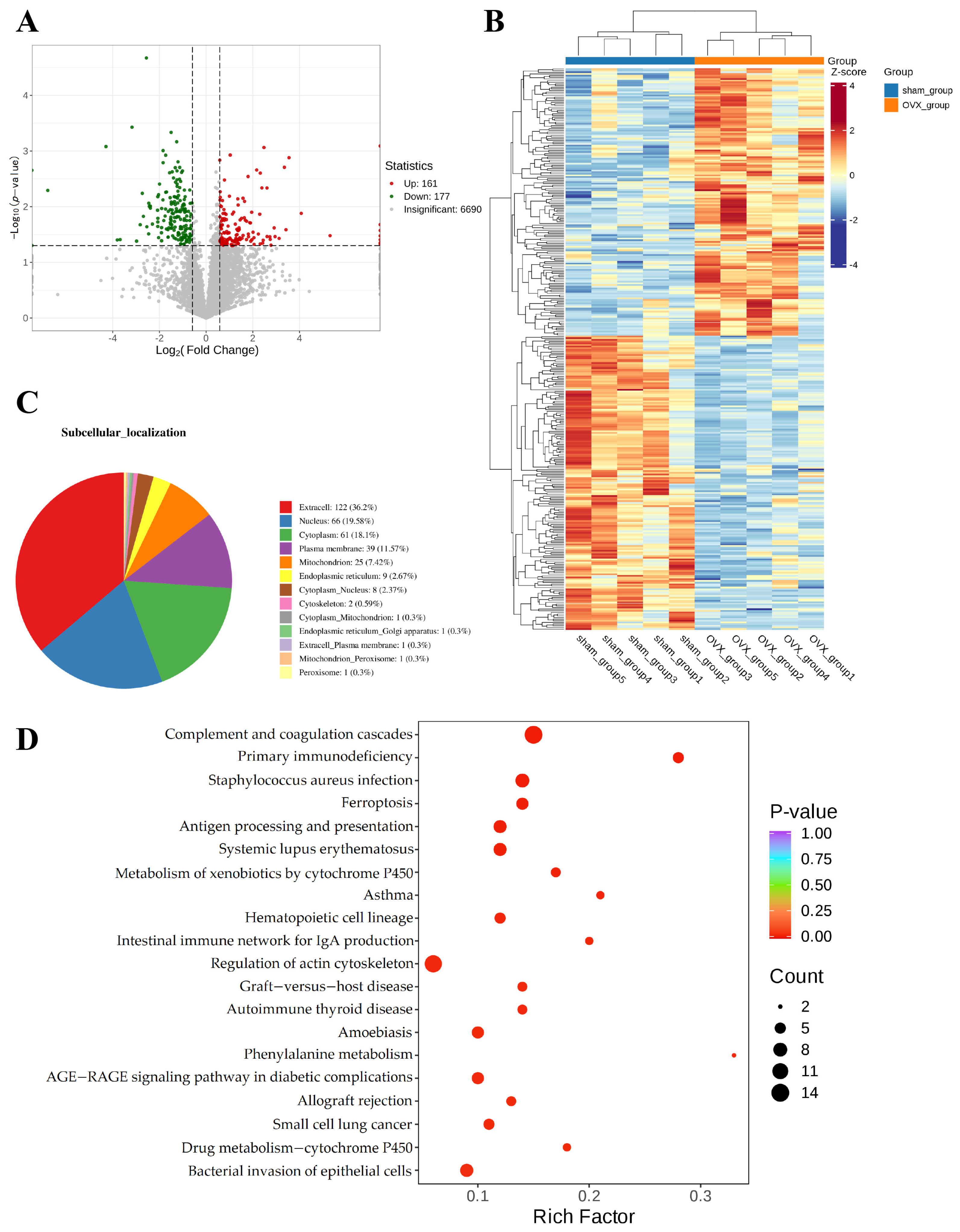
To explore the underlying reasons for the increased progression of ovariectomy and atherosclerosis, this study examined the plasma proteome of mice at 300 days after oophorectomy. Compared with the sham group, the expression levels of 161 proteins in the plasma of OVX mice increased, while those of 177 protein groups decreased (Figure 1A,B), with extracellular-related proteins accounting for the highest proportion (Figure 1C). KEGG enrichment analysis showed changes in the expression of proteins related to multiple biological pathways, with the highest number of proteins related to complement and coagulation cascade function (Figure 1D). Please refer to Supplementary Materials Tables S1 and S2 for the raw data of the proteomics diagram in Figure 1.

3.2. The 300-Day OCA Intervention Attenuates Platelet Activation and Atherosclerosis Progression After OVX in Mice

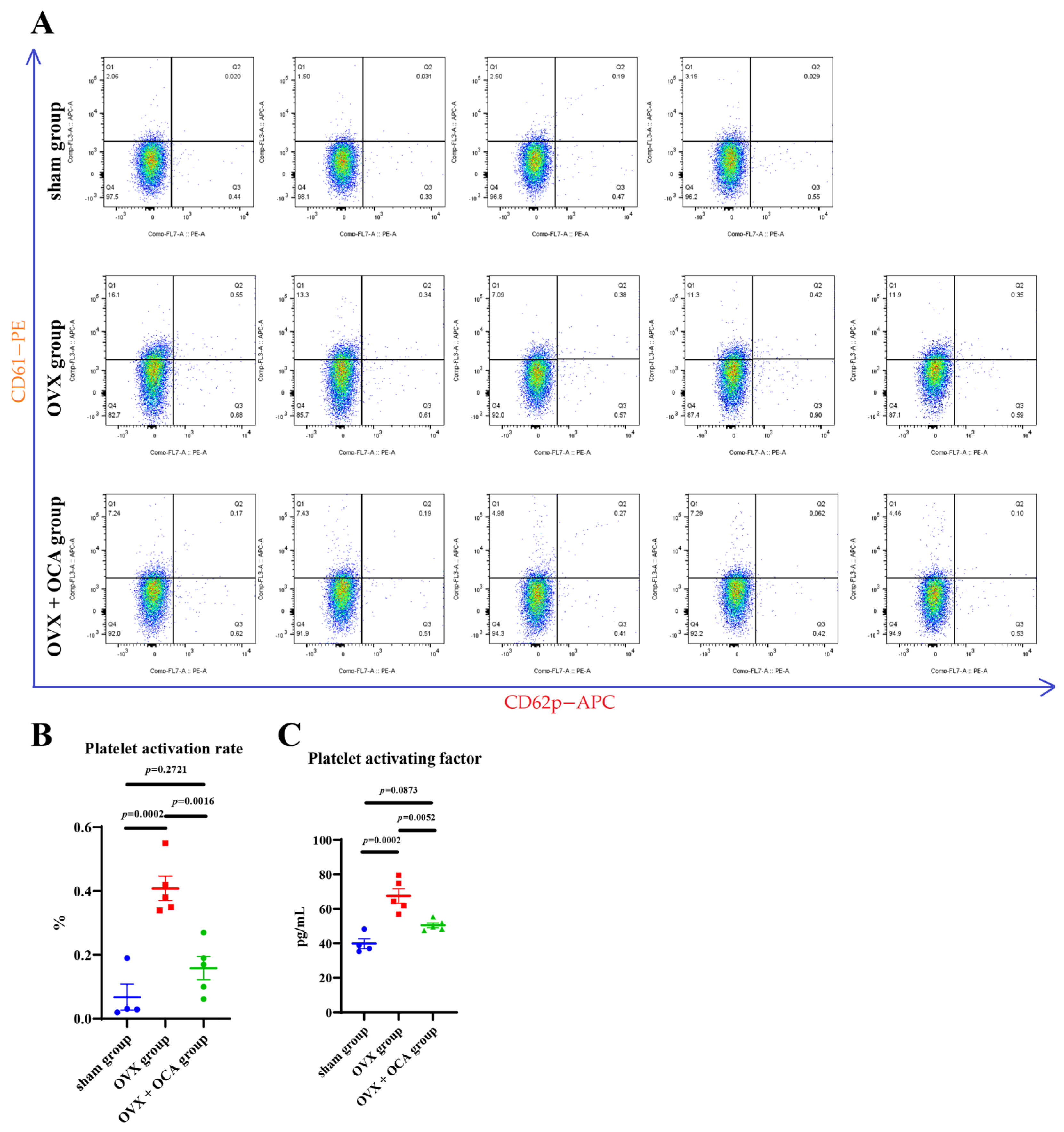
As the proteomic data show that OVX significantly alters complement and coagulation cascade-related protein levels in mouse plasma, considering the correlation between coagulation cascade and platelet activation rate [55,56,57], combined with clinical research findings indicating that platelet activation rates significantly increase after menopause [58], we speculate that platelet activation levels may be altered in OVX mice. Considering the view that OCA can inhibit platelet activation [44], we detected the platelet activation rate in mice after ovariectomy and administration of OCA. In the flow cytometry data, the horizontal axis and the vertical axis represent the PE fluorescence intensity of the CD61 antibody and the APC fluorescence intensity of the CD62p antibody, respectively (the same applies below). The data show that the platelet activation rate of mice significantly increased after OVX, while OCA intervention significantly reduced this increase in platelet activation rate (Figure 2A,B) and decreased plasma platelet-activating factor levels (Figure 2C).
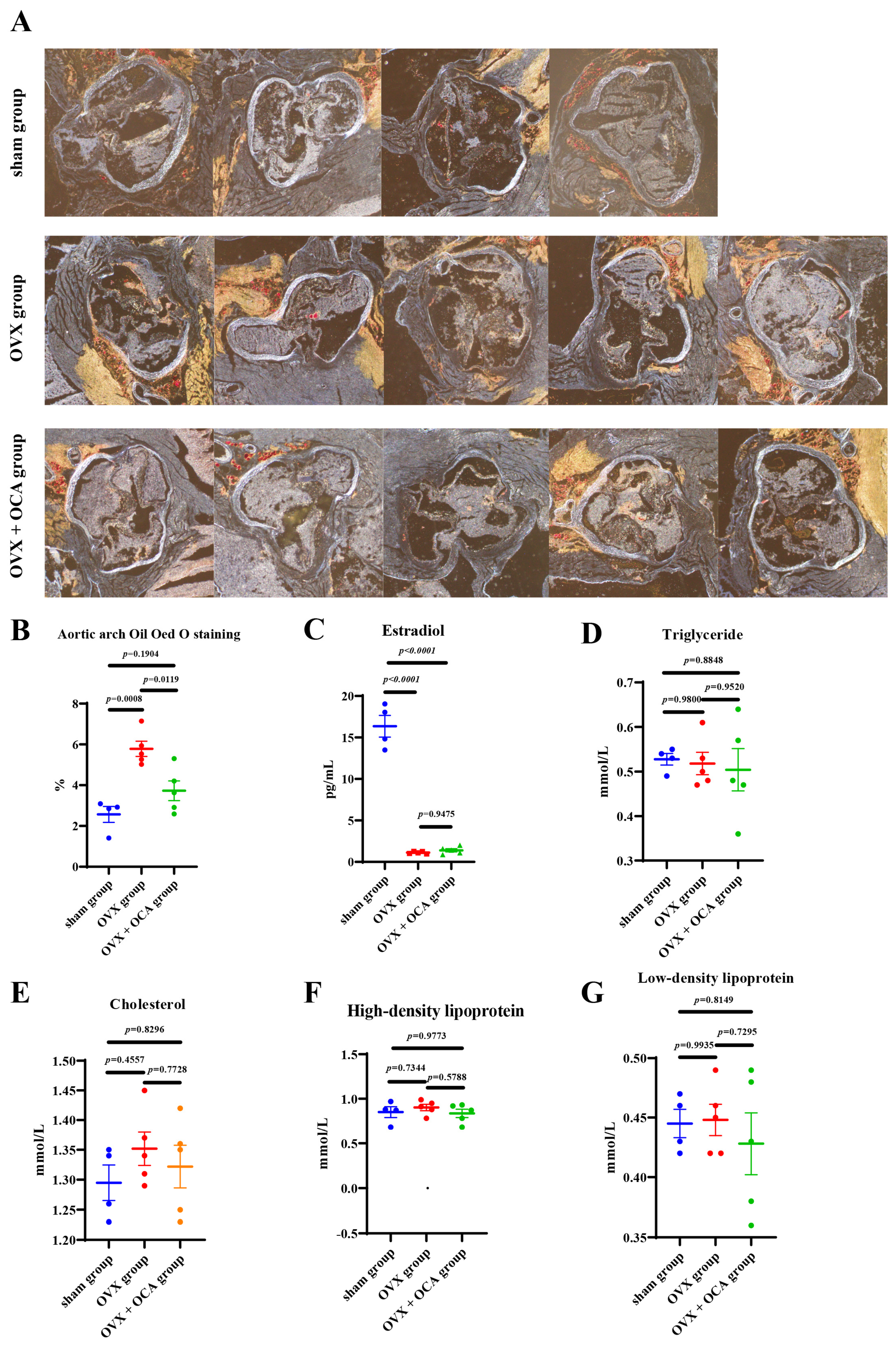
To evaluate the impact of OCA on the risk of postmenopausal atherosclerosis, in this study, we first simulated the postmenopausal physiological state by constructing an OVX mouse model. The mice were treated with OCA by oral gavage for 300 consecutive days, following which lipid deposition at the aortic arch was detected through Oil Red O staining. The results showed that OVX significantly increased lipid deposition in the aortic arch, while OCA treatment significantly reduced lipid accumulation in the OVX mice (Figure 3A,B).
Menopause has been found to cause a decrease in estradiol levels [35]. In this study, plasma estradiol concentrations were measured in mice, and the data showed that OVX significantly reduced estradiol levels in mouse plasma, while OCA did not significantly alter the estradiol reduction caused by OVX (Figure 3C). Further detection revealed that OCA administration had no significant effect on the blood lipids of OVX mice (Figure 3D–G), suggesting that atherosclerosis progression in mice after OCA-reduced OVX may be influenced by factors other than blood lipids.

3.3. A 7-Day OCA Intervention Can Reduce the Platelet Activation Rate in Mice After Ovariectomy

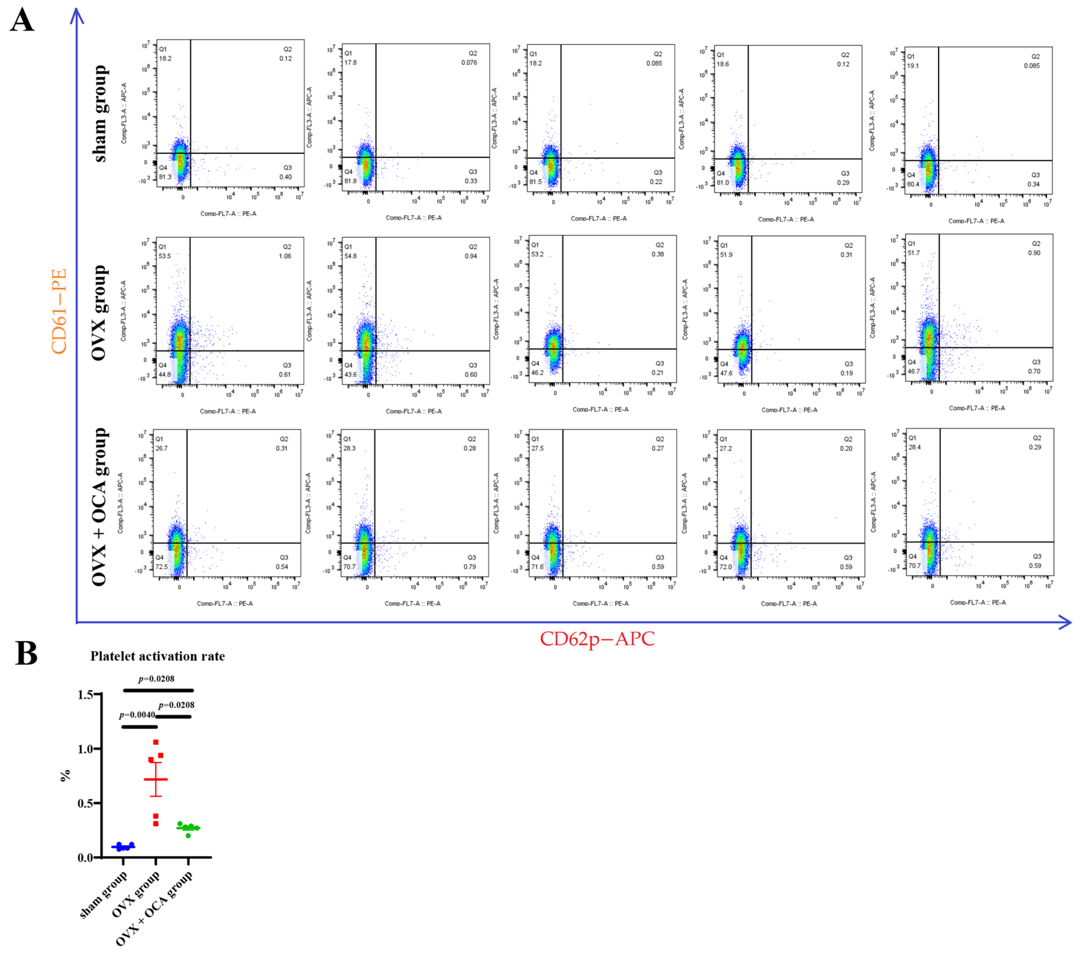
To further confirm the effect of elevated platelet activation rate on atherosclerosis progression in mice after OVX, in this study, we conducted an intragastric administration of OCA in mice 8–14 days after OVX and measured their platelet activation rate. The results showed that the platelet activation rate significantly increased after 7 days of OVX, while OCA intervention significantly inhibited this phenomenon (Figure 4A,B).

3.4. The 300-Day OCA Intervention Enhances the Intestinal Barrier in OVX Mice

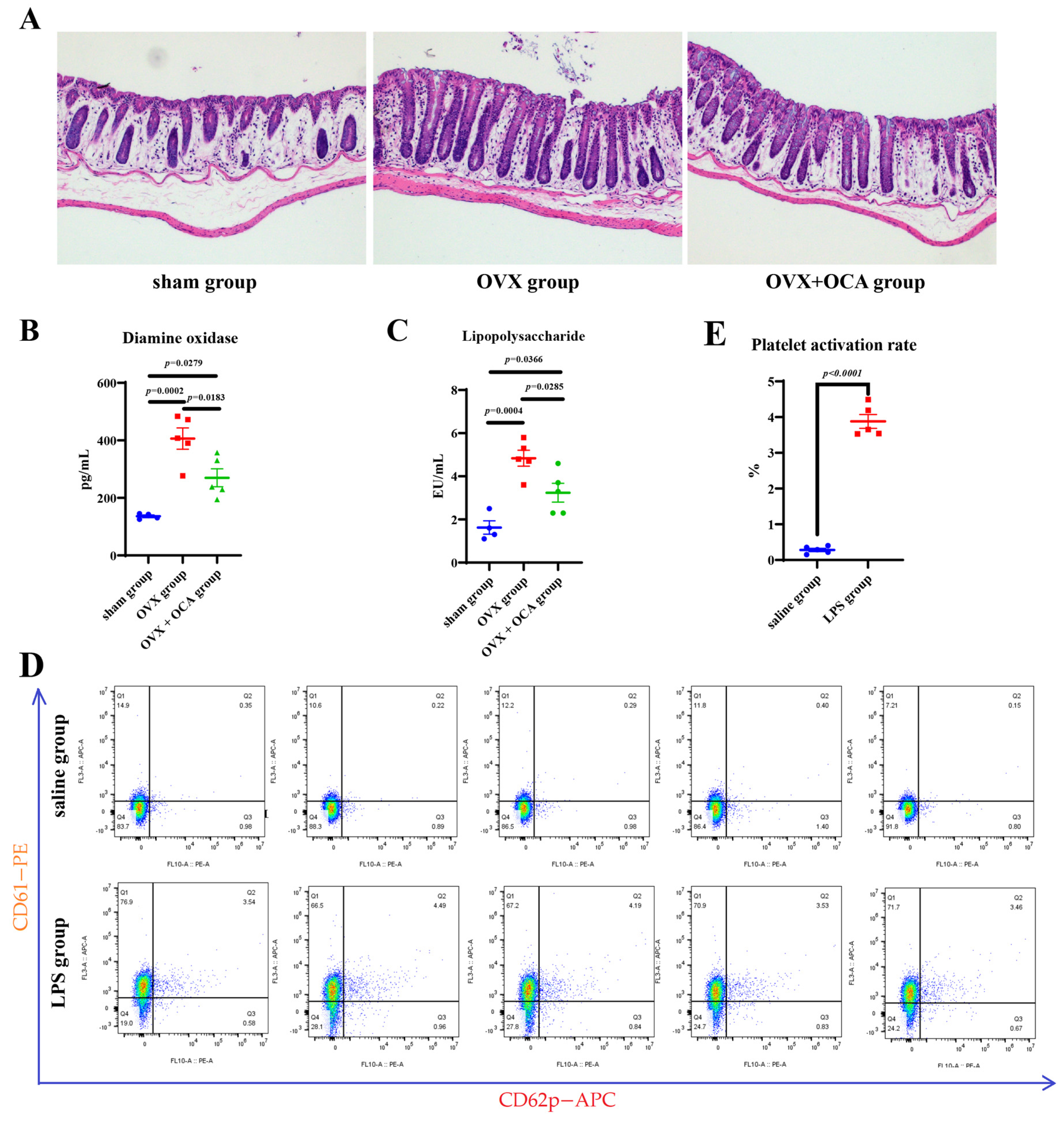
Menopause can lead to intestinal barrier damage [35], which in turn increases the level of LPS in the blood [59]. Because OCA can enhance the intestinal barrier, in this study, we further examined the intestinal morphology and barrier markers in each group of mice. HE staining showed the intestinal morphology of each group (Figure 5A), and plasma DAO detection (intestinal permeability index [60,61,62] showed that OCA significantly reduced the increase in DAO after 300 days of OVX (Figure 5B), indicating that OCA slowed down the intestinal barrier damage caused by OVX.
Intestinal barrier injury can lead to the invasion of gut microbiota into the bloodstream [59,63]. As a component of Gram-negative bacteria, LPS can enhance the platelet activation rate [64] and reflect bacterial levels in the blood [65]. Here, we further detected the LPS concentration in the plasma of each group of mice after 300 days of OVX. The data show that, at 300 days after OVX, the level of LPS in mouse plasma significantly increased, while OCA intervention significantly reduced these levels after OVX (Figure 5C).
To directly detect the effect of LPS on platelet activation, we detected the platelet activation rate in mice after intraperitoneal injection of LPS for 30 min, with the data showing that the platelet activation rate significantly increased (Figure 5D,E).

3.5. Chronic LPS Exposure Increases Platelet Activation and Atherosclerosis in Mice

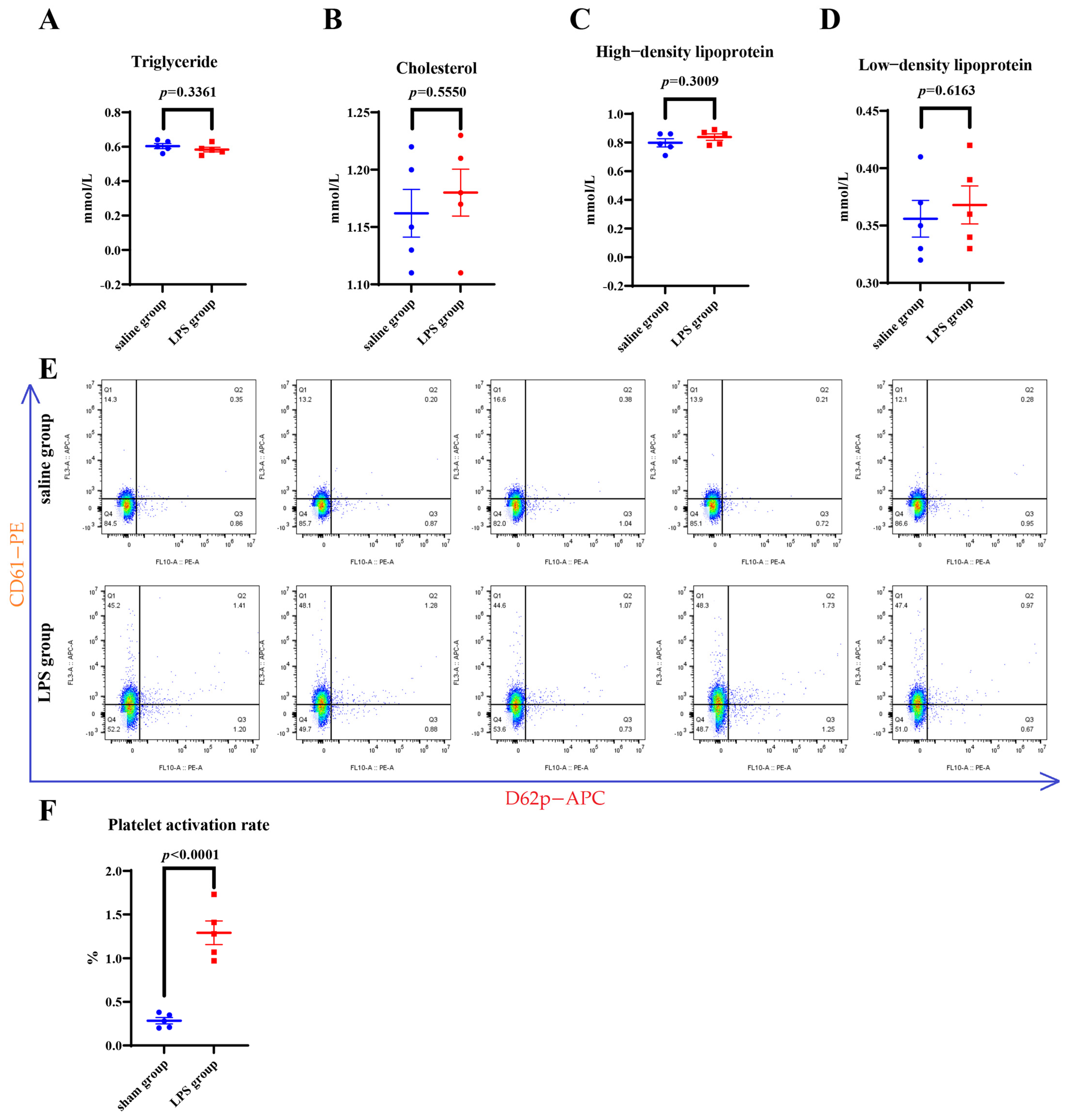
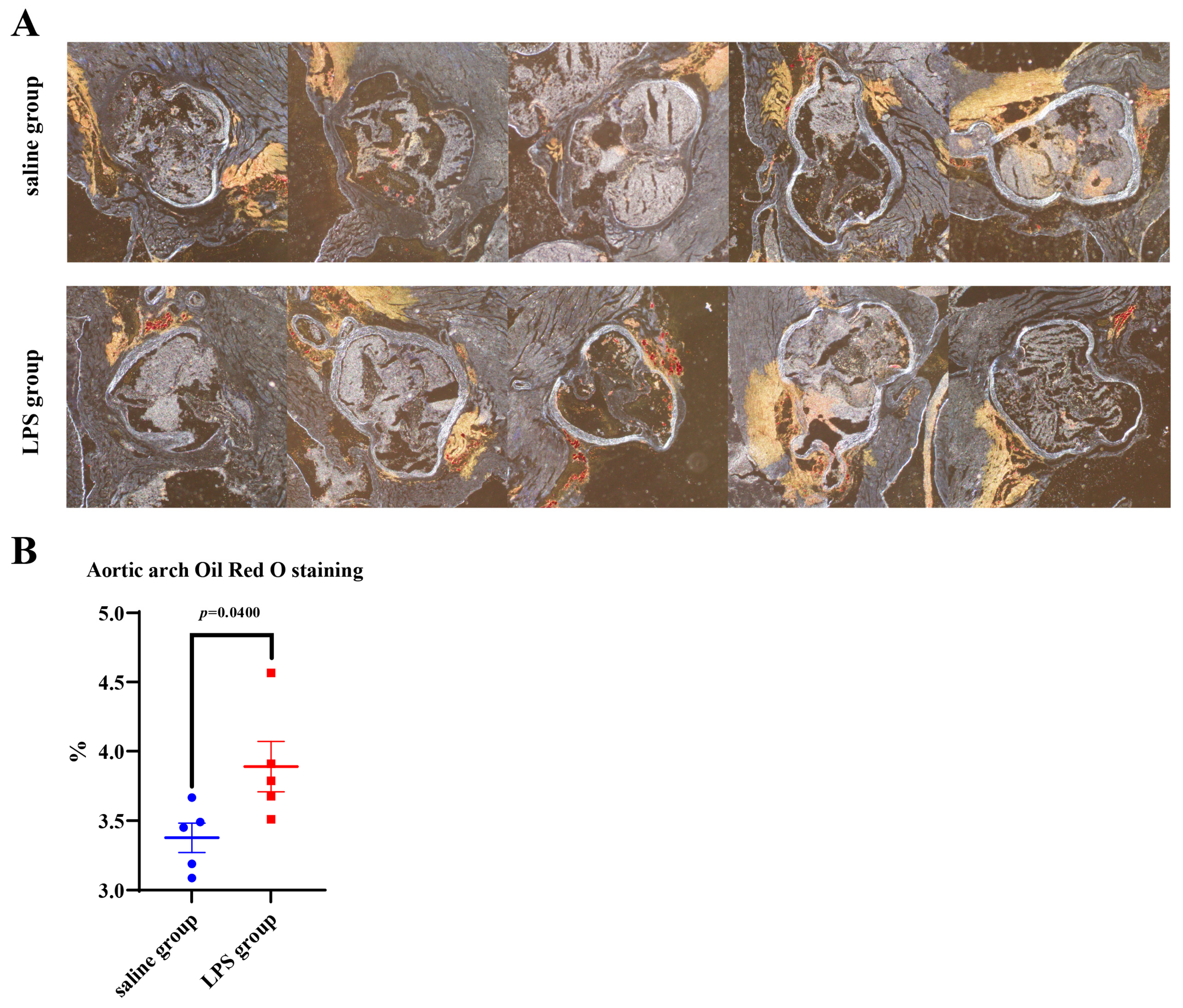
To further examine the role of elevated plasma LPS in postmenopausal atherosclerosis, we embedded a slow-release pump into the abdominal cavity to keep LPS in the blood. After testing the blood lipids of mice 30 days later, it was found that intraperitoneal slow-release LPS did not significantly change the blood lipid level of mice (Figure 6A–D), but significantly increased the platelet activation rate (Figure 6E,F) and increased the development of atherosclerosis (Figure 7A,B).

4. Discussion

In this study, we detected changes in the plasma proteome of mice after OVX through proteome analysis, and the effects of OCA on atherosclerosis, blood lipids, intestinal barrier, platelet-activating factor, and platelet activation rate in mice after OVX through oral intervention with OCA. We also detected the effects of LPS on platelet activation rate in mice through intraperitoneal injection and intraperitoneal sustained release of LPS, and the effects of intraperitoneal sustained release of LPS on blood lipids and atherosclerosis progression in mice.
The data from this study showed that OVX leads to intestinal barrier dysfunction and increased platelet activation rate, which is consistent with clinical studies showing intestinal barrier damage [35,39,66] and elevated platelet activation rate in postmenopausal women [67]. Repairing the intestinal barrier can reduce excessive activation of platelets [68]; the data from this study show that continuous OCA administration enhanced the intestinal barrier of mice after OVX, reduced the platelet activation rate and atherosclerosis progression, in alleviating the progression of atherosclerosis, which is consistent with the research conclusion that OCA can alleviate vascular calcification in ApoE−/− mice [69].
At the same time, the level of LPS in the plasma of mice after OVX increased. The data show that LPS entering the bloodstream can increase the platelet activation rate, which is in agreement with multiple studies [70,71,72]. Continuous intraperitoneal sustained release of LPS can also increase the platelet activation rate in mice and promote the progression of atherosclerosis, which is consistent with the clinical observation of systemic endotoxemia and the risk of thrombosis [73,74]. Combined with the view that excessive activation of platelets can promote the progression of atherosclerosis [40,41], this study speculated that the increase in atherosclerosis induced by ovariectomy in mice may be related to the abnormal platelet overactivation after menopause.
Menopause not only increases the risk of atherosclerosis [75,76] but is also associated with multiple comorbidities, including anxiety [2], Alzheimer’s disease [77,78], osteoporosis [79,80,81], periodontitis [82], and sarcopenia [83,84], which are closely related to changes in intestinal microbiota composition [83,85,86,87,88]. The diversity of intestinal microorganisms changes in postmenopausal atherosclerosis patients [89]. Given that OCA modulates gut microbial diversity [49], its potential impact on postmenopause-related diseases deserves further investigation. The atherosclerosis formation mechanism is complex, and individual risk assessment is particularly important in women [90]. In addition to drug intervention, another important research direction to explore is reliable biomarkers [91] and effective diagnostic methods [92,93] for early detection of postmenopausal atherosclerosis.
Inflammation is an important risk factor for atherosclerosis [94,95], and postmenopausal women have elevated systemic inflammatory markers [96,97,98]; in this context, it has been reported that restoration of intestinal barrier function can reduce systemic inflammation [99,100]. In this study, we did not evaluate inflammatory cytokines, nor did we describe the detailed molecular pathways involved in OCA-mediated protection, which are significant limitations. This study found that the plasma protein expression in mice changed significantly after OVX, but no further research was conducted regarding the impacts of these protein expression changes on the risk of atherosclerosis. The sample size of this study, being relatively small, is a further limitation of this study. Although this study found that OCA can enhance the intestinal barrier of OVX mice and reduce the level of LPS in the plasma of OVX mice, it was not confirmed whether the increased LPS levels in the plasma of OVX mice come from the intestinal barrier. Furthermore, the data of this study showed that OCA significantly reduced the platelet activation rate and the progress of atherosclerosis in OVX mice, but we did not conduct in-depth research on platelet aggregation in atherosclerotic plaque, which is another limitation. Further research addressing the abovementioned limitations may provide new insights into the formation and prevention of atherosclerosis after menopause.

5. Conclusions

This study described the phenomenon of OCA being able to reduce the progression of atherosclerosis in mice after bilateral oophorectomy. However, this cannot be considered as definitive evidence of a fully established mechanism, and further research is still needed.

Supplementary Materials

The following supporting information can be downloaded at: https://www.mdpi.com/article/10.3390/jgg74030017/s1, Table S1: KEGG annotation; Table S2: KEGG enrichmen.

Author Contributions

Conceptualization, J.S. and D.Q.; methodology, J.S., Y.Y. and Y.J.; validation, J.S. and Y.Y.; formal analysis, J.S.; investigation, J.S.; resources, J.S. and D.Q.; data curation, J.S., Y.Y. and Y.J.; writing—original draft preparation, J.S.; writing—review and editing, J.S. and D.Q.; visualization, J.S. and Y.Y.; supervision, project administration and funding acquisition, D.Q. All authors have read and agreed to the published version of the manuscript.

Funding

This research and the APC was funded by the Natural Science Foundation of Shandong Province (ZR2021QH254) support.

Institutional Review Board Statement

The study was conducted in accordance with the Declaration of Helsinki, and approved by the Ethics Committee of Qingdao University Medical College on 25 August 2021 (No. 20210825C573520220315179).

Informed Consent Statement

Not applicable.

Data Availability Statement

The author declares that all raw data for this study can be provided by contacting the corresponding author.

Acknowledgments

We would like to thank Meng Zhang, Yunlong Luo and Changcheng Li from Beijing Tsingke Biotech Co., Ltd. for them assistance in data interpretation.

Conflicts of Interest

The authors declare no conflicts of interest.

Abbreviations

The following abbreviations are used in this manuscript:
OCAObeticholic Acid
OVXBilateral ovariectomy
DAODiamine Oxidase
LPSLipopolysaccharide
HE stainingHematoxylin-eosin staining
FXRFarnesoid X Receptor

References

  1. Schattner, A.; Glick, Y.; Dubin, I. An Underappreciated Embolic Source: Aortic Arch Floating Thrombus. Am. J. Med. 2020, 133, e430–e431. [Google Scholar] [CrossRef] [Scilit] [PubMed]
  2. Ren, Y.; Pan, Z.; Zuo, X.; Jiang, X. The correlation of vaginal flora and anxiety in perimenopause women: A prospective study. BMC Women’s Health 2025, 25, 459. [Google Scholar] [CrossRef] [Scilit] [PubMed]
  3. Koros, R.; Domouzoglou, E.M.; Papafaklis, M.I. Metabolically “healthy” obesity in postmenopausal women: Unmasking the cardiovascular risk. World J. Cardiol. 2025, 17, 110228. [Google Scholar] [CrossRef] [Scilit] [PubMed]
  4. Ogata, J.; Yamanishi, H.; Ishibashi-Ueda, H. Review: Role of cerebral vessels in ischaemic injury of the brain. Neuropathol. Appl. Neurobiol. 2011, 37, 40–55. [Google Scholar] [CrossRef] [Scilit] [PubMed]
  5. Getie, A.; Gedfew, M.; Kitaw, T.A.; Yilak, G.; Bimerew, M. Mortality rate of stroke and its determinants in Africa: An umbrella review of systematic review and meta-analysis. Glob. Epidemiol. 2025, 10, 100225. [Google Scholar] [CrossRef] [Scilit] [PubMed]
  6. Han, C.; Yang, M.; Pan, Y.; Li, H.; Liu, L.; Meng, X.; Wang, Y.; Wang, Y. Disabling Neurologic Deficits and Antiplatelet Therapy in Acute Minor Stroke. J. Am. Heart Assoc. 2025, 14, e029734. [Google Scholar] [CrossRef] [Scilit] [PubMed]
  7. Martinez, R.; Muñoz-Venturelli, P.; Ordunez, P.; Fregni, F.; Abanto, C.; Alet, M.; Alvarez, T.F.; Amaya, P.; Ameriso, S.; Arauz, A.; et al. Risk and impact of stroke across 38 countries and territories of the Americas from 1990 to 2021: A population-based trends analysis from the Global Burden of Disease Study 2021. Lancet Reg. Health Am. 2025, 43, 101017. [Google Scholar] [CrossRef] [Scilit] [PubMed]
  8. Gasaly, N.; de Vos, P.; Hermoso, M.A. Impact of Bacterial Metabolites on Gut Barrier Function and Host Immunity: A Focus on Bacterial Metabolism and Its Relevance for Intestinal Inflammation. Front. Immunol. 2021, 12, 658354. [Google Scholar] [CrossRef] [Scilit] [PubMed]
  9. Bouchareychas, L.; Duong, P.; Covarrubias, S.; Alsop, E.; Phu, T.A.; Chung, A.; Gomes, M.; Wong, D.; Meechoovet, B.; Capili, A.; et al. Macrophage Exosomes Resolve Atherosclerosis by Regulating Hematopoiesis and Inflammation via MicroRNA Cargo. Cell Rep. 2020, 32, 107881. [Google Scholar] [CrossRef] [Scilit] [PubMed]
  10. Dregan, A.; Charlton, J.; Chowienczyk, P.; Gulliford, M.C. Chronic inflammatory disorders and risk of type 2 diabetes mellitus, coronary heart disease, and stroke: A population-based cohort study. Circulation 2014, 130, 837–844. [Google Scholar] [CrossRef] [Scilit] [PubMed]
  11. Appiah, D.; Schreiner, P.J.; Pankow, J.S.; Brock, G.; Tang, W.; Norby, F.L.; Michos, E.D.; Ballantyne, C.M.; Folsom, A.R. Long-term changes in plasma proteomic profiles in premenopausal and postmenopausal Black and White women: The Atherosclerosis Risk in Communities study. Menopause 2022, 29, 1150–1160. [Google Scholar] [CrossRef] [Scilit] [PubMed]
  12. Värri, M.; Niskanen, L.; Tuomainen, T.-P.; Honkanen, R.; Kröger, H.; Tuppurainen, M.T. Metabolite Profiling of Osteoporosis and Atherosclerosis in Postmenopausal Women: A Cross-Sectional Study. Vasc. Health Risk Manag. 2020, 16, 515–524. [Google Scholar] [CrossRef] [Scilit] [PubMed]
  13. Brown, P.A. Genes Differentially Expressed Across Major Arteries Are Enriched in Endothelial Dysfunction-Related Gene Sets: Implications for Relative Inter-artery Atherosclerosis Risk. Bioinform. Biol. Insights 2024, 18, 11779322241251563. [Google Scholar] [CrossRef] [Scilit] [PubMed]
  14. Park, H.-J.; Kim, Y.; Kim, M.-K.; Kim, H.J.; Bae, S.-K.; Bae, M.-K. Regulation of Vascular Calcification by M1-Type Macrophage-Derived Semaphorin 4D. Int. J. Mol. Sci. 2025, 26, 5071. [Google Scholar] [CrossRef] [Scilit] [PubMed]
  15. Strömberg, S.; Stomby, A.; Engvall, J.; Östgren, C.J. Systematic Coronary Risk Evaluation 2 (SCORE2), arterial stiffness, and subclinical coronary atherosclerosis in a population-based study. Scand. J. Prim. Health Care 2025, 43, 455–462. [Google Scholar] [CrossRef] [Scilit] [PubMed]
  16. Valentin Cabrera, A.M.; Ashbrook, S.K.; Hutcheson, J.D. Considerations on the Development of Therapeutics in Vascular Calcification. J. Cardiovasc. Dev. Dis. 2025, 12, 206. [Google Scholar] [CrossRef] [Scilit] [PubMed]
  17. Roeters van Lennep, J.E.; Tokgözoğlu, L.S.; Badimon, L.; Dumanski, S.M.; Gulati, M.; Hess, C.N.; Holven, K.B.; Kavousi, M.; Kayıkçıoğlu, M.; Lutgens, E.; et al. Women, lipids, and atherosclerotic cardiovascular disease: A call to action from the European Atherosclerosis Society. Eur. Heart J. 2023, 44, 4157–4173. [Google Scholar] [CrossRef] [Scilit] [PubMed]
  18. Chaudhry, A.; Ikram, K.; Ayesha, K.; Waheed, M.; Ulain, N.; Tariq, A.; Khalid, T. The Comparative Study of Serum Estrogen and Lipid Profile in Pre- and Post-menopausal Women as Atherosclerosis Risk Factors in Pakistan. Cureus 2024, 16, e65604. [Google Scholar] [CrossRef] [Scilit] [PubMed]
  19. Ridker, P.M.; Devalaraja, M.; Baeres, F.M.M.; Engelmann, M.D.M.; Hovingh, G.K.; Ivkovic, M.; Lo, L.; Kling, D.; Pergola, P.; Raj, D.; et al. IL-6 inhibition with ziltivekimab in patients at high atherosclerotic risk (RESCUE): A double-blind, randomised, placebo-controlled, phase 2 trial. Lancet 2021, 397, 2060–2069. [Google Scholar] [CrossRef] [Scilit] [PubMed]
  20. Brunner, F.J.; Waldeyer, C.; Ojeda, F.; Salomaa, V.; Kee, F.; Sans, S.; Thorand, B.; Giampaoli, S.; Brambilla, P.; Tunstall-Pedoe, H.; et al. Application of non-HDL cholesterol for population-based cardiovascular risk stratification: Results from the Multinational Cardiovascular Risk Consortium. Lancet 2019, 394, 2173–2183. [Google Scholar] [CrossRef] [Scilit] [PubMed]
  21. Yu, H.; Rimbert, A.; Palmer, A.E.; Toyohara, T.; Xia, Y.; Xia, F.; Ferreira, L.M.R.; Chen, Z.; Chen, T.; Loaiza, N.; et al. GPR146 Deficiency Protects against Hypercholesterolemia and Atherosclerosis. Cell 2019, 179, 1276–1288.e14. [Google Scholar] [CrossRef] [Scilit] [PubMed]
  22. Mortensen, M.B.; Nordestgaard, B.G. Elevated LDL cholesterol and increased risk of myocardial infarction and atherosclerotic cardiovascular disease in individuals aged 70–100 years: A contemporary primary prevention cohort. Lancet 2020, 396, 1644–1652. [Google Scholar] [CrossRef] [Scilit] [PubMed]
  23. Zhang, X.; Zhang, Y.; Wang, P.; Zhang, S.Y.; Dong, Y.; Zeng, G.; Yan, Y.; Sun, L.; Wu, Q.; Liu, H.; et al. Adipocyte Hypoxia-Inducible Factor 2alpha Suppresses Atherosclerosis by Promoting Adipose Ceramide Catabolism. Cell Metab. 2019, 30, 937–951.e935. [Google Scholar] [CrossRef] [Scilit] [PubMed]
  24. Gencer, B.; Marston, N.A.; Im, K.; Cannon, C.P.; Sever, P.; Keech, A.; Braunwald, E.; Giugliano, R.P.; Sabatine, M.S. Efficacy and safety of lowering LDL cholesterol in older patients: A systematic review and meta-analysis of randomised controlled trials. Lancet 2020, 396, 1637–1643. [Google Scholar] [CrossRef] [Scilit] [PubMed]
  25. Everson-Rose, S.A.; Barinas-Mitchell, E.J.M.; El Khoudary, S.R.; Huang, H.H.; Wang, Q.; Janssen, I.; Thurston, R.C.; Jackson, E.A.; Lewis, M.E.; Karvonen-Gutierrez, C.; et al. Adipokines and Subclinical Cardiovascular Disease in Post-Menopausal Women: Study of Women’s Health Across the Nation. J. Am. Heart Assoc. 2021, 10, e019173. [Google Scholar] [CrossRef] [Scilit] [PubMed]
  26. Nafakhi, H.; Al-Mosawi, A.A.; Alkhalil, Q.Q.M. Cardiovascular risk factors and pericardial fat volume as predictors of coronary atherosclerosis burden in postmenopausal women: A retrospective, single center study. Health Sci. Rep. 2023, 6, e1155. [Google Scholar] [CrossRef] [Scilit] [PubMed]
  27. Clayton, G.L.; Soares, A.G.; Kilpi, F.; Fraser, A.; Welsh, P.; Sattar, N.; Nelson, S.M.; Tilling, K.; Lawlor, D.A. Cardiovascular health in the menopause transition: A longitudinal study of up to 3892 women with up to four repeated measures of risk factors. BMC Med. 2022, 20, 299. [Google Scholar] [CrossRef] [Scilit] [PubMed]
  28. Machuca, J.N.; Rosales-Alvarez, C.P. Cardiovascular Disease in Women and the Role of Hormone Replacement Therapy. Cureus 2024, 16, e69752. [Google Scholar] [CrossRef] [Scilit] [PubMed]
  29. Honigberg, M.C.; Zekavat, S.M.; Niroula, A.; Griffin, G.K.; Bick, A.G.; Pirruccello, J.P.; Nakao, T.; Whitsel, E.A.; Farland, L.V.; Laurie, C.; et al. Premature Menopause, Clonal Hematopoiesis, and Coronary Artery Disease in Postmenopausal Women. Circulation 2021, 143, 410–423. [Google Scholar] [CrossRef] [Scilit] [PubMed]
  30. Chen, I.J.; Shoupe, D.; Karim, R.; Stanczyk, F.Z.; Kono, N.; Sriprasert, I.; Hodis, H.N.; Mack, W.J. The association of hysterectomy with or without ovarian conservation with subclinical atherosclerosis progression in healthy postmenopausal women. Menopause 2023, 30, 692–702. [Google Scholar] [CrossRef] [Scilit] [PubMed]
  31. D’Costa, Z.; Spertus, E.; Hingorany, S.; Patil, R.; Horwich, T.; Press, M.C.; Shah, J.; Watson, K.E.; Jafari, L. Cardiovascular Risk Associated with Menopause and Menopause Hormone Therapy: A Review and Contemporary Approach to Risk Assessment. Curr. Atheroscler. Rep. 2025, 27, 100. [Google Scholar] [CrossRef] [Scilit] [PubMed]
  32. Xiang, D.; Liu, Y.; Zhou, S.; Zhou, E.; Wang, Y.; D’Adamo, S. Protective Effects of Estrogen on Cardiovascular Disease Mediated by Oxidative Stress. Oxidative Med. Cell. Longev. 2021, 2021, 5523516. [Google Scholar] [CrossRef] [Scilit] [PubMed]
  33. Yoon, B.-K.; Kang, Y.-H.; Oh, W.-J.; Roh, C.-R.; Kim, D.-K.; Kang, C.-D. 17β-Estradiol Inhibits Lysophosphatidylcholine-Induced Apoptosis in Cultured Vascular Smooth Muscle Cells. J. Menopausal Med. 2020, 26, 1–8. [Google Scholar] [CrossRef] [Scilit] [PubMed]
  34. Buscato, M.; Davezac, M.; Zahreddine, R.; Adlanmerini, M.; Métivier, R.; Fillet, M.; Cobraiville, G.; Moro, C.; Foidart, J.-M.; Lenfant, F.; et al. Estetrol prevents Western diet–induced obesity and atheroma independently of hepatic estrogen receptor α. Am. J. Physiol. Endocrinol. Metab. 2021, 320, E19–E29. [Google Scholar] [CrossRef] [Scilit] [PubMed]
  35. Grishina, I. Gender Differences, Aging and Hormonal Status in Mucosal Injury and Repair. Aging Dis. 2014, 5, 160–169. [Google Scholar] [CrossRef] [Scilit] [PubMed]
  36. Davezac, M.; Buscato, M.; Zahreddine, R.; Lacolley, P.; Henrion, D.; Lenfant, F.; Arnal, J.-F.; Fontaine, C. Estrogen Receptor and Vascular Aging. Front. Aging 2021, 2, 727380. [Google Scholar] [CrossRef] [Scilit] [PubMed]
  37. Sriprasert, I.; Hodis, H.N.; Bernick, B.; Mirkin, S.; Mack, W.J. Determinants of attained estradiol levels in response to oral estradiol plus progesterone therapy. Climacteric 2020, 24, 194–199. [Google Scholar] [CrossRef] [Scilit] [PubMed]
  38. Miller, V.M.; Taylor, H.S.; Naftolin, F.; Manson, J.E.; Gleason, C.E.; Brinton, E.A.; Kling, J.M.; Cedars, M.I.; Dowling, N.M.; Kantarci, K.; et al. Lessons from KEEPS: The Kronos Early Estrogen Prevention Study. Climacteric 2020, 24, 139–145. [Google Scholar] [CrossRef] [Scilit] [PubMed]
  39. Shieh, A.; Epeldegui, M.; Karlamangla, A.S.; Greendale, G.A. Gut permeability, inflammation, and bone density across the menopause transition. J. Clin. Investig. 2020, 5, 134092. [Google Scholar] [CrossRef] [Scilit] [PubMed]
  40. Lee, H.; Joo, J.-Y.; Kang, J.; Yu, Y.; Kim, Y.H.; Park, H.R. Single-cell analysis of platelets from patients with periodontitis and diabetes. Res. Pract. Thromb. Haemost. 2023, 7, 100099. [Google Scholar] [CrossRef] [Scilit] [PubMed]
  41. Ishida, M.; Sakai, C.; Kobayashi, Y.; Ishida, T. Cigarette Smoking and Atherosclerotic Cardiovascular Disease. J. Atheroscler. Thromb. 2024, 31, 189–200. [Google Scholar] [CrossRef] [Scilit] [PubMed]
  42. Yan, M.; Hou, L.; Cai, Y.; Wang, H.; Ma, Y.; Geng, Q.; Jiang, W.; Tang, W. Effects of Intestinal FXR-Related Molecules on Intestinal Mucosal Barriers in Biliary Tract Obstruction. Front. Pharmacol. 2022, 13, 906452. [Google Scholar] [CrossRef] [Scilit] [PubMed]
  43. Cai, Y.; Chen, X.; Wang, H.; Hou, L.; Zheng, R.; Wang, Y.; Jiang, W.; Tang, W. Investigating intestinal farnesoid X receptor functions at the intestinal mucosal barrier and in the intestinal microbiota in a biliary obstruction mouse model. Am. J. Physiol. Gastrointest. Liver Physiol. 2025, 329, G313–G327. [Google Scholar] [CrossRef] [Scilit] [PubMed]
  44. Maligłówka, M.; Dec, A.; Bułdak, Ł.; Okopień, B. The Effects of Inclisiran on the Subclinical Prothrombotic and Platelet Activation Markers in Patients at High Cardiovascular Risk. J. Cardiovasc. Dev. Dis. 2025, 12, 355. [Google Scholar] [CrossRef] [Scilit] [PubMed]
  45. Tung, Y.-T.; Hsu, Y.-J.; Chien, Y.-W.; Huang, C.-C.; Huang, W.-C.; Chiu, W.-C. Tea Seed Oil Prevents Obesity, Reduces Physical Fatigue, and Improves Exercise Performance in High-Fat-Diet-Induced Obese Ovariectomized Mice. Molecules 2019, 24, 980. [Google Scholar] [CrossRef] [Scilit] [PubMed]
  46. Fischer, V.; Kalbitz, M.; Müller-Graf, F.; Gebhard, F.; Ignatius, A.; Liedert, A.; Haffner-Luntzer, M. Influence of Menopause on Inflammatory Cytokines during Murine and Human Bone Fracture Healing. Int. J. Mol. Sci. 2018, 19, 2070. [Google Scholar] [CrossRef] [Scilit] [PubMed]
  47. Zhang, Q.-Y.; Gong, H.-B.; Jiang, M.-Y.; Jin, F.; Wang, G.; Yan, C.-Y.; Luo, X.; Sun, W.-Y.; Ouyang, S.-H.; Wu, Y.-P.; et al. Regulation of enzymatic lipid peroxidation in osteoblasts protects against postmenopausal osteoporosis. Nat. Commun. 2025, 16, 758. [Google Scholar] [CrossRef] [Scilit] [PubMed]
  48. Qian, S.; Su, Z.; Lin, J.; Hou, Q.; Wang, X.; Li, Y.; Wang, J.; Huang, C.; Wang, Z.; Cubero, F.J.; et al. Inhibition of Farnesoid-x-receptor signaling during abdominal sepsis by dysbiosis exacerbates gut barrier dysfunction. Cell Commun. Signal. 2025, 23, 236. [Google Scholar] [CrossRef] [Scilit] [PubMed]
  49. Zhang, D.-Y.; Zhu, L.; Liu, H.-N.; Tseng, Y.-J.; Weng, S.-Q.; Liu, T.-T.; Dong, L.; Shen, X.-Z. The protective effect and mechanism of the FXR agonist obeticholic acid via targeting gut microbiota in non-alcoholic fatty liver disease. Drug Des. Dev. Ther. 2019, 13, 2249–2270. [Google Scholar] [CrossRef] [Scilit] [PubMed]
  50. Sekio, M.; Seki, K. Lipopolysaccharide-Induced Depressive-Like Behavior is Associated with α1-Adrenoceptor Dependent Downregulation of the Membrane GluR1 Subunit in the Mouse Medial Prefrontal Cortex and Ventral Tegmental Area. Int. J. Neuropsychopharmacol. 2015, 18, pyu005. [Google Scholar] [CrossRef] [Scilit] [PubMed]
  51. Choi, J.; Jang, J.; Son, D.; Im, H.-S.; Kim, J.; Park, J.; Choi, W.; Han, S.-B.; Hong, J. Antarctic Krill Oil Diet Protects against Lipopolysaccharide-Induced Oxidative Stress, Neuroinflammation and Cognitive Impairment. Int. J. Mol. Sci. 2017, 18, 2554. [Google Scholar] [CrossRef] [Scilit] [PubMed]
  52. Chen, W.A.; Fletcher, H.M.; Payne, K.J.; Aka, S.; Thornburg, M.B.; Gheorghe, J.D.; Safi, S.B.; Shavlik, D.; Oyoyo, U.; Boskovic, D.S. Platelet and neutrophil responses to Porphyromonas gingivalis in human whole blood. Mol. Oral Microbiol. 2021, 36, 202–213. [Google Scholar] [CrossRef] [Scilit] [PubMed]
  53. Gąsecka, A.; Banaszkiewicz, M.; Nieuwland, R.; van der Pol, E.; Hajji, N.; Mutwil, H.; Rogula, S.; Rutkowska, W.; Pluta, K.; Eyileten, C.; et al. Prostacyclin Analogues Inhibit Platelet Reactivity, Extracellular Vesicle Release and Thrombus Formation in Patients with Pulmonary Arterial Hypertension. J. Clin. Med. 2021, 10, 1024. [Google Scholar] [CrossRef] [Scilit] [PubMed]
  54. Bian, Y.; Jin, Q.; He, J.; Ngo, T.; Bae, O.-N.; Xing, L.; Pi, J.; Chung, H.Y.; Xu, Y. Biomedical application of TiO2NPs can cause arterial thrombotic risks through triggering procoagulant activity, activation and aggregation of platelets. Cell Biol. Toxicol. 2024, 40, 67. [Google Scholar] [CrossRef] [Scilit] [PubMed]
  55. Brata, V.D.; Crisan, D.A.; Cozma, A.; Gerdanovics, C.-A.; Popa, S.L.; Milaciu, M.V.; Orășan, O.H. From Inflammation to Thrombosis: The Prothrombotic State and Cardiovascular Risk in Inflammatory Bowel Disease. Medicina 2026, 62, 270. [Google Scholar] [CrossRef] [Scilit] [PubMed]
  56. Majin, S.; Malik, A.; Poudel, P.; Faustino, E.V.S.; Zuverza-Mena, N.; Hwa, J.; Takyar, S.; Shea, S.; Amoako, K.A. Drug-loaded nanoparticles reduced platelet aggregation and blood coagulation. Front. Med. Technol. 2026, 7, 1690389. [Google Scholar] [CrossRef] [Scilit] [PubMed]
  57. Martin, M.; Tschirhart, M.; Auger, C.; Toti, F.; Helms, J.; Choulier, L. Emerging role of nucleic acid aptamers in sepsis-induced coagulopathy: Future perspectives in diagnostics and therapeutics. Ann. Intensive Care 2026, 16, 100014. [Google Scholar] [CrossRef] [Scilit] [PubMed]
  58. Santos-Marcos, J.A.; Barroso, A.; Rangel-Zuñiga, O.A.; Perdices-Lopez, C.; Haro, C.; Sanchez-Garrido, M.A.; Molina-Abril, H.; Ohlsson, C.; Perez-Martinez, P.; Poutanen, M.; et al. Interplay between gonadal hormones and postnatal overfeeding in defining sex-dependent differences in gut microbiota architecture. Aging 2020, 12, 19979–20000. [Google Scholar] [CrossRef] [Scilit] [PubMed]
  59. Ivnitsky, J.J.; Schäfer, T.V.; Rejniuk, V.L.; Vakunenkova, O.A. Secondary Dysfunction of the Intestinal Barrier in the Pathogenesis of Complications of Acute Poisoning. J. Evol. Biochem. Physiol. 2022, 58, 1075–1098. [Google Scholar] [CrossRef] [Scilit] [PubMed]
  60. Shao, Y.; Li, Y.; Xie, S.; Zhang, B.; Li, J.; Wang, Y.; Gao, Y.; Ma, J.; Kishawy, A.T.Y.; Gu, L.; et al. Effects of 4-hydroxyphenylacetic acid, a phenolic acid compound from Yucca schidigera extract, on immune function and intestinal health in meat pigeons. Poult. Sci. 2025, 104, 105543. [Google Scholar] [CrossRef] [Scilit] [PubMed]
  61. Wang, N.; Yang, H.; Tong, X.; Xu, T.; Zhao, J.; Li, Y.-k. Ginkgolide B modulates the gut-bone axis to ameliorate bone loss in ovariectomized mice. J. Orthop. Surg. Res. 2025, 20, 804. [Google Scholar] [CrossRef] [Scilit] [PubMed]
  62. Zhang, W.; Li, C.; Bi, R.; Lu, Y.; Zhang, Y.; Shi, C.; Qiao, Z.; Sun, Y.; Chen, J.; Wang, P.; et al. Lactoferrin and Osteopontin Cooperatively Promote Intestinal Epithelial Maturation in Neonatal Mice by Activating the Brg1/Notch1/Hes1 Pathway. Nutrients 2025, 17, 3176. [Google Scholar] [CrossRef] [Scilit] [PubMed]
  63. Chen, Y.; Pu, W.; Maswikiti, E.P.; Tao, P.; Li, X.; Wang, D.; Gu, B.; Yu, Y.; Gao, L.; Zhao, C.; et al. Intestinal congestion and reperfusion injury: Damage caused to the intestinal tract and distal organs. Biosci. Rep. 2021, 41, BSR20211560. [Google Scholar] [CrossRef] [Scilit] [PubMed]
  64. Hernández-Cuellar, E.; Tsuchiya, K.; Medina-Contreras, O.; Valle-Ríos, R. The Role of Inflammasomes in LPS and Gram-Negative Bacterial Sepsis. J. Clin. Med. 2025, 14, 7102. [Google Scholar] [CrossRef] [Scilit] [PubMed]
  65. Potrykus, M.; Stanisławowski, M.; Czaja-Stolc, S.; Potrykus, A.; Stankiewicz, M.; Owczarzak, A.; Guzek, M.; Szymański, M.; Łoniewski, I.; Adrych, K.; et al. 12-week preoperative probiotic supplementation versus placebo: Effects on inflammation, endotoxemia, adipokines, and gastrointestinal peptides in patients six months after bariatric surgery—A double-blind, randomized, placebo-controlled clinical trial. Nutr. J. 2025, 24, 156. [Google Scholar] [CrossRef] [Scilit] [PubMed]
  66. Collins, F.L.; Rios-Arce, N.D.; Atkinson, S.; Bierhalter, H.; Schoenherr, D.; Bazil, J.N.; McCabe, L.R.; Parameswaran, N. Temporal and regional intestinal changes in permeability, tight junction, and cytokine gene expression following ovariectomy-induced estrogen deficiency. Physiol. Rep. 2017, 5, e13263. [Google Scholar] [CrossRef] [Scilit] [PubMed]
  67. Yamaguchi, A.; Stanger, L.; Freedman, J.C.; Prieur, A.; Thav, R.; Tena, J.; Holman, T.R.; Holinstat, M. Supplementation with omega-3 or omega-6 fatty acids attenuates platelet reactivity in postmenopausal women. Clin. Transl. Sci. 2022, 15, 2378–2391. [Google Scholar] [CrossRef] [Scilit] [PubMed]
  68. Cheng, B.; Du, M.; He, S.; Yang, L.; Wang, X.; Gao, H.; Chang, H.; Gao, W.; Li, Y.; Wang, Q.; et al. Inhibition of platelet activation suppresses reactive enteric glia and mitigates intestinal barrier dysfunction during sepsis. Mol. Med. 2022, 28, 127. [Google Scholar] [CrossRef] [Scilit] [PubMed]
  69. Miyazaki-Anzai, S.; Levi, M.; Kratzer, A.; Ting, T.C.; Lewis, L.B.; Miyazaki, M. Farnesoid X Receptor Activation Prevents the Development of Vascular Calcification in ApoE−/− Mice With Chronic Kidney Disease. Circ. Res. 2010, 106, 1807–1817. [Google Scholar] [CrossRef] [Scilit] [PubMed]
  70. Tan, L.; Zhang, C.; Kou, X.; Zhao, L.; Wu, D.; Li, J.; Yu, C.; Xu, T.; Gao, L.; Mao, X.; et al. Apoptotic Vesicles Attenuate Acute Lung Injury via CD73-Mediated Inhibition of Platelet Activation and NETosis. Int. J. Nanomed. 2025, 20, 91–107. [Google Scholar] [CrossRef] [Scilit] [PubMed]
  71. Wang, L.; Mi, L.-y.; Chen, X.-y.; He, H.-W.; Long, Y. Effects of different antiplatelet therapy drugs on platelet activation and platelet-leukocyte aggregate formation in early septic ARDS. BMC Pharmacol. Toxicol. 2025, 26, 4. [Google Scholar] [CrossRef] [Scilit] [PubMed]
  72. Hagisawa, K.; Ishida, O.; Nakashima, H.; Takeoka, S.; Morimoto, Y.; Kinoshita, M. Post-treatment with H12-(ADP)-liposomes after LPS challenge ameliorated coagulopathy and critical organ injury in rats. Intensive Care Med. Exp. 2026, 14, 61. [Google Scholar] [CrossRef] [Scilit] [PubMed]
  73. Szeto, C.-C.; Kwan, B.C.-H.; Chow, K.-M.; Lai, K.-B.; Chung, K.-Y.; Leung, C.-B.; Li, P.K.-T. Endotoxemia is Related to Systemic Inflammation and Atherosclerosis in Peritoneal Dialysis Patients. Clin. J. Am. Soc. Nephrol. 2008, 3, 431–436. [Google Scholar] [CrossRef] [Scilit] [PubMed]
  74. McIntyre, C.W.; Harrison, L.E.A.; Eldehni, M.T.; Jefferies, H.J.; Szeto, C.-C.; John, S.G.; Sigrist, M.K.; Burton, J.O.; Hothi, D.; Korsheed, S.; et al. Circulating Endotoxemia. Clin. J. Am. Soc. Nephrol. 2011, 6, 133–141. [Google Scholar] [CrossRef] [Scilit] [PubMed]
  75. Han, T.-W.; Liu, Y.-Q.; Dong, W.; Bai, X.-J.; Liu, Y.-Y.; Su, X.; Li, Y.-M.; Qian, J.-Y.; Xiang, M.-X.; Cai, L.; et al. Poor cardiovascular health status among Chinese women. BMC Cardiovasc. Disord. 2020, 20, 497. [Google Scholar] [CrossRef] [Scilit] [PubMed]
  76. Chu, J.H.; Michos, E.D.; Ouyang, P.; Vaidya, D.; Blumenthal, R.S.; Budoff, M.J.; Blaha, M.J.; Whelton, S.P. Coronary artery calcium and atherosclerotic cardiovascular disease risk in women with early menopause: The Multi-Ethnic Study of Atherosclerosis (MESA). Am. J. Prev. Cardiol. 2022, 11, 100362. [Google Scholar] [CrossRef] [Scilit] [PubMed]
  77. Brayne, C.; Gleason, C.E.; Dowling, N.M.; Kara, F.; James, T.T.; Salazar, H.; Ferrer Simo, C.A.; Harman, S.M.; Manson, J.E.; Hammers, D.B.; et al. Long-term cognitive effects of menopausal hormone therapy: Findings from the KEEPS Continuation Study. PLoS Med. 2024, 21, e1004435. [Google Scholar] [CrossRef] [Scilit] [PubMed]
  78. Conley, A.C.; Albert, K.M.; McDonald, B.C.; Saykin, A.J.; Dumas, J.A.; Newhouse, P.A. Estradiol treatment in young postmenopausal women with self-reported cognitive complaints: Effects on cholinergic-mediated cognitive performance. Hum. Psychopharmacol. Clin. Exp. 2022, 37, e2838. [Google Scholar] [CrossRef] [Scilit] [PubMed]
  79. Yang, H.J.; Kim, M.J.; Qiu, J.Y.; Zhang, T.; Wu, X.; Jang, D.-J.; Park, S. Rice Porridge Containing Welsh Onion Root Water Extract Alleviates Osteoarthritis-Related Pain Behaviors, Glucose Levels, and Bone Metabolism in Osteoarthritis-Induced Ovariectomized Rats. Nutrients 2019, 11, 1503. [Google Scholar] [CrossRef] [Scilit] [PubMed]
  80. Wang, S.; Ding, P.; Xia, X.; Chen, X.; Mi, D.; Sheng, S.; Gu, F.; Li, Z.; Su, K.; Li, Y. Bugan Rongjin decoction alleviates inflammation and oxidative stress to treat the postmenopausal knee osteoarthritis through Wnt signaling pathway. Biomed. Eng. OnLine 2021, 20, 103. [Google Scholar] [CrossRef] [Scilit] [PubMed]
  81. Damani, J.J.; De Souza, M.J.; Strock, N.C.A.; Koltun, K.J.; Williams, N.I.; Weaver, C.; Rogers, C.J. Associations Between Inflammatory Mediators and Bone Outcomes in Postmenopausal Women: A Cross-Sectional Analysis of Baseline Data from the Prune Study. J. Inflamm. Res. 2023, 16, 639–663. [Google Scholar] [CrossRef] [Scilit] [PubMed]
  82. Tjahajawati, S.; Rafisa, A.; Aldrin, N.V.K.; Pramesti, H.T. Oral Conditions, Salivary pH, Flow Rate, Phosphate Level, and Phosphorus Intake of Pre- and Postmenopausal Women. Eur. J. Dent. 2024, 19, 979–984. [Google Scholar] [CrossRef] [Scilit] [PubMed]
  83. Huang, P.-X.; Yeh, C.-L.; Yang, S.-C.; Shirakawa, H.; Chang, C.-L.; Chen, L.-H.; Chiu, Y.-S.; Chiu, W.-C. Rice Bran Supplementation Ameliorates Gut Dysbiosis and Muscle Atrophy in Ovariectomized Mice Fed with a High-Fat Diet. Nutrients 2023, 15, 3514. [Google Scholar] [CrossRef] [Scilit] [PubMed]
  84. Akaras, E.; Pan, N.; Krasowska, K.; Zbigniew, O. Effect of short-term exercise with different programs on prevention of sarcopenia in postmenopausal women: A Quasi-Randomized Controlled Trial. PLoS ONE 2025, 20, e0333171. [Google Scholar] [CrossRef] [Scilit] [PubMed]
  85. You, Y.; Xiang, T.; Yang, C.; Xiao, S.; Tang, Y.; Luo, G.; Ling, Z.; Luo, F.; Chen, Y. Interactions between the gut microbiota and immune cell dynamics: Novel insights into the gut-bone axis. Gut Microbes 2025, 17, 2545417. [Google Scholar] [CrossRef] [Scilit] [PubMed]
  86. Wang, X.; Hu, M.; Wu, W.; Lou, X.; Gao, R.; Ma, T.; Dheen, S.T.; Cheng, J.; Xiong, J.; Chen, X.; et al. Indole derivatives ameliorated the methamphetamine-induced depression and anxiety via aryl hydrocarbon receptor along “microbiota-brain” axis. Gut Microbes 2025, 17, 2470386. [Google Scholar] [CrossRef] [Scilit] [PubMed]
  87. Pan, M.; Qian, C.; Huo, S.; Wu, Y.; Zhao, X.; Ying, Y.; Wang, B.; Yang, H.; Yeerken, A.; Wang, T.; et al. Gut-derived lactic acid enhances tryptophan to 5-hydroxytryptamine in regulation of anxiety via Akkermansia muciniphila. Gut Microbes 2025, 17, 2447834. [Google Scholar] [CrossRef] [Scilit] [PubMed]
  88. Hwang, H.-G.; Park, J.-W.; Lee, H.-J.; Ko, M.Y.; Ka, M.; Lee, Y.K.; Choi, J.; In, S.-A.; Lee, Y.-E.; Lee, S.; et al. Akkermansia muciniphila reverses neuronal atrophy in Negr1 knockout mice with depression-like phenotypes. Gut Microbes 2025, 17, 2508424. [Google Scholar] [CrossRef] [Scilit] [PubMed]
  89. Meng, Q.; Ma, M.; Zhang, W.; Bi, Y.; Cheng, P.; Yu, X.; Fu, Y.; Chao, Y.; Ji, T.; Li, J.; et al. The gut microbiota during the progression of atherosclerosis in the perimenopausal period shows specific compositional changes and significant correlations with circulating lipid metabolites. Gut Microbes 2021, 13, 1880220. [Google Scholar] [CrossRef] [Scilit] [PubMed]
  90. Freaney, P.M.; Khan, S.S.; Lloyd-Jones, D.M.; Stone, N.J. The Role of Sex-Specific Risk Factors in the Risk Assessment of Atherosclerotic Cardiovascular Disease for Primary Prevention in Women. Curr. Atheroscler. Rep. 2020, 22, 46. [Google Scholar] [CrossRef] [Scilit] [PubMed]
  91. Giannelou, M.; Skarlis, C.; Stamouli, A.; Antypa, E.; Moutsopoulos, H.M.; Mavragani, C.P. Atherosclerosis in SLE: A potential role for serum parathormone levels. Lupus Sci. Med. 2020, 7, e000393. [Google Scholar] [CrossRef] [Scilit] [PubMed]
  92. Karim, R.; Xu, W.; Kono, N.; Li, Y.; Yan, M.; Stanczyk, F.Z.; Hodis, H.N.; Mack, W.J. Comparison of Cardiovascular Disease Risk Factors Between 2 Subclinical Atherosclerosis Measures in Healthy Postmenopausal Women. J. Ultrasound Med. 2022, 42, 35–44. [Google Scholar] [CrossRef] [Scilit] [PubMed]
  93. van Rosendael, S.E.; Bax, A.M.; Lin, F.Y.; Achenbach, S.; Al-Mallah, M.H.; Andreini, D.; Budoff, M.J.; Cademartiri, F.; Callister, T.Q.; Chinnaiyan, K.; et al. Sex and age-specific interactions of coronary atherosclerotic plaque onset and prognosis from coronary computed tomography. Eur. Heart J. Cardiovasc. Imaging 2023, 24, 1180–1189. [Google Scholar] [CrossRef] [Scilit] [PubMed]
  94. Nikolajević, J.; Šabovič, M. Inflammatory, Metabolic, and Coagulation Effects on Medial Arterial Calcification in Patients with Peripheral Arterial Disease. Int. J. Mol. Sci. 2023, 24, 3132. [Google Scholar] [CrossRef] [Scilit] [PubMed]
  95. Zhang, K.; Zeng, Y.; Li, J.; Huang, Y.; Zhang, N.; Gong, Y.; Xiao, K.; Chen, J.; Chen, T.; Qiu, H.; et al. Inulin alleviates atherosclerosis through improving lipid metabolism, inflammation, and gut microbiota in ApoE-knockout mice: The short-chain is more efficacious. Front. Pharmacol. 2024, 15, 1445528. [Google Scholar] [CrossRef] [Scilit] [PubMed]
  96. Perry, C.D.; Alekel, D.L.; Ritland, L.M.; Bhupathiraju, S.N.; Stewart, J.W.; Hanson, L.N.; Matvienko, O.A.; Kohut, M.L.; Reddy, M.B.; Van Loan, M.D.; et al. Centrally located body fat is related to inflammatory markers in healthy postmenopausal women. Menopause 2008, 15, 619–627. [Google Scholar] [CrossRef] [Scilit] [PubMed]
  97. Prather, A.A.; Carroll, J.E.; Fury, J.M.; McDade, K.K.; Ross, D.; Marsland, A.L. Gender differences in stimulated cytokine production following acute psychological stress. Brain Behav. Immun. 2009, 23, 622–628. [Google Scholar] [CrossRef] [Scilit] [PubMed]
  98. Barreto, G.E.; Abildgaard, J.; Tingstedt, J.; Zhao, Y.; Hartling, H.J.; Pedersen, A.T.; Lindegaard, B.; Dam Nielsen, S. Increased systemic inflammation and altered distribution of T-cell subsets in postmenopausal women. PLoS ONE 2020, 15, e0235174. [Google Scholar] [CrossRef] [Scilit] [PubMed]
  99. Huang, H.; Li, J.; Zuo, J.; Qiu, Y.; Li, B.; Hu, R.; Bai, Y.; Zhang, J. Ferulic Acid Alleviates Intestinal Inflammatory Damage in Mice, Associated with Ameliorating Intestinal Barrier Damage and Gut Microbiota. Animals 2025, 15, 2698. [Google Scholar] [CrossRef] [Scilit] [PubMed]
  100. Liu, X.; Wang, H.; Lu, Y.; Jin, G.; Fu, L.; Mao, H.; Wu, Y.; Liu, P. Dietary Supplementation With Yeast Cell Wall Modulates Gut Microbiota and SCFAs Production to Improve Intestinal Health in Adult Cats. Food Sci. Nutr. 2025, 13, e71007. [Google Scholar] [CrossRef] [Scilit] [PubMed]
Figure 1. At 300 days after OVX, the expression of proteins is significantly altered in mouse plasma. (A) The volcano plot shows the difference in expression levels of differentially expressed proteins between two groups of samples (the horizontal axis represents the logarithm of the difference multiplier with a base of 2, the vertical axis represents the logarithm of the p-value with a base of 10, and the red and green scatter dots represent the up- and downregulated differentially expressed proteins, respectively). (B) Cluster heatmap of differentially expressed proteins (rows represent differentially expressed proteins, columns represent samples, and shorter clustering branches indicate higher similarity). (C) Pie chart of differentially expressed protein subcellular localization results (different colors represent different subcellular structures; numbers outside parentheses represent the number of differentially expressed proteins annotated with corresponding subcellular structures, and the ratios of them to all differentially expressed proteins annotated with subcellular structures are inside parentheses). (D) KEGG enrichment analysis result bubble plot (selecting the 20 most significant pathway entries from the enrichment results and displaying them in the plot, with the vertical axis representing KEGG pathways and the horizontal axis representing enrichment factors, the ratio of the number of differentially expressed proteins enriched in this entry to the number of annotated proteins; the larger the enrichment factor, the higher the degree of differentially expressed protein enrichment, and the smaller the p-value, the more significant the enrichment).
Jgg 74 00017 g001
Figure 2. The 300-day OCA intervention reduces platelet activation in OVX mice. (A,B) Flow cytometric analysis of platelet activation rate in each group (the detection occurred 300 days after OCA administration). The one-way ANOVA results of the sham group, OVX group, and OVX + OCA group showed that there was a significant difference between the three groups (F = 21.04 and p = 0.0002), and post hoc tests showed that there was a statistically significant difference between the sham group and the OVX group, as well as between the OVX group and the OVX + OCA group (p < 0.05), while there was no statistically significant difference between the sham group and the OVX + OCA group (p = 0.2721; differences were considered significant at p < 0.01). In the flow cytometry data, the horizontal axis represents the fluorescence intensity and corresponding CD61 protein particle count, and the vertical axis represents the fluorescence intensity and corresponding CD62p protein particle count. (C) Plasma platelet-activating factor levels in each group. The one-way ANOVA results of the sham group, OVX group, and OVX + OCA group showed that there was a significant difference between the three groups (F = 19.78 and p = 0.2721), and post hoc tests showed that there was a statistically significant difference between the sham group and the OVX group, as well as between the OVX group and the OVX + OCA group (p < 0.01), while there was no statistically significant difference between the sham group and the OVX + OCA group (p = 0.0873; differences were considered significant at p < 0.05).
Jgg 74 00017 g002
Figure 3. The 300-day OCA intervention reduces atherosclerosis progression in OVX mice. (A,B) Oil Red O staining of the aortic arch showing atherosclerotic lesions in each group (the detection occurred 300 days after OCA administration, 100× magnification, the red area within the outer wall of the blood vessel divided by the total area within the outer wall is an indicator of lipid deposition progression). The one-way ANOVA results of the sham group, OVX group, and OVX + OCA group showed that there was a significant difference between the three groups (F = 14.33 and p = 0.0009), and post hoc tests showed that there was a statistically significant difference between the sham group and the OVX group, as well as between the OVX group and the OVX + OCA group (p < 0.05), while there was no statistically significant difference between the sham group and the OVX + OCA group, (p = 0.1904; differences were considered significant at p < 0.05). (C) Plasma estradiol levels in sham group, OVX group, and OVX + OCA group. The one-way ANOVA results of the sham group, OVX group, and OVX + OCA group showed that there was a significant difference between the three groups (F = 166.9, p < 0.0001), and post hoc tests showed that there was a statistically significant difference in the e-index between the sham group, OVX group, sham group, and OVX + OCA group (p < 0.0001). There was no statistically significant difference between the OVX group and the OVX + OCA group (p = 0.9475). (DG) Plasma triglyceride, cholesterol, and high-density lipoprotein and low-density lipoprotein levels in each group. The one-way ANOVA results based on the triglyceride data of the sham group, OVX group, and OVX + OCA group showed that there was no statistically significant difference between the three groups (F = 0.1161 and p = 0.8915). The results of one-way ANOVA based on the triglyceride data of the sham group, OVX group, and OVX + OCA group showed that there was no statistically significant difference between the three groups (F = 0.7761 and p = 0.4838). The one-way ANOVA results based on the high-density lipoprotein data of the sham group, OVX group, and OVX + OCA group showed that there was no statistically significant difference between the three groups (F = 0.5721 and p = 0.5802). The one-way ANOVA results based on the low-density lipoprotein data of the sham group, OVX group, and OVX + OCA group showed that there was no statistically significant difference between the three groups (F = 0.3360 and p = 0.7217).
Jgg 74 00017 g003
Figure 4. A 7-day OCA intervention reduces platelet activation rate in OVX mice. (A,B) Flow cytometric analysis of the platelet activation rate in each group after 7 days of OCA intervention. The one-way ANOVA results of the sham group, OVX group, and OVX + OCA group showed that there was a significant difference between the three groups (F = 12.60 and p = 0.0011), and post hoc tests showed that there was a statistically significant difference between the sham group and the OVX group, between the sham group and the OVX + OCA group, and between the OVX group and the OVX + OCA group (p < 0.05). (The detection occurred 7 days after the OCA intervention. In the flow cytometry data, the horizontal axis represents the fluorescence intensity and corresponding CD61 protein particle count, and the vertical axis represents the fluorescence intensity and corresponding CD62p protein particle count, t-test; differences were considered significant at p < 0.05.).
Jgg 74 00017 g004
Figure 5. The 300-day OCA intervention enhances the intestinal barrier and reduces plasma LPS in OVX mice. (A) Representative HE staining images showing intestinal morphology in each group (the detection occurred 300 days after OCA administration, 100× magnification). (B) Plasma diamine oxidase (DAO) levels in different groups. The one-way ANOVA results of the sham group, OVX group, and OVX + OCA group showed that there was a significant difference between the three groups (F = 18.94 and p < 0.0003), and post hoc tests showed that differences between the sham group and OVX group, sham group and OVX + OCA group, and OVX group and OVX + OCA group were statistically significant (p < 0.05). (C) LPS levels in different groups. The one-way ANOVA results of the sham group, OVX group, and OVX + OCA group showed that there was a significant difference between the three groups (F = 16.54 and p = 0.0005), and the difference between the three groups was statistically significant; post hoc tests showed that differences between the sham group and OVX group, sham group and OVX + OCA group, and OVX group and OVX + OCA group were statistically significant (p < 0.05; differences were considered significant at p < 0.05). (D,E) Flow cytometric analysis of platelet activation rate. In the flow cytometry data, the horizontal axis represents the fluorescence intensity and corresponding CD61 protein particle count, and the vertical axis represents the fluorescence intensity and corresponding CD62p protein particle count (the detection occurred 30 min after intraperitoneal injection of saline or LPS, t-test; differences were considered significant at p < 0.05).
Jgg 74 00017 g005
Figure 6. Chronic LPS release increases platelet activation rate without altering plasma lipid profiles in mice. (AD) Plasma triglyceride, cholesterol, and high-density lipoprotein and low-density lipoprotein levels in each group (t-test, differences were considered significant at p < 0.05). (E,F) Flow cytometric analysis of platelet activation rate. (The detection occurred 30 days after continuous intraperitoneal LPS release. In the flow cytometry data, the horizontal axis represents the fluorescence intensity and corresponding CD61 protein particle count, and the vertical axis represents the fluorescence intensity and corresponding CD62p protein particle count, t-test; differences were considered significant at p < 0.05).
Jgg 74 00017 g006
Figure 7. Chronic LPS release promotes atherosclerosis progression in mice. (A,B) Oil Red O staining of the aortic arch showing atherosclerotic lesion development in each group, 100× magnification; the red area within the outer wall of the blood vessel divided by the total area within the outer wall is an indicator of lipid deposition progression (the detection occurred 30 days after continuous intraperitoneal LPS release, t-test; differences were considered significant at p < 0.05).
Jgg 74 00017 g007
Disclaimer/Publisher’s Note: The statements, opinions and data contained in all publications are solely those of the individual author(s) and contributor(s) and not of MDPI and/or the editor(s). MDPI and/or the editor(s) disclaim responsibility for any injury to people or property resulting from any ideas, methods, instructions or products referred to in the content.