
Post-Mortem Diagnosis of Pediatric Dengue Using Minimally Invasive Autopsy during the COVID-19 Pandemic in Brazil

 , , ,
, , ,  , ,
, , 
Abstract
:1. Introduction
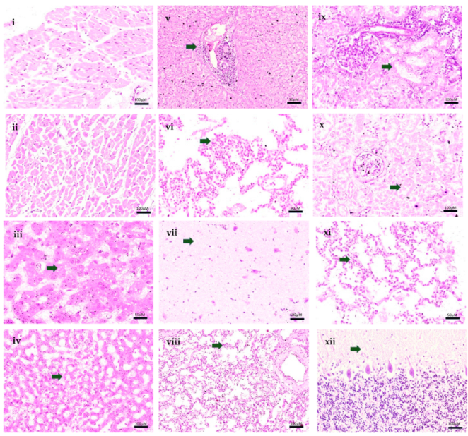
2. Case Report
3. Conclusions
Author Contributions
Funding
Institutional Review Board Statement
Informed Consent Statement
Data Availability Statement
Conflicts of Interest
References
- Yang, X.; Quam, M.B.M.; Zhang, T.; Sang, S. Global burden for dengue and the evolving pattern in the past 30 years. J. Travel Med. 2021, 28, taab146. [Google Scholar] [CrossRef] [PubMed]
- Du, M.; Jing, W.; Liu, M.; Liu, J. The Global Trends and Regional Differences in Incidence of Dengue Infection from 1990 to 2019: An Analysis from the Global Burden of Disease Study 2019. Infect. Dis. Ther. 2021, 10, 1625–1643. [Google Scholar] [CrossRef] [PubMed]
- Martins, A.B.S.; Alencar, C.H. Ecoepidemiology of dengue in Brazil: From the virus to the environment. One Health Implement. Res. 2022, 2, 1–14. [Google Scholar] [CrossRef]
- Cavalcanti, L.P.D.G.; Castiglioni, M.; da Silva, L.M.A.; Malta, D.L.; Pereira, R.A.D.C.; Silva-Junior, J.U.; Aguiar, M.G.; Pompeu, M.M.D.L.; Araújo, F.M.D.C.; Braga, D.N.D.M. Postmortem Diagnosis of Dengue as an Epidemiological Surveillance Tool. Am. J. Trop. Med. Hyg. 2016, 94, 187–192. [Google Scholar] [CrossRef] [Green Version]
- Maixenchs, M.; Anselmo, R.; Sanz, A.; Castillo, P.; Macete, E.; Carrilho, C.; Ordi, J.; Menéndez, C.; Bassat, Q.; Munguambe, K. Healthcare providers’ views and perceptions on post-mortem procedures for cause of death determination in Southern Mozambique. PLoS ONE 2018, 13, e0200058. [Google Scholar] [CrossRef] [Green Version]
- Maixenchs, M.; Anselmo, R.; Zielinski-Gutiérrez, E.; Odhiambo, F.O.; Akello, C.; Ondire, M.; Zaidi, S.S.H.; Soofi, S.B.; Bhutta, Z.A.; Diarra, K.; et al. Willingness to Know the Cause of Death and Hypothetical Acceptability of the Minimally Invasive Autopsy in Six Diverse African and Asian Settings: A Mixed Methods Socio-Behavioural Study. PLoS Med. 2016, 13, e1002172. [Google Scholar] [CrossRef] [Green Version]
- Castillo, P.; Ussene, E.; Ismail, M.R.; Jordao, D.; Lovane, L.; Carrilho, C.; Lorenzoni, C.; Lacerda, M.V.; Palhares, A.E.M.; Rodríguez-Carunchio, L.; et al. Pathological Methods Applied to the Investigation of Causes of Death in Developing Countries: Minimally Invasive Autopsy Approach. PLoS ONE 2015, 10, e0132057. [Google Scholar] [CrossRef]
- Bassat, Q.; Castillo, P.; Alonso, P.L.; Ordi, J.; Menéndez, C. Resuscitating the Dying Autopsy. PLoS Med. 2016, 13, e1001927. [Google Scholar] [CrossRef] [Green Version]
- Bassat, Q.; Castillo, P.; Martinez, M.J.; Jordao, D.; Lovane, L.; Hurtado, J.C.; Nhampossa, T.; Ritchie, P.S.; Bandeira, S.; Sambo, C.; et al. Validity of a minimally invasive autopsy tool for cause of death determination in pediatric deaths in Mozambique: An observational study. PLoS Med. 2017, 14, e1002317. [Google Scholar] [CrossRef] [Green Version]
- Menéndez, C.; Castillo, P.; Martinez, M.J.; Jordao, D.; Lovane, L.; Ismail, M.R.; Carrilho, C.; Lorenzoni, C.; Fernandes, F.; Nhampossa, T.; et al. Validity of a minimally invasive autopsy for cause of death determination in stillborn babies and neonates in Mozambique: An observational study. PLoS Med. 2017, 14, e1002318. [Google Scholar] [CrossRef] [Green Version]
- Schwartz, D.A. Autopsy and Postmortem Studies Are Concordant: Pathology of Zika Virus Infection Is Neurotropic in Fetuses and Infants With Microcephaly Following Transplacental Transmission. Arch. Pathol. Lab. Med. 2016, 141, 68–72. [Google Scholar] [CrossRef] [PubMed] [Green Version]
- Duarte-Neto, A.N.; Monteiro, R.; Johnsson, J.; Cunha, M.D.P.; POUR, S.Z.; Saraiva, A.C.; Ho, Y.-L.; Da Silva, L.F.F.; Mauad, T.; Zanotto, P.M.D.A.; et al. Ultrasound-guided minimally invasive autopsy as a tool for rapid post-mortem diagnosis in the 2018 Sao Paulo yellow fever epidemic: Correlation with conventional autopsy. PLoS Negl. Trop. Dis. 2019, 13, e0007625. [Google Scholar] [CrossRef] [PubMed] [Green Version]
- Argueta, V. La importancia de la autopsia en epidemias. Rev Me´d (Col Me´d Cir Guatem) 2020, 159, 2–3. [Google Scholar] [CrossRef]
- Martínez, M.J.; Massora, S.; Mandomando, I.; Ussene, E.; Jordao, D.; Lovane, L.; Muñoz-Almagro, C.; Castillo, P.; Mayor, A.; Rodriguez, C.; et al. Infectious cause of death determination using minimally invasive autopsies in developing countries. Diagn. Microbiol. Infect. Dis. 2016, 84, 80–86. [Google Scholar] [CrossRef]
- Melo, D.N.; Coelho, T.M.; Lima, G.R.P.; Fernandes, C.G.; Alves, B.C.F.D.B.; Araújo, F.M.D.C.; Monteiro, R.A.D.A.; Ordi, J.; Saldiva, P.H.D.N.; Cavalcanti, L.P.D.G. Use of minimally invasive autopsy during the COVID-19 pandemic and its possibilities in the context of developing countries. PLoS Negl. Trop. Dis. 2021, 15, e0009629. [Google Scholar] [CrossRef]
- Saldiva, P.H.N. Minimally invasive autopsies: A promise to revive the procedure. Autops. Case Rep. 2014, 4, 1–3. [Google Scholar] [CrossRef]
- Rakislova, N.; Marimon, L.; Ismail, M.; Carrilho, C.; Fernandes, F.; Ferrando, M.; Castillo, P.; Rodrigo-Calvo, M.; Guerrero, J.; Ortiz, E.; et al. Minimally Invasive Autopsy Practice in COVID-19 Cases: Biosafety and Findings. Pathogens 2021, 10, 412. [Google Scholar] [CrossRef]
- Reyes-Ruiz, J.M.; Campuzano-Vences, R.; Osuna-Ramos, J.F.; De Jesús-González, L.A.; Pérez-Méndez, M.J.; González-González, C.; Farfan-Morales, C.N.; Rivas-Tovar, L.; Dávila-González, E.; del Ángel, R.M.; et al. Case Report: Extrapulmonary Manifestations of COVID-19 and Dengue Coinfection. Am. J. Trop. Med. Hyg. 2021, 105, 363–367. [Google Scholar] [CrossRef]
- Araújo, F.; Brilhante, R.; Cavalcanti, L.; Rocha, M.; Cordeiro, R.; Perdigão, A.; Miralles, I.; Araújo, L.; Lima, E.; Sidrim, J. Detection of the dengue non-structural 1 antigen in cerebral spinal fluid samples using a commercially available enzyme-linked immunosorbent assay. J. Virol. Methods 2011, 177, 128–131. [Google Scholar] [CrossRef] [Green Version]
- Póvoa, T.F.; Alves, A.M.B.; Oliveira, C.A.B.; Nuovo, G.J.; Chagas, V.L.A.; Paes, M. The Pathology of Severe Dengue in Multiple Organs of Human Fatal Cases: Histopathology, Ultrastructure and Virus Replication. PLoS ONE 2014, 9, e83386. [Google Scholar] [CrossRef] [Green Version]
- Araujo, F.M.C.; Araujo, M.S.; Nogueira, R.M.R.; Brilhante, R.S.N.; Oliveira, D.N.; Rocha, M.F.G.; Cordeiro, R.A.; Sidrim, J.J.C. Central nervous system involvement in dengue: A study in fatal cases from a dengue endemic area. Neurology 2012, 78, 736–742. [Google Scholar] [CrossRef] [PubMed]


| Exam | 27 June 2021 (3 Days of Symptom) | 28 June 2021 (4 Days of Symptom) | Reference Values |
|---|---|---|---|
| Red Cells | 4.29 million/mm3 | 4.11 million/mm3 | 4.1 to 5.3 million/mm3 |
| Haemoglobin | 12.3 g/dL | 11.9 g/dL | 12 to 14.5 g/dL |
| Haematocrit | 35.9% | 35% | 36 to 43% |
| Leukocytes | 2300/mm3 | 5600/mm3 | 3400 to 10,800/mm3 |
| Neutrophils | 1955/mm3 | 4424/mm3 | 1500 to 8500/mm3 |
| Rod Neutrophils | 69/mm3 | 224/mm3 | 0 to 860/mm3 |
| Segmented Neutrophils | 1886/mm3 | 4200/mm3 | 1500 to 8500/mm3 |
| Eosinophils | 0/mm3 | 56/mm3 | 0 to 500/mm3 |
| Lymphocytes | 92/mm3 | 672/mm3 | 1500 to 6500/mm3 |
| Monocytes | 253/mm3 | 336/mm3 | 0 to 800/mm3 |
| Basophils | 0/mm3 | 0/mm3 | 0 to 200/mm3 |
| Platelets | 57,000/mm3 | 20,000/mm3 | 150 to 450 mil/mm3 |
| Atypical Lymphocytes | - | 112/mm3 | 0% |
| MPV | 8.3 fL | 7.5 fL | 9.2 to 12.6 fL |
| Ultrasensitive C-reactive protein | 2.96 mg/dL | 2.37 mg/dL | <0.10 mg/dL |
| Magnesium | - | 2.02 mg/dL | 2.02 to 2.75 mg/dL |
| Potassium | 4.0 mmol/L | 6.8 mmol/L | 3.5 to 5.1 mmol/L |
| Sodium | 136 mmol/L | 139 mmol/L | 136 to 145 mmol/L |
| AST | 83.3 U/L | 741.1 U/L | 17 to 33 U/L |
| ALT | 27.3 U/L | 248.9 U/L | 9 to 23 U/L |
| Urinary Urobilinogen | 3.0 mg/dL | - | < 1.0 mg/dL |
| Creatinine | - | 0.82 mg/dL | 0.32 to 0.61 mg/dL |
| Urea | - | 28.9 mg/dL | 19.2 to 46.2 mg/dL |
| TAP—prothrombin time | - | 16.8 s | 9.4 to 12.5 s |
| APTT- activated partial thromboplastin | - | 49.8 s | 25.1 to 36.5 s |
| Laboratory tests performed after death | |||
| Blood culture RT-PCR for SARS-CoV-2 qRT-PCR for dengue qRT-PCR for Zika qRT-PCR for chikungunya NS1 antigen | No microbial growth Not Detectable Positive Negative Negative Positive | ||
Publisher’s Note: MDPI stays neutral with regard to jurisdictional claims in published maps and institutional affiliations. |
© 2022 by the authors. Licensee MDPI, Basel, Switzerland. This article is an open access article distributed under the terms and conditions of the Creative Commons Attribution (CC BY) license (https://creativecommons.org/licenses/by/4.0/).
Share and Cite
Melo, D.N.; Lima, G.R.P.; Fernandes, C.G.; Teixeira, A.C.; Filho, J.B.; Araújo, F.M.C.; Araújo, L.C.; Siqueira, A.M.; Farias, L.A.B.G.; Monteiro, R.A.A.; et al. Post-Mortem Diagnosis of Pediatric Dengue Using Minimally Invasive Autopsy during the COVID-19 Pandemic in Brazil. Trop. Med. Infect. Dis. 2022, 7, 123. https://doi.org/10.3390/tropicalmed7070123
Melo DN, Lima GRP, Fernandes CG, Teixeira AC, Filho JB, Araújo FMC, Araújo LC, Siqueira AM, Farias LABG, Monteiro RAA, et al. Post-Mortem Diagnosis of Pediatric Dengue Using Minimally Invasive Autopsy during the COVID-19 Pandemic in Brazil. Tropical Medicine and Infectious Disease. 2022; 7(7):123. https://doi.org/10.3390/tropicalmed7070123
Chicago/Turabian StyleMelo, Deborah N., Giovanna R. P. Lima, Carolina G. Fernandes, André C. Teixeira, Joel B. Filho, Fernanda M. C. Araújo, Lia C. Araújo, André M. Siqueira, Luís A. B. G. Farias, Renata A. A. Monteiro, and et al. 2022. "Post-Mortem Diagnosis of Pediatric Dengue Using Minimally Invasive Autopsy during the COVID-19 Pandemic in Brazil" Tropical Medicine and Infectious Disease 7, no. 7: 123. https://doi.org/10.3390/tropicalmed7070123
APA StyleMelo, D. N., Lima, G. R. P., Fernandes, C. G., Teixeira, A. C., Filho, J. B., Araújo, F. M. C., Araújo, L. C., Siqueira, A. M., Farias, L. A. B. G., Monteiro, R. A. A., Ordi, J., Martinez, M. J., Saldiva, P. H. N., & Cavalcanti, L. P. G. (2022). Post-Mortem Diagnosis of Pediatric Dengue Using Minimally Invasive Autopsy during the COVID-19 Pandemic in Brazil. Tropical Medicine and Infectious Disease, 7(7), 123. https://doi.org/10.3390/tropicalmed7070123