Article

Multiplex Recombinase Polymerase Amplification Assay for Simultaneous Detection of Treponema pallidum and Haemophilus ducreyi in Yaws-Like Lesions

by
Michael Frimpong
1,2,*,
Shirley Victoria Simpson
3,
Hubert Senanu Ahor
1,2,
Abigail Agbanyo
2,
Solomon Gyabaah
2,
Bernadette Agbavor
2,
Ivy Brago Amanor
3,
Kennedy Kwasi Addo
3,
Susanne Böhlken-Fascher
4,
Jonas Kissenkötter
4,
Ahmed Abd El Wahed
4,5 and
Richard Odame Phillips
2
1
Department of Molecular Medicine, College of Health Sciences, Kwame Nkrumah University of Science and Technology, Kumasi AK-448, Ghana
2
Kumasi Centre for Collaborative Research in Tropical Medicine, Kwame Nkrumah University of Science and Technology, Kumasi AK-312, Ghana
3
Bacteriology Department, Noguchi Memorial Institute of Medical Research, University of Ghana, Accra GA-337, Ghana
4
Division of Microbiology and Animal Hygiene, Georg-August University, D-37077 Goettingen, Germany
5
Institute of Animal Hygiene and Veterinary Public Health, University of Leipzig, D-04103 Leipzig, Germany
*
Author to whom correspondence should be addressed.
Trop. Med. Infect. Dis. 2020, 5(4), 157; https://doi.org/10.3390/tropicalmed5040157
Submission received: 25 August 2020 / Revised: 28 September 2020 / Accepted: 29 September 2020 / Published: 6 October 2020

Abstract

:
Yaws is a skin debilitating disease caused by Treponema pallidum subspecies pertenue with most cases reported in children. World Health Organization (WHO) aims at total eradication of this disease through mass treatment of suspected cases followed by an intensive follow-up program. However, effective diagnosis is pivotal in the successful implementation of this control program. Recombinase polymerase amplification (RPA), an isothermal nucleic acid amplification technique offers a wider range of differentiation of pathogens including those isolated from chronic skin ulcers with similar characteristics such as Haemophilus ducreyi (H. ducreyi). We have developed a RPA assay for the simultaneous detection of Treponema pallidum (T. pallidum) and H. ducreyi (TPHD-RPA). The assay demonstrated no cross-reaction with other pathogens and enable detection of T. pallidum and H. ducreyi within 15 min at 42 °C. The RPA assay was validated with 49 clinical samples from individuals confirmed to have yaws by serological tests. Comparing the developed assay with commercial multiplex real-time PCR, the assay demonstrated 94% and 95% sensitivity for T. pallidum and H. ducreyi, respectively and 100% specificity. This simple novel TPHD-RPA assay enables the rapid detection of both T. pallidum and H. ducreyi in yaws-like lesions. This test could support the yaws eradication efforts by ensuring reliable diagnosis, to enable monitoring of program success and planning of follow-up interventions at the community level.

1. Introduction

Yaws, most common of the three endemic non-venereal treponemal infections is caused by Treponema pallidum sp. pertenue [1,2]. Yaws presents as lesions of the skin (papilloma), which ulcerates and progresses to cause destructive lesions of bone and cartilage leading to chronic disfigurement and disability when left untreated. The transmission is via direct (non-sexual) contact with fluid exudate from the lesion of an infected person [3]. The disease mostly affects children living in poor communities of West Africa, Latin America, Asia, and the Pacific where access to healthcare is limited [3]. Estimates in 2012 show that over 89 million people are affected with Yaws in 15 countries in Africa, Southeast Asia, and the Pacific region [4].
The WHO aimed to achieve total eradication of yaws by 2020 through a comprehensive large-scale treatment strategy, called the Morges strategy [5]. This strategy consists of an initial mass drug therapy with oral intake of azithromycin in endemic communities followed by an active surveillance every 6 months to actively detect and treat remaining cases and their contacts. Pilot implementation of this strategy in endemic countries such as Ghana, Congo, Papua New Guinea, Vanuatu, and the Solomon Islands have shown promising results, with the need for scale-up [6]. However, the success of the Morges strategy is hinged on the selection of the most appropriate diagnostic assay at each stage of the eradication effort to ensure a complete halt in the transmission of yaws [7]. The occurrence of Haemophilus ducreyi, as a common cause of chronic skin ulcers similar to yaws in endemic areas also compounds the challenges of diagnosing yaws [8,9]. H. ducreyi is a common cause of genital ulcers called chancroid, and it is now increasingly recognized as a cause of non-genital skin ulcers in children in developing countries [8]. In patients with yaws-like lesions, differential diagnosis other than those cause by T. pallidum pertenue is important for accurate reporting and management of cases.
Traditional serological tests such as Treponema Pallidum Particle Agglutination assay (TPPA) and Rapid Plasma Reagin test (RPR) have remained the common diagnostic tool for yaws, over the years [1]. These tests are encumbered with issues of low sensitivity and specificity hence are not appropriate for use during the follow-up stage of the eradication strategy [10]. The Dual Path Platform (DPP) Syphilis Screen-and-Confirm assay (Chembio Diagnostic Systems, Inc., Medford, NY, USA), which enables the simultaneous detection of antibodies to treponemal and non-treponemal antigens, is accurate for the confirmation of clinically suspected cases. This point-of-care (POC) test has increased sensitivity in high seropositive individuals and low sensitivity in individuals with low antibody titre [10]. In addition, issues of co-infections cannot be overlooked as reactive syphilis serology caused by latent yaws has been observed in ulcers with H. ducreyi [8]. The WHO recommends the use of molecular diagnostic tests as part of the case definition in yaws eradication effort [7]. This highlights the need for a sensitive and specific diagnostic test in confirming yaws suspected cases even after mass drug administration (MDA).
Nucleic acid amplification techniques, especially polymerase chain reaction (PCR), can address the issues of diagnosis of yaws, due to their high clinical sensitivity and specificity to T. pallidum and H. ducreyi. This DNA-based diagnostic tests could play a central role in the yaws eradication strategy by ensuring effective diagnosis and surveillance after MDA [7,8,11]. However, PCR techniques can only be applied in a reference laboratory and not at the point of care (POC) in endemic communities. As such, the need for diagnostic techniques that do not involve costly equipment and can be used in remote or resource limited areas have been indicated [12]. The use of isothermal amplification techniques such as Loop-mediated amplification (LAMP), for on-site screening has been developed as a possible POC test for chronic skin ulcers caused by T. pallidum and H. ducreyi [13,14,15]. These LAMP assays for T. pallidum and H. ducreyi demonstrated high sensitivity (85−100%) and specificity (85−100%). Nevertheless, the use of unextracted DNA reduced the sensitivity of T. pallidum [15]. LAMP assays are generally performed at 60−65 °C and results are obtained after 30−60 min., LAMP uses four to six primers which are not easy to design especially for highly variable pathogens under field conditions. In contrast, recombinase polymerase amplification (RPA), has emerged as a better field-deployable molecular diagnostic tool, since it yields readily readable results within a shorter turnaround time (less than 15 min) and it is less complicated to run at a lower temperature (37–42 °C) compared to techniques such as LAMP [16,17]. RPA have been performed at low resource settings in a mobile suitcase laboratory coupled with field-friendly DNA extraction techniques such as SpeedXtract (Qiagen, Hilden, Germany) for effective diagnosis of infectious diseases [18,19,20]. The technique opens the door to extending the application of DNA amplification in the field or point-of-need to differentiate between pathogens commonly isolated from chronic skin ulcers [21]. This isothermal nucleic acid amplification technique can be used at all stages of yaws eradication efforts, especially during the follow-up phase, monitoring of azithromycin resistance as well as identifying individuals with Treponema pallidum and H. ducreyi co-infection in low resource settings. In this study, we have developed a RPA assay for the simultaneous detection of T. pallidum and H. ducreyi (TPHD-RPA) in DPP confirmed yaws patients.

2. Materials and Methods

2.1. Clinical Samples

Samples were obtained from an ongoing skin neglected tropical diseases (NTDs) project (Yaws, Leprosy and Buruli ulcer) community outreach program being conducted in Wassa Amenfi East, Upper Denkyira East, Upper Denkyira West, Upper West Akyem, Akwapim North and Sekyere afram plains districts of Ghana. In brief, clinically suspected yaws patients were first screened with immunochromatographic assay (SD Bioline Syphilis 3.0 RDT kit, Standard Diagnostics Inc., Suwon, South Korea) for the qualitative detection of antibodies of all isotypes (IgG, IgM, IgA) against Treponema pallidum (TP). Positive samples by this test were then tested with the DPP Syphilis Screen-and-Confirm assay (Chembio Diagnostic Systems, Inc., Medford, NY, USA), according to manufacturer instructions for the detection of antibodies to treponemal and non-treponemal antigens. Swab samples were taken for both ulcers and wet papilloma, while scab was removed from dry or closed papilloma from patients who were DPP positive. Samples were placed in a sterile patient ID pre-labeled cryotubes containing 700 μL cell lysis solution (CLS, Qiagen, Hilden, Germany) and transported to Kumasi Center for Collaborative Research in Tropical Medicine (Kumasi, Ghana) and Noguchi Memorial Institute for Medical Research (Accra, Ghana) depending on the proximity of the district to the reference laboratory for further molecular analysis.
This study used archived samples with consent from study participants for future use of samples. Approval was also sort from the Committee for Human Research Ethics and Publication (Ref: CHRPE/AP/122/17), School of Medical Sciences, Kwame Nkrumah University of Science and Technology (Kumasi, Ghana).

2.2. DNA Extraction

DNA extraction was performed using the Gentra Puregene Tissue Kit (Qiagen GmbH, Hilden, Germany) according to manufacturer’s instructions. Briefly, samples stored in lysis buffer were vortexed for 2 min, swab sticks removed from the solution and then centrifuged for 1 min at 13,000 rpm to pellet the cells. Supernatant was carefully discarded. A total of 300 µL of CLS was added to the pellet after which 15 µL of lysozyme was added and mixed by inverting 25 times. It was then incubated at 37 °C for 30 min and at 80 °C for 5 min to lyse the cells. Proteins were precipitated by adding 100 μL of protein precipitation solution (Qiagen, Hilden, Germany) to the sample, vortexed 20 s and centrifuge at 13,000 rpm to pellet. Supernatant was transferred into a pre-labelled 1.5 mL tube (Eppendorf AG, Hamburg, Germany), containing 300 μL isopropanol and 2 μL glycogen. This mixture was mixed by inverting gently 50 times and then centrifuged for 1 min at 13,000 rpm to pellet DNA. The supernatant was carefully discarded and 300 µL of 70% ethanol added to wash the pelleted DNA and then centrifuged for 1 min. Alcohol supernatant was carefully discarded and DNA air dried for 5−30 min. The extracted DNA was resuspended in 100 µL DNA Hydration Solution (Qiagen, Hilden, Germany), vortex for 5 s and incubated at 65 °C for 1 hr. Extracted DNA was stored at –20 °C till it was used for T. pallidum and H. ducreyi RPA (TPHD-RPA) assay and the multiplex real-time PCR (qPCR).

2.3. T. pallidum and H. ducreyi Multiplex qPCR Assay

The multiplex qPCR assay was performed with RealCycler® universal kit (Progenie molecular, Valencia, Spain) for the detection of the PolA and HhdA specific genes for T. pallidum and H. ducreyi, respectively, on a Bio-Rad CFX96 Real-time PCR detection system (Bio-Rad Laboratories, Paris, France) according to manufacturer instructions. Briefly, 14 µL of RealCycler® Universal AmpliMix (qPCR mastermix reagent) was pipetted into reaction tubes and 6 µL of DNA template was used to yield a reaction volume of 20 µL. Positive and negative controls were also included in each run. Cycling condition of PCR are as follows one (1) cycle of initial denaturing at 95 °C for 15 min followed by 45 cycles of 95 °C (denaturing) for 5 s, 60 °C (annealing) for 30 s and 72 °C (extension) for 30 s.

2.4. T. pallidum and H. ducreyi RPA (TPHD-RPA) Assay

2.4.1. Molecular Standard

Molecular DNA standard of 600 bp, made up of 300 bp T. pallidum PolA gene (GenBank: U57757.1, range 1836−2135) and 300 bp of H. ducreyi hemolytic cytotoxin HhdA gene (GenBank: U32175.1, range 5031−5330) was synthesized by GeneArt (Invitrogen, Darmstadt, Germany). The number of DNA molecules per microliter was determined using the equation described previously [22]. Serial dilutions between 106 and 100 of the molecular standard were prepared.

2.4.2. TPHD-RPA Primers and Probes

PolA and HhdA genes of T. pallidum and H. ducreyi respectively, were used as target sequences for RPA detection. RPA primers and probes were designed in accordance with the TwistDx manufacturer’s recommendations (TwistDx Limited, Maidenhead, United Kingdom). Three forward primers (FPs), three reverse primers (RPs) and one exo probe (P) each for T. pallidum and H. ducreyi (Supplementary File S1: Table S1) were designed by using the Molecular Evolution Genetics Analysis version 7 software (MEGA, State College, PA, USA). In silico BLAST analysis of these oligonucleotide demonstrated high specificity to the T. pallidum and H. ducreyi. All the primers were produced by Eurofins Genomics (Ebersberg, Germany) and the exo probe by TibMolBiol (Berlin, Germany).

2.4.3. TPHD-RPA Assay

Preliminary screening of three forward primers, three reverse primers and one exo probe combinations were tested with 105 molecules/5 µL of molecular standard in singleplex RPA assay for T. pallidum and H. ducreyi using the TwistAmp Exo kits according to the manufacturer’s instructions in a final reaction volume of 50 µL (TwistDX, Cambridge, UK). The reaction for each target contained 2.1 µL each of 10 µM forward and reverse primers, 0.8 µL each of 10 µM probe, 29.5 µL rehydration buffer, 8.0 µL DNase-free water, 2.5 µL of 280 mM Magnesium Acetate (MgOAc). Reaction tubes were incubated in an isothermal fluorometer (T8-ISO Axxin Pty Ltd., Victoria, Australia). Using the T8-ISO Desktop application, we programmed the fluorometer to detect the lowest dilutions that met criteria for distinguishing positive samples from negative controls as indicated previously [21]. All tests were run at 42 °C for 15 min with mixing after 4 min. To be considered positive, a sample required either a gradient or an amplitude (plateau florescence) of at least 900 mV over a 40 s sliding window during the amplification phase of the test (5−15 min). The best primer and probe combination for T. pallidum and H. ducreyi (Table 1) were later produced as mix for subsequent RPA assay using TwistAmp Exo kit, with minor modification of manufacturer’s instructions. The reaction volume was 50 µL and contained 2.5 µL each of TP and HD 10 µM primer/probe mix, 29.5 µL rehydration buffer, 8.0 µL DNase-free water, 2.5 µL of 280 mM Magnesium Acetate (MgOAc). Five microliters of DNA extract were used.

2.4.4. Analytical Singleplex RPA Assay Sensitivity and Specificity

The sensitivity of the singleplex RPA assay was evaluated using as serial dilations (106 to 100 copies/µL) of TP/HD DNA molecular standard in eight separate reaction in triplicate. The threshold time and copies of DNA molecules detected was plotted, and a semi logarithm (log) regression was calculated using PRISM 8.0 software (GraphPad Software, San Diego, CA, USA). The specificity of TPHD-RPA assay was evaluated using genomic DNA of other pathogens (Supplementary File S2: Table S2).

2.4.5. Clinical Performance of TPHD-RPA Assay

The clinical performance of the TPHD-RPA assay was evaluated with forty-nine (49) DPP positive yaws suspected samples. The result of the TPHD-RPA assay was compared with the results T. pallidum and H. ducreyi multiplex qPCR assay.

2.4.6. Statistical Analysis

All data were entered in Microsoft Excel 2016 while GraphPad Prism v.8 (GraphPad software, San Diego, CA, USA) was used for data analysis and drawing graphs. The limit of detection (LOC) of both qPCR and singleplex RPA assay was determined using probit regression analysis (GraphPad Prism v.8). Descriptive information of patients recruited such as percentages, frequency, median and interquartile ranges were obtained using descriptive statistics. Contingency tables were used to determine the sensitivity, specificity and the predictive values of the TPHD-RPA assay, using qPCR results as the gold standard.

3. Results

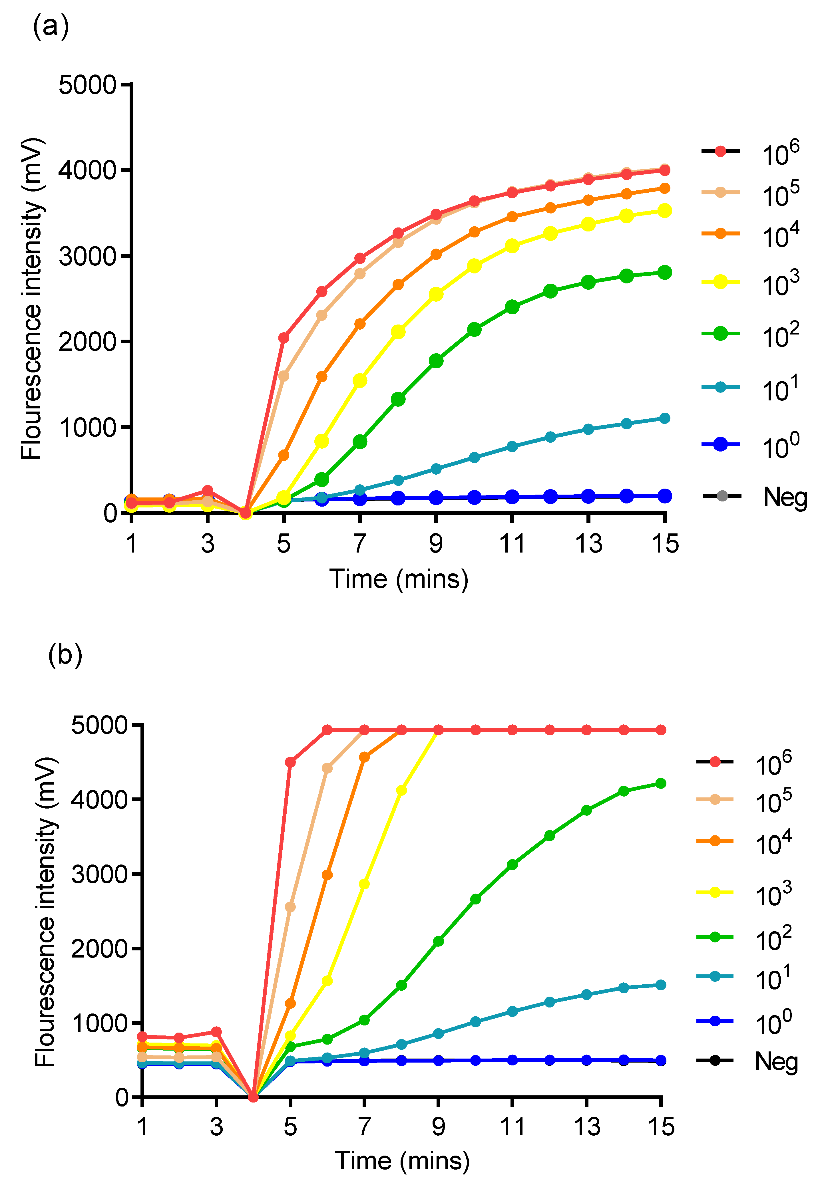
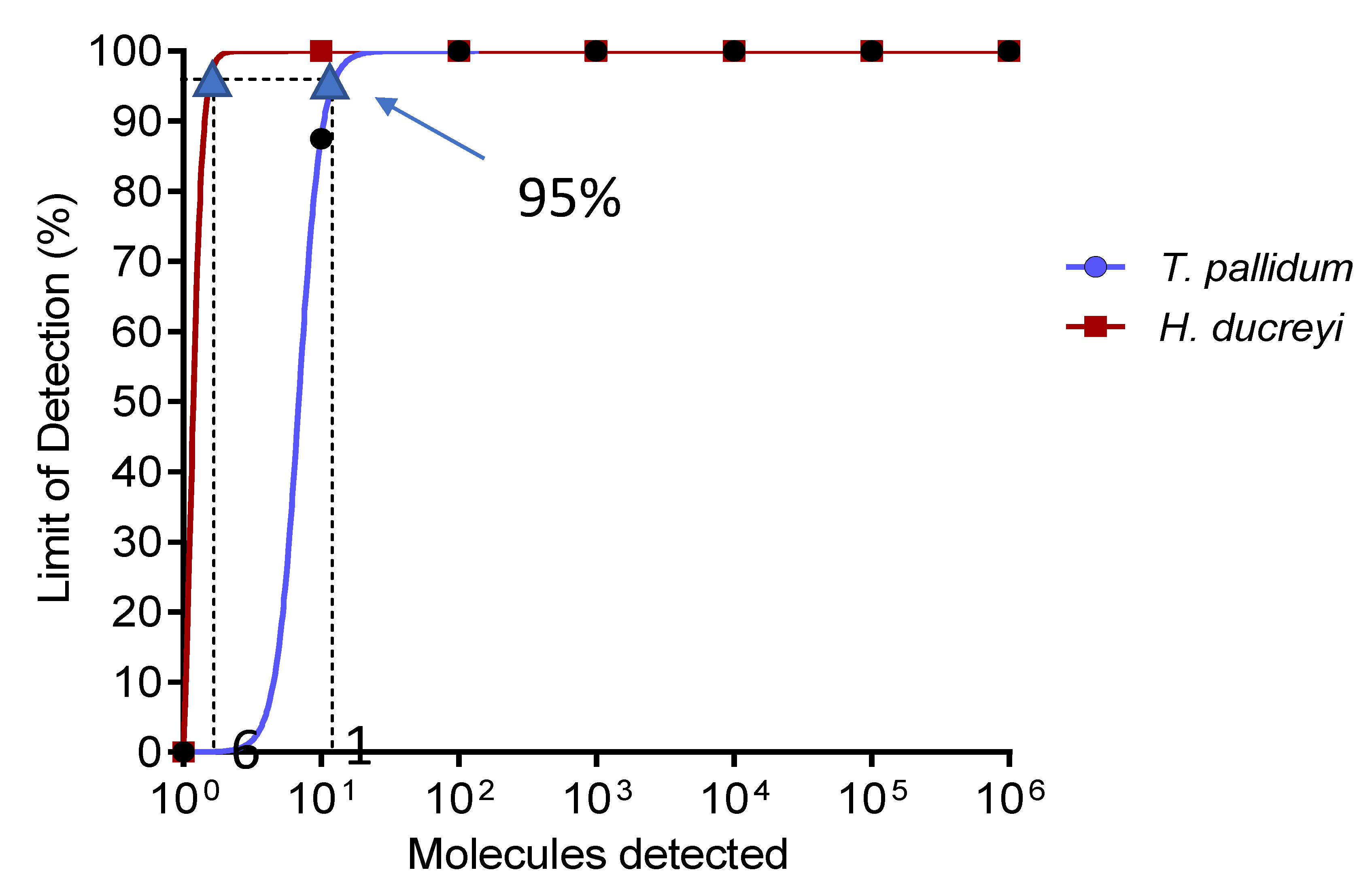
3.1. Analytical Sensitivity of Singleplex RPA Assay and Specificity of TPHD-RPA Assay

The nine oligonucleotide combinations for each assay were screened with the molecular DNA standard. Primers for TP (TP_RPA_FP3 and TP_RPA_RP2) and HD (Hd_RPA_FP3 and Hd_RPA_RP1) when combined TP_RPA_P1 and Hd_RPA_P1 respectively (Table 1) were able to amplify down to ten (10) of DNA molecules (Figure 1).
The gradient or an amplitude florescence of at least 900 mV signal over a 40 s sliding window for both singleplex RPA assay for 106 to 103 copies of DNA molecular standard was at 5−6 min, 6−7 min for 102 copies, and 8−9 min for 10 copies (Figure 1).
Using probit analysis, the limit of detection of the singleplex RPA assay at 95% probability was 11 and 6 copies of DNA molecular standard for T. pallidum and H. ducreyi respectively (Figure 2). The assay showed no cross-reactivity when tested with a panel of 29 genomic DNA of bacterial and parasite species (Supplementary File S2: Table S2).

3.2. Clinical Performance of TPHD-RPA Assay

Forty-nine sample from DPP positive participants were included for the evaluation of the TPHD-RPA assay for T. pallidum and H. ducreyi with majority being males (N = 31, 63%). The median age of participants was 10 years. They presented with ulcers (N = 35, 71%), papillomas (N = 12, 25%) and both (N = 2, 4%). Using qPCR, 10 lesions were identified as T. pallidum lesions, 14 as H. ducreyi lesions, 7 lesions coinfected with T. pallidum and H. ducreyi, and 18 (37%) negative for both pathogens.
The TPHD-RPA assay also correctly identified all samples containing only one pathogen and five samples (71%) being coinfected. Two (4%) samples positive for both pathogens by qPCR, were either positive for either T. pallidum or H. ducreyi and not both. Using qPCR as the gold standard test, the diagnostic sensitivity of the TPHD-RPA assay for T. pallidum and H. ducreyi was 94% and 95%, respectively (Table 2). Kappa coefficients (κ), ranging from 0.95 to 1 for the detection of T. pallidum and H. ducreyi demonstrate an excellent diagnostic agreement between qPCR and our developed TPHD-RPA. Additionally, excellent agreement between qPCR and TPHD-RPA (κ = 0.9) was demonstrated for lesions coinfected with both pathogens. All negative qPCR samples were negative by TPHD-RPA assay demonstrating 100% specificity (Table 2).

4. Discussion

Eradication of yaws is a major priority by WHO with the detection of H. ducreyi, an essential component in the management of yaws patients. In yaws endemic communities, clinical diagnosis combined with serological testing is the sole means of diagnosing suspected yaws cases. Nucleic acid amplification techniques can help overcome the limitations of serological tests, by enabling the determination of whether yaws like lesions are caused by either T. pallidum or H. ducreyi or both. Hence these tests are needed in the WHO yaws eradication strategy [7,8,10,11].
In this study, we developed a real-time recombinase polymerase assay for the simultaneous detection of T. pallidum and H. ducreyi (TPHD-RPA) in yaws-like lesions. The developed TPHD-RPA assay was highly specific to the target organisms while the limit of detection of singleplex RPA assay was 11 and 6 copies of DNA for T. pallidum and H. ducreyi respectively. These analytical sensitivities are comparable to multiplex qPCR whose sensitivity is 100 and 1 copy for T. pallidum and H. ducreyi respectively (RealCycler® universal TPHD-U TPHD-G protocol V.4). The turnaround time of 15 min for the developed TPHD-RPA assay, suggest that this assay could provide rapid molecular diagnosis of T. pallidum and H. ducreyi lesions as well as a distinction between these two pathogens in areas where co-infection has been reported to enhance case management [8,9].
Polymerase chain reaction provides high sensitivity and specificity for diagnosing yaws and H. ducreyi lesions [7,8,11]. Operating this technique at the point of care is very challenging. This is because of its requirement for sophisticated laboratories, equipped with expensive equipment such as a thermocycler [12], which can cost more than 15 times as much as isothermal fluorometers required for the performance RPA. RPA performance requires less costly equipment and can be performed in less resource settings in a mobile suitcase laboratory [18,20]. As such, TPHD-RPA could be an alternative molecular diagnostic tool which can be essential in the yaws eradication strategy.
Recently, the use of Loop-mediated amplification (LAMP) for on-site screening has been proposed as a future point of care test for chronic skin ulcers caused by T. pallidum and H. ducreyi (TPHD-LAMP) [13,14]. These TPHD-LAMP assays had a limit of detection of 300−600 DNA copies with a diagnostic performance of 85−92% sensitivity and 85−96% specificity [14]. A T. pallidum LAMP assay has also been developed with 100% sensitivity and specificity reported [15]. The sensitivity of this singleplex assay was reduced (67%) when evaluated with unextracted DNA samples. In our study, the developed TPHD-RPA assay showed 86−100% sensitivity and 100% specificity. These diagnostic performances of both TPHD-RPA and TPHD-LAMP suggest the potential use of these amplification techniques as an alternative to PCR for confirming clinically suspected yaws cases. However, RPA has an added advantage of rapidly amplifying nucleic acid target at 39–42 °C within a short test time of 15 min compared to LAMP which requires a higher temperature of 60−65 °C with a turnaround time of 30 min to an hour [13,14,15]. All critical RPA reagents thus enzymes/proteins and deoxy-nucleoside triphosphates are lyophilized into a single pellet which ensures the stability of reagents at room temperature for at least six months, reduces contamination, facilitates easy transportation of reagents and simplifies RPA applicability in low resource settings [23,24]. In addition, RPA assays have been evaluated with field friendly extraction techniques such as simple boiling of samples [24,25,26], SpeedXtract (Qiagen, Germany) [18,27,28] and GenoLyse DNA extraction Kit (Hain Life science, Nehren, Germany) [21] with little or no effect on RPA diagnostic performance. These DNA extraction techniques are less labor intensive with only one/two manipulation or pipetting steps and do not require sophisticated laboratory settings and hence applicable in low resource settings or mobile suitcase laboratories [18,27,28]. This suggests that the developed TPHD-RPA can be used with such DNA extraction techniques in low resource settings.
This study used DNA extracted with Gentra Puregene Tissue commercial Kit (Qiagen GmbH, Hilden, Germany) in a well-equipped laboratory. As such the novel TPHD assay should be evaluated with the above-named field friendly extraction procedures. This would help ascertain the full diagnostic performance of the developed assay.

5. Conclusions

We have developed a simple novel TPHD-RPA assay which can enable rapid differential detection of T. pallidum and H. ducreyi in yaws-like lesions. The diagnostic performance of the TPHD-RPA suggests its potential to increase access to molecular diagnosis of yaws, especially in low resource yaws endemic communities. This test, when combine with an effective field friendly extraction technique could support all yaws eradication efforts by ensuring reliable diagnosis, enabling monitoring of eradication effort success and planning of follow-up interventions at the community level.

Supplementary Materials

The following are available online at https://www.mdpi.com/2414-6366/5/4/157/s1, Figure S1: Primer combination testing for PolA of T. pallidum, Figure S2: Primer combination testing for hhdA of H. ducreyi, Table S1: Sequences for all Primers and Probes Used in the Assay Development, Table S2: List of Bacteria and Parasite Species and Strains Used for Determining the Cross-Reactivity of the Duplex RPA Assay.

Author Contributions

Conceptualization, M.F. and A.A.E.W.; methodology, M.F., S.V.S., H.S.A., S.B.-F. and J.K.; software, A.A.E.W.; validation, M.F., J.K., K.K.A., and R.O.P.; formal analysis, M.F., S.V.S., H.S.A.. and S.G.; investigation, S.V.S., A.A., S.G., B.A., I.B.A., and S.B.-F.; resources, A.A.E.W., K.K.A., and R.O.P.; data curation, M.F., S.V.S., A.A., S.G., B.A., and I.B.A.; writing—original draft preparation, M.F. and H.S.A.; writing—review and editing, S.V.S., A.A., S.G., B.A., I.B.A., S.B.-F., J.K., K.K.A., A.A.E.W., and R.O.P.; visualization, M.F. and A.A.E.W.; supervision, K.K.A., and R.O.P.; project administration, M.F., K.K.A., and R.O.P.; funding acquisition, M.F. Addo K.K., A.A.E.W., and R.O.P. All authors have read and agreed to the published version of the manuscript.

Funding

This research was part of the laboratory support for the control and elimination of NTDs in Ghana project funded by Fundación Anesvad. The lead author was funded by the second European and Developing Countries Clinical Trials Partnership (EDCTP2) career development fellowship and TWAS-DFG Cooperation Visits Programme for Scientists from sub-Saharan Africa.

Acknowledgments

This project was made possible by the joint efforts of the various District Health Directorates, the field technicians and the National Buruli ulcer/Yaws Control Programme for their technical and administrative support. We also want to acknowledge the immense contribution of the community volunteers and the participants whose samples were included in the evaluation of these assay.

Conflicts of Interest

The authors declare no conflict of interest. The funders had no role in the design of the study; in the collection, analyses, or interpretation of data; in the writing of the manuscript, or in the decision to publish the result.

References

  1. Mitjà, O.; Asiedu, K.; Mabey, D. Yaws. Lancet 2013, 381, 763–773. [Google Scholar] [CrossRef]
  2. Marks, M.; Solomon, A.W.; Mabey, D.C. Endemic treponemal diseases. Trans. R. Soc. Trop. Med. Hyg. 2014, 108, 601–607. [Google Scholar] [CrossRef] [PubMed]
  3. WHO. WHO Fact Sheet on Yaws 2017. Available online: http://www.who.int/mediacentre/factsheets/fs316/en/ (accessed on 8 November 2017).
  4. Mitjà, O.; Marks, M.; Konan, D.J.P.; Ayelo, G.; Gonzalez-Beiras, C.; Boua, B.; Houinei, W.; Kobara, Y.; Tabah, E.N.; Nsiire, A.; et al. Global epidemiology of yaws: A systematic review. Lancet Glob. Health 2015, 3, e324–e331. [Google Scholar] [CrossRef] [Green Version]
  5. WHO. Eradication of yaws—The Morges Strategy. Wkly. Epidemiol. Rec. 2012, 87, 189–200. [Google Scholar]
  6. Mitjà, O.; Houinei, W.; Moses, P.; Kapa, A.; Paru, R.; Hays, R.; Lukehart, S.; Godornes, C.; Bieb, S.V.; Grice, T.; et al. Mass Treatment with Single-Dose Azithromycin for Yaws. N. Engl. J. Med. 2015, 372, 703–710. [Google Scholar] [CrossRef] [Green Version]
  7. Marks, M.; Mitjà, O.; Vestergaard, L.S.; Pillay, A.; Knauf, S.; Chen, C.; Bassat, Q.; Martin, D.L.; Fegan, D.; Taleo, F.; et al. Challenges and key research questions for yaws eradication. Lancet Infect. Dis. 2015, 3099, 1–6. [Google Scholar] [CrossRef] [Green Version]
  8. Mitjà, O.; Lukehart, S.A.; Pokowas, G.; Moses, P.; Kapa, A.; Godornes, C.; Robson, J.; Cherian, S.; Houinei, W.; Kazadi, W.; et al. Haemophilus ducreyi as a cause of skin ulcers in children from a yaws-endemic area of Papua New Guinea: A prospective cohort study. Lancet Glob. Health 2014, 2, 235–241. [Google Scholar] [CrossRef] [Green Version]
  9. Marks, M.; Chi, K.H.; Vahi, V.; Pillay, A.; Sokana, O.; Pavluck, A.; Mabey, D.C.; Chen, C.Y.; Solomon, A.W. Haemophilus ducreyi associated with skin ulcers among children, Solomon Islands. Emerg. Infect. Dis. 2014, 20, 1705–1707. [Google Scholar] [CrossRef] [Green Version]
  10. Marks, M.; Goncalves, A.; Vahi, V.; Sokana, O.; Puiahi, E.; Zhang, Z.; Dalipanda, T.; Bottomley, C.; Mabey, D.; Solomon, A.W. Evaluation of a Rapid Diagnostic Test for Yaws Infection in a Community Surveillance Setting. PLoS Negl. Trop. Dis. 2014, 8, 1–5. [Google Scholar] [CrossRef] [Green Version]
  11. Chi, K.H.; Danavall, D.; Taleo, F.; Pillay, A.; Ye, T.; Nachamkin, E.; Kool, J.L.; Fegan, D.; Asiedu, K.; Vestergaard, L.S.; et al. Molecular differentiation of treponema pallidum subspecies in skin ulceration clinically suspected as yaws in vanuatu using real-time multiplex PCR and serological methods. Am. J. Trop. Med. Hyg. 2015, 92, 134–138. [Google Scholar] [CrossRef] [Green Version]
  12. Pahil, S.; Goyal, K. Rapid molecular diagnosis of chronic skin ulcers. Lancet Glob. Health 2014, 2, e385. [Google Scholar] [CrossRef] [Green Version]
  13. Becherer, L.; Bakheit, M.; Frischmann, S.; Stinco, S.; Borst, N.; Zengerle, R.; Von Stetten, F. Simplified Real-Time Multiplex Detection of Loop-Mediated Isothermal Amplification Using Novel Mediator Displacement Probes with Universal Reporters. Anal. Chem. 2018, 90, 4741–4748. [Google Scholar] [CrossRef] [PubMed]
  14. Becherer, L.; Knauf, S.; Marks, M.; Lueert, S.; Frischmann, S.; Borst, N.; Von Stetten, F.; Bieb, S.; Adu-Sarkodie, Y.; Asiedu, K.; et al. Multiplex mediator displacement loop-mediated isothermal amplification for detection of treponema pallidum and haemophilus ducreyi. Emerg. Infect. Dis. 2020, 26, 282–288. [Google Scholar] [CrossRef] [PubMed]
  15. Basing, L.A.W.; Simpson, S.V.; Adu-Sarkodie, Y.; Linnes, J.C. A Loop-Mediated Isothermal Amplification Assay for the Detection of Treponema pallidum subsp. pertenue. Am. J. Trop. Med. Hyg. 2020, 103, 253–259. [Google Scholar] [CrossRef]
  16. Piepenburg, O.; Williams, C.H.; Stemple, D.L.; Armes, N.A. DNA Detection using Recombination Proteins. PLoS Biol. 2006, 4, 1115–1121. [Google Scholar] [CrossRef] [PubMed]
  17. Daher, R.K.; Stewart, G.; Boissinot, M.; Bergeron, M.G. Recombinase Polymerase Amplification for Diagnostic Applications. Clin. Chem. 2016, 62, 947–958. [Google Scholar] [CrossRef] [PubMed]
  18. Mondal, D.; Ghosh, P.; Khan, M.A.A.; Hossain, F.; Böhlken-Fascher, S.; Matlashewski, G.; Kroeger, A.; Olliaro, P.; Abd El Wahed, A. Mobile Suitcase Laboratory for Rapid Detection of Leishmania donovani using Recombinase Polymerase Amplification Assay. Parasit. Vectors 2016, 9, 281. [Google Scholar] [CrossRef] [Green Version]
  19. Abd El Wahed, A.; Patel, P.; Faye, O.; Thaloengsok, S.; Heidenreich, D.; Matangkasombut, P.; Manopwisedjaroen, K.; Sakuntabhai, A.; Sall, A.A.; Hufert, F.T.; et al. Recombinase polymerase amplification assay for rapid diagnostics of dengue infection. PLoS ONE 2015, 10, e0129682. [Google Scholar] [CrossRef]
  20. Weidmann, M.; Faye, O.; Faye, O.; Abd El Wahed, A.; Patel, P.; Batejat, C.; Manugerra, J.C.; Adjami, A.; Niedrig, M.; Hufert, F.T.; et al. Development of Mobile Laboratory for Viral Hemorrhagic Fever Detection in Africa. J. Infect. Dis. 2018, 218, 1622–1630. [Google Scholar] [CrossRef] [Green Version]
  21. Frimpong, M.; Ahor, H.S.; Abd El Wahed, A.; Agbavor, B.; Sarpong, F.N.; Laing, K.; Wansbrough-Jones, M.; Phillips, R.O. Rapid detection of mycobacterium ulcerans with isothermal recombinase polymerase amplification assay. PLoS Negl. Trop. Dis. 2019, 13, e0007155. [Google Scholar] [CrossRef] [Green Version]
  22. Zhong, C.; Peng, D.; Ye, W.; Chai, L.; Qi, J.; Yu, Z.; Ruan, L.; Sun, M. Determination of plasmid copy number reveals the total plasmid DNA amount is greater than the chromosomal DNA amount in Bacillus thuringiensis YBT-1520. PLoS ONE 2011, 6, e16025. [Google Scholar] [CrossRef] [PubMed] [Green Version]
  23. Boyle, D.S.; Lehman, D.A.; Lillis, L.; Peterson, D.; Singhal, M.; Armes, N.; Parker, M.; Piepenburg, O.; Overbaugh, J. Rapid detection of HIV-1 proviral DNA for early infant diagnosis using recombinase polymerase amplification. MBio 2013, 4, e00135-13. [Google Scholar] [CrossRef] [PubMed] [Green Version]
  24. Lobato, I.M.; O’Sullivan, C.K. Recombinase polymerase amplification: Basics, applications and recent advances. Trends Anal. Chem. 2018, 98, 19–35. [Google Scholar] [CrossRef] [PubMed]
  25. Castellanos-Gonzalez, A.; Saldarriaga, O.A.; Tartaglino, L.; Gacek, R.; Temple, E.; Sparks, H.; Melby, P.C.; Travi, B.L. A Novel Molecular Test to Diagnose Canine Visceral Leishmaniasis at the Point of Care. Am. J. Trop. Med. Hyg. 2015, 93, 970–975. [Google Scholar] [CrossRef] [Green Version]
  26. Rohrman, B.; Richards-Kortum, R. Inhibition of Recombinase Polymerase Amplification by Background DNA: A lateral Flow-Based Method for Enriching Target DNA. Anal. Chem. 2015, 87, 1963–1967. [Google Scholar] [CrossRef]
  27. Gunaratna, G.; Manamperi, A.; Böhlken-Fascher, S.; Wickremasinge, R.; Gunawardena, K.; Yapa, B.; Pathirana, N.; Pathirana, H.; de Silva, N.; Sooriyaarachchi, M.; et al. Evaluation of rapid extraction and isothermal amplification techniques for the detection of Leishmania donovani DNA from skin lesions of suspected cases at the point of need in Sri Lanka. Parasit. Vectors 2018, 11, 665. [Google Scholar] [CrossRef] [Green Version]
  28. Hansen, S.; Roller, M.; Alslim, L.M.A.; Böhlken-fascher, S.; Fechner, K.; Czerny, C.; Ahmed Abd El, W. Development of Rapid Extraction Method of Mycobacterium avium Subspecies paratuberculosis DNA from Bovine Stool Samples. Diagnostics 2019, 9, 36. [Google Scholar] [CrossRef] [Green Version]
Figure 1. Sensitivity analysis of the singleplex RPA assay using synthetic molecular DNA standard. The assay showed a sensitivity of 10 copies/reaction in T. pallidum and H. ducreyi. RPA reaction tubes were removed and quick vortexed/mixed four minutes after state of amplification and hence no florescence recorded. Sensitivity test for T. pallidum (a) and H. ducreyi (b). “Neg” is negative control or no template control (NTC).
Tropicalmed 05 00157 g001
Figure 2. Probit regression analysis. The limit of detection at 95% probability is 11 and 6 DNA molecules for T. pallidum and H. ducreyi, respectively. CI: confidence interval.
Tropicalmed 05 00157 g002
Table 1. RPA Primer and Probe for T. pallidum and H. ducreyi Detection.
NameSequenceAmplicon Size
TP_RPA_FP3GTAACACTAATGTCGAGACTGAAAAGGAGTGC144 bp
TP_RPA_RP2GAATGATGAGACGCTCACACTTGTTATGC
TP_RPA_P1GTATCTGCATCTGCTGTGCAGGATCCGGCA (BHQ2-dT) (X-dSpacer) (ROX-dT)GTCCAAGCTGTCATG-PH
Hd_RPA_FP3TACTCAGGCAACGGATACCCAACATGCA169 bp
Hd_RPA_RP1GAGGTAAATCAGGCTGTTACAGGTCATTTA
Hd_RPA_P1TACGCCTAAATCGTTAACTGCGGGATTAGG (BHQ2-dT) (X-dSpacer) (FAM-dT)AGATGGCCATGGTAG-PH
FP: Forward primer, RP: Reverse primer, P: probe, FAM-dT and ROX-dT: thymidine nucleotide carrying Fluorochrome; dSpacer: tetrahydrofuran basic site mimic, BHQ2-dT: thymidine nucleotide carrying Black Hole quencher, PH: 3′ phosphate group.
Table 2. Diagnostic Performance of TPHD-RPA Assay as Compared to Multiplex qPCR for Each Pathogen.
Target OrganismSamples Category Sensitivity %Specificity %PPV %NPV %NTPHD-RPAqPCR
(95% CI)(95% CI)(95% CI)(95% CI) posneg
T. pallidumAll samples941001009749pos160
(73−100)(89−100)(81−100)(85−100)neg132
Samples containing a single organism10010010010042pos100
(72−100)(89−100)(72−100)(89−100)neg032
Samples containing both pathogens861001009739pos60
(49−100)(89−100)(61−100)(84−100)neg132
H. ducreyiAll samples951001009749pos200
(77−100)(88−100)(84−100)(83−100)neg128
Samples containing a single organism10010010010042pos140
(78−100)(88−100)(78−100)(88−100)neg028
Samples containing both pathogens861001009739pos60
(49−100)(89−100)(61−100)(85−100)neg132
CI: confidence interval, PPV: Positive predictive value, NPV: Negative predictive value, n: Number of samples tested, pos: positive, neg: negative.

