Review

Intestinal Schistosomiasis and Giardiasis Co-Infection in Sub-Saharan Africa: Can a One Health Approach Improve Control of Each Waterborne Parasite Simultaneously?

1
Wolfson Wellcome Biomedical Laboratories, Department of Zoology, Natural History Museum, Cromwell Road, London SW7 5BD, UK
2
Department of Tropical Disease Biology, Liverpool School of Tropical Medicine, Pembroke Place, Liverpool L3 5QA, UK
3
Department of Tropical Infectious Diseases, Ministry of Health, Asir District, Abha 61411, Saudi Arabia
4
Department of Clinical Research, London School of Hygiene and Tropical Medicine, Keppel Street, London WC1E 7HT, UK
5
Vector Control Division, Ministry of Health, Kampala 759125, Uganda
*
Author to whom correspondence should be addressed.
Trop. Med. Infect. Dis. 2020, 5(3), 137; https://doi.org/10.3390/tropicalmed5030137
Submission received: 6 July 2020 / Revised: 16 August 2020 / Accepted: 19 August 2020 / Published: 25 August 2020
(This article belongs to the Special Issue One Health and Neglected Tropical Diseases)

Abstract

:
Both intestinal schistosomiasis and giardiasis are co-endemic throughout many areas of sub-Saharan Africa, significantly impacting the health of millions of children in endemic areas. While giardiasis is not considered a neglected tropical disease (NTD), intestinal schistosomiasis is formally grouped under the NTD umbrella and receives significant advocacy and financial support for large-scale control. Although there are differences in the epidemiology between these two diseases, there are also key similarities that might be exploited within potential integrated control strategies permitting tandem interventions. In this review, we highlight these similarities and discuss opportunities for integrated control of giardiasis in low and middle-income countries where intestinal schistosomiasis is co-endemic. By applying new, advanced methods of disease surveillance, and by improving the provision of water, sanitation and hygiene (WASH) initiatives, (co)infection with intestinal schistosomiasis and/or giardiasis could not only be more effectively controlled but also better understood. In this light, we appraise the suitability of a One Health approach targeting both intestinal schistosomiasis and giardiasis, for if adopted more broadly, transmission of both diseases could be reduced to gain improvements in health and wellbeing.

1. Introduction

Throughout many tropical and sub-tropical low- and middle-income countries (LMICs) where provision of water, sanitation and hygiene (WASH) infrastructure is inadequate, communities of people are often found to be co-infected with multiple parasitic diseases acquired through ingestion of, or contact with, contaminated water and food [1,2]. Notably, intestinal schistosomiasis and giardiasis are both waterborne parasitic diseases highly prevalent and co-endemic in these regions.
Intestinal schistosomiasis is a debilitating neglected tropical disease (NTD) caused by infection with parasitic blood flukes of the species Schistosoma mansoni, S. japonicum, S. intercalatum, S. mekongi and S. guineensis [3]. This disease is highly prevalent throughout many areas of sub-Saharan Africa and locally endemic in some areas of South America and the Caribbean, where the vast majority of cases are caused by infection with S. mansoni. Intestinal schistosomiasis compromises the general integrity of the small bowel via egg-induced perforations with associated local and systemic inflammation [3].
Giardiasis, another debilitating but underreported intestinal parasitic disease, is caused by infection with the single-celled eukaryotic diplomonad Giardia duodenalis, a flagellated protist [4]. While cosmopolitan in distribution, giardiasis prevalence is particularly high in low and middle-income countries [5,6]. Like intestinal schistosomiasis, giardiasis also compromises the general integrity of the digestive tract. It does this through a major disruption of the gut microbiota, causing a variety of debilitating pathologies such as dehydration and anemia [7,8]. Unlike intestinal schistosomiasis, however, giardiasis is not considered an NTD, although there have been previous discussions proposing its inclusion [9,10].
Whilst there are differences in the transmission biology and epidemiology between both intestinal schistosomiasis and giardiasis, there are also key similarities that might be exploited within potential integrated control strategies, allowing for control of both diseases in tandem. By highlighting these similarities, and by outlining opportunities for integrated control of giardiasis in areas where intestinal schistosomiasis is co-endemic, we aim to diminish the detrimental effects of (co)infection, thereby improving the health and wellbeing of those, particularly children, in endemic areas. To do this, an integrated ‘One Health’ approach is needed that requires a detailed knowledge of the transmission biology of each parasite, appropriate use of reliable point-of-care (POC) diagnostics, mitigation of environmental and zoonotic transmission through improved WASH infrastructure and effective use of anti-parasitic chemotherapies. Each of these should be carefully considered and applied simultaneously to improve public health outcomes [11,12,13,14].

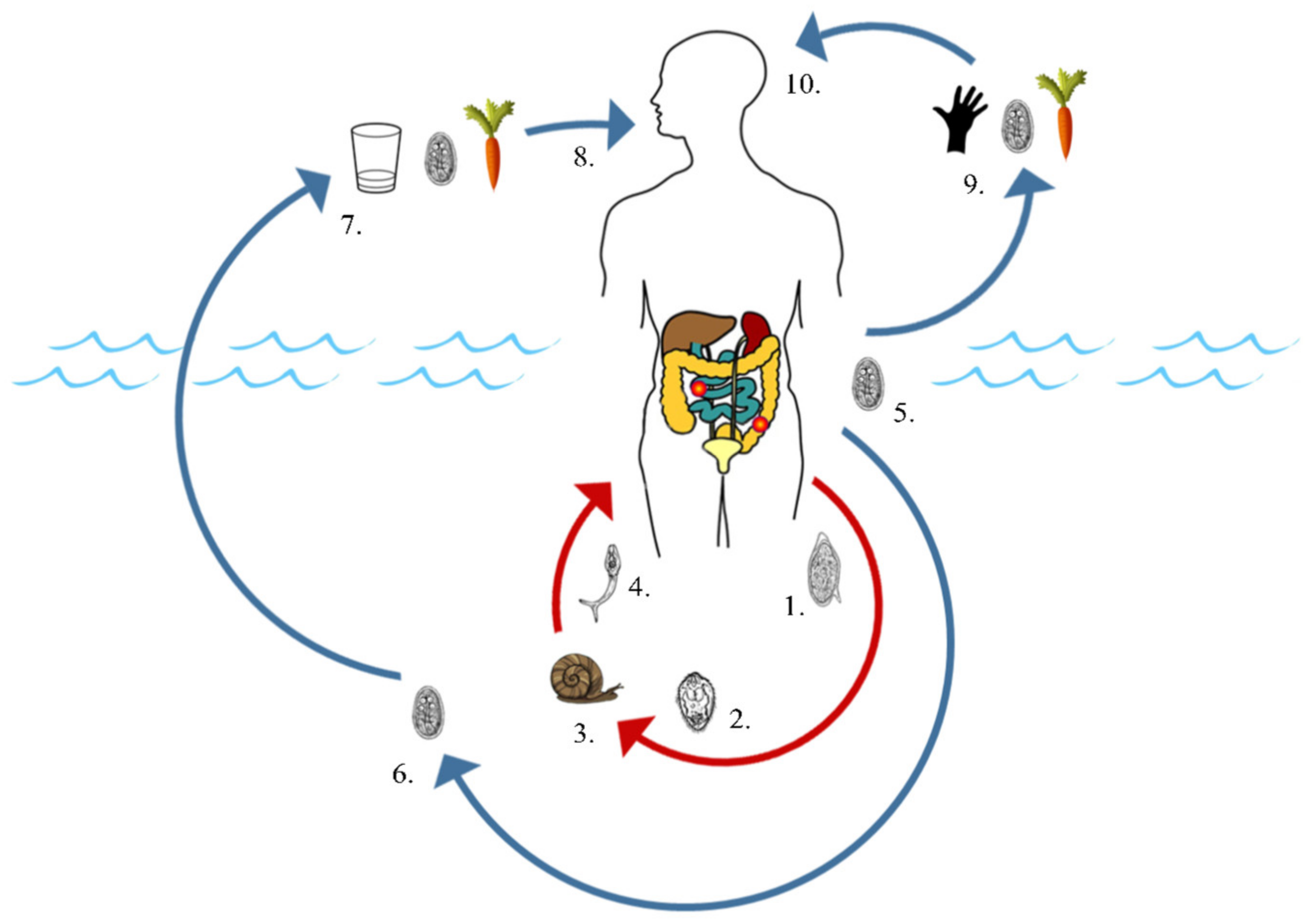
2. Intestinal Schistosomiasis and Giardiasis: Pathology and Epidemiology

Intestinal schistosomiasis disproportionally afflicts school-aged children between the ages of six and fifteen years old, where pathology can be both acute and chronic [3]. As based on ‘classic’ age-infection profiles and measured using faecal egg counts, the intensity of infection typically begins to decline in late adolescence while morbidity associated with S. mansoni, such as multi-organ fibrosis, accumulates. This decline in egg-patent prevalence is due to a variety of factors such as partial-immunity to infection, notwithstanding extensive fibrosis of the bowel itself which can occlude egg exit sites, giving rise to granulomatous masses known as intestinal ‘bilharzomas’ [15,16].
Pathologies associated with intestinal schistosomiasis occur primarily as a result of the body’s response to the copious number of eggs produced by female adult worms inhabiting the mesenteric veins surrounding the intestines. Rather than being passed in stool (or occasionally in urine) to perpetuate the parasites lifecycle, a large proportion of eggs will instead become sequestered throughout the venous bloodstream of the intestinal and hepatoportal tracts. Many eggs will then enter into general venous circulation and subsequently become lodged in other major organs. Once eggs become trapped, for example in the intestinal wall and/or liver sinuses, a range of clinical systemic and organ-specific morbidity ensues, including acute abdominal pain, stunted growth, environmental enteropathy, presence of faecal occult blood and overt hepato/splenomegaly [17,18].
Human giardiasis is caused by infection with G. duodenalis (syn. Giardia intestinalis, Giardia lamblia). While G. duodenalis is the only human-infecting Giardia species, eight distinct evolutionary assemblages based on multi-locus genotyping, named A through H, are known to exist [19]. Of these eight, the vast majority of human infections are caused by assemblages A and B, although human infections with assemblages C, D, E and F do also occur, albeit rarely [19,20].
Unlike the distribution of S. mansoni, which is intrinsically linked to its Biomphalaria spp. intermediate freshwater snail hosts, the distribution of G. duodenalis is truly cosmopolitan. Giardiasis prevalence in humans is particularly high; however, in LMICs lacking access to clean, safe drinking water and associated WASH infrastructures, including many areas of sub-Saharan Africa and South America, where S. mansoni is also endemic [5,6]. A notable feature of giardiasis, in humans, can be asymptomatic infections, although acute and/or chronic and debilitating pathologies owing to infection are well described. These include diarrhoea, dehydration, malabsorption, tropical enteropathy, stunted growth, impaired cognitive development, anaemia and chronic fatigue [7,8]. The primary cause of these pathologies is a major disruption to the gut microbiota, a complex community of symbiotic microbes responsible for vitamin production, nutrient absorption and regulation of lipid metabolism, brought about through G. duodenalis invading, inhabiting and multiplying within the intestinal tract [21,22,23]. Importantly, severe morbidity is most often observed in certain high-risk groups including children, the elderly, those with physical disabilities and the immunocompromised [24,25].

2.1. Common Modes of Environmental Contamination

A major factor linking the transmission of both intestinal schistosomiasis and giardiasis is their transfaecal environmental contamination routes via the excretion of schistosome eggs (S. mansoni) or cysts (G. duodenalis) into a viable body of freshwater. Although waterborne transmission is not required for G. duodenalis to complete its life cycle, and while not all S. mansoni eggs or G. duodenalis cysts will successfully reach a viable freshwater habitat, in a disease-endemic setting, many environmental water bodies will undergo some extent of direct or indirect faecal contamination with both parasites (Figure 1) [3,4,26,27]. Indirect faecal contamination of freshwater can occur, for example, while bathing, through infected stools deposited on the banks of rivers and ponds being washed into these waters by heavy rains or floods, through overflowing pit latrines, and possibly through animals such as cattle walking through sites of defecation and transporting faecal matter to bodies of water on their hooves [28,29]. S. mansoni eggs can survive up to approximately eight days in the stool post-defecation and before reaching freshwater, whereas G. duodenalis cysts can survive up to eight weeks in the environment [4,30] (Table 1).
Once exposed to freshwater, S. mansoni eggs (Figure 1, (1)) will hatch to release free-swimming ciliated miracidia (Figure 1, (2)) that will then employ a range of morphological adaptations and host-seeking behaviours to locate and penetrate the soft tissues of its freshwater snail intermediate host, Biomphalaria spp. (Figure 1, (3)) [31,32,33]. Miracidia are ephemeral, living only a short period of time, typically less than six hours, before dying as their glycogen energy reserves are exhausted (Table 1).
Miracidia that successfully invade a suitable intermediate snail host metamorphose into mother sporocysts, which, in turn, produce daughter sporocysts. These daughter sporocysts then differentiate upon sporogenesis, producing numerous cercariae that are shed from the snail approximately one month after initial invasion by the miracidium (Figure 1, (4)). Once established, cercarial production and shedding from Biomphalaria spp. snail hosts occur daily and typically continue over the remainder of the snails’ lives. Over the course of an infected snail’s life, tens of thousands of cercariae can be liberated [3,34].
Shed cercariae will then go on to infect humans and other mammalian definitive hosts, primarily through cutaneous penetration (Figure 1, (4)), although infection may also occur through penetration of the buccal cavity when consuming contaminated water [30]. Like miracidia, cercariae are ephemeral in freshwater as their glycogen energy reserves are quickly depleted, lasting no longer than three days (Table 1). In addition, survival of both miracidia and cercariae is highly dependent on favourable biotic and abiotic environmental conditions. Freshwater too high in salinity or too polluted, for example, can prevent the hatching of eggs into miracidia and can be lethal to both miracidia and cercariae [32,33,35,36].
G. duodenalis has a direct, faecal–oral life cycle that can be completed either through waterborne transmission when consuming water contaminated with cysts (or foods/utensils washed using contaminated water without sufficient soaps) (Figure 1, (5–8)), or through the consumption of contaminated foods or unwashed hands (Figure 1, (9–10)) [4,37]. Unlike S. mansoni, which will asexually reproduce within an intermediate host, and although Giardia cysts may survive and even accumulate within certain filter-feeding freshwater invertebrates (for example, oysters), G. duodenalis does not require an intermediate host for transmission [38]. Cysts passed in the stool are, however, extremely resilient and can remain viable within the stool or in freshwater for up to eight weeks after excretion (Table 1) [4,37,39].
Maintained transmission of intestinal schistosomiasis is therefore dependent on the continued contamination of freshwater and continued exposure to contaminated/infested water, whereas giardiasis transmission is heavily exacerbated by the continued contamination of freshwater and continued consumption of contaminated water. There are a variety of ways in which an individual can be exposed to and infected with both parasites, for example, when water is used for consumption, sanitation purposes, income generation from fishing or farming and/or recreation [40,41]. As such, transmission of both diseases is particularly high in impoverished areas lacking adequate WASH infrastructures, such as access to functional pit latrines and clean drinking water, as well as behavioural impediments in those unaware of how to avoid contamination and infection [42,43].

2.2. Transmission via Non-Human Hosts

The transmission of both intestinal schistosomiasis and giardiasis is also exacerbated by a range of non-human definitive hosts acting as either major or minor reservoirs of infection, although the precise extent to which these hosts contribute to human transmission is not fully understood (Table 2). Further to the significant health and economic impact of infection with African schistosomes and/or Giardia on, for example livestock, animal reservoirs of both parasites also pose challenges in controlling and reducing human transmission as each parasite follows similar routes of infection, contamination and, ultimately, environmental transmission via non-human hosts [5,44,45].
To reduce human transmission effectively, animal reservoirs and the degree to which they contribute to and maintain disease transmission must therefore be carefully considered when developing, implementing and monitoring any disease control strategies. Moreover, particular attention is needed on those animal hosts able to reintroduce parasites into viable bodies of freshwater following prior control or elimination campaigns [52]. Limiting contact of cattle with freshwater, for example, as well as limiting run-off from fields on which cattle manure has been spread and disposing of animal waste away from bodies of freshwater are known to reduce transmission of Giardia, as well as non-human infecting Schistosoma species [53,54]. Doing so, however, can be extremely challenging to implement and maintain through time.
In light of recent findings, additional consideration should also be given to the potential emergence of schistosome hybrids and their impact on schistosomiasis transmission [55,56,57]. Schistosoma mansoni, for example, can form hybrids with rodent-infecting species S. rodhani, which have been observed along the shoreline of Lake Victoria, one of the Great East African lakes. However, S. rodhaini appears exclusive to rodents, together with the S. mansoni-rodhaini hybrids, but with many gaps in routine surveillance this appraisal may be incomplete [58,59]. Uniquely among trematodes, schistosomes are dioecious, and so adult worms form inter-species copulatory pairs which facilitate permissive introgression(s). In nature, pre- and post-zygotic reproductive isolating barriers, such as host specificity, anatomical site of infection, geographical distribution, mating preference, worm competition and snail incompatibility, are thought to prevent prolific inter-species admixture. Recently, however, and owing to advanced methods of molecular analysis on schistosome larval stages from snail-intermediate and mammalian-definitive hosts, surprising inter-species hybrid forms are now being identified in several endemic African countries [58]. Such hybrids, resulting from interactions between human- and animal-infecting species, not only raise concerns about zoonotic transmission, but also the expanded host ranges and increased transmission potential acquired through heritable traits [59].
Changes to natural landscapes can readily lead to the formation of new freshwater bodies, snail habitats and multi-host transmission sites, breaking down the ecological barriers between species and leading to further inter-species interactions. Although the full impact that these hybridization events may have on human disease epidemiology and disease pathology is currently unknown, hybridization certainly suggests that future schistosomiasis control may warrant an expanded One Health approach with more tailored interventions specific to local settings and schistosome epidemiology [56,58,60].

3. Intestinal Schistosomiasis and Giardiasis: Surveillance and Control

Highlighting these key similarities in disease transmission, biology and epidemiology between S. mansoni and Giardia presents clear opportunities for integrated surveillance and control of both diseases, particularly with regard to disease diagnosis, surveillance and control.

3.1. Diagnosis: Parasitological, Immunological and Molecular Methods

Owing to its low cost, portability and high specificity, light microscopy for the detection of S. mansoni eggs in faecal samples is widely used during disease surveillance programmes to detect infection with S. mansoni in sub-Saharan Africa [61]. Using microscopy, routine parasitological surveillance to assess endemicity and prevalence of intestinal schistosomiasis, as well as other intestinal helminth infections, in a community is typically carried out via the Kato-Katz technique using faecal samples provided by school-aged children [62]. While inexpensive and portable, the sensitivity of Kato-Katz is, however, severely reduced in low-intensity infections, hampering its use in areas of low disease endemicity or in areas having undergone successful disease control intervention(s) [63,64,65].
Giardiasis cannot be reliably detected using the Kato-Katz method, and so alternative methods, such as formalin/ether concentration techniques, flotation techniques or immunofluorescent antibody microscopy, are needed [11,66,67,68]. Unfortunately, however, none of these techniques are straightforward or inexpensive to carry out under rural field conditions, particularly as it has been reported that formalin/ether concentration techniques have a higher sensitivity in detecting infection with S. mansoni than the more field-deployable Kato-Katz technique [69,70]. Moreover, these techniques can also often be insufficiently sensitive to reliably detect giardiasis infection [68].
For these reasons, a variety of immunological and molecular diagnostic assays with improved sensitivity in detecting both S. mansoni and Giardia infection using non-invasive urine and faecal samples have been developed (Table 3).
Though highly sensitive, immunoassays such as the enzyme-linked immunosorbent assay (ELISA) and molecular assays such as PCR/qPCR require specialist laboratory infrastructure seldom available in disease-endemic areas, preventing their use at POC [84,85]. As such, several rapid and field-deployable RDTs have also been developed to detect trace levels of parasite-derived antigens and parasite-derived DNA in urine and faecal samples. Some examples include straightforward lateral-flow dipsticks to detect S. mansoni circulating cathodic antigen (CCA) in urine samples and G. duodenalis (with or without Cryptosporidium spp.) cyst antigen in faecal samples, as well as loop-mediated isothermal amplification (LAMP) and recombinase polymerase amplification (RPA) assays to detect species-specific Schistosoma- and Giardia-derived DNA in urine and faecal samples [11,64,85].
While POC-RDTs have many advantages over light-microscopy, microscopy remains less financially expensive to carry out and so it is the favoured method of diagnosis during routine monitoring and control programmes with only limited financial resources available [68,75,78]. In addition, and though promising, assays such as LAMP and RPA to detect species-specific parasite DNA currently require further assessment and validation before their upscaled and routine use in such control programmes [81,85]. Nevertheless, continued development, assessment and validation of POC-RDTs is widely advocated as affordable and sensitive point-of-care diagnostics, capable of detecting low levels of infection within individuals able to maintain disease transmission, are sorely needed [86]. Given these challenges in reliably detecting infection using human samples, particularly in low-endemicity settings, alternative methods of detecting and monitoring disease transmission within endemic foci, such as parasite host surveillance and use of environmental DNA (eDNA), have also been explored.

3.2. Exploring the One Health InterFace with Increased Host Surveillance

Intermediate hosts and definitive reservoir hosts, such as Biomphalaria freshwater snails (S. mansoni) and rodents or cattle (S. mansoni and G. duodenalis, respectively), offer an alternative means of detecting and monitoring disease transmission in areas where detecting transmission through human diagnosis may be unreliable [87]. Collecting freshwater snails capable of transmitting schistosomes and carrying out shedding analyses to assess cercarial emergence, for example, may help identify active transmission sites [87,88]. This approach, however, can also be unreliable, as very few snails are typically found to be shedding cercariae [89].
For this reason, highly sensitive molecular xenomonitoring approaches to detect Schistosoma DNA in snail host tissues have also been developed and assessed [90]. Using PCR to detect Schistosoma DNA within snail hosts, for example, can identify prepatent infections and is not affected by diurnal fluctuations in cercarial shedding in the same way that cercarial shedding is; allowing a more reliable assessment of schistosome presence in a given locality than shedding analyses can allow. An added advantage of molecular xenomonitoring by use of PCR is the ability to genotype parasite and snail DNA, providing valuable opportunities to better understand disease transmission and molecular epidemiology, such as more reliable species identification of human-infecting cercariae and snail intermediate hosts than can be achieved using morphological analysis and the detection of Schistosoma hybridisation events [55]. In addition, collecting and screening freshwater snail hosts for the presence of parasite DNA can be more straightforward and more lucrative than collecting and screening human faecal samples for parasite DNA [90]. Currently, however, mass collection and molecular screening of freshwater snail hosts using PCR remains logistically, technically and financially demanding, and so development of a high-throughput methodology, possibly incorporating use of rapid and POC DNA amplification technologies such as LAMP or RPA, or pooling of snail samples, should also be further explored and assessed [91,92].
Similarly, molecular detection of S. mansoni and G. duodenalis DNA in DNA extracted from faeces collected from definitive reservoir hosts capable of perpetuating transmission, such as rodents (S. mansoni) and cattle (Giardia), has also been used to monitor disease transmission with success [49,58]. An added benefit of collecting and analysing faecal samples in this way is that this method also provides an opportunity to assess and better understand wild-type Schistosoma hybridisation events and zoonotic transmission of human-infecting G. duodenalis through genotyping [55,93,94]. Again, however, this approach too requires significant financial and technological resources. As such, it is unlikely to be widely integrated into control programmes undertaken in low-resource areas such as rural regions of sub-Saharan Africa without further development and use of field-deployable DNA amplification technologies.

3.3. Detecting Parasitic Contamination through Water Sampling and by Environmental DNA (eDNA) Analysis

Extensive screening of environmental water samples for contamination with Giardia cysts using the United States Environmental Protection Agency method 1623 (US-EPA method 1623) has been carried out in many areas of the world, such as the USA [95]. Through collection and filtration of water samples, protozoan cysts can be detected and quantified, allowing viable transmission sites to be identified [96]. Although straightforward to carry out, this method does not differentiate between morphologically identical Giardia assemblages A-H and so identification of human-infecting Giardia transmission sites, specifically, is not possible. As such, revised methods of detecting and monitoring assemblage-specific Giardia-contaminated water sources, such as through detection and genotypic analysis of parasite-derived eDNA, have been developed.
Assessing and monitoring disease transmission within a given focus through the detection of parasite-derived eDNA rather than, or in conjunction with, using human bodily samples, has been explored with respect to a range of waterborne pathogens, including both schistosomiasis and giardiasis [97,98,99]. Dependence of both parasites on freshwater provides an ideal target for sample collection and assessment using PCR/qPCR, LAMP or RPA assays [83,100,101]. In addition, collection of water samples to detect eDNA derived from Schistosoma freshwater snail hosts to identify and monitor the presence of snail species capable of transmitting infection within a given waterbody has also been assessed [102,103].
Again, though promising, the upscaled and routine use of molecular assays to detect parasite- and/or parasite host-derived eDNA remains beyond the financial reach of most LMIC control programmes and too requires further methodological development, assessment and validation. Nevertheless, continued development of this approach to better understand the potential of eDNA as an effective monitoring tool and to reduce associated financial costs has been encouraged [75]. In particular, and like molecular xenomonitoring approaches, the monitoring of eDNA to identify disease transmission may prove extremely useful in areas of low disease endemicity where identifying infection in individual patients may be challenging.

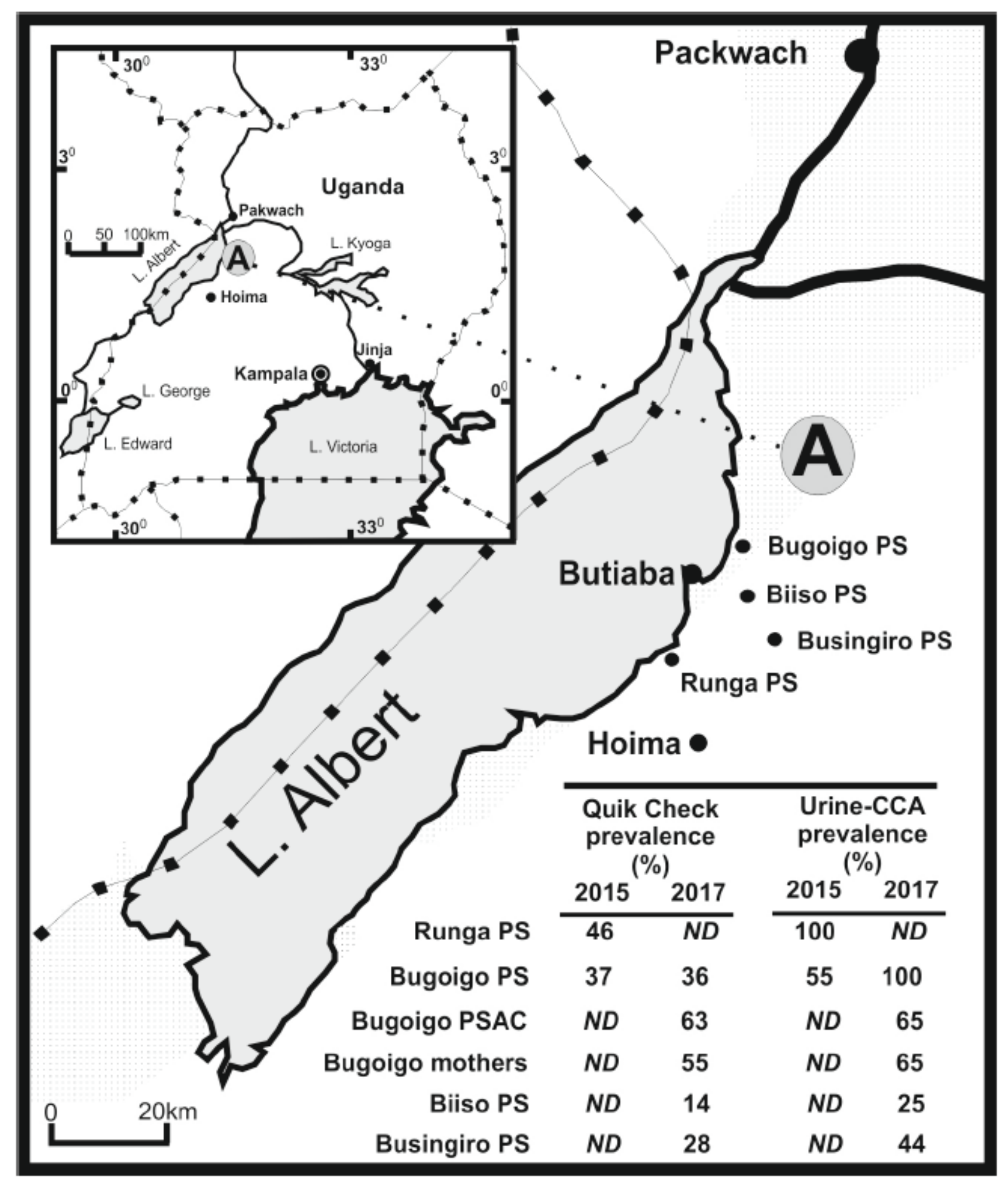
3.4. A Case Example of Co-Infection and Morbidity Surveillance in Uganda

Given these key similarities in disease transmission biology and co-endemicity of both intestinal schistosomiasis and giardiasis throughout much of sub-Saharan Africa, co-infection with both parasites is likely commonplace, yet only little formal attention has been given towards co-surveillance of both diseases. This is despite each parasite potentially influencing reciprocal infection susceptibilities and disease-associated pathologies, before and after anti-parasitic treatment(s). As an example, it is possible that S. mansoni egg-induced perforations to the bowel with associated mucosal bleeding, inflammation and bacterial translocation may influence an individual’s susceptibility to chronic Giardia infection. The extent to which this occurs, however, is currently unknown.
This lack of attention on co-infection and co-surveillance may be, in part, due to an unfortunate division within parasitology that often siloes macro-parasite (helminth) and micro-parasite (protist) research. Though sparse, recent epidemiological studies are now beginning to shed more detailed light on the prevalence of co-infection of intestinal schistosomiasis and giardiasis, with detection of associated morbidities, throughout rural areas of sub-Saharan Africa [11,104,105]. A suitable example arises from two recent studies assessing co-infection in school-aged children along the shoreline of Lake Albert, Uganda, which, despite ongoing preventive chemotherapy for intestinal schistosomiasis, can still be considered hyper-endemic for S. mansoni today (Figure 2) [88]. Here, initial infection with S. mansoni occurs very soon after birth, with all ages vulnerable to infection and chronic disease [106].
Beginning in 2015, Al-Shehri et al. [8] conducted a novel attempt to integrate surveillance for intestinal schistosomiasis, giardiasis and malaria using available POC rapid diagnostic tests (RDTs) combined with later real-time qPCR analysis of stool and finger-prick collected blood with parasite-specific TaqMan DNA® probes. This was the first attempt to quantify giardiasis with the POC Quik Chek RDT (TechLab, USA), finding 42% of children attending Runga and Bugoigo primary schools to be positive (Figure 2). Upon qPCR analysis of ethanol preserved stool using an 18S rDNA Giardia-specific TaqMan® probe, up to 87.0% of children were found excreting Giardia DNA. Notably, the prevalence of heavy infection by real-time PCR (Ct ≤ 19) was 19.5% and strongly associated with Quik-Chek RDTs, as well as postively correlated with increasing intensities of egg-patent schistosomiasis and host anaemia [11].
Giardia species assemblages present were also later identified and characterised with specific triose phosphate isomerase (TPI) Taqman® probes and by sequence characterisation of the β-giardin gene [107]. While less sensitive than the 18S rDNA assay, general prevalence by TPI probes was 52%, with prevalence by taxon assemblage of 8% (assemblage A), 36% (assemblage B) and 8% co-infection (A and B assemblages), and while assemblage B was dominant across the sample, proportions of assemblages A and B, and co-infections thereof, varied by school and by age of child. Mixed infections were particularly common at Runga school and in children aged 6 and under. Most importantly, infection with assemblage B was associated with underweight children. The presence of each assemblage was also confirmed by sequence analysis of the β-giardin gene finding sub-assemblage AII and further genetic diversity within assemblage B; also of note was the absence cryptosporidiosis, another pertinent water-borne disease, concurrently detectable by the same Quik Chek RDT.
To assess any changes through time, a repeat epidemiological survey was undertaken in 2017 which included reinspection of Bugoigo school and expanded point-of-care testing with Quik Chek (Figure 2). The prevalence of giardiasis at Bugoigo primary school was shown to be identical with a third of children examined positive by Quik Chek, with even higher local prevalence in pre-school-age children (63%) and their mothers (55%), good evidence for pervasive nature of giardiasis across all ages. Away from the lake at Biiso and Busingiro, the prevalence of giardiasis and intestinal schistosomiasis declined, suggesting that the risk of infection is perhaps higher on the lake shoreline. This study also attempted to evaluate a new POC-RPA RDT onsite, as well as a pilot assessment of giardiasis in local livestock and companion animals [81]. Ultimately, the RPA assay did not perform as well as expected, being in need for further optimisation of stool DNA extraction protocols.
Further to human-surveillance, screening for S. mansoni transmission has also taken place along the same shoreline by collection and cercarial shedding of Biomphalaria freshwater snail hosts with success [88,108]. Future surveillance should also be carried out through molecular xenomonitoring of collected snails (and so omitting the need for shedding analyses), collection and screening of non-human definitive hosts to identify S. mansoni and G. duodenalis DNA and through collection and screening of surface water samples to identify S. mansoni and G. duodenalis eDNA.

3.5. Access to Treatment and Large-Scale Campaigns

In areas where schistosomiasis transmission is identified, preventive chemotherapy through repeated mass drug administration (MDA) of the donated anthelmintic drug praziquantel (40 mg/kg body weight) is the principal strategy for disease control [109]. Because the highest burden of infection is typically seen in children and young adolescents, MDA is customarily carried out in schools, but aims to limit overall transmission within a community through a reduced human reservoir of infection while also reducing overall disease morbidity [110]. Though praziquantel’s mechanism of action is not currently fully understood, significant reductions in disease prevalence and morbidity have been seen globally since MDA programmes began in 2001 [111,112]. Reinfection of schistosomiasis following treatment is, however, commonplace owing to communities’ reliance on freshwater, and so MDA must be repeated annually or biannually, depending on disease prevalence, to achieve a sustained impact.
Severe adverse effects are seen only very rarely when distributing praziquantel, making it well suited for mass distribution. Praziquantel, however, typically does not achieve 100% infection clearance primarily because dosing is usually based only on height and so does not account for differences in body mass. As a result, treatment success varies between individuals meaning many are still able to continue maintaining transmission [113]. In addition, and while local school systems provide a viable means of mass-distributing praziquantel, important human reservoirs of infection, including pre-school-aged children and adults, typically remain untreated [106,114].
The need for repeated annual or biannual distribution of MDA in this way has also raised regular concerns about the development of praziquantel resistance in schistosomes; particularly as there is currently no known efficacious alternative treatment to replace praziquantel if Schistosoma populations were to become more drug-tolerant or resistant [115,116]. A significant reduction in praziquantel efficacy, identified by a decreased reduction in Schistosoma egg output from infected individuals pre- and post-praziquantel treatment, has already been reported in S. mansoni populations in many communities across sub-Saharan Africa that have undergone repeated rounds of MDA [113]. This reduced efficacy may be a direct result of selection pressure placed on schistosomes during repeated and prolonged MDA campaigns, highlighting an urgent need to consider alternative methods of disease control outside of MDA.
A variety of drugs can be used to treat giardiasis [117,118]. Of these, metronidazole is the most predominantly used and most thoroughly studied owing to its straightforward oral administration and relatively low price. Like with schistosomiasis, reinfection with giardiasis is also commonplace; however, repeated mass drug administration to alleviate giardiasis transmission is not seen as a feasible strategy because the drug is not currently involved in any donation scheme, severe adverse effects of treatment are often seen, and metronidazole has only limited efficacy in clearing infection [117]. As an example, it has been reported that just one course of treatment has only an approximately 60% clearance rate, and so repeated treatment is needed to significantly clear infection [119]. Repeated treatment, however, not only significantly increases the likelihood of severe adverse events but is difficult to carry out during MDA campaigns [5]. In addition, the potential emergence of giardiasis resistance to treatment with metronidazole has also recently been reported, and while alternative and more efficacious chemotherapies, such as tinidazole exist, these can also cause adverse events [117,118,119]. Albendazole, a broad-spectrum and efficacious anthelmintic treatment used in MDA campaigns to reduce transmission of soil-transmitted helminth and some filarial nematode infections, can also be used to treat giardiasis [117,120,121]. To significantly reduce Giardia infection, however, a minimum dosage of 200–800 mg/day albendazole is needed for at least three concurrent days which, again, is difficult to carry out in the context of MDA campaigns and, by having limited donated stocks, also diminishes albendazole availability for anti-helminth control programmes [117,118,122].
Though treatment of schistosomiasis and giardiasis using praziquantel and metronidazole are important components of disease control, aligning treatment of both diseases in tandem during control campaigns may be challenging, owing to differences in drug type, dosages and treatment courses. In addition, as neither drug can safely and reliably achieve a 100% clearance rate using just one dosage, it is now widely accepted that alternative methods of control to reduce transmission and overall prevalence must be implemented alongside treatment campaigns if disease elimination targets are to be met. One such example is the implementation of WASH initiatives in communities where both diseases are endemic. The extent to which WASH provision, when used in conjunction with MDA, can successfully reduce schistosomiasis transmission is now beginning to be understood, and although only minimal data has been reported on the impact of WASH provision on giardiasis transmission in sub-Saharan Africa, it is widely assumed that improved WASH infrastructure would help significantly reduce giardiasis transmission [1,123,124].

3.6. Water, Sanitation and Hygeine (WASH)

WASH provision and infrastructure is extremely inadequate throughout many areas of rural sub-Saharan Africa [125]. In 2012, the World Health Assembly (WHA) formally advocated for the integration of WASH provision and education initiatives into amenable NTD control and elimination programmes; subsequently publishing guidance on ways in which these can be integrated [126,127]. Since, much attention has been given towards how WASH initiatives can be tailored for use, specifically, in schistosomiasis control programmes and the impact such initiatives have had when used in tandem with routine strategies such as preventive chemotherapy [2,30,128,129].
WASH initiatives relevant to schistosomiasis control, such as the adequate provision of safe drinking water, fully functional and properly maintained pit latrines and improved community hygiene education, effectively reduce disease transmission by minimising the direct and indirect contamination of freshwater by infected individuals and animals, by reducing contact with/consumption of infectious waters by human and non-human hosts and by helping communities better understand human and non-human disease transmission [30,130] (Figure 3).
Like intestinal schistosomiasis, giardiasis is widely prevalent throughout many rural and impoverished regions of sub-Saharan Africa and is intrinsically linked to contact with contaminated and unsafe water in areas lacking adequate water, sanitation and hygiene (WASH) infrastructure [4,41]. Giardiasis, however, receives only relatively little attention with regard to disease control, surveillance and elimination throughout sub-Saharan Africa.
As an example, despite numerous clear advantages of implementing WASH initiatives on reducing schistosomiasis transmission and despite these key similarities between schistosomiasis and giardiasis with regard to disease transmission biology, surprisingly little attention has been given to the impact of improved WASH provision and education on giardiasis transmission in sub-Saharan Africa [43]. This oversight presents not only a missed opportunity with regard to better understanding, and reducing, giardiasis transmission and its associated pathological impact on some of the world’s most disadvantaged communities, but also presents the question: why is giardiasis ignored in intestinal schistosomiasis monitoring and control programmes?

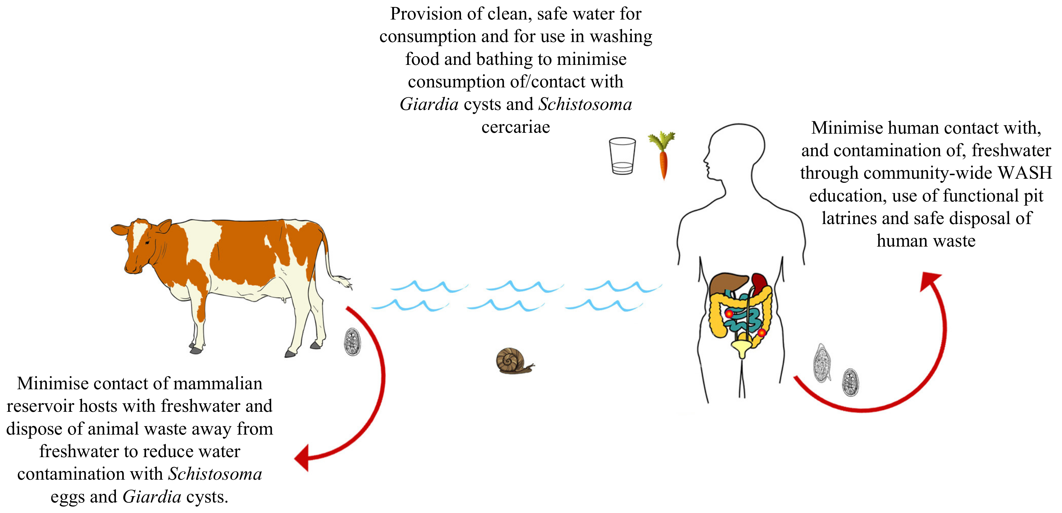
4. Intestinal Schistosomiasis and Giardiasis: Towards a One Health Approach

Research funding opportunities for NTDs are limited when compared to those for other diseases such as human immunodeficiency virus (HIV), malaria or tuberculosis [131]. One way in which the impact of NTD control programmes can be significantly increased, however, is by appropriate integration with other disease surveillance, control, research and policy efforts. Successful examples of this integrated One Health approach can be seen when integrating lymphatic filariasis surveillance and elimination efforts into malaria elimination programmes, as well as by integrating soil-transmitted helminth and schistosomiasis control and elimination efforts [128,132,133,134,135].
In keeping with this integrated One Health approach, here, we propose a variety of ways in which the transmission of, and pathologies associated with, co-infection of intestinal schistosomiasis and giardiasis can be better understood, monitored and reduced via the integration of giardiasis control efforts into existing schistosomiasis control programmes. These include:
  • Integrating screening of giardiasis endemicity and infection prevalence into existing schistosomiasis control programmes by using stool samples used for diagnosing infection with S. mansoni, and other intestinal parasites, to also record and report levels of Giardia infection in school-aged children. This can be conducted with POC-RDTs such as the Quik Chek immunoassay or using PCR/qPCR. In addition, the continued development, assessment and application of sensitive and straightforward POC-RDTs able to detect low-levels of infection in asymptomatic individuals capable of maintaining transmission of both parasites, such as the RPA, is encouraged.
  • Further development and application of sensitive molecular assays to detect trace levels of species/assemblage-specific parasite DNA within freshwater snail intermediate hosts of human-infecting Schistosoma, and in faecal samples from non-human animal definitive hosts of both diseases. Further development and application of sensitive molecular assays to detect trace levels of species-/assemblage-specific parasite DNA from human-infecting Schistosoma cercariae and Giardia cysts in water samples easily collected from viable transmission sites is also encouraged.
  • The upscaled provision of water, sanitation and hygiene (WASH) infrastructure and education initiatives to communities afflicted by both schistosomiasis and giardiasis to reduce environmental contamination events and to reduce contact with/consumption of contaminated water, simultaneously reducing transmission of both diseases.
  • Monitoring Giardia disease prevalence and associated morbidities in tandem with schistosomiasis surveillance in school-aged children following any control programme intervention to better understand how giardiasis transmission and related pathologies can be reduced.
  • An increased focus on understanding how the transmission of intestinal schistosomiasis and giardiasis, as well as immune responses and morbidities related to both diseases, interact and are potentially exacerbated by co-infection.

5. Conclusions

Here, we have highlighted various potential opportunities to improve the health and wellbeing of individuals in low- and middle-income countries where intestinal schistosomiasis and giardiasis are co-endemic by exploiting key similarities between both diseases with regard to disease transmission biology, epidemiology, surveillance and control. In addition, future steps needed to develop and implement an integrated, One Health approach for intestinal schistosomiasis and giardiasis co-infection surveillance, control and elimination strategies, are also outlined. In adopting this One Health approach, and by integrating giardiasis surveillance, control and elimination efforts into existing schistosomiasis elimination programmes, not only can the debilitating pathological impacts of intestinal schistosomiasis/giardiasis co-infection be better understood, but a reduction in co-infection and concurrent reduction in disease-related morbidities experienced by the world’s most disadvantaged communities can also be achieved.

Author Contributions

Concept of the study (J.A., A.L.B. & J.R.S.), literature searching and review (J.A., L.O., S.S., M.S.), fieldwork (J.A., H.A.-S., N.B.K., A.A., M.A. (Moses Adriko), M.A. (Moses Arianaitwe), E.J.L., A.L.B., J.R.S.), and molecular analyses performed by (H.A.-S., T.B., B.L.W.). All authors have read and agreed to the published version of the manuscript.

Funding

J.A. is funded by an MRC-DTP studentship. Fieldwork reported here was supported by HEFC through Liverpool School of Tropical Medicine, Education Department MSc project funding and a PhD studentship awarded by the Ministry of Health, Saudi Arabia to H.A.-S.

Acknowledgments

J.A. would like to thank Michael Fowler of EH Studios for support with figures.

Conflicts of Interest

The authors declare that they have no competing interest.
Ethical Standards: Approvals for the work conducted in Uganda were granted by The Ugandan Council for Science and Technology (April 2015) and the Liverpool School of Tropical Medicine, UK (M09-17).

Abbreviations

eDNAenvironmental DNA
ELISAenzyme-linked immunosorbent assay
FGSfemale genital schistosomiasis
LAMPloop-mediated isothermal reaction
LMIClower-middle-income countries
MDAmass drug administration
NTDneglected tropical disease
PCRpolymerase chain reaction
POCpoint-of-care
qPCRquantitative polymerase chain reaction
RDTrapid diagnostic test
RPArecombinase polymerase amplification
Th1T-helper 1
Th2T-helper 2
WASHwater, sanitation and hygiene
WHAworld health assembly

References

  1. Omarova, A.; Tussupova, K.; Berndtsson, R.; Kalishev, M.; Sharapatova, K. Protozoan parasites in drinking water: A system approach for improved water, sanitation and hygiene in developing countries. Int. J. Environ. Res. Public Health 2018, 15, 495. [Google Scholar] [CrossRef] [PubMed] [Green Version]
  2. Campbell, S.J.; Biritwum, N.K.; Woods, G.; Velleman, Y.; Fleming, F.; Stothard, J.R. Tailoring water, sanitation, and hygiene (WASH) Targets for toil-transmitted helminthiasis and schistosomiasis control. Trends Parasitol. 2018, 34, 53–63. [Google Scholar] [CrossRef] [PubMed] [Green Version]
  3. Colley, D.G.; Bustinduy, A.L.; Secor, W.E.; King, C.H. Human schistosomiasis. Lancet 2014, 383, 2253–2264. [Google Scholar] [CrossRef]
  4. Thompson, R.C.A.; Reynoldson, J.A.; Sciences, B. Giardia and giardiasis. Adv. Parasitol. 1993, 32, 71–160. [Google Scholar]
  5. Squire, S.A.; Ryan, U. Cryptosporidium and Giardia in Africa: Current and future challenges. Parasites Vectors 2017, 10, 1–32. [Google Scholar] [CrossRef] [Green Version]
  6. Nkrumah, B.; Nguah, S. Giardia lamblia: A major parasitic cause of childhood diarrhoea in patients attending a district hospital in Ghana. Parasites Vectors 2011, 4, 1–7. [Google Scholar] [CrossRef] [Green Version]
  7. Bartelt, L.A.; Sartor, R.B. Advances in understanding Giardia: Determinants and mechanisms of chronic sequelae. F1000Prime Rep. 2015, 7, 1–14. [Google Scholar] [CrossRef] [Green Version]
  8. Naess, H.; Nyland, M.; Hausken, T.; Follestad, I.; Nyland, H.I. Chronic fatigue syndrome after Giardia enteritis: Clinical characteristics, disability and long-term sickness absence. BMC Gastroenterol. 2012, 12. [Google Scholar] [CrossRef] [Green Version]
  9. Chifunda, K.; Kelly, P. Parasitic infections of the gut in children. Paediatr. Int. Child Health 2019, 39, 65–72. [Google Scholar] [CrossRef]
  10. Savioli, L.; Smith, H.; Thompson, A. Giardia and Cryptosporidium join the “Neglected Diseases Initiative”. Trends Parasitol. 2006, 22, 203–208. [Google Scholar] [CrossRef]
  11. Al-Shehri, H.; Stanton, M.C.; LaCourse, J.E.; Atuhaire, A.; Arinaitwe, M.; Wamboko, A.; Adriko, M.; Kabatereine, N.B.; Stothard, J.R. An extensive burden of giardiasis associated with intestinal schistosomiasis and anaemia in school children on the shoreline of Lake Albert, Uganda. Trans. R. Soc. Trop. Med. Hyg. 2016, 110, 597–603. [Google Scholar] [CrossRef] [PubMed]
  12. La Hoz, R.M.; Morris, M.I. Intestinal parasites including Cryptosporidium, Cyclospora, Giardia, and Microsporidia, Entamoeba histolytica, Strongyloides, Schistosomiasis, and Echinococcus: Guidelines from the American Society of Transplantation Infectious Diseases Community of Pract. Clin. Transplant. 2019, 33, 1–16. [Google Scholar] [CrossRef] [PubMed]
  13. Zhou, X.N. Prioritizing research for “One health—One world”. Infect. Dis. Poverty 2012, 1, 1–5. [Google Scholar] [CrossRef] [PubMed] [Green Version]
  14. Mackenzie, J.S.; Jeggo, M. One Health and Zoonoses; MDPI Books: Basel, Switzeland, 2019; ISBN 9783039212958. [Google Scholar] [CrossRef] [Green Version]
  15. Samuels, A.M.; Matey, E.; Mwinzi, P.N.M.; Wiegand, R.E.; Muchiri, G.; Ireri, E.; Hyde, M.; Montgomery, S.P.; Karanja, D.M.S.; Secor, W.E. Schistosoma mansoni morbidity among school-aged children: A SCORE Project in Kenya. Am. J. Trop. Med. Hyg. 2012, 87, 874–882. [Google Scholar] [CrossRef] [PubMed] [Green Version]
  16. De Moira, A.P.; Fulford, A.J.C.; Kabatereine, N.B.; Ouma, J.H.; Booth, M.; Dunne, D.W. Analysis of complex patterns of human exposure and immunity to Schistosomiasis mansoni: The influence of age, sex, ethnicity and IgE. PLoS Negl. Trop. Dis. 2010, 4, e820. [Google Scholar] [CrossRef] [Green Version]
  17. Costain, A.H.; MacDonald, A.S.; Smits, H.H. Schistosome egg migration: Mechanisms, pathogenesis and host immune responses. Front. Immunol. 2018, 9, 3042. [Google Scholar] [CrossRef] [Green Version]
  18. Olveda, David Bilharzia: Pathology, diagnosis, management and control. Trop. Med. Surg. 2013, 1, 1–19. [CrossRef]
  19. Heyworth, M.F. Giardia duodenalis genetic assemblages and hosts. Parasite 2016, 23. [Google Scholar] [CrossRef] [Green Version]
  20. Sprong, H.; Cacciò, S.M.; Van Der Giessen, J.W.B. Identification of zoonotic genotypes of Giardia duodenalis. PLoS Negl. Trop. Dis. 2009, 3, 1–12. [Google Scholar] [CrossRef] [Green Version]
  21. Fink, M.Y.; Singer, S.M. The intersection of immune responses, microbiota, and pathogenesis in giardiasis. Trends Parasitol. 2017, 33, 901–913. [Google Scholar] [CrossRef]
  22. Keselman, A.; Li, E.; Maloney, J.; Singer, S.M. The microbiota contributes to CD8+ T cell activation and nutrient malabsorption following intestinal infection with Giardia duodenalis. Infect. Immun. 2016, 84, 2853–2860. [Google Scholar] [CrossRef] [PubMed] [Green Version]
  23. Allain, T.; Amat, C.B.; Motta, J.P.; Manko, A.; Buret, A.G. Interactions of Giardia sp. with the intestinal barrier: Epithelium, mucus, and microbiota. Tissue Barriers 2017, 5, 1–16. [Google Scholar] [CrossRef] [PubMed] [Green Version]
  24. Mmbaga, B.T.; Houpt, E.R. Cryptosporidium and Giardia infections in children: A Review. Pediatr. Clin. N. Am. 2017, 64, 837–850. [Google Scholar] [CrossRef] [PubMed]
  25. Lane, S.; Lloyd, D. Current trends in research into the waterborne parasite Giardia. Crit. Rev. Microbiol. 2002, 28, 123–147. [Google Scholar] [CrossRef]
  26. Centers for Disease Control and Prevention (CDC), USA: Schistosomiasis, About, Life Cycle. Available online: https://www.cdc.gov/parasites/schistosomiasis/biology.html (accessed on 1 July 2020).
  27. Centers for Disease Control and Prevention (CDC), USA: Giardiasis, About, Life Cycle. Available online: https://www.cdc.gov/dpdx/giardiasis/index.html#:~:text=The spectrum varies from asymptomatic carriage to severe,include diarrhea%2C abdominal pain%2C bloating%2C nausea%2C and vomiting. (accessed on 1 July 2020).
  28. Sow, S.; Polman, K.; Vereecken, K.; Vercruysse, J.; Gryseels, B.; de Vlas, S.J. The role of hygienic bathing after defecation in the transmission of Schistosoma mansoni. Trans. R. Soc. Trop. Med. Hyg. 2008, 102, 542–547. [Google Scholar] [CrossRef]
  29. Vercruysse, J.; Shaw, D.J.; De Bont, J. Index of potential contamination for schistosomiasis. Trends Parasitol. 2001, 17, 256–261. [Google Scholar] [CrossRef]
  30. Grimes, J.E.; Croll, D.; Harrison, W.E.; Utzinger, J.; Freeman, M.C.; Templeton, M.R. The roles of water, sanitation and hygiene in reducing schistosomiasis: A review. Parasites Vectors 2015, 8, 1–16. [Google Scholar] [CrossRef] [Green Version]
  31. Wright, C.A. Chapter 4: Fluke Life-Cycles. In Flukes and Snails; George Allen and Unwin LTD: London, UK, 1971. [Google Scholar]
  32. Galaktionov, K.V.; Dobrovolskij, A. The Biology and Evolution of Trematodes. An Essay on the Biology, Morphology, Life Cycles, Transmission, and Evolution of Digenetic Trematodes. Chapter 2: The Trematode Life Cycle as a System of Adaptations; Kluwer Academic Publishers: Dordrecht, The Netherlands; Boston, MA, USA, 2003. [Google Scholar]
  33. Fried, B.T.; Graczyk, T. Chapter 7: Host Recognition by Trematode Miracidia and Cercariae. In Advances in Trematode Biology; CRC Press: New York, NY, USA, 1997. [Google Scholar]
  34. Lockyer, A.E.; Jones, C.S.; Noble, L.R.; Rollinson, D. Trematodes and snails: An intimate association. Can. J. Zool. 2004, 82, 251–269. [Google Scholar] [CrossRef]
  35. Théron, A. Chronobiology of Trematode Cercarial Emergence: From Data Recovery to Epidemiological, Ecological and Evolutionary Implications; Elsevier Ltd.: Amsterdam, The Netherlands, 2015; Volume 88. [Google Scholar]
  36. Frandsen, F.; Christensen, N. An introductory guide to the identification of cercariae from African freshwater snails with special reference to cercariae of trematode species of medical and veterinary importance. Acta Trop. 1984. [Google Scholar] [CrossRef]
  37. Einarsson, E.; Ma’ayeh, S.; Svärd, S.G. An up-date on Giardia and giardiasis. Curr. Opin. Microbiol. 2016, 34, 47–52. [Google Scholar] [CrossRef]
  38. Mohammed Mahdy, A.K.; Lim, Y.A.L.; Surin, J.; Wan, K.L.; Al-Mekhlafi, M.S.H. Risk factors for endemic giardiasis: Highlighting the possible association of contaminated water and food. Trans. R. Soc. Trop. Med. Hyg. 2008, 102, 465–470. [Google Scholar] [CrossRef] [PubMed]
  39. Ankarklev, J.; Jerlström-Hultqvist, J.; Ringqvist, E.; Troell, K.; Svärd, S.G. Behind the smile: Cell biology and disease mechanisms of Giardia species. Nat. Rev. Microbiol. 2010, 8, 413–422. [Google Scholar] [CrossRef] [PubMed]
  40. Huang, Y.; Manderson, L. Schistosomiasis and the social patterning of infection. Acta Trop. 1992, 51, 175–194. [Google Scholar] [CrossRef]
  41. Ahmed, S.A.; Guerrero Flórez, M.; Karanis, P. The impact of water crises and climate changes on the transmission of protozoan parasites in Africa. Pathog. Glob. Health 2018, 112, 281–293. [Google Scholar] [CrossRef] [PubMed]
  42. Esrey, S.A.; Collett, J.; Miliotis, M.D.; Koornhof, H.J.; Makhale, P. The risk of infection from Giardia lamblia due to drinking water supply, use of water, and latrines among preschool children in rural Lesotho. Int. J. Epidemiol. 1989, 18, 248–253. [Google Scholar] [CrossRef] [Green Version]
  43. Campbell, S.J.; Nery, S.V.; D’Este, C.A.; Gray, D.J.; McCarthy, J.S.; Traub, R.J.; Andrews, R.M.; Llewellyn, S.; Vallely, A.J.; Williams, G.M.; et al. Water, sanitation and hygiene related risk factors for soil-transmitted helminth and Giardia duodenalis infections in rural communities in Timor-Leste. Int. J. Parasitol. 2016, 46, 771–779. [Google Scholar] [CrossRef]
  44. Robinson, M.W.; Dalton, J.P. Zoonotic helminth infections with particular emphasis on fasciolosis and other trematodiases. Philos. Trans. R. Soc. B Biol. Sci. 2009, 364, 2763–2776. [Google Scholar] [CrossRef] [Green Version]
  45. Chomel, B.B. Control and prevention of emerging parasitic zoonoses. Int. J. Parasitol. 2008, 38, 1211–1217. [Google Scholar] [CrossRef]
  46. MARTINS, A.V. Non-human vertebrate hosts of Schistosoma haematobium and Schistosoma mansoni. Bull. World Health Organ. 1958, 18, 931–944. [Google Scholar]
  47. Standley, C.J.; Dobson, A.; Dobson, A.P.; Stothard, J.R. Out of Animals and Back again: Schistosomiasis as a Zoonosis in Africa. Schistosomiasis; Rokni, M.B., Ed.; InTech Europe: Rijeka, Croatia, 2012; Available online: http://www.intechopen.com/books/schistosomiasis/out-of-animals-andback-again-schistosomiasis-as-a-zoonosis-in-africa (accessed on 13 January 2012)ISBN ISBN 978-953-307-852-6.
  48. Ryan, U.; Cacciò, S.M. Zoonotic potential of Giardia. Int. J. Parasitol. 2013, 43, 943–956. [Google Scholar] [CrossRef]
  49. Yaoyu, F.; Xiao, L. Zoonotic potential and molecular epidemiology of Giardia species and giardiasis. Clin. Microbiol. Rev. 2011, 24, 110–140. [Google Scholar] [CrossRef] [Green Version]
  50. Thompson, R.C.A. The zoonotic significance and molecular epidemiology of Giardia and giardiasis. Vet. Parasitol. 2004, 126, 15–35. [Google Scholar] [CrossRef]
  51. Sak, B.; Petrzelkova, K.J.; Kvetonova, D.; Mynarova, A.; Shutt, K.A.; Pomajbikova, K.; Kalousova, B.; Modry, D.; Benavides, J.; Todd, A.; et al. Long-term monitoring of Microsporidia, Cryptosporidium and Giardia Infections in western lowland gorillas (Gorilla gorilla gorilla) at different stages of habituation in Dzanga Sangha protected areas, Central African Republic. PLoS ONE 2013, 8, e71840. [Google Scholar] [CrossRef] [PubMed]
  52. Hanelt, B.; Mwangi, I.N.; Kinuthia, J.M.; Maina, G.M.; Agola, L.E.; Mutuku, M.W.; Steinauer, M.L.; Agwanda, B.R.; Kigo, L.; Mungai, B.N.; et al. Schistosomes of small mammals from the Lake Victoria Basin, Kenya: New species, familiar species, and implications for schistosomiasis control. Parasitology 2010, 137, 1109–1118. [Google Scholar] [CrossRef] [Green Version]
  53. De Bont, J.; Vercruysse, J. The epidemiology and control of cattle schistosomiasis. Parasitol. Today 1997, 13, 255–262. [Google Scholar] [CrossRef]
  54. Olson, M.E.; O’Handley, R.M.; Ralston, B.J.; McAllister, T.A.; Thompson, R.C.A. Update on Cryptosporidium and Giardia infections in cattle. Trends Parasitol. 2004, 20, 185–191. [Google Scholar] [CrossRef]
  55. Savassi, B.A.E.S.; Mouahid, G.; Lasica, C.; Mahaman, S.D.K.; Garcia, A.; Courtin, D.; Allienne, J.F.; Ibikounlé, M.; Moné, H. Cattle as natural host for Schistosoma haematobium (Bilharz, 1852) Weinland, 1858 × Schistosoma bovis Sonsino, 1876 interactions, with new cercarial emergence and genetic patterns. Parasitol. Res. 2020, 1–17. [Google Scholar] [CrossRef]
  56. Sene-Wade, M.; Marchand, B.; Rollinson, D.; Webster, B.L. Urogenital schistosomiasis and hybridization between Schistosoma haematobium and Schistosoma bovis in adults living in Richard-Toll, Senegal. Parasitology 2018, 145, 1723–1726. [Google Scholar] [CrossRef]
  57. Standley, C.J.; Stothard, J.R. DNA Barcoding of schistosome cercariae reveals a novel sub-lineage within Schistosoma rodhaini from Ngamba Island chimpanzee sanctuary, Lake Victoria. J. Parasitol. 2012, 98, 1049–1051. [Google Scholar] [CrossRef] [Green Version]
  58. Catalano, S.; Sène, M.; Diouf, N.D.; Fall, C.B.; Borlase, A.; Léger, E.; Bâ, K.; Webster, J.P. Rodents as natural hosts of zoonotic schistosoma species and hybrids: An epidemiological and evolutionary perspective from West Africa. J. Infect. Dis. 2018, 218, 429–433. [Google Scholar] [CrossRef] [Green Version]
  59. Steinauer, M.L.; Hanelt, B.; Mwangi, I.N.; Maina, G.M.; Agola, L.E.; Kinuthia, J.M.; Mutuku, M.W.; Mungai, B.N.; Wilson, W.D.; Mkoji, G.M.; et al. Introgressive hybridization of human and rodent schistosome parasites in western Kenya. Mol. Ecol. 2008, 17, 5062–5074. [Google Scholar] [CrossRef] [PubMed]
  60. Huyse, T.; Webster, B.L.; Geldof, S.; Stothard, J.R.; Diaw, O.T.; Polman, K.; Rollinson, D. Bidirectional introgressive hybridization between a cattle and human schistosome species. PLoS Pathog. 2009, 5, e1000571. [Google Scholar] [CrossRef] [PubMed] [Green Version]
  61. World Health Organisation. Research Priorities for Helminth Infections; WHO Technical Report Series No. 972; World Health Organisation: Geneva, Switzerland, 2012; Available online: https://apps.who.int/iris/bitstream/handle/10665/75922 (accessed on 30 June 2020).
  62. Katz, N.; Chaves, A.; Pellegrino, J. A simple device for quantitative stool thick-smear technique in Schistosomiasis mansoni. Rev. Inst. Med. Trop. Sao Paulo 1972, 14, 397–400. [Google Scholar] [PubMed]
  63. Lamberton, P.H.L.; Kabatereine, N.B.; Oguttu, D.W.; Fenwick, A.; Webster, J.P. Sensitivity and specificity of multiple Kato-Katz thick smears and a circulating cathodic antigen test for Schistosoma mansoni diagnosis pre- and post-repeated-praziquantel treatment. PLoS Negl. Trop. Dis. 2014, 8. [Google Scholar] [CrossRef] [Green Version]
  64. Adeyemo, F.E.; Singh, G.; Reddy, P.; Stenström, T.A. Methods for the detection of Cryptosporidium and Giardia: From microscopy to nucleic acid based tools in clinical and environmental regimes. Acta Trop. 2018, 184, 15–28. [Google Scholar] [CrossRef] [PubMed]
  65. Zahan, N. A Comparison of microscopy and enzyme linked immunosorbent assay for diagnosis of Giardia lamblia in human faecal specimens. J. Clin. Diagn. Res. 2014, 8, 10–12. [Google Scholar] [CrossRef] [PubMed]
  66. Barda, B.D.; Rinaldi, L.; Ianniello, D.; Zepherine, H.; Salvo, F.; Sadutshang, T.; Cringoli, G.; Clementi, M.; Albonico, M. Mini-FLOTAC, an innovative direct diagnostic technique for intestinal parasitic infections: Experience from the field. PLoS Negl. Trop. Dis. 2013, 7, e2344. [Google Scholar] [CrossRef] [Green Version]
  67. Barda, B.; Ianniello, D.; Zepheryne, H.; Rinaldi, L.; Cringoli, G.; Burioni, R.; Albonico, M. Parasitic infections on the shore of Lake Victoria (East Africa) detected by Mini-FLOTAC and standard techniques. Acta Trop. 2014, 137, 140–146. [Google Scholar] [CrossRef]
  68. Hooshyar, H.; Rostamkhani, P.; Mohsen Arbabi, M.D. Giardia lamblia infection: Review of current diagnostic strategies. Gastroenterol. Hepatol. 2019, 95, 347–349. [Google Scholar] [CrossRef]
  69. Glinz, D.; Silué, K.D.; Knopp, S.; Lohourignon, L.K.; Yao, K.P.; Steinmann, P.; Rinaldi, L.; Cringoli, G.; N’Goran, E.K.; Utzinger, J. Comparing diagnostic accuracy of Kato-Katz, Koga agar plate, ether-concentration, and FLOTAC for Schistosoma mansoni and soil-transmitted helminths. PLoS Negl. Trop. Dis. 2010, 4, e754. [Google Scholar] [CrossRef]
  70. Taye, S. Comparison of Kato-Katz and formol-ether concentration methods for the diagnosis of intestinal helminthic infections among school children of Wonji Shoa town, Eastern Ethiopia: A school based cross-sectional study. Am. J. Heal. Res. 2014, 2, 271. [Google Scholar] [CrossRef] [Green Version]
  71. Utzinger, J.; Becker, S.L.; van Lieshout, L.; van Dam, G.J.; Knopp, S. New diagnostic tools in schistosomiasis. Clin. Microbiol. Infect. 2015, 21, 529–542. [Google Scholar] [CrossRef] [PubMed] [Green Version]
  72. Archer, J.; Lacourse, E.J.; Webster, L.B.; Stothard, J.R. An update on non-invasive urine diagnostics for human-infecting parasitic helminths: What more could be done and how? Parasitology 2019, 147, 873–888. [Google Scholar] [CrossRef] [PubMed] [Green Version]
  73. Colley, D.G.; Binder, S.; Campbell, C.; King, C.H.; Tchuenté, L.A.T.; N’Goran, E.K.; Erko, B.; Karanja, D.M.S.; Kabatereine, N.B.; Van Lieshout, L.; et al. A five-country evaluation of a point-of-care circulating cathodic antigen urine assay for the prevalence of Schistosoma mansoni. Am. J. Trop. Med. Hyg. 2013, 88, 426–432. [Google Scholar] [CrossRef] [PubMed]
  74. Weerakoon, K.G.; Gordon, C.A.; McManus, D.P. DNA diagnostics for schistosomiasis control. Trop. Med. Infect. Dis. 2018, 3, 81. [Google Scholar] [CrossRef] [Green Version]
  75. Minetti, C.; LaCourse, E.J.; Reimer, L.; Stothard, J.R. Focusing nucleic acid-based molecular diagnostics and xenomonitoring approaches for human helminthiases amenable to preventive chemotherapy. Parasitol. Open 2016, 2. [Google Scholar] [CrossRef] [Green Version]
  76. Mwangi, I.N.; Agola, E.L.; Mugambi, R.M.; Shiraho, E.A.; Mkoji, G.M. Development and evaluation of a loop-mediated isothermal amplification assay for diagnosis of Schistosoma mansoni infection in faecal samples. J. Parasitol. Res. 2018, 2018. [Google Scholar] [CrossRef]
  77. Poulton, K.; Webster, B. Development of a lateral flow recombinase polymerase assay for the diagnosis of Schistosoma mansoni infections. Anal. Biochem. 2018, 546, 65–71. [Google Scholar] [CrossRef]
  78. Koehler, A.V.; Jex, A.R.; Haydon, S.R.; Stevens, M.A.; Gasser, R.B. Giardia/giardiasis—A perspective on diagnostic and analytical tools. Biotechnol. Adv. 2014, 32, 280–289. [Google Scholar] [CrossRef]
  79. Alexander, L.C.; Niebel, M.; Jones, B. The rapid detection of Cryptosporidium and Giardia species in clinical stools using the Quik Chek immunoassay. Parasitol. Int. 2013, 62, 552–553. [Google Scholar] [CrossRef]
  80. Silva, R.K.N.R.; Pacheco, F.T.F.; Martins, A.S.; Menezes, J.F.; Costa-Ribeiro, H.; Ribeiro, T.C.M.; Mattos, Â.P.; Oliveira, R.R.; Soares, N.M.; Teixeira, M.C.A. Performance of microscopy and ELISA for diagnosing Giardia duodenalis infection in different pediatric groups. Parasitol. Int. 2016, 65, 635–640. [Google Scholar] [CrossRef] [PubMed]
  81. Gonzalez, S.J.M.; Bhattacharyya, T.; Alshehri, H.R.; Poulton, K.; Allen, S.; Miles, M.A.; Arianitwe, M.; Tukahebwa, E.M.; Webster, B.; Stothard, J.R. Application of a recombinase polymerase amplification (RPA) assay and pilot field testing for Giardia duodenalis at Lake Albert, Uganda. Parasit. Vectors 2020, 13, 1–9. [Google Scholar] [CrossRef]
  82. Crannell, Z.A.; Cabada, M.M.; Castellanos-Gonzalez, A.; Irani, A.; White, A.C.; Richards-Kortum, R. Recombinase polymerase amplification-based assay to diagnose Giardia in stool samples. Am. J. Trop. Med. Hyg. 2015, 92, 583–587. [Google Scholar] [CrossRef]
  83. Plutzer, J.; Karanis, P. Rapid identification of Giardia duodenalis by loop-mediated isothermal amplification (LAMP) from faecal and environmental samples and comparative findings by PCR and real-time PCR methods. Parasitol. Res. 2009, 104, 1527–1533. [Google Scholar] [CrossRef] [PubMed]
  84. De Dood, C.J.; Hoekstra, P.T.; Mngara, J.; Kalluvya, S.E.; Van Dam, G.J.; Downs, J.A.; Corstjens, P.L.A.M. Refining diagnosis of Schistosoma haematobium infections: Antigen and antibody detection in urine. Front. Immunol. 2018, 9, 1–9. [Google Scholar] [CrossRef] [Green Version]
  85. Weerakoon, K.G.A.D.; Gobert, G.N.; Cai, P.; McManus, D.P. Advances in the diagnosis of human schistosomiasis. Clin. Microbiol. Rev. 2015. [Google Scholar] [CrossRef] [Green Version]
  86. Amoah, A.S.; Hoekstra, P.T.; Casacuberta-Partal, M.; Coffeng, L.E.; Corstjens, P.L.A.M.; Greco, B.; van Lieshout, L.; Lim, M.D.; Markwalter, C.F.; Odiere, M.R.; et al. Sensitive diagnostic tools and targeted drug administration strategies are needed to eliminate schistosomiasis. Lancet Infect. Dis. 2020, 3099, 1–8. [Google Scholar] [CrossRef]
  87. Pennance, T.; Person, B.; Muhsin, M.A.; Khamis, A.N.; Muhsin, J.; Khamis, I.S.; Mohammed, K.A.; Kabole, F.; Rollinson, D.; Knopp, S. Urogenital schistosomiasis transmission on Unguja Island, Zanzibar: Characterisation of persistent hot-spots. Parasites Vectors 2016, 9, 1–13. [Google Scholar] [CrossRef] [Green Version]
  88. Stothard, J.R.; Archer, J.; Gyapong, M.; Tchuem-Tchuenté, L.A.; Bustinduy, A.L.; Kabatereine, N.B.; Al-Shehri, H. A centenary of Robert T. Leiper’s lasting legacy on schistosomiasis and a COUNTDOWN on control of neglected tropical diseases. Parasitology 2016, 144, 1602–1612. [Google Scholar] [CrossRef]
  89. King, C.H.; Sturrock, R.F.; Kariuki, H.C.; Hamburger, J. Transmission control for schistosomiasis-why it matters now. Trends Parasitol. 2006, 22, 575–582. [Google Scholar] [CrossRef]
  90. Allan, F.; Ame, S.M.; Tian-Bi, Y.-N.T.; Hofkin, B.V.; Webster, B.L.; Diakité, N.R.; N’Goran, E.K.; Kabole, F.; Khamis, I.S.; Gouvras, A.N.; et al. Snail-related contributions from the Schistosomiasis Consortium for Operational Research and Evaluation program including xenomonitoring, focal mollusciciding, biological control, and modeling. Am. J. Trop. Med. Hyg. 2020. [Google Scholar] [CrossRef] [PubMed]
  91. Abbasi, I.; King, C.H.; Muchiri, E.M.; Hamburger, J. Detection of Schistosoma mansoni and Schistosoma haematobium DNA by loop-mediated isothermal amplification: Identification of infected snails from early prepatency. Am. J. Trop. Med. Hyg. 2010, 83, 427–432. [Google Scholar] [CrossRef] [PubMed] [Green Version]
  92. Gandasegui, J.; Fernández-Soto, P.; Hernández-Goenaga, J.; López-Abán, J.; Vicente, B.; Muro, A. Biompha-LAMP: A new rapid loop-mediated isothermal amplification assay for detecting Schistosoma mansoni in Biomphalaria glabrata snail host. PLoS Negl. Trop. Dis. 2016, 10, 1–14. [Google Scholar] [CrossRef] [PubMed]
  93. Bartley, P.M.; Roehe, B.K.; Thomson, S.; Shaw, H.J.; Peto, F.; Innes, E.A.; Katzer, F. Detection of potentially human infectious assemblages of Giardia duodenalis in fecal samples from beef and dairy cattle in Scotland. Parasitology 2019, 146, 1123–1130. [Google Scholar] [CrossRef]
  94. Sawitri, D.H.; Wardhana, A.H.; Martindah, E.; Ekawasti, F.; Dewi, D.A.; Utomo, B.N.; Shibahara, T.; Kusumoto, M.; Tokoro, M.; Sasai, K.; et al. Detections of gastrointestinal parasites, including Giardia intestinalis and Cryptosporidium spp., in cattle of Banten province, Indonesia. J. Parasit. Dis. 2020, 44, 174–179. [Google Scholar] [CrossRef]
  95. DiGiorgio, C.L.; Gonzalez, D.A.; Huitt, C.C. Cryptosporidium and Giardia recoveries in natural waters by using environmental protection agency method 1623. Appl. Environ. Microbiol. 2002, 68, 5952–5955. [Google Scholar] [CrossRef] [Green Version]
  96. EPA United States Environmental Protection Agency. Method 1623: Cryptosporidium and Giardia in Water by filtration/IMS/FA. Available online: https://www.epa.gov/sites/production/files/2015-07/documents/epa-1623.pdf (accessed on 24 July 2020).
  97. Bass, D.; Stentiford, G.D.; Littlewood, D.T.J.; Hartikainen, H. Diverse applications of environmental DNA methods in parasitology. Trends Parasitol. 2015, 31, 499–513. [Google Scholar] [CrossRef] [Green Version]
  98. Sengupta, M.E.; Hellström, M.; Kariuki, H.C.; Olsen, A.; Thomsen, P.F.; Mejer, H.; Willerslev, E.; Mwanje, M.T.; Madsen, H.; Kristensen, T.K.; et al. Environmental DNA for improved detection and environmental surveillance of schistosomiasis. Proc. Natl. Acad. Sci. USA 2019, 116, 8931–8940. [Google Scholar] [CrossRef] [Green Version]
  99. Baque, R.H.; Gilliam, A.O.; Robles, L.D.; Jakubowski, W.; Slifko, T.R. A real-time RT-PCR method to detect viable Giardia lamblia cysts in environmental waters. Water Res. 2011, 45, 3175–3184. [Google Scholar] [CrossRef]
  100. Alzaylaee, H.; Collins, R.A.; Rinaldi, G.; Shechonge, A.; Ngatunga, B.; Morgan, E.R.; Genner, M.J. Schistosoma species detection by environmental DNA assays in african freshwaters. PLoS Negl. Trop. Dis. 2020, 14, 1–19. [Google Scholar] [CrossRef] [Green Version]
  101. Lass, A.; Szostakowska, B.; Korzeniewski, K.; Karanis, P. Detection of Giardia intestinalis in water samples collected from natural water reservoirs and wells in northern and north-eastern Poland using LAMP, real-time PCR and nested PCR. J. Water Health 2017, 15, 775–787. [Google Scholar] [CrossRef] [PubMed] [Green Version]
  102. Alzaylaee, H.; Collins, R.A.; Shechonge, A.; Ngatunga, B.P.; Morgan, E.R.; Genner, M.J. Environmental DNA-based xenomonitoring for determining Schistosoma presence in tropical freshwaters. Parasites Vectors 2020, 13, 1–11. [Google Scholar] [CrossRef] [PubMed]
  103. Mulero, S.; Boissier, J.; Allienne, J.; Quilichini, Y.; Foata, J.; Pointier, J.; Rey, O. Environmental DNA for detecting Bulinus truncatus: A new environmental surveillance tool for schistosomiasis emergence risk assessment. Environ. DNA 2020, 2, 161–174. [Google Scholar] [CrossRef] [Green Version]
  104. Coulibaly, G.; Ouattara, M.; Dongo, K.; Hürlimann, E.; Bassa, F.K.; Koné, N.; Essé, C.; Yapi, R.B.; Bonfoh, B.; Utzinger, J.; et al. Epidemiology of intestinal parasite infections in three departments of south-central Côte d’Ivoire before the implementation of a cluster-randomised trial. Parasite Epidemiol. Control 2018, 3, 63–76. [Google Scholar] [CrossRef] [PubMed]
  105. Fofana, H.K.M.; Schwarzkopf, M.; Doumbia, M.N.; Saye, R.; Nimmesgern, A.; Landouré, A.; Traoré, M.S.; Mertens, P.; Utzinger, J.; Sacko, M.; et al. Prevalence of Giardia intestinalis infection in schistosomiasis-endemic areas in south-central Mali. Trop. Med. Infect. Dis. 2019, 4, 86. [Google Scholar] [CrossRef] [Green Version]
  106. Stothard, J.R.; Sousa-Figueiredo, J.C.; Betson, M.; Bustinduy, A.; Reinhard-Rupp, J. Schistosomiasis in African infants and preschool children: Let them now be treated! Trends Parasitol. 2013, 29, 197–205. [Google Scholar] [CrossRef] [Green Version]
  107. Al-Shehri, H.; James LaCourse, E.; Klimach, O.; Kabatereine, N.B.; Stothard, J.R. Molecular characterisation and taxon assemblage typing of giardiasis in primary school children living close to the shoreline of Lake Albert, Uganda. Parasite Epidemiol. Control 2019, 4, e00074. [Google Scholar] [CrossRef]
  108. Levitz, S.; Standley, C.J.; Adriko, M.; Kabatereine, N.B.; Stothard, J.R. Environmental epidemiology of intestinal schistosomiasis and genetic diversity of Schistosoma mansoni infections in snails at Bugoigo village, Lake Albert. Acta Trop. 2013, 128, 284–291. [Google Scholar] [CrossRef]
  109. Tchuem Tchuenté, L.A.; Momo, S.C.; Stothard, J.R.; Rollinson, D. Efficacy of praziquantel and reinfection patterns in single and mixed infection foci for intestinal and urogenital schistosomiasis in Cameroon. Acta Trop. 2013, 128, 275–283. [Google Scholar] [CrossRef]
  110. Stothard, J.R.; Sousa-Figueiredo, J.C.; Navaratnam, A.M.D. Advocacy, policies and practicalities of preventive chemotherapy campaigns for African children with schistosomiasis. Expert Rev. Anti. Infect. Ther. 2013, 11, 733–752. [Google Scholar] [CrossRef] [Green Version]
  111. Park, S.K.; Marchant, J.S. The journey to discovering a flatworm target of praziquantel: A long TRP. Trends Parasitol. 2020, 36, 182–194. [Google Scholar] [CrossRef]
  112. Lo, N.C.; Addiss, D.G.; Hotez, P.J.; King, C.H.; Stothard, J.R.; Evans, D.S.; Colley, D.G.; Lin, W.; Coulibaly, J.T.; Bustinduy, A.L.; et al. A call to strengthen the global strategy against schistosomiasis and soil-transmitted helminthiasis: The time is now. Lancet Infect. Dis. 2017, 17, e64–e69. [Google Scholar] [CrossRef] [Green Version]
  113. Wang, W.; Wang, L.; Liang, Y.S. Susceptibility or resistance of praziquantel in human schistosomiasis: A review. Parasitol. Res. 2012, 111, 1871–1877. [Google Scholar] [CrossRef]
  114. Bustinduy, A.L.; Friedman, J.F.; Kjetland, E.F.; Ezeamama, A.E.; Kabatereine, N.B.; Stothard, J.R.; King, C.H. Expanding praziquantel (PZQ) access beyond mass drug administration programs: Paving a way forward for a pediatric PZQ formulation for schistosomiasis. PLoS Negl. Trop. Dis. 2016, 10, 1–7. [Google Scholar] [CrossRef] [Green Version]
  115. Bergquist, R.; Utzinger, J.; Keiser, J. Controlling schistosomiasis with praziquantel: How much longer without a viable alternative? Infect. Dis. Poverty 2017, 6, 1–10. [Google Scholar] [CrossRef] [PubMed] [Green Version]
  116. Doenhoff, M.J.; Cioli, D.; Utzinger, J. Praziquantel: Mechanisms of action, resistance and new derivatives for schistosomiasis. Curr. Opin. Infect. Dis. 2008, 21, 659–667. [Google Scholar] [CrossRef]
  117. Hill, D.R.; Timothy, B.G. Treatment of giardiasis. Curr. Treat. Options Gastroenterol. 2005, 8, 13–17. [Google Scholar] [CrossRef]
  118. Ce, G.; Reveiz, L.; Lg, U.; Cp, C. Drugs for treating giardiasis. Cochrane Database Syst. Rev. 2012, 12. [Google Scholar] [CrossRef]
  119. Carter, E.R.; Nabarro, L.E.; Hedley, L.; Chiodini, P.L. Nitroimidazole-refractory giardiasis: A growing problem requiring rational solutions. Clin. Microbiol. Infect. 2018, 24, 37–42. [Google Scholar] [CrossRef] [PubMed] [Green Version]
  120. Vercruysse, J.; Behnke, J.M.; Albonico, M.; Ame, S.M.; Angebault, C.; Bethony, J.M.; Engels, D.; Guillard, B.; Hoa, N.T.V.; Kang, G.; et al. Assessment of the anthelmintic efficacy of albendazole in school children in seven countries where soil-transmitted helminths are endemic. PLoS Negl. Trop. Dis. 2011, 5, e948. [Google Scholar] [CrossRef] [PubMed]
  121. Hoerauf, A.; Pfarr, K.; Mand, S.; Debrah, A.Y.; Specht, S. Filariasis in Africa-treatment challenges and prospects. Clin. Microbiol. Infect. 2011, 17, 977–985. [Google Scholar] [CrossRef] [Green Version]
  122. Solaymani-Mohammadi, S.; Genkinger, J.M.; Loffredo, C.A.; Singer, S.M. A meta-analysis of the effectiveness of albendazole compared with metronidazole as treatments for infections with Giardia duodenalis. PLoS Negl. Trop. Dis. 2010, 4, e682. [Google Scholar] [CrossRef] [Green Version]
  123. Pickering, A.J.; Njenga, S.M.; Steinbaum, L.; Swarthout, J.; Lin, A.; Arnold, B.F.; Stewart, C.P.; Dentz, H.N.; Mureithi, M.; Chieng, B.; et al. Effects of single and integrated water, sanitation, handwashing, and nutrition interventions on child soil-transmitted helminth and Giardia infections: A cluster-randomized controlled trial in rural Kenya. PLoS Med. 2019, 16, 1–21. [Google Scholar] [CrossRef] [Green Version]
  124. Aw, J.Y.H.; Clarke, N.E.; McCarthy, J.S.; Traub, R.J.; Amaral, S.; Huque, M.H.; Andrews, R.M.; Gray, D.J.; Clements, A.C.A.; Vaz Nery, S. Giardia duodenalis infection in the context of a community-based deworming and water, sanitation and hygiene trial in Timor-Leste. Parasites Vectors 2019, 12, 4–13. [Google Scholar] [CrossRef]
  125. Roche, R.; Bain, R.; Cumming, O. A long way to go-Estimates of combined water, sanitation and hygiene coverage for 25 sub-Saharan African countries. PLoS ONE 2017, 12, 1–24. [Google Scholar] [CrossRef] [Green Version]
  126. World Health Organization (WHO). Water, Sanitation & Hygiene for Accelerating and Sustaining Progress on Neglected Tropical Diseases. Available online: https://www.who.int/water_sanitation_health/publications/wash-and-ntd-strategy/en/ (accessed on 2 July 2020).
  127. World Health Organisation. Integrating Neglected Tropical Diseases into Global Health and Development; World Health Organisation: Geneva, Switzerland, 2017; Available online: https://apps.who.int/iris/bitstream/handle/10665/255011/9789241565448-eng.pdf (accessed on 17 June 2020).
  128. Campbell, S.J.; Savage, G.B.; Gray, D.J.; Atkinson, J.A.M.; Soares Magalhães, R.J.; Nery, S.V.; McCarthy, J.S.; Velleman, Y.; Wicken, J.H.; Traub, R.J. Water, sanitation, and hygiene (WASH): A critical component for sustainable soil-transmitted helminth and schistosomiasis control. PLoS Negl. Trop. Dis. 2014, 8, 1–5. [Google Scholar] [CrossRef] [Green Version]
  129. Spear, R.C. Commentary by Spear, R. on “Integration of water, sanitation, and hygiene for the prevention and control of Neglected Tropical Diseases: A rationale for inter-sectoral collaboration:” Can the control of NTDs profit from a good WASH? PLoS Negl. Trop. Dis. 2013, 7, e2473. [Google Scholar] [CrossRef]
  130. Rollinson, D.; Knopp, S.; Levitz, S.; Stothard, J.R.; Tchuem Tchuenté, L.A.; Garba, A.; Mohammed, K.A.; Schur, N.; Person, B.; Colley, D.G.; et al. Time to set the agenda for schistosomiasis elimination. Acta Trop. 2013, 128, 423–440. [Google Scholar] [CrossRef]
  131. Reed, S.L.; Mckerrow, J.H. Why funding for Neglected Tropical Diseases should be a global priority. Clin. Infect. Dis. 2018, 18, 323–326. [Google Scholar] [CrossRef] [Green Version]
  132. Van den Berg, H.; Kelly-Hope, L.A.; Lindsay, S.W. Malaria and lymphatic filariasis: The case for integrated vector management. Lancet Infect. Dis. 2013, 13, 89–94. [Google Scholar] [CrossRef]
  133. Kelly-Hope, L.A.; Molyneux, D.H.; Bockarie, M.J. Can malaria vector control accelerate the interruption of lymphatic filariasis transmission in Africa; Capturing a window of opportunity? Parasites Vectors 2013, 6, 1–12. [Google Scholar] [CrossRef] [Green Version]
  134. Knipes, A.K.; Lemoine, J.F.; Monestime, F.; Fayette, C.R.; Direny, A.N.; Desir, L.; Beau de Rochars, V.E.; Streit, T.G.; Renneker, K.; Chu, B.K.; et al. Partnering for impact: Integrated transmission assessment surveys for lymphatic filariasis, soil transmitted helminths and malaria in Haiti. PLoS Negl. Trop. Dis. 2017, 11, e0005387. [Google Scholar] [CrossRef] [Green Version]
  135. Bronzan, R.N.; Dorkenoo, A.M.; Agbo, Y.M.; Halatoko, W.; Layibo, Y.; Adjeloh, P.; Teko, M.; Sossou, E.; Yakpa, K.; Tchalim, M.; et al. Impact of community-based integrated mass drug administration on schistosomiasis and soil-transmitted helminth prevalence in Togo. PLoS Negl. Trop. Dis. 2018, 12, e0006551. [Google Scholar] [CrossRef] [Green Version]
Figure 1. Transmission routes of S. mansoni (red, 1–4) and G. duodenalis (blue, 5–8 and 9–10). Both parasites are transmitted through faecal contamination of freshwater (S. mansoni: 1, G. duodenalis: 5). Infection with S. mansoni primarily occurs through cercarial penetration of the skin upon contact with contaminated water (4). Waterborne infection with G. duodenalis occurs through consumption of contaminated water or foods washed with contaminated water (7–8). Infection with G. duodenalis can also occur through consumption of contaminated foods or unwashed hands (9–10). Adapted from [26,27].
Tropicalmed 05 00137 g001
Figure 2. High prevalence of intestinal schistosomiasis (assessed using urine-CCA POC-RDT) and giardiasis co-infection (assessed using Quik Chek POC-RDT (TechLab, USA)) in school-aged children across multiple communities along the shoreline of Lake Albert, Uganda in 2015 and 2017 [8].
Tropicalmed 05 00137 g002
Figure 3. Examples of water, sanitation and hygiene (WASH) initiatives implemented to prevent the contamination of freshwater with S. mansoni eggs and G. duodenalis cysts, as well as to prevent contact with and consumption of contaminated water. Adapted from [26,27].
Tropicalmed 05 00137 g003
Table 1. Survival and viability of S. mansoni eggs and G. duodenalis cysts in the environment post-defecation.
SpeciesLife-StageSurvival Post-Defecation in the Stool or in FreshwaterReference(s)
S. mansoniEggs~8 days in stool prior to reaching freshwater[30]
Miracidia<6 h in freshwater[31,32,33]
Cercariae~1–3 days in freshwater[32,33,35,36]
G. duodenalis
(Assemblages A and B)
CystsUp to eight weeks in stool or in freshwater[4,37,39]
Table 2. Primary reservoir hosts of S. mansoni and G. duodenalis (assemblages A-H). ‘+’ denotes known primary reservoir host; ‘-‘ denotes no known primary reservoir host.
SpeciesHumansNon-Human PrimatesRuminantsRodentsOther MammalsFishReferences
S. mansoni++-+--[46,47]
G. duodenalis
(assemblage A)
++++++[20,48,49,50,51]
G. duodenalis
(assemblage B)
++++++[20,48,49,50]
G. duodenalis
(assemblage C)
- *
-+-+-[19,20]
G. duodenalis
(assemblage D)
- *
-+-+-[19,20]
G. duodenalis
(assemblage E)
- *
-+-+-[19,20]
G. duodenalis
(assemblage F)
- *
---+-[19,20]
G. duodenalis
(assemblage G)
---+--[19,20]
G. duodenalis
(assemblage H)
----+-[19,20]
* Human infection possible but rarely observed [20].
Table 3. Overview of primary diagnostic assays to detect infection with Schistosoma mansoni and Giardia duodenalis.
SpeciesDirect DiagnosisAntigen DetectionMolecular Diagnosis
S. mansoniIdentification of ova in concentrated faecal smear via Kato-Katz technique [62,71]Detection of circulating cathodic antigen (CCA) or circulating anodic antigen (CAA) in urine samples using ELISA or lateral-flow test strips [72,73]Detection and amplification of species-specific DNA in faecal samples using PCR/qPCR [74], (LAMP and RPA assays have also been developed [71,75,76,77]).
G. duodenalisIdentification of cysts in concentrated faecal smear via formalin/ether concentration techniques, flotation techniques or immunofluorescent antibody microscopy [68]Detection of species-specific antigens in faecal samples using ELISA or lateral-flow test strips [11,78,79,80]Detection and amplification of species-specific DNA in faecal samples using PCR/qPCR [78], (LAMP and RPA assays have also been developed [81,82,83]).

