Strongyloides Hyperinfection Associated with Enterococcus faecalis Bacteremia, Meningitis, Ventriculitis and Gas-Forming Spondylodiscitis: A Case Report


Abstract
:1. Introduction
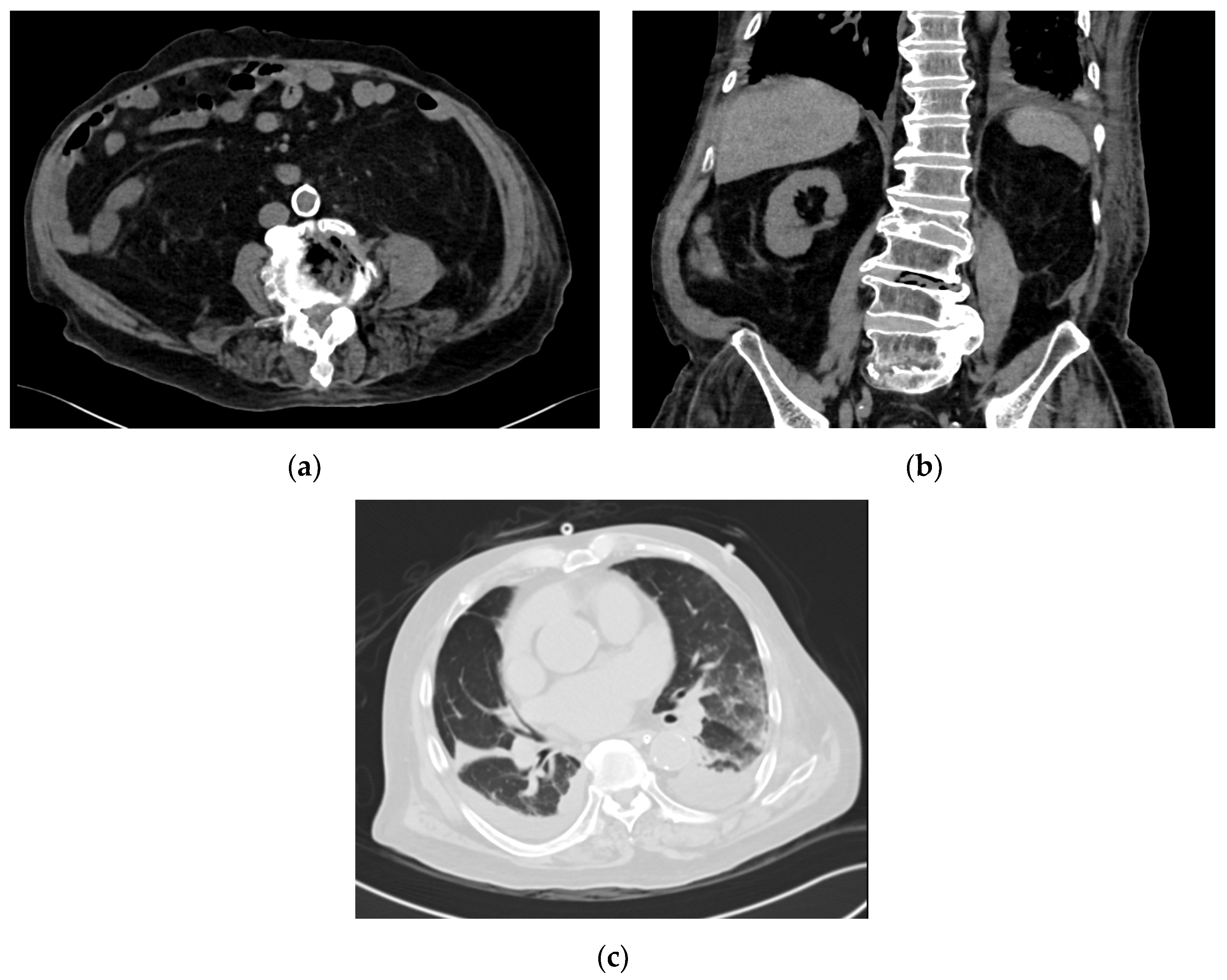
2. Case Report
3. Discussion
Author Contributions
Funding
Acknowledgments
Conflicts of Interest
References
- Vasquez-Rios, G.; Pineda-Reyes, R.; Pineda-Reyes, J.; Marin, R.; Ruiz, E.F.; Terashima, A. Strongyloides stercoralis hyperinfection syndrome: A deeper understanding of a neglected disease. J. Parasit. Dis. 2019, 43, 167–175. [Google Scholar] [CrossRef] [PubMed]
- Nutman, T.B. Human infection with Strongyloides stercoralis and other related Strongyloides species. Parasitology 2017, 144, 263–273. [Google Scholar] [CrossRef] [PubMed] [Green Version]
- Buonfrate, D.; Requena-Mendez, A.; Angheben, A.; Muñoz, J.; Gobbi, F.; Van Den Ende, J.; Bisoffi, Z. Severe strongyloidiasis: A systematic review of case reports. BMC Infect. Dis. 2013, 13, 78. [Google Scholar] [CrossRef] [PubMed] [Green Version]
- Koh, M.S.; Leng, P.H.; Eng, P.; Hwang, J. An unusual cause of pulmonary haemorrhage in a patient with rheumatoid arthritis. Ann. Acad. Med. Singap. 2004, 33, 365–367. [Google Scholar] [PubMed]
- Oh, C.C.; Ong, T.H.; Busmanis, I.; Wijaya, L. An 81-year-old man with cutaneous periumbilical purpura. Chest 2012, 141, 818–821. [Google Scholar] [CrossRef] [PubMed]
- Seet, R.C.; Lau, L.G.; Tambyah, P.A. Strongyloides hyperinfection and hypogammaglobulinemia. Clin. Diagn. Lab. Immunol. 2005, 12, 680–682. [Google Scholar] [CrossRef] [PubMed] [Green Version]
- Robson, D.; Beeching, N.J.; Gill, G.V. Strongyloides hyperinfection syndrome in British veterans. Ann. Trop. Med. Parasitol. 2009, 103, 145–148. [Google Scholar] [CrossRef] [PubMed]
- Zeana, C.; Kubin, C.J.; Della-Latta, P.; Hammer, S.M. Vancomycin-resistant Enterococcus faecium meningitis successfully managed with linezolid: Case report and review of the literature. Clin. Infect. Dis. 2001, 33, 477–482. [Google Scholar] [CrossRef] [PubMed] [Green Version]
- Abu Omar, M.; Abu Ghanimeh, M.; Kim, S.; Howell, G. Strongyloides hyperinfection syndrome and VRE pneumonia. BMJ Case Rep. 2017, bcr2016216634. [Google Scholar] [CrossRef] [PubMed]
- Schein, F.; Fouillet, L.; Lutz, M.F.; Daguenet, E.; Botelho-Nevers, E.; Cornillon, J. Recurrent Enterococcus faecalis meningitis in a patient presenting with Strongyloides hyperinfection syndrome during HTLV-1-induced T-cell lymphoma. Med. Mal. Infect. 2018, 48, 428–430. [Google Scholar] [CrossRef] [PubMed]
- Bang, J.H.; Cho, K.T. Rapidly Progressive Gas-containing Lumbar Spinal Epidural Abscess. Korean J. Spine 2015, 12, 139–142. [Google Scholar] [CrossRef] [PubMed] [Green Version]
- Ono, R.; Uehara, K.; Kitagawa, I. Emphysematous Osteomyelitis of the Spine: A Case Report and Literature Review. Intern. Med. 2018, 57, 2081–2087. [Google Scholar] [CrossRef] [Green Version]
- Khanduri, S.; Singh, M.; Goyal, A.; Singh, S. Emphysematous osteomyelitis: Report of two cases and review of literature. Indian J. Radiol. Imaging 2018, 28, 78–80. [Google Scholar]
- Kim, Y.K.; Jo, K.M.; Jang, J.H.; Heo, C.M.; Lee, J.H.; Park, J.H.; Kim, S.; Jang, H.J.; Kim, H.K.; Kiem, S. Rapidly Fatal Emphysematous Osteomyelitis with Multiple Septic Emboli and Liver Abscess Caused by Klebsiella pneumoniae. Infect. Chemother. 2018, 50, 268–273. [Google Scholar] [CrossRef]
- Aghaei Lasboo, A.; Walker, M.T.; Hijaz, T.A. An unusual appearance of discitis due to gas-forming Escherichia coli with associated pneumocephalus. Spine 2010, 35, E257–E259. [Google Scholar] [CrossRef] [PubMed]

| Date | 2002 | 2004 | March–April 2016 | 2018 | January 2019 | July 2019 | December 2019 | ||||||||
|---|---|---|---|---|---|---|---|---|---|---|---|---|---|---|---|
| Eosinophil count (×109/L) | 0.8 | 0.8 | 3.9 | 2.8 | 0.8 | 0.6 | 0.5 | 0.0 | 0.8 | 0.2 | 0.3 | 1.7 | 2.2 | 1.1 | 0.8 |
| Eosinophil % | 9.5 | 6.5 | 25.4 | 31.1 | 4.7 | 6.3 | 4.2 | 0.1 | 8.4 | 1.9 | 1.3 | 8.1 | 16.6 | 12 | 5.4 |
| Event | Routine primary care visit | Admitted for bleeding gastric ulcer | Admitted for left upper limb cellulitis and deconditioning in March 2016; transferred to a community hospital for rehabilitation in April 2016. Had two documented episodes of E.coli urinary tract infection | Routine primary care visit | Routine primary care visit | Routine primary care visit | Routine primary care visit | Admitted in mid-December 2019 for fever and altered mental status, diagnosed with Strongyloides hyperinfection (stool positive from D4-D8 of admission) Started on daily oral ivermectin from D4-D11 of admission. | |||||||
| Steroid usage | Not known | Traditional Chinese medicine | Started on hydrocortisone 20 mg once every morning, 10 mg once every evening for adrenal insufficiency on 16/3/16; dose subsequently reduced further to 10 mg daily | Remains on maintenance hydrocortisone 10 mg once daily | Switched to prednisolone 5 mg once daily from June 2019 and stopped from September 2019, but patient inadvertently took hydrocortisone 10 mg once daily and prednisolone 5 mg once daily up till December 2019 admission. | ||||||||||
© 2020 by the authors. Licensee MDPI, Basel, Switzerland. This article is an open access article distributed under the terms and conditions of the Creative Commons Attribution (CC BY) license (http://creativecommons.org/licenses/by/4.0/).
Share and Cite
Wee, L.E.; Hnin, S.W.K.; Xu, Z.; Lee, L.S.-U. Strongyloides Hyperinfection Associated with Enterococcus faecalis Bacteremia, Meningitis, Ventriculitis and Gas-Forming Spondylodiscitis: A Case Report. Trop. Med. Infect. Dis. 2020, 5, 44. https://doi.org/10.3390/tropicalmed5010044
Wee LE, Hnin SWK, Xu Z, Lee LS-U. Strongyloides Hyperinfection Associated with Enterococcus faecalis Bacteremia, Meningitis, Ventriculitis and Gas-Forming Spondylodiscitis: A Case Report. Tropical Medicine and Infectious Disease. 2020; 5(1):44. https://doi.org/10.3390/tropicalmed5010044
Chicago/Turabian StyleWee, Liang En, Su Wai Khin Hnin, Zheyu Xu, and Lawrence Soon-U Lee. 2020. "Strongyloides Hyperinfection Associated with Enterococcus faecalis Bacteremia, Meningitis, Ventriculitis and Gas-Forming Spondylodiscitis: A Case Report" Tropical Medicine and Infectious Disease 5, no. 1: 44. https://doi.org/10.3390/tropicalmed5010044
APA StyleWee, L. E., Hnin, S. W. K., Xu, Z., & Lee, L. S.-U. (2020). Strongyloides Hyperinfection Associated with Enterococcus faecalis Bacteremia, Meningitis, Ventriculitis and Gas-Forming Spondylodiscitis: A Case Report. Tropical Medicine and Infectious Disease, 5(1), 44. https://doi.org/10.3390/tropicalmed5010044