Article

Can the Cyanobacterium Nostoc commune Exert In Vitro Biocontrol on Fusarium oxysporum, Causal Agent of Wilt in Banana (Musa AAB)?

by
Ana Isabel Pico-González
1,
Juan de Dios Jaraba-Navas
2,
Alfredo Jarma-Orozco
2,
Dairo Javier Pérez-Polo
2,
Diana Sofia Herazo-Cárdenas
3,
Adriana Vallejo-Isaza
3,
Alberto Antonio Angulo-Ortíz
4,
Yirlis Yadeth Pineda-Rodríguez
1,
Anthony Ricardo Ariza-González
1,
Daniela Vegliante Arrieta
1 and
Luis Alfonso Rodríguez-Páez
2,*
1
Department of Agronomic Engineering and Rural Development, Faculty of Agricultural Sciences, Universidad de Córdoba, Montería 230002, Colombia
2
Phytopathology Practical Laboratory, Faculty of Agricultural Sciences, Universidad de Córdoba, Monteria 230002, Colombia
3
Laboratory of Aquaculture Health and Water Quality, Faculty of Veterinary Medicine, Universidad de Córdoba, Montería 230002, Colombia
4
Chemistry Department, Faculty of Basic Sciences, Universidad de Córdoba, Montería 230002, Colombia
*
Author to whom correspondence should be addressed.
Sci 2025, 7(3), 115; https://doi.org/10.3390/sci7030115
Submission received: 28 May 2025 / Revised: 18 July 2025 / Accepted: 28 July 2025 / Published: 18 August 2025
(This article belongs to the Section Biology Research and Life Sciences)

Abstract

Fusarium wilt, caused by Fusarium oxysporum f. sp. cubense tropical race 4 (Foc TR4), threatens banana and plantain production throughout South America. Because Colombian biosafety regulations restrict in vitro work with Foc TR4, we tested the antifungal activity of Nostoc commune against F. oxysporum race 2 isolated from cv. ‘Manzano’ (Musa AAB). An ethanolic extract of the cyanobacterium (EEC) was profiled by gas chromatography and evaluated with a Kirby–Bauer assay (1000–4000 ppm; n = 4). Synthetic Sico® and botanical Timorex® served as positive controls, and solvent-free plates were the negative control. Growth reduction (GR) and percentage inhibition of radial growth (PIRG) were analysed with Student’s t-test (α = 0.05). Forty-two compounds—mainly fatty and carboxylic acids associated with antifungal activity—were detected. Sico achieved complete inhibition (100 ± 0%), Timorex suppressed 76 ± 2%, and 4 000 ppm EEC curtailed mycelial expansion by 45 ± 3% (p < 0.01). Although less potent than commercial fungicides, EEC impeded F. oxysporum growth, demonstrating that N. commune synthesises bioactive metabolites. Optimising cyanobacterial cultivation and formulation could yield a sustainable biocontrol alternative for managing Fusarium wilt in the region.

Graphical Abstract

1. Introduction

Fusarium wilt of bananas is one of the most devastating vascular diseases affecting edible Musaceae, including bananas, plantains, and bluggoe. Initially reported in the banana cultivar ‘Gros Michel’ (Musa AAA, Gros Michel subgroup), the disease is caused by the fungus Fusarium oxysporum f. sp. cubense (Foc) Race 1. By the late 1950s, this pathogen had decimated banana plantations worldwide, leading to the replacement of ‘Gros Michel’ with the resistant ‘Cavendish’ cultivar (Musa AAA) [1,2]. However, the emergence of Tropical Race 4 (Foc TR4) poses a significant threat to global banana and plantain production, as it can infect both crops and exhibits remarkable resilience and persistence in contaminated soils. Foc TR4 is considered one of the top ten most impactful diseases in agricultural history due to its ability to survive for decades in the absence of host plants, rendering contaminated lands unsuitable for Musaceae cultivation [3,4,5,6]. By 2040, an estimated 1.7 million hectares of banana plantations are projected to be affected by this pathogen [7,8].
In Latin America, Foc TR4 has been detected in Colombia (2019), Perú (2021), and Venezuela (2023). In Colombia, the pathogen was reported in Cavendish banana plantations in La Guajira and Magdalena [1,9]. Similarly, in Peru, the disease was identified in the Piura region [10], while in Venezuela, it was found affecting plantain crops (Musa AAB) in the state of Aragua [11]. The rapid dissemination of Foc TR4 across these regions is attributed to its ability to spread through contaminated soil, water, farm tools, and even footwear, posing significant challenges for disease control. Consequently, banana and plantain growers in these areas face substantial economic losses and reduced productivity [12,13,14]. Small- and medium-scale producers, who collectively manage 105,574 hectares of banana and 279,229 hectares of plantain [15], are especially vulnerable because they lack the capital required for intensive quarantine and sanitation programmes. Because Colombian biosafety regulations currently prohibit in vitro work with Foc TR4, direct pathogenicity assays with this race were not possible. We therefore used a Fusarium oxysporum strain isolated from symptomatic ‘Manzano Criollo’ banana (Musa AAB) plants as a surrogate in all experiments.
In response, the Colombian government has implemented various containment measures, including strict quarantine protocols, disinfection checkpoints, eradication of infected plants, and the promotion of clean planting materials [16]. Simultaneously, ongoing research focuses on developing resistant banana cultivars and exploring alternative management strategies, such as biological control using microorganisms [17]. Among these strategies, microbial biocontrol agents, including Bacillus spp., Pseudomonas spp., Rhizobium spp., Stenotrophomonas spp., and Trichoderma spp., have shown promising results in controlling soilborne phytopathogens [18,19,20,21]. Additionally, certain cyanobacteria species have emerged as potential biocontrol agents [22].
Cyanobacteria, belonging to the phylum Cyanobacteria, are a diverse group of prokaryotic, photosynthetic microorganisms commonly found in freshwater and marine ecosystems [23]. These microorganisms have gained attention in agriculture due to their ability to produce bioactive secondary metabolites with antifungal and antimicrobial properties. Cyanobacteria have shown efficacy against economically significant pathogens like Fusarium oxysporum, making them valuable tools for sustainable disease management [24,25,26,27,28,29,30].
Nostoc commune is a filamentous cyanobacterium characterised by olive-green to dark yellowish-brown colonies, spherical to flattened cells, heterocysts for nitrogen fixation, and akinetes that function as resistance structures [31,32]. These attributes enable N. commune to adapt to diverse environmental conditions, enhancing its resilience [33]. The biocontrol potential of N. commune is primarily attributed to its ability to synthesise secondary metabolites, including phenols, terpenes, flavonoids, fatty acids, lactones, carotenoids, and tannins [34,35]. These compounds exhibit antifungal properties, providing a natural defence mechanism against pathogenic fungi [35].
Previous studies by Kajiyama et al. [36], Hernández-Carlos and Gamboa-Angulo [37], and Pooja and Niveshika [38], have demonstrated the antifungal efficacy of N. commune-derived compounds, highlighting their potential to activate plant defence mechanisms and confer resistance against both biotic and abiotic stress factors [39]. Kim and Kim [40], reported that Nostoc commune extracts inhibited the growth of Fusarium oxysporum as effectively as the fungicide mancozeb in both in planta and in vitro assays. In subsequent comparative tests, N. commune outperformed other cyanobacteria—Anabaena solitaria, Oscillatoria angustissima, Nodularia sp., and Calothrix brevissima—against Alternaria alternata, Botrytis cinerea, Colletotrichum gloeosporioides, and F. oxysporum [41]. This superior activity is likely rooted in the unique chemical structures, ecological functions, and bioactivities of the antifungal secondary metabolites produced by N. commune, which differ markedly from those described in Anabaena and Microcystis [42].
Because N. commune can be grown in inexpensive, non-agitated semi-closed systems, scales readily to industrial production, and tolerates harsh tropical conditions, it is an appealing candidate for biocontrol formulations targeting fungal diseases in tropical agro-ecosystems [43]. Therefore, this study sought to assess, under in vitro conditions, the antifungal activity of metabolites produced by Nostoc commune against Fusarium oxysporum f. sp. cubense Race 2.

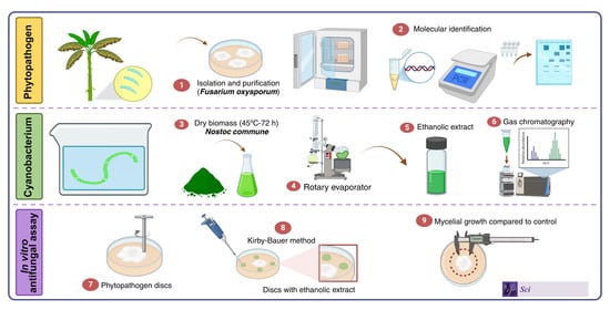
2. Materials and Methods

2.1. Location

The experiment was conducted in 2023 in the Laboratories of Aquaculture Health and Water Quality, Phytopathology, and Chemistry of Natural Products at the University of Córdoba, Colombia (8°47′37″ N; 75°51′51″ W, 15 m.a.s.l.).

2.2. Cultivation and Obtention of Ethanolic Extract of Cyanobacteria (EEC)

The cyanobacterium used in this study was donated by the company Microalgas Oleas S.A., Mexico. It was cultivated in BG11 medium in the Laboratory of Aquaculture Health and Water Quality at the University of Córdoba under continuous illumination with white LED light (40–50 µmol photons m−2 s−1). The microorganism was grown at room temperature 28 ± 2 °C, in 50 L plastic containers with constant aeration, maintaining a pH of 10 (Figure 1). The optical density at harvest was measured at 1.315 using a ThermoScientific™ Multiskan™ GO spectrophotometer (Missouri City, TX, USA). After 20 days of cultivation, the cyanobacterial biomass was harvested by centrifugation at 10,000 rpm for 15 min. The supernatant was discarded, and the biomass was dried at 45 °C in a Memmert® heating and drying oven for 72 h [44]. Once dried, the biomass was ground into a fine powder using a porcelain mortar.
The ethanolic extract was obtained in the Laboratory of Natural Products at the Faculty of Basic Sciences. Twenty grams of the powdered biomass were mixed with 200 mL of ethanol (96%) (Sigma-Aldrich, St. Louis, MO, USA, part number 64175) and left to macerate under laboratory conditions (24 °C) for five days. After the maceration period, the mixture was filtered through a Whatman filter paper (125 mm in diameter). Residual ethanol was removed from the filtrate by rotary evaporation under reduced pressure (Heidolph Hei-VAP Expert; 55 rpm, 45 °C) until the extract formed a homogeneous paste.

2.3. Preliminary Chemical Analysis

A sample of 50 mg of the EEC was analysed to determine the presence of secondary metabolites. The analysis was based on observable changes, such as colour reactions, formation of precipitates, or gas release.

2.4. Derivatization of Ethanolic Extract of Cyanobacteria (EEC)

The ethanolic extract of Nostoc commune (EEC) was evaporated to dryness under a gentle stream of nitrogen at 40 °C and transferred to pre-silanized 1.5 mL glass microvials. For silylation, 50 µL of N,O-bis(trimethyl-silyl)trifluoro-acetamide (BSTFA) containing 1% (v/v) trimethyl-chlorosilane (TMCS) (≥99% purity, Sigma-Aldrich) were added to each vial. The mixture was vortexed for 10 s and incubated in a thermostatted ultrasonic bath at 60 °C for 15 min to ensure complete derivatization of free hydroxyl and carboxyl groups. After the reaction, samples were cooled to room temperature and centrifuged at 10,000× g for 2 min to remove particulates. A 200 µL aliquot of the clear supernatant was transferred to a 2 mL autosampler vial fitted with a PTFE/silicone septum and diluted with 300 µL of anhydrous n-hexane (HPLC grade). Derivatized extract was stored at −20 °C, protected from light, and analysed by GC-MS within 48 h to prevent hydrolytic or thermal degradation.

2.5. Gas Chromatographic–Mass-Spectrometric Analysis

Derivatised aliquots were analysed on a Shimadzu GCMS-TQ system (GC-2010 Plus gas chromatograph coupled to a triple-quadrupole MS) fitted with a DB-5 ms fused-silica capillary column (30 m × 0.25 mm i.d., 0.25 µm film, 5% phenyl/95% dimethyl-polysiloxane). Injection and carrier gas. A 1.0 µL volume was introduced with the autosampler in split mode (1:15) at 280 °C. Ultra-high-purity helium (≥99.999%) served as the carrier gas at a constant-flow rate of 1.0 mL min−1 under electronic pressure control. Oven programme. The oven was held at 60 °C for 0 min, ramped to 310 °C at 22 °C min−1, and held for 5 min, giving a total runtime of 15.4 min. Mass-spectrometer parameters. Ionisation was by electron impact (EI, 70 eV). The transfer line and quadrupole assemblies were maintained at 280 °C, and the ion source at 200 °C. Full-scan data were acquired from m/z 40–550 at 5 scans s−1. Data-dependent production (MS/MS) spectra were obtained with argon (1.5 mTorr) as collision gas for structural confirmation. Compound identification and quality control. Tentative identifications were assigned by matching full-scan and MS/MS spectra against the NIST 17 and Wiley 12 libraries, accepting hits with a similarity index ≥ 800 and a retention-index deviation ≤ 10 units relative to a C7–C40 n-alkane series analysed under identical conditions. Where available, assignments were corroborated authentic standards. Solvent blanks and pooled quality-control samples were injected every ten runs to monitor carry-over, retention-time stability (RSD < 0.2%) and spectral reproducibility. Derivatised extract was stored at −20 °C, protected from light, and injected within 48 h to minimise hydrolytic or thermal degradation.

2.6. Obtaining the Phytopathogenic Fungus

Due to the strict sanitary regulations enforced by the Colombian government to prevent the spread of Fusarium oxysporum f. sp. cubense tropical race 4, an isolate of Fusarium oxysporum f. sp. cubense race 2 isolate FB INVEPAR was used in this research. The fungus was isolated from symptomatic banana plants (cv. Manzano, Musa AAB) exhibiting characteristic symptoms of banana wilt disease, including wilting, chlorotic leaves, and vascular discoloration. The isolation and purification procedures were carried out in the Phytopathology Laboratory at the Faculty of Agricultural Sciences, University of Córdoba, following the methodology described by García-Bastidas et al. [45]. Rectangular tissue Sections (2 cm long × 0.5 cm wide) were obtained from the pseudostem of infected plants. These tissue samples were surface disinfected by immersion in 1.5% sodium hypochlorite for 2 min, followed by a one-minute rinse in sterile distilled water. Subsequently, the samples were briefly washed with 70% ethanol and then subjected to three additional rinses in sterile distilled water, each lasting one minute.
The disinfected tissue sections were transferred onto Potato Dextrose Agar (PDA) medium and incubated at 25 °C for 3 to 6 days, during which fungal growth was observed. Mycelial fragments from actively growing colonies were subcultured onto fresh PDA plates to obtain pure cultures. These pure cultures were examined under a light microscope to identify the presence of macroconidia, microconidia, and chlamydospores, which are key morphological structures for the identification of F. oxysporum.
To further ensure purity and reproducibility, monosporic cultures were prepared using the spore isolation technique with vertical incubation. Briefly, a portion of the fungal mycelium was suspended in 10 mL of sterile distilled water, and the suspension was poured onto water agar medium. The plates were placed vertically and incubated for 24 h, allowing single germinated spores to be carefully transferred onto PDA plates for further culturing [45].
The monosporic cultures were subsequently used for both morphological and molecular characterisation of the fungus. Morphological identification was performed using the pictorial keys of Barnett and Hunter [46], while molecular identification was conducted through Internal Transcribed Spacer (ITS) sequencing.

2.7. Molecular Identification of the Study Microorganisms

Genomic DNA was extracted from monosporic mycelia of Fusarium oxysporum f. sp. cubense race 2 (FB-INVEPAR) and from axenic cultures of Nostoc commune strain F56 using the PureLink™ Plant Genomic DNA Kit (Invitrogen) following the manufacturer’s instructions. Extraction, PCR amplification and sequencing protocols were tailored to each organism to accommodate their distinct cell-wall architectures. All procedures were performed in the Applied Molecular Biology Laboratory, University of Córdoba.

2.7.1. Fusarium oxysporum f. sp. Cubense Race 2 (FB-INVEPAR)

DNA extraction. Approximately 100 mg of fresh monosporic mycelium were processed with the PureLink™ Plant Genomic DNA Mini Kit (Invitrogen™, Thermo Fisher Scientific, Missouri, TX, USA) following the manufacturer’s instructions. Yields averaged 120–160 ng µL−1, with A260/A280 ratios of 1.8–1.9. PCR reactions were conducted in a BIO-RAD T100™ thermocycle. Each 50 µL reaction mixture contained template DNA, primers, Taq polymerase, dNTPs, and the appropriate buffer for optimal enzymatic activity (Table 1).
The success of the PCR amplification was confirmed through electrophoresis on a 2% agarose gel prepared with 1X TBE buffer. A 5 µL aliquot of the PCR product was mixed with loading dye and loaded onto the gel alongside a 1 µL molecular weight marker for fragment size determination. The electrophoresis was run at 100 volts for 60 min, and the gel was subsequently visualised under UV light using a Labnet U1001 photodocumentation system.

2.7.2. Nostoc commune Strain F56 (GenBank PV865569)

Axenic cultures were grown to OD750 ≈ 1.2, pelleted (6000× g, 10 min, 4 °C), washed twice with TE buffer (10 mM Tris-HCl, 1 mM EDTA, pH 8.0), flash-frozen, lyophilised (24 h), and cryopulverised; 20 mg of powder were processed with the PureLink™ Genomic DNA Mini Kit (Invitrogen) following the Pbact-P protocol optimised for cyanobacteria [47], which includes a 30 min lysis at 65 °C with periodic vortexing and the addition of 10 µL 10% (w/v) PVP-10 to sequester polyphenols. The eluate yielded 140–180 ng µL−1 DNA (A260/A280 ≈ 1.8; A260/A230 ≥ 1.8) of high integrity, confirmed on 1% agarose-TBE gels. Nearly full-length 16S rRNA genes were amplified with primers 27F (5′-AGAGTTTGATCMTGGCTCAG-3′) and 1492R (5′-TACGGYTACCTTGTTACGACTT-3′) in 50 µL reactions containing 25 µL DreamTaq™ Hot Start PCR Master Mix (2×; Thermo Fisher Scientific), 1 µL of each primer (10 µM), 2 µL template (~157 ng µL−1), and nuclease-free water. Amplicons were resolved on 1% agarose gels, visualised with an Enduro GDS system, and sized against a 1 kb ladder (Sigma-Aldrich).
The PCR products were sent to the Institute of Genetics and Biotechnology (IBUN) at the National University of Colombia in Bogotá for sequencing. Raw sequence data were analysed using 4Peaks software (version 1.8) (available at https://nucleobytes.com/4peaks/ accessed on 23 March 2025) for quality assessment and nucleotide visualisation. The sequences were subsequently compared with those available in the GenBank database at the National Center for Biotechnology Information (NCBI) (available at https://www.ncbi.nlm.nih.gov accessed on 24 March 2025) using the BLAST v 2.17.0 (Basic Local Alignment Search Tool) algorithm (available at https://blast.ncbi.nlm.nih.gov/Blast.cgi). The identification of the fungal isolate was confirmed based on sequence similarity, and the sequence was deposited in the GenBank database to ensure accessibility for future studies.

2.8. In Vitro Antifungal Assay

The antifungal potential of the cyanobacterial ethanolic extract (EEC) was evaluated against Fusarium oxysporum isolated from banana cultivar Manzano (Musa AAB) under in vitro conditions at the Plant Pathology Laboratory of the University of Córdoba. The assay was performed using the disc diffusion method (Kirby-Bauer). PDA was used as the growth medium in 10 cm diameter Petri dishes. The EEC was dissolved in 10% dimethyl sulfoxide (DMSO) to prepare four concentrations: 1000, 2000, 3000, and 4000 ppm. Selection of those concentrations were based on a previous assay where 2000 ppm showed to have antifungal activity (data not published). Sterile paper filter discs (Double Ring, 0.6 cm diameter) were saturated with 50 UL of each concentration using a micropipette, for 5 min. Subsequently, a 5 mm diameter fungal disc, obtained from a 10-day-old culture of F. oxysporum, was placed in the centre of the Petri dish. Four filter discs saturated with the same concentration of EEC were arranged equidistantly from the fungal disc.
Positive controls included discs saturated with the chemical fungicide Difenoconazole (SICO 250 EC, Syngenta, Bogota, Colombia, part number: 119446-68-3) or the biological fungicide Thimorex Gold EC (Syngenta, Stockton, Israel, part number: 68647-73-4), based on tea tree extract (Melaleuca alternifolia L.). Since the best solubilization of the EEC was obtained using Dimethyl sulfoxide (DMSO), the influence of DMSO on fungus growth was determined using discs saturated with 10% of this solvent (negative control). The ethanol was extracted during the rotoevaporation process; consequently, fungal discs without any treatment were used as absolute control. The plates were incubated at room temperature in a completely randomised design (CRD). Fungal radial colony growth was measured every 24 h for 240 h, by which time the fungal growth in the absolute control had covered the entire Petri dish. Growth rates (GR) and the percentage inhibition of radial growth (PIRG%) were calculated using ImageJ® software v. 1.54k as follows [48]:
GR = [(Cf − Ci)/(Tf − Ti)]
where Cf: Final growth diameter (cm), Ci: Initial growth diameter (cm), Tf: Final growth time, and Ti: Initial measurement time [49].
PIRG = [(R1  R2)/R1 ∗ 100]
where R1 = Radial growth (cm) of the F. oxysporum in absolute control plates, and R2 = Radial growth (cm) of the F. oxysporum in treatment plates

2.9. Statistical Analysis of Data

All statistical analyses were conducted in R v 4.5.1 (https://www.r-project.org). Radial growth data were interrogated with four a priori two sample Student’s t tests (α = 0.05): (i) absolute control versus every treatment, (ii) the ethanolic extract of Nostoc commune pooled across four concentrations (1000, 2000, 3000, 4000 ppm) versus the fungicides Sico 250 EC® and Thimorex Gold® EC, (iii) Sico 250 EC® versus Thimorex Gold® EC, and (iv) low (1000 + 2000 ppm) versus high (3000 + 4000 ppm) extract doses. Family wise type I error was controlled with the Holm adjustment. Percentage inhibition of radial growth (PIRG) violated normality and homoscedasticity even after Box–Cox and arcsine square root transformations; consequently, a Kruskal–Wallis test followed by one way non parametric Tukey type multiple comparisons was performed with the nparcomp package [50]; Treatment medians were visualised on their original scale with ggplot2 [51], and statistically separable groups were denoted by distinct lowercase letters. Finally, principal component analysis of centred, standardised variables (FactoMineR, factoextra) was used to uncover multivariate patterns and correlations among treatments and response variables. Components with eigenvalues greater than one were retained for biological interpretation.

3. Results

3.1. Preliminary Chemical Analysis

The qualitative chemical analysis of the ethanolic extract of Nostoc commune revealed the presence of several secondary metabolites, as determined through specific colorimetric reactions, gas evolution, or precipitate formation (Table 2). Alkaloids, sterols, terpenoids, naphthoquinones, and anthraquinones were identified, with alkaloids showing high positivity across all tests conducted. In contrast, flavonoids, anthocyanidins, cardiac glycosides, coumarins, terpenic lactones, tannins, and saponins were not detected. The intensity of the colorimetric responses indicated varying levels of metabolite presence, with alkaloids presenting the most significant results. These findings justified the subsequent analysis via gas chromatography to further characterise the ethanolic extract’s chemical profile.

3.2. Gas Chromatography (GC-MS)

Gas chromatography analysis of the EEC, identified 42 distinct compounds, characterised by their peak number (PK), retention time (RT), percentage area, and probability of identification (Table 3). Fatty acids, carboxilic acids, alcoholes, aminoacids, hydrocarbures and other compounds with pharmacological, nutritional or antimicrobian activity were detected in the EEC. This extract showed high contents lactic acid (peaks 9) and fatty acids sucha as palmitic and oleic (peaks 35, 37, respectively) (Figure 2). Diethyl Phthalate was also detected, provably due to plastic containers used during the growth of the N. comunne.

3.3. Identification of the Phytopathogenic Fungus

The isolated and purified strain of the phytopathogenic fungus, cultured on potato PDA medium, displayed morphological characteristics consistent with Fusarium oxysporum. The strain produced abundant macroconidia that were falcate to slightly curved, with thin walls and 3 to 5 septa. These structures were borne on monophialides or branched conidiophores arranged in sporodochia. Additionally, abundant microconidia were observed; these were non-septate, hyaline, oval-shaped, and formed on false heads on short monophialides. Chlamydospores were present, occurring singly or in chains, and were both terminal and intercalary (Figure 3). The morphological characteristics align with previous descriptions of F. oxysporum [46,52,53].

3.4. Molecular Identification

3.4.1. Fusarium oxysporum f. sp. Cubense Race 2 (FB-INVEPAR)

Both molecular markers corroborated the taxonomic assignment of isolate FB-INVEPAR to Fusarium oxysporum f. sp. cubense race 2. The 900 bp RPB2 consensus returned BLASTn hits with 100% query coverage, E = 0, and ≥99.4% pairwise identity (895/900 bp) to canonical race-2 alleles—including MH972578.1, ON316739.1 and the Colombian isolate OM100609.1 [54]. The ribosomal ITS fragment likewise displayed ≥99.5% identity to race-2 references (GenBank OR704561). When the two loci were concatenated, a maximum-likelihood analysis (Kimura-2-parameter, 1000 bootstrap replicates, MEGA 11) clustered FB-INVEPAR firmly within the race-2 clade, supported by a 96% bootstrap value. The ITS sequence is archived in GenBank under accession OR704561; the RPB2 sequence was deposited via BankIt (submission ID 2782225) and will be assigned a permanent accession number immediately after GenBank’s review.

3.4.2. Nostoc commune Strain F56

The near-full-length 16S rRNA gene (1463 bp) obtained from strain F56 produced BLASTn hits with 100% query coverage, E = 0, and ≥99.4% pairwise identity to authenticated Nostoc commune sequences; the top matches were MK247968.1 and MK247967.1 (clone ACT709, 99.45% identity), followed by AB694927.1 and AB101003.1 (≥98% identity). Maximum-likelihood analysis (Kimura two-parameter model, 1000 bootstrap replicates, MEGA 11) positioned F56 robustly within the N. commune sensu stricto clade (bootstrap = 98%). The 16S rRNA gene sequence has been deposited in the GenBank database under accession number PV865569.1 and is publicly available through the National Center for Biotechnology Information (NCBI) at the following link: https://www.ncbi.nlm.nih.gov/nuccore/PV865569 (accessed on 7 July 2025).

3.5. In Vitro Assays on Fusarium oxysporum

3.5.1. Growth Diameter (cm)

Student’s t-test contrasts (α = 0.05 and 0.01) showed statistically significant differences in radial mycelial growth between every treatment and the untreated control from the first evaluation onward (Figure 4). The synthetic fungicide Sico completely inhibited F. oxysporum (100% suppression), while the botanical formulation Timorex achieved 76% inhibition. The ethanolic extract of Nostoc commune (EEC) displayed a clear dose-dependent response: 4000 ppm curtailed growth by 45%, whereas 2000 ppm and 1000 ppm suppressed only 17.4% and 7.0%, respectively. These results confirm that although the EEC is less potent than commercial fungicides, its antifungal efficacy increases proportionally with concentration.

3.5.2. Percentage of Inhibition of Radial Growth (PIRG)

The effect of the six treatments on the radial growth of Fusarium oxysporum showed statistically significant differences across the five evaluation periods: 48 h (Figure 5A), 96 h (Figure 5B), 144 h (Figure 5C), 192 h (Figure 5D), and 240 h (Figure 5E). At 48 h, the commercial fungicides SICO 250 EC and Timorex Gold EC achieved inhibition levels close to 95–100%, forming the highest statistical group (“a”). In contrast, all concentrations of the ethanolic extract of Nostoc commune (EEC 1000–4000 ppm) clustered in the lowest group (“b”), exhibiting inhibition values ≤ 30%. From 96 h onward, the extract displayed a clear dose-dependent response, with PIRG values progressively increasing with concentration. The 4000 ppm dose (35–45%) was statistically distinct from the intermediate concentrations (2000–3000 ppm, 20–30%) and the lowest dose (1000 ppm, ≤10%). Nevertheless, even at its highest concentration, the extract remained statistically inferior to Timorex Gold EC (≈70–80% PIRG) and SICO 250 EC (≥95% PIRG) at all time points.
These findings consistently position SICO 250 EC as the most effective treatment, with Timorex Gold EC as a reliable second tier. In contrast, the ethanolic extract of N. commune exhibited a moderate, concentration-dependent fungistatic effect, which may hold potential as a complementary or additive agent but remains insufficient to replace commercial fungicides under the in vitro conditions tested.

3.5.3. Growth Rate (cm Day−1)

Figure 6 reveals that the treatments with EEC (4000 ppm), SICO 250 EC and Timorex Gold EC, were the most effective in reducing the growth rate of Fusarium oxysporum. Specifically, the fungicides SICO 250 EC and Timorex Gold demonstrated greater efficiency in suppressing fungal growth compared to the lower concentrations of EEC (1000 and 2000 ppm). This dose-dependent response indicates that the inhibitory effect of EEC on fungal growth becomes more pronounced as its concentration increases, consistent with the observations of mycelial growth reduction across different evaluation times. Additionally, the growth of the fungus with the dissolvent DMSO was similar to the growth of the absolutely control, indicating that this type of dissolvent had not fungical effect.

3.5.4. Principal Component Analysis (PCA)

The biplot generated from the PCA (Figure 7) showed that the first principal component accounted for 99% of the total variability. Treatments involving SICO 250 EC and Timorex Gold were strongly associated with high PIRG values, reflecting their superior efficiency in inhibiting fungal growth. In contrast, the EEC was characterised by higher values of growth diameter and growth rate. Notably, the growth diameter exhibited a negative correlation with PIRG, indicating that treatments with lower growth diameter values corresponded to higher PIRG values. Similarly, growth rate correlated positively with growth diameter and negatively with PIRG, as treatments with lower growth rates demonstrated higher PIRG values. These relationships underscore the effectiveness of treatments with high PIRG values in reducing fungal growth and provide a comprehensive understanding of the interactions between the evaluated variables.

4. Discussion

The present study demonstrates that an ethanolic extract of Nostoc commune (EEC) exhibits a dose-dependent but moderate fungistatic effect against Fusarium oxysporum under in vitro conditions. At the highest concentration tested (4000 ppm), the extract achieved a mean PIRG of ~45%, which is significantly lower than the inhibition provided by Timorex Gold EC (70–85%) and SICO 250 EC (≥95%) across all evaluation times (48–240 h). Accordingly, while EEC exhibits clear bioactivity, it cannot yet match the performance of commercial products. It should be viewed as a complementary component within an integrated disease-management framework, rather than as a direct replacement. The identification of fatty acids, alkanes, sterols, terpenoids, naphthoquinones, and anthraquinones in the ethanolic extract of N. commune aligns with previous research highlighting the diverse metabolic capabilities of cyanobacteria [55,56]. These compounds, known for their antimicrobial properties, underscore the potential of cyanobacteria as a natural source of bioactive molecules for managing plant diseases [35]. However, the efficacy of these compounds is highly dependent on environmental conditions, cultivation methods, and extraction techniques, which must be carefully optimised to maximise their potential. The antimicrobial activity of cyanobacteria has been extensively documented, with numerous studies demonstrating their ability to inhibit the growth of phytopathogenic fungi [57,58]. Consistent with prior reports of Nostoc-derived antifungal activity, the extract curtailed the radial growth of Fusarium oxysporum in a clear dose-dependent manner, with PIRG reaching ≈45% at 4000 ppm.
This fungistatic, rather than fungicidal, effect indicates that the extract slows pathogen proliferation without fully eradicating it. This observation aligns with the work of Rodríguez-Palacio [59], who also reported a reduction in microorganism growth without complete inhibition. The antifungal efficacy of ethanolic extracts from Nostoc spp. is well established. Asimakis et al. [60], demonstrated that such extracts suppress Fusarium oxysporum f. sp. melonis, Penicillium expansum, Rhizoctonia solani, Rosellinia sp., Sclerotinia sclerotiorum, and Verticillium alboatrum. Águila-Carricondo et al. [61], found that N. commune achieved the highest inhibition rates against F. oxysporum compared with other cyanobacteria. Abed et al. [62], attributed this activity to metabolites such as nostodione A, fischerellin A, tolytoxin, nostocyclamide, and hapalindole. Ferrinho et al. [63], further noted that Nostocales—the order that includes N. commune—has recorded production of roughly 924 secondary metabolites, far exceeding the totals for Oscillatoriales, Chroococcales, and Synechococcales. This richness likely reflects the larger genome size of Nostocales members and underpins their superior antifungal spectra. In our study, gas-chromatographic profiling detected 42 compounds in the EEC. The discrepancy between this figure and the larger metabolite inventories reported elsewhere probably stems from specific culture conditions and extraction protocols rather than from intrinsic metabolic limitations of N. commune. Parambil and Yusuf [64], used GC–MS to profile crude extracts of Nostoc spp. and identified phytol, heptadecane, and 10-octadecenoic acid—constituents also detected in our ethanolic extract. Desbois et al. [65], underscored the antimicrobial value of polyunsaturated fatty acids, including 10-octadecenoic (oleic) acid. Abdel-Hafez et al. [66], quantified antifungal metabolites in Anabaena oryzae, Arthrospira sp., Nostoc spp., and Oscillatoria sp., attributing significant inhibitory activity to phytol and oleic acid, both present in our extract. Concordantly, Farghl et al. [67], demonstrated that phytol enhances the antifungal potency of N. carneum, producing clear inhibition zones against F. oxysporum. Beyond its defensive role, phytol performs essential cellular functions in cyanobacteria, which may account for its prevalence and recurrent bioactivity [68,69].
Another compound class detected in the EEC comprised the amino acids L-alanine, L-valine, and L-isoleucine. Renganathan et al. [70], observed that cyanobacterial polysaccharides, proteins, and amino acids provide notable phytoprotective benefits. Cyanobacteria are also recognised for their exceptional protein yields [71], and their ability to synthesise bioactive peptides, vitamins, and minerals [72], attributes that underpin their expanding use in pharmaceutical and nutritional applications [73]. In agreement, Lucato et al. [74], reported that biomass produced by Nostoc spp. is especially rich in these amino acids, underscoring its high protein quality and nutritional value.
The antifungal potential of the EEC is strongly linked to its fatty-acid, alkane, and terpenoid fractions. Saturated fatty acids—particularly hexadecanoic (palmitic) and nonadecanoic acids—can disrupt fungal and bacterial membranes, thereby inhibiting growth [75,76]. Given the abundance of fatty acids in cyanobacterial metabolites, López-Arellanes et al. [77], demonstrated that these compounds suppress spore germination and compromise cell-wall integrity in Fusarium spp. Qiu et al. [78], proposed that, once fatty acids penetrate the cell wall and interact with ergosterol, they disturb membrane dynamics and cellular homeostasis. Palmitic acid, detected in our extract (Table 3) and previously isolated from the methanolic fraction of N. calcicola by El-Sheekh et al. [24] markedly reduced F. oxysporum growth. Likewise, Perveen et al. [79], reported that hexadecanoic acid—also present in the EEC—impairs both radial expansion and hyphal morphology in Fusarium spp. Chen et al. [80], therefore emphasised that fatty acids merit further investigation, as they appear to be the most potent bioactive metabolites limiting the development of this pathogen.
Similarly, alkanes like heptadecane and octadecane, although their exact role remains unclear, are believed to play a role in maintaining cellular integrity and defence mechanisms [81,82]. These findings are supported by studies from Gheda and Ismail [83] and Chu [84], who also identified these compounds in cyanobacterial extracts. The variability in metabolite production among different cyanobacterial species and strains is a critical factor to consider. As noted by Shakeel et al. [85], environmental conditions such as salinity, temperature, and nutrient availability can significantly influence the metabolic profile of cyanobacteria. This variability may explain the differences in antifungal activity observed in our study compared to others, such as Kim and Kim [40], who reported a more pronounced antifungal effect under optimal cultivation conditions. These discrepancies highlight the need for standardised cultivation and extraction protocols to ensure consistent results across studies. The implications of these findings extend beyond the laboratory, offering potential applications in the field of sustainable agriculture.
The use of cyanobacterial extracts as biocontrol agents could provide an eco-friendly alternative to chemical fungicides, reducing the environmental impact of conventional disease management practices. This is particularly relevant in the context of banana cultivation, where Fusarium oxysporum poses a significant threat to crop yields. By harnessing the natural antimicrobial properties of cyanobacteria, farmers could potentially reduce their reliance on synthetic chemicals, thereby promoting more sustainable agricultural practices. Moreover, the ability of cyanobacteria to produce a wide range of bioactive compounds under varying environmental conditions suggests that they could be tailored to specific agricultural contexts [86].
For instance, in regions where water salinity or temperature fluctuations are common, cyanobacterial strains that thrive under these conditions could be selected for biocontrol applications. This adaptability makes cyanobacteria a versatile tool in the fight against plant pathogens, particularly in the face of climate change, which is expected to exacerbate disease pressures in many agricultural systems [87]. However, the practical application of cyanobacterial extracts in the field will require further research to address several challenges. For example, the stability and persistence of these compounds in the environment need to be evaluated to ensure their long-term efficacy. Additionally, the potential for resistance development in target pathogens must be considered, as over-reliance on any single biocontrol agent could lead to reduced effectiveness over time. These considerations underscore the importance of integrating cyanobacterial-based solutions into a broader integrated pest management (IPM) strategy, where they can be used in conjunction with other control methods to maximise their impact [88,89,90]. The role of cyanobacteria in sustainable agriculture extends beyond their antimicrobial properties [91,92,93,94,95]. As photosynthetic organisms, cyanobacteria contribute to soil health by fixing atmospheric nitrogen and producing organic matter, which can enhance soil fertility and structure [96]. This dual role as both biocontrol agents and soil enhancers makes cyanobacteria a valuable component of sustainable farming systems, particularly in regions where soil degradation and nutrient depletion are pressing concerns [97].
In the broader context of global agricultural challenges, the findings of this study contribute to the growing body of evidence supporting the use of microbiological solutions to enhance crop resilience [98]. As the world faces increasing pressures from climate change, biodiversity loss, and food insecurity, the need for innovative and sustainable agricultural practices has never been greater. Cyanobacteria, with their diverse metabolic capabilities and adaptability, offer a promising avenue for addressing these challenges. Furthermore, the integration of cyanobacterial-based solutions into agricultural systems could have far-reaching implications for food security and rural livelihoods. By reducing the need for chemical inputs, these solutions could lower production costs for farmers, making agriculture more economically viable in resource-limited settings. Additionally, the use of locally available cyanobacterial strains could empower communities to develop context-specific solutions, fostering greater resilience and self-reliance. While this study provides valuable insights into the antimicrobial potential of cyanobacteria, several questions remain unanswered, highlighting the need for further research. One key area of investigation is the molecular mechanisms underlying the antimicrobial activity of cyanobacterial compounds. Understanding how these compounds interact with pathogen cells at the molecular level could provide valuable insights into their mode of action and inform the development of more effective biocontrol strategies. Another important direction for future research is the optimisation of cultivation and extraction techniques to maximise the production of bioactive compounds [98,99]. The projected production cost in this study was USD 1.38 g−1 of dry biomass. Although relatively high for bulk commodities, this figure is competitive for the manufacture of high-value antifungal metabolites; [100], estimated production costs of USD 1.5–1.8 g−1 in closed photobioreactor systems designed specifically for bioactive compounds.
As Kim and Kim [40], showed, culture conditions strongly modulate the metabolite profile of cyanobacteria. Future work should therefore assess closed bioreactors and other precisely controlled systems to maximise antimicrobial yields, as well as greener solvents and advanced extraction techniques to expand both the diversity and concentration of metabolites in the final extract [75]. In parallel, the potential synergism between cyanobacterial extracts and complementary biocontrol agents merits systematic evaluation [61].
The best control in the fungal growth on the isolate of F. oxysporum used in this study was performed by Sico 250 EC and Timorex Gold EC, both commercial products used to control many plant disease caused by fungus. However, the EEC showed to be able to suppress the mycelial growth of the fungus (45%). This type of EEC could be improved and be part, alone, or combined with other products, of a strategy to do an integrate management of Fusarium wilt disease. For example, combining cyanobacterial extracts with beneficial microbes or plant-derived compounds could enhance their overall efficacy and reduce the likelihood of resistance development in target pathogens. This approach could also provide a more holistic solution to disease management, addressing multiple aspects of the plant-pathogen interaction simultaneously. Finally, the long-term environmental impact of using cyanobacterial extracts in agriculture must be carefully evaluated. While these compounds are naturally derived, their introduction into agricultural systems could have unintended consequences for non-target organisms and ecosystem dynamics [101]. Long-term field studies are needed to assess the ecological risks and benefits of using cyanobacterial-based biocontrol agents, ensuring that their use is both effective and environmentally sustainable.
Gas-chromatographic profiling revealed trace diethyl phthalate in the EEC (Table 3). Phthalate esters frequently occur in plant and microbial extracts yet seldom display antifungal activity. For example, Rubini and Sivakumar [102], found bis(2-ethylhexyl) phthalate to be the dominant constituent of an ethanolic extract of Andrographis stellulata but reported no antimicrobial effect. Likewise, Ahuactzin-Pérez et al. [103], supplemented culture medium with dibutyl phthalate and observed no reduction in Fusarium culmorum mycelial growth. González-Márquez et al. [104], demonstrated that Lentinula edodes cultivated on substrates enriched with di(2-ethylhexyl) phthalate grew and sporulated normally, indicating no negative impact on fungal development. To date, therefore, no study has shown direct antifungal activity of diethyl or related phthalates against F. oxysporum. As Pace et al. [105], note, phthalates often originate from external sources—plastic tubing, solvents, or other laboratory consumables—used during extraction and analysis, making contamination difficult to trace. Consequently, the diethyl phthalate detected here is most likely an artefact and unlikely to contribute to the antifungal activity observed in this study.

5. Conclusions

This work confirms that an ethanolic extract of Nostoc commune (EEC) exerts a dose dependent but moderate fungistatic effect against Fusarium oxysporum, restricting radial mycelial growth by ~45% at 4000 ppm—the most effective concentration evaluated. GC MS profiling revealed a chemically heterogeneous mixture dominated by fatty acids (≈19%), together with minor fractions of hydrocarbons, terpenoids and amino acid derivatives. However, the analytical approach employed cannot unambiguously identify the specific metabolite(s) driving inhibition, nor can it exclude the presence of additional, more polar compounds that elude GC-MS detection.
Consequently, future work should (i) employ complementary LC-MS workflows to capture a broader spectrum of metabolites, (ii) isolate or purchase authentic standards of the major constituents and test their individual and combined effects on F. oxysporum, and (iii) elucidate potential synergistic interactions. Parallel optimisation of N. commune cultivation (light regime, nutrients, salinity) and extraction parameters—while avoiding plasticisers through the exclusive use of inert glass, aluminium, or stainless steel vessels—will be critical to maximise the yield of bioactive molecules.
From an applied perspective, higher EEC doses, stabilising carrier formulations, and alternative delivery systems warrant evaluation under controlled environment assays (mesh house, greenhouse), followed by multi-location field trials. Given the relatively low production cost of N. commune in semi closed facilities, such advances could provide small banana and plantain producers with an affordable, eco-compatible complement to synthetic fungicides within integrated Fusarium wilt management programmes.

Supplementary Materials

The following supporting information can be downloaded at: https://www.mdpi.com/article/10.3390/sci7030115/s1.

Author Contributions

Conceptualization, A.I.P.-G. and J.d.D.J.-N.; methodology, A.I.P.-G. and J.d.D.J.-N.; software, D.J.P.-P. and Y.Y.P.-R.; validation, J.d.D.J.-N., A.J.-O., D.J.P.-P., D.S.H.-C. and A.V.-I.; research, Y.Y.P.-R., A.R.A.-G. and D.V.A.; chemical analysis, A.A.A.-O.; molecular analysis, Y.Y.P.-R. and L.A.R.-P.; resources, A.J.-O., D.S.H.-C., A.V.-I. and J.d.D.J.-N.; data curation, D.J.P.-P.; writing—original draft preparation, A.I.P.-G., L.A.R.-P., J.d.D.J.-N. and A.J.-O.; writing—review and editing, A.I.P.-G., L.A.R.-P., J.d.D.J.-N. and A.J.-O.; supervision, A.J.-O., LAR-P., J.d.D.J.-N., D.J.P.-P., D.S.H.-C. and A.V.-I.; project administration, J.d.D.J.-N. All authors have read and agreed to the published version of the manuscript.

Funding

This research was funded by University of Córdoba, Contingent Recovery Financing Agreement, Grants 80740-440-2020.

Institutional Review Board Statement

Not applicable.

Informed Consent Statement

Not applicable.

Data Availability Statement

The original contributions presented in this study are included in the article/Supplementary Materials. Further inquiries can be directed to the corresponding author(s).

Acknowledgments

To the Ministry of Science, Technology and Innovation (Minciencias), of Colombia, for providing the necessary resources for this research and to the network of laboratories of the University of Córdoba, which facilitated the realisation of the activities contemplated in this research.

Conflicts of Interest

The authors declare no conflicts of interest.

References

  1. Martínez-Solórzano, G.E.; Rey-Brina, J.C.; Pargas-Pichardo, R.E.; Manzanilla, E.E. Fusarium Wilt Tropical Race 4: Current Status and Presence on the American Continent. Agron. Mesoam. 2019, 31, 259–276. [Google Scholar] [CrossRef]
  2. Maryani, N.; Lombard, L.; Poerba, Y.S.; Subandiyah, S.; Crous, P.W.; Kema, G.H.J. Phylogeny and Genetic Diversity of the Banana Fusarium Wilt Pathogen Fusarium oxysporum f. sp. cubense in the Indonesian Centre of Origin. Stud. Mycol. 2019, 92, 155–194. [Google Scholar] [CrossRef] [PubMed]
  3. Leiva, A.M.; Rouard, M.; Lopez-Alvarez, D.; Cenci, A.; Breton, C.; Acuña, R.; Rojas, J.C.; Dita, M.; Cuellar, W.J. Draft Genome Sequence of Fusarium oxysporum f. sp. cubense Tropical Race 4 from Peru, Obtained by Nanopore and Illumina Hybrid Assembly. Microbiol. Resour. Announc. 2022, 11, e00347-22. [Google Scholar] [CrossRef] [PubMed]
  4. García-Bastidas, F.A.; Quintero-Vargas, J.C.; Ayala-Vasquez, M.; Schermer, T.; Seidl, M.F.; Santos-Paiva, M.; Noguera, A.M.; Aguilera-Galvez, C.; Wittenberg, A.; Hofstede, R.; et al. First Report of Fusarium Wilt Tropical Race 4 in Cavendish Bananas Caused by Fusarium odoratissimum in Colombia. Plant Dis. 2020, 104, 994. [Google Scholar] [CrossRef]
  5. Dita, M.; Teixeira, L.A.J.; O’Neill, W.; Pattison, A.B.; Weinert, M.P.; Li, C.Y.; Zheng, S.J.; Staver, C.; Thangavelu, R.; Viljoen, A. Current State of Fusarium Wilt of Banana in the Subtropics. Acta Hortic. 2020, 1272, 45–56. [Google Scholar] [CrossRef]
  6. Dita, M.; Barquero, M.; Heck, D.; Mizubuti, E.S.G.; Staver, C.P. Fusarium Wilt of Banana: Current Knowledge on Epidemiology and Research Needs Toward Sustainable Disease Management. Front. Plant Sci. 2018, 9, 1468. [Google Scholar] [CrossRef]
  7. Ploetz, R.C. Fusarium Wilt of Banana Is Caused by Several Pathogens Referred to as Fusarium oxysporum f. sp. cubense. Phytopathology 2006, 96, 653–656. [Google Scholar] [CrossRef]
  8. Scheerer, L.; Pemsl, D.; Dita, M.; Vicente, L.P.; Staver, C. A Quantified Approach to Projecting Losses Caused by Fusarium Wilt Tropical Race 4. Acta Hortic. 2018, 1196, 211–218. [Google Scholar] [CrossRef]
  9. Instituto Colombiano Agropecuario (ICA). Actualización de La Condición de Fusarium oxysporum f. sp. cubense Raza 4 Tropical—Foc R4T 2021. Available online: https://www.ica.gov.co/areas/agricola/servicios/epidemiologia-agricola/saf/notificacion-oficial/detalle-notificacion-oficial/actualizacion-de-la-condicion-de-fusarium-oxysporu (accessed on 15 October 2023).
  10. Quevedo, M. (2021, abril 11). Declaran Emergencia Fitosanitaria En Todo El Territorio Nacional Ante La Presencia de La Plaga Fusarium oxysporum f. sp. cubense Raza 4 Tropical. Diario Oficial del Bicentenario El Peruano. Available online: https://busquedas.elperuano.pe/dispositivo/NL/1942722-1 (accessed on 22 June 2023).
  11. Instituto Nacional de Salud Agrícola Integral (INSAI). Comunicado Oficial. Available online: https://sigesai.insai.gob.ve/assets/files/fusarium_oxysporum.pdf (accessed on 8 February 2024).
  12. Olivares, B.O.; Rey, J.C.; Lobo, D.; Navas-Cortés, J.A.; Gómez, J.A.; Landa, B.B. Fusarium Wilt of Bananas: A Review of Agro-Environmental Factors in the Venezuelan Production System Affecting Its Development. Agronomy 2021, 11, 986. [Google Scholar] [CrossRef]
  13. Izquierdo-García, L.F.; Carmona, S.L.; Zuluaga, P.; Rodríguez, G.; Dita, M.; Betancourt, M.; Soto-Suárez, M. Efficacy of Disinfectants against Fusarium oxysporum f. sp. cubense Tropical Race 4 Isolated from La Guajira, Colombia. J. Fungi 2021, 7, 297. [Google Scholar] [CrossRef]
  14. Van Westerhoven, A.C.; Meijer, H.J.G.; Seidl, M.F.; Kema, G.H.J. Uncontained Spread of Fusarium Wilt of Banana Threatens African Food Security. PLoS Pathog. 2022, 18, e1010769. [Google Scholar] [CrossRef]
  15. Organización de las Naciones Unidas para la Alimentación y la Agricultura (FAO). FAOSTAT, Cultivos y Productos de Ganadería. Available online: https://www.fao.org/faostat/es/#data/QCL (accessed on 20 February 2025).
  16. Instituto Colombiano Agropecuario (ICA). RESOLUCIÓN No.00002081 (11/03/2024). Available online: https://www.ica.gov.co/getattachment/fc57b4b9-ab28-4c1a-83c6-1c1bbbbf2d8f/2024R00002081.aspx (accessed on 16 July 2024).
  17. García-Bastidas, F.A.; Arango-Isaza, R.; Rodriguez-Cabal, H.A.; Seidl, M.F.; Cappadona, G.; Segura, R.; Salacinas, M.; Kema, G.H.J. Induced Resistance to Fusarium Wilt of Banana Caused by Tropical Race 4 in Cavendish Cv Grand Naine Bananas after Challenging with Avirulent Fusarium spp. PLoS ONE 2022, 17, e0273335. [Google Scholar] [CrossRef] [PubMed]
  18. Xue, C.; Ryan Penton, C.; Shen, Z.; Zhang, R.; Huang, Q.; Li, R.; Ruan, Y.; Shen, Q. Manipulating the Banana Rhizosphere Microbiome for Biological Control of Panama Disease. Sci. Rep. 2015, 5, 11124. [Google Scholar] [CrossRef] [PubMed]
  19. Köberl, M.; Dita, M.; Martinuz, A.; Staver, C.; Berg, G. Members of Gammaproteobacteria as Indicator Species of Healthy Banana Plants on Fusarium Wilt-Infested Fields in Central America. Sci. Rep. 2017, 7, 45318. [Google Scholar] [CrossRef] [PubMed]
  20. Shen, Z.; Xue, C.; Penton, C.R.; Thomashow, L.S.; Zhang, N.; Wang, B.; Ruan, Y.; Li, R.; Shen, Q. Suppression of Banana Panama Disease Induced by Soil Microbiome Reconstruction through an Integrated Agricultural Strategy. Soil Biol. Biochem. 2019, 128, 164–174. [Google Scholar] [CrossRef]
  21. Pisciotta, J.M.; Zou, Y.; Baskakov, I.V. Light-Dependent Electrogenic Activity of Cyanobacteria. PLoS ONE 2010, 5, e10821. [Google Scholar] [CrossRef]
  22. Righini, H.; Francioso, O.; Martel Quintana, A.; Roberti, R. Cyanobacteria: A Natural Source for Controlling Agricultural Plant Diseases Caused by Fungi and Oomycetes and Improving Plant Growth. Horticulturae 2022, 8, 58. [Google Scholar] [CrossRef]
  23. Morsy, K.M. Biological Control of Damping-off, Root Rot and Wilt Diseases of Faba Bean by Cyanobacteria (Blue-Green Algal) Culture Filtrate. Egypt J. Phytopathol. 2011, 39, 159–171. [Google Scholar] [CrossRef]
  24. El-Sheekh, M.M.; Deyab, M.A.; Hasan, R.S.A.; Ahmed, S.E.A.; Elsadany, A. Biological Controls of Fusarium Tomato-Wilt Disease by the Blue-Green Alga Nostoc spp. Arch. Microbiol. 2022, 204, 116. [Google Scholar] [CrossRef]
  25. Sharma, H.S.S.; Fleming, C.; Selby, C.; Rao, J.R.; Martin, T. Plant Biostimulants: A Review on the Processing of Macroalgae and Use of Extracts for Crop Management to Reduce Abiotic and Biotic Stresses. J. Appl. Phycol. 2014, 26, 465–490. [Google Scholar] [CrossRef]
  26. López-Padrón, I.; Martínez-González, L.; Pérez-Dominguez, G.; Reyes-Guerrero, Y.; Núñez-Vásquez, M.; Cabrera-Rodríguez, J. Algae and Their Uses in Agriculture. An Updated Vision. Cultiv. Trop. 2020, 41, e10. Available online: https://www.redalyc.org/journal/1932/193264539010/ (accessed on 20 February 2025).
  27. Volk, R.-B.; Furkert, F.H. Antialgal, Antibacterial and Antifungal Activity of Two Metabolites Produced and Excreted by Cyanobacteria during Growth. Microbiol. Res. 2006, 161, 180–186. [Google Scholar] [CrossRef]
  28. Alwathnani, H.; Perveen, K. Biological Control of Fusarium Wilt of Tomato by Antagonist Fungi and Cyanobacteria. Afr. J. Biotechnol. 2012, 11, 1100–1105. [Google Scholar] [CrossRef]
  29. Hrouzek, P.; Šimek, M.; Komárek, J. Nitrogenase Activity (Acetylene Reduction Activity) and Diversity of Six Soil Nostoc Strains. Algol. Stud. Für Hydrobiol. Suppl. Vol. 2003, 108, 87–101. [Google Scholar] [CrossRef]
  30. Rajaniemi, P.; Hrouzek, P.; Kaštovská, K.; Willame, R.; Rantala, A.; Hoffmann, L.; Komárek, J.; Sivonen, K. Phylogenetic and Morphological Evaluation of the Genera Anabaena, Aphanizomenon, Trichormus and Nostoc (Nostocales, Cyanobacteria). Int. J. Syst. Evol. Microbiol. 2005, 55, 11–26. [Google Scholar] [CrossRef] [PubMed]
  31. Singh, P.; Šnokhousová, J.; Saraf, A.; Suradkar, A.; Elster, J. Phylogenetic Evaluation of the Genus Nostoc and Description of Nostoc neudorfense sp. Nov., from the Czech Republic. Int. J. Syst. Evol. Microbiol. 2020, 70, 2740–2749. [Google Scholar] [CrossRef] [PubMed]
  32. Sathasivam, R.; Radhakrishnan, R.; Hashem, A.; Abd_Allah, E.F. Microalgae Metabolites: A Rich Source for Food and Medicine. Saudi J. Biol. Sci. 2019, 26, 709–722. [Google Scholar] [CrossRef]
  33. Sreenikethanam, A.; Raj, S.; J, R.B.; Gugulothu, P.; Bajhaiya, A.K. Genetic Engineering of Microalgae for Secondary Metabolite Production: Recent Developments, Challenges, and Future Prospects. Front. Bioeng. Biotechnol. 2022, 10, 836056. [Google Scholar] [CrossRef]
  34. Li, H.; Xu, J.; Liu, Y.; Ai, S.; Qin, F.; Li, Z.; Zhang, H.; Huang, Z. Antioxidant and Moisture-Retention Activities of the Polysaccharide from Nostoc Commune. Carbohydr. Polym. 2011, 83, 1821–1827. [Google Scholar] [CrossRef]
  35. Singh, N.K.; Dhar, D.W.; Tabassum, R. Role of Cyanobacteria in Crop Protection. Proc. Natl. Acad. Sci. India Sect. B Biol. Sci. 2016, 86, 1–8. [Google Scholar] [CrossRef]
  36. Kajiyama, S.; Kanzaki, H.; Kawazu, K.; Kobayashi, A. Nostofungicidine, an Antifungal Lipopeptide from the Field-Grown Terrestrial Blue-Green Alga Nostoc Commune. Tetrahedron Lett. 1998, 39, 3737–3740. [Google Scholar] [CrossRef]
  37. Hernández-Carlos, B.; Gamboa-Angulo, M.M. Metabolites from Freshwater Aquatic Microalgae and Fungi as Potential Natural Pesticides. Phytochem. Rev. 2011, 10, 261–286. [Google Scholar] [CrossRef]
  38. Pooja, S.; Niveshika, N. Insight into the Potential Cyanobacterial Metabolites and Their Screening Strategies. Biosci. Biotechnol. Res. Asia 2022, 19, 255–279. [Google Scholar] [CrossRef]
  39. Poveda, J. Cyanobacteria in Plant Health: Biological Strategy against Abiotic and Biotic Stresses. Crop Prot. 2021, 141, 105450. [Google Scholar] [CrossRef]
  40. Kim, J.-Y.; Kim, J.-D. Inhibitory Effect of Algal Extracts on Mycelial Growth of the Tomato-Wilt Pathogen, Fusarium oxysporum f. sp. lycopersici. Mycobiology 2008, 36, 242–248. [Google Scholar] [CrossRef] [PubMed]
  41. Kim, J.-D. Screening of Cyanobacteria (Blue-Green Algae) from Rice Paddy Soil for Anti-Fungal Activity against Plant Pathogenic Fungi. Mycobiology 2006, 34, 138–142. [Google Scholar] [CrossRef]
  42. Kar, J.; Ramrao, D.P.; Zomuansangi, R.; Lalbiaktluangi, C.; Singh, S.M.; Joshi, N.C.; Kumar, A.; Kaushalendra; Mehta, S.; Yadav, M.K.; et al. Revisiting the Role of Cyanobacteria-Derived Metabolites as Antimicrobial Agent: A 21st Century Perspective. Front. Microbiol. 2022, 13, 1034471. [Google Scholar] [CrossRef]
  43. Mouga, T.; Pereira, J.; Moreira, V.; Afonso, C. Unveiling the Cultivation of Nostoc sp. under Controlled Laboratory Conditions. Biology 2024, 13, 306. [Google Scholar] [CrossRef]
  44. Herazo-Cárdenas, D.; Vallejo-Isaza, A.; Vegliante-Arrieta, D.; Pineda-Rodríguez, Y.; Jarma-Orozco, A.; Jaraba-Navas, J.; Ariza-González, A.; Pico-González, A. Cultivo de Cianobacterias: Aspectos Prácticos, 1st ed.; Fondo Editorial Universidad de Córdoba: Montería, Córdoba, 2023; ISBN 978-958-5104-72-3. [Google Scholar]
  45. García-Bastidas, F.; Pachacama-Gualotuña, S.F.; Jarrín-Escudero, D.A.; Iza-Arteaga, M.L.; Ayala-Vasquez, M.; Ortiz, H.E.; Dix-Luna, O.J.; Echegaray, J.; Farfán, D.; Bartolini, I.; et al. Andean Guide for the Diagnosis of Fusarium Race 4 Tropical (R4T). Fusarium Oxysporum f.Sp. Cubense (Syn. Fusarium Odoratissimum) Causal Agent of Fusarium Wilt in Musaceae (Bananas and Plantains), 1st ed.; General Secretariat of CAN: Andean community: Lima, Perú, 2020; ISBN 978-612-4054-37-2. [Google Scholar]
  46. Barnett, H.; Hunter, B. Ilustrated Genera of Imperfect Fungi, 4th ed.; APS Press: Saint Paul, MN, USA, 1998. [Google Scholar]
  47. Pineda-Rodriguez, Y.Y.; Pompelli, M.F.; Jarma-Orozco, A.; Rodríguez, N.V.; Rodriguez-Paez, L.A. A New and Profitable Protocol to DNA Extraction in Limnospira Maxima. Methods Protoc. 2023, 6, 62. [Google Scholar] [CrossRef]
  48. Benítez, S.; Bentley, J.; Bustamante, P.; Sánchez, L.; Corrales, L. Isolation of Culturable Microorganisms from the Rhizosphere of Ornithogalum Umbellatum and Evaluation of the Potential Biocontrol Effect on Two Soil Pathogens. NOVA Publ. Científica En Cienc. Bioméd. 2007, 5, 147–153. [Google Scholar]
  49. Astiti, N.P.; Suprapta, D.N. Antifungal Activity of Teak (Tectona grandis L.F) Leaf Extractagainst Arthrinium Phaeospermum (Corda) M.B. Ellis, the Cause of Wood Decay on Albizia falcataria (L.) Fosberg. J. Int. Soc. Southeast Asian Agric. Sci. 2012, 18, 62–69. [Google Scholar]
  50. Konietschke, F.; Placzek, M.; Schaarschmidt, F.; Hothorn, L.A. nparcomp: An R Software Package for Nonparametric Multiple Comparisons and Simultaneous Confidence Intervals. J. Stat. Softw. 2015, 64, 1–17. [Google Scholar] [CrossRef]
  51. Wickham, H. Ggplot2: Elegant Graphics for Data Analysis, 2nd ed.; Springer: Berlin/Heidelberg, Germany, 2016. [Google Scholar]
  52. Sun, E.J.; Su, H.J.; Ko, W.H. Identification of Fusarium oxysporum f. sp. Cubense Race 4 from Soil or Host Tissue by Cultural Characters. Phytopathology 1978, 68, 1672–1673. [Google Scholar] [CrossRef]
  53. Fourie, G.; Steenkamp, E.T.; Ploetz, R.C.; Gordon, T.R.; Viljoen, A. Current Status of the Taxonomic Position of Fusarium oxysporum Formae Specialis cubense within the Fusarium oxysporum Complex. Infect. Genet. Evol. 2011, 11, 533–542. [Google Scholar] [CrossRef]
  54. Jaraba-Navas, J.D.D.A.; Pico-González, A.I.; Vargas-Acosta, J.E.A.; Jarma-Orozco, A.J.; Combatt-Caballero, E.A.; Herazo-Cárdenas, D.S.A.; Vallejo-Isaza, A.; Pineda-Rodríguez, Y.Y.A.; Rodríguez-Páez, L.A.A. Fusarium Oxysporum Strain FB INVEPAR 5.8S Ribosomal RNA Gene, Partial Sequence; Internal Transcribed Spacer 2, Complete Sequence; and Large Subunit Ribosomal RNA Gene, Partial Sequence. 2023. Available online: https://www.ncbi.nlm.nih.gov/nuccore/OR704561 (accessed on 20 February 2025).
  55. Dittmann, E.; Gugger, M.; Sivonen, K.; Fewer, D.P. Natural Product Biosynthetic Diversity and Comparative Genomics of the Cyanobacteria. Trends Microbiol. 2015, 23, 642–652. [Google Scholar] [CrossRef] [PubMed]
  56. Burja, A.M.; Banaigs, B.; Abou-Mansour, E.; Grant Burgess, J.; Wright, P.C. Marine Cyanobacteria—A Prolific Source of Natural Products. Tetrahedron 2001, 57, 9347–9377. [Google Scholar] [CrossRef]
  57. Biondi, N.; Piccardi, R.; Margheri, M.C.; Rodolfi, L.; Smith, G.D.; Tredici, M.R. Evaluation of Nostoc Strain ATCC 53789 as a Potential Source of Natural Pesticides. Appl. Environ. Microbiol. 2004, 70, 3313–3320. [Google Scholar] [CrossRef]
  58. Zulpa, G.; Zaccaro, M.C.; Boccazzi, F.; Parada, J.L.; Storni, M. Bioactivity of Intra and Extracellular Substances from Cyanobacteria and Lactic Acid Bacteria on “Wood Blue Stain” Fungi. Biol. Control 2003, 27, 345–348. [Google Scholar] [CrossRef]
  59. Rodríguez-Palacio, M.C. Laboratorio de Ficología Aplicada, Departamento de Hidrobiología, División de Ciencias Biológicas y de la Salud, Universidad Autónoma Metropolitana-Iztapalapa Potential Use of Some Species of Microalgae and Cyanobacteria as Erythrocyte Agglutinators and Bactericides. Hidrobiológica 2022, 32, 17–24. [Google Scholar] [CrossRef]
  60. Asimakis, E.; Shehata, A.A.; Eisenreich, W.; Acheuk, F.; Lasram, S.; Basiouni, S.; Emekci, M.; Ntougias, S.; Taner, G.; May-Simera, H.; et al. Algae and Their Metabolites as Potential Bio-Pesticides. Microorganisms 2022, 10, 307. [Google Scholar] [CrossRef]
  61. Águila-Carricondo, P.; Román, R.; Marín-Guirao, J.I.; Cantón, Y.; De Cara, M. Native Biocrust Cyanobacteria Strains Showing Antagonism against Three Soilborne Pathogenic Fungi. Pathogens 2024, 13, 579. [Google Scholar] [CrossRef] [PubMed]
  62. Abed, R.M.M.; Dobretsov, S.; Sudesh, K. Applications of Cyanobacteria in Biotechnology. J. Appl. Microbiol. 2009, 106, 1–12. [Google Scholar] [CrossRef] [PubMed]
  63. Ferrinho, S.; Connaris, H.; Mouncey, N.J.; Goss, R.J.M. Compendium of Metabolomic and Genomic Datasets for Cyanobacteria: Mined the Gap. Water Res. 2024, 256, 121492. [Google Scholar] [CrossRef] [PubMed]
  64. Parambil, F.V.; Yusuf, A. Biochemical Characterization and Phylogenetic Analysis of Nostoc Species Using Gas Chromatography-Mass Spectroscopy (GC-MS) Analysis. Nova Hedwig. 2025, 000, 106615. Available online: https://www.schweizerbart.de/papers/nova_hedwigia/detail/prepub/106615/Biochemical_characterization_and_phylogenetic_analysis_of_Nostoc_species_using_Gas_Chromatography_Mass_Spectroscopy_GC_MS_analysis (accessed on 20 February 2025).
  65. Desbois, A.P.; Mearns-Spragg, A.; Smith, V.J. A Fatty Acid from the Diatom Phaeodactylum tricornutum Is Antibacterial Against Diverse Bacteria Including Multi-Resistant Staphylococcus aureus (MRSA). Mar. Biotechnol. 2009, 11, 45–52. [Google Scholar] [CrossRef]
  66. Abdel-Hafez, S.I.I.; Abo-Elyousr, K.A.M.; Abdel-Rahim, I.R. Fungicidal Activity of Extracellular Products of Cyanobacteria against Alternaria porri. Eur. J. Phycol. 2015, 50, 239–245. [Google Scholar] [CrossRef]
  67. Farghl, A.; El-Sheekh, M.; Mousa, A. Extraction and Characterization of Antimicrobial Active Substance from Cyanobacteria Nostoc carneum and Anabaena circinalis. Fresenius Environ. Bull. 2019, 28, 5481–5490. Available online: https://www.researchgate.net/publication/334401092_EXTRACTION_AND_CHARACTERIZATION_OF_ANTIMICROBIAL_ACTIVE_SUBSTANCE_FROM_CYANOBACTERIA_NOSTOC_CARNEUM_AND_ANABAENA_CIRCINALIS (accessed on 20 February 2025).
  68. Vavilin, D.; Vermaas, W. Continuous Chlorophyll Degradation Accompanied by Chlorophyllide and Phytol Reutilization for Chlorophyll Synthesis in Synechocystis sp. PCC 6803. Biochim. Biophys. Acta BBA Bioenerg. 2007, 1767, 920–929. [Google Scholar] [CrossRef]
  69. Gupta, D.; Ip, T.; Summers, M.L.; Basu, C. 2-Methyl-3-Buten-2-Ol (MBO) Synthase Expression in Nostoc punctiforme Leads to over Production of Phytols. Bioengineered 2015, 6, 33–41. [Google Scholar] [CrossRef]
  70. Renganathan, P.; Gaysina, L.A.; Holguín-Peña, R.J.; Sainz-Hernández, J.C.; Ortega-García, J.; Rueda-Puente, E.O. Phycoremediated Microalgae and Cyanobacteria Biomass as Biofertilizer for Sustainable Agriculture: A Holistic Biorefinery Approach to Promote Circular Bioeconomy. Biomass 2024, 4, 1047–1077. [Google Scholar] [CrossRef]
  71. Gouveia, L.; Marques, A.E.; Sousa, J.M.; Moura, P.; Bandarra, N.M. Microalgae—Source of Natural Bioactive Molecules as Functional Ingredients. Food Sci. Technol. Bull. Funct. Foods 2010, 7, 21–37. [Google Scholar] [CrossRef]
  72. Li, Z.; Guo, M. Healthy Efficacy of Nostoc commune Vaucher. Oncotarget 2018, 9, 14669–14679. [Google Scholar] [CrossRef]
  73. Corrales, M.; Villalobos, A.; Rodríguez, K.; Muñóz, N.; Umaña, R. Identification and Molecular Characterization of Tropical Cyanobacteria of the Genera Nostoc, Calothrix, Tolypothrix and Scytonema (Nostocales: Nostocaceae), with Possible Biotechnological Potential. Res. J. 2017, 9, 280–288. [Google Scholar]
  74. Lucato, V.; Ihadjadene, Y.; Sut, S.; Dall’Acqua, S.; Sforza, E.; Krujatz, F. Optimization of Nostoc sp. Biomass and Protein Composition in High-Density Cultivator Based on DoE. Algal Res. 2025, 89, 104088. [Google Scholar] [CrossRef]
  75. Martínez, J. Bioprospecting of the Antimicrobial and Biotoxic Activity of Cyanobacterial and Microalgae Extracts. Master’s Thesis, Ensenada Center for Scientific Research and Higher Education (CICESE), Ensenada, Baja California, México, 2012. [Google Scholar]
  76. Leão, P.N.; Martins, T.P.; Abt, K.; Reis, J.P.A.; Figueiredo, S.; Castelo-Branco, R.; Freitas, S. Incorporation and Modification of Fatty Acids in Cyanobacterial Natural Products Biosynthesis. Chem. Commun. 2023, 59, 4436–4446. [Google Scholar] [CrossRef] [PubMed]
  77. López-Arellanes, M.E.; López-Pacheco, L.D.; Elizondo-Luevano, J.H.; González-Meza, G.M. Algae and Cyanobacteria Fatty Acids and Bioactive Metabolites: Natural Antifungal Alternative Against Fusarium sp. Microorganisms 2025, 13, 439. [Google Scholar] [CrossRef]
  78. Qiu, M.; Wang, Y.; Sun, L.; Deng, Q.; Zhao, J. Fatty Acids and Oxylipins as Antifungal and Anti-Mycotoxin Agents in Food: A Review. Toxins 2021, 13, 852. [Google Scholar] [CrossRef]
  79. Perveen, K.; Bukhari, N.A.; Al Masoudi, L.M.; Alqahtani, A.N.; Alruways, M.W.; Alkhattaf, F.S. Antifungal Potential, Chemical Composition of Chlorella vulgaris and SEM Analysis of Morphological Changes in Fusarium oxysporum. Saudi J. Biol. Sci. 2022, 29, 2501–2505. [Google Scholar] [CrossRef]
  80. Chen, W.; Li, T.; Du, S.; Chen, H.; Wang, Q. Microalgal Polyunsaturated Fatty Acids: Hotspots and Production Techniques. Front. Bioeng. Biotechnol. 2023, 11, 1146881. [Google Scholar] [CrossRef]
  81. Valentine, D.L.; Reddy, C.M. Latent Hydrocarbons from Cyanobacteria. Proc. Natl. Acad. Sci. USA 2015, 112, 13434–13435. [Google Scholar] [CrossRef]
  82. Patrikainen, P.; Carbonell, V.; Thiel, K.; Aro, E.-M.; Kallio, P. Comparison of Orthologous Cyanobacterial Aldehyde Deformylating Oxygenases in the Production of Volatile C3-C7 Alkanes in Engineered E. coli. Metab. Eng. Commun. 2017, 5, 9–18. [Google Scholar] [CrossRef]
  83. Gheda, S.F.; Ismail, G.A. Natural Products from Some Soil Cyanobacterial Extracts with Potent Antimicrobial, Antioxidant and Cytotoxic Activities. An. Acad. Bras. Ciênc. 2020, 92, e20190934. [Google Scholar] [CrossRef]
  84. Chu, W.-L. Biotechnological Applications of Microalgae. Int. E-J. Sci. Med. Educ. 2012, 6, S24–S37. [Google Scholar] [CrossRef]
  85. Shakeel, T.; Fatma, Z.; Fatma, T.; Yazdani, S.S. Heterogeneity of Alkane Chain Length in Freshwater and Marine Cyanobacteria. Front. Bioeng. Biotechnol. 2015, 3, 34. [Google Scholar] [CrossRef] [PubMed]
  86. Jaki, B.; Heilmann, J.; Sticher, O. New Antibacterial Metabolites from the Cyanobacterium Nostoc commune (EAWAG 122b). J. Nat. Prod. 2000, 63, 1283–1285. [Google Scholar] [CrossRef]
  87. Ramírez, L.S.; Marin-Castaño, D. Methodologies to Evaluate in Vitro Antibacterial Activity of Plant-Derived Compounds. Sci. Tech. 2009, 15, 263–268. [Google Scholar]
  88. Rosales-Loaiza, N.; Hassanhi, M.; Morales, E. Biological Activity of Extracts of Two Strains of the Cyanobacterium Nostoc. Biol. Res. Cent. Newsl. 2012, 46, 45–62. [Google Scholar]
  89. Galal, H.R.M.; Salem, W.M.; Nasr El-De, F. Biological Control of Some Pathogenic Fungi Using Marine Algae Extracts. Res. J. Microbiol. 2011, 6, 645–657. [Google Scholar] [CrossRef]
  90. Al-Nazwani, M.S.; Aboshosha, S.S.; El-Saedy, M.A.M.; Ghareeb, R.Y.; Komeil, D.A. Antifungal Activities of Chlorella vulgaris Extract on Black Scurf Disease, Growth Performance and Quality of Potato. Arch. Phytopathol. Plant Prot. 2021, 54, 2171–2190. [Google Scholar] [CrossRef]
  91. Jennifer, S.; Ronny, F.; Echeverría, I. Evaluación de La Actividad Antibacteriana de Extractos de Microalgas Usando Cepas ATCC. Quím. Cent. 2018, 6, 57–66. [Google Scholar] [CrossRef]
  92. Pawar, S.T.; Puranik, P.R. Screening of Terrestrial and Freshwater Halotolerant Cyanobacteria for Antifungal Activities. World J. Microbiol. Biotechnol. 2008, 24, 1019–1025. [Google Scholar] [CrossRef]
  93. Marí Parra, M. Optimización de un Método para Evaluar la Capacidad Antifúngica de Extractos de Cianobacterias. Tesis de Licenciatura, Universidad Miguel Hernández, San Juan de Alicante, España, 2018. Available online: https://dspace.umh.es/handle/11000/8533 (accessed on 18 December 2024).
  94. Toribio Gallardo, A. Aplicación de Cianobacterias como Agentes Biofertilizantes, Bioestimulantes y Bioplaguicidas en Etapas Tempranas del Desarrollo Vegetal. Tesis Doctoral, Universidad de Almería, Almería, España, 2021. Available online: https://repositorio.ual.es/bitstream/handle/10835/12764/01.%20Tesis.pdf?sequence=1 (accessed on 18 December 2023).
  95. Asghari, S.; Zeinalzadeh, K.; Kheirfam, H.; Habibzadeh Azar, B. The Impact of Cyanobacteria Inoculation on Soil Hydraulic Properties at the Lab-Scale Experiment. Agric. Water Manag. 2022, 272, 107865. [Google Scholar] [CrossRef]
  96. Massey, M.S.; Davis, J.G. Beyond Soil Inoculation: Cyanobacteria as a Fertilizer Replacement. Nitrogen 2023, 4, 253–262. [Google Scholar] [CrossRef]
  97. Chamizo, S.; Mugnai, G.; Rossi, F.; Certini, G.; De Philippis, R. Cyanobacteria Inoculation Improves Soil Stability and Fertility on Different Textured Soils: Gaining Insights for Applicability in Soil Restoration. Front. Environ. Sci. 2018, 6, 49. [Google Scholar] [CrossRef]
  98. Singh, J.S.; Kumar, A.; Rai, A.N.; Singh, D.P. Cyanobacteria: A Precious Bio-Resource in Agriculture, Ecosystem, and Environmental Sustainability. Front. Microbiol. 2016, 7, 529. [Google Scholar] [CrossRef] [PubMed]
  99. Haque, F.; Banayan, S.; Yee, J.; Chiang, Y.W. Extraction and Applications of Cyanotoxins and Other Cyanobacterial Secondary Metabolites. Chemosphere 2017, 183, 164–175. [Google Scholar] [CrossRef] [PubMed]
  100. Rezvani, S.; Saadaoui, I.; Al Jabri, H.; Moheimani, N.R. Techno-Economic Modelling of High-Value Metabolites and Secondary Products from Microalgae Cultivated in Closed Photobioreactors with Supplementary Lighting. Algal Res. 2022, 65, 102733. [Google Scholar] [CrossRef]
  101. Acea, M. Cyanobacterial Inoculation of Heated Soils: Effect on Microorganisms of C and N Cycles and on Chemical Composition in Soil Surface. Soil Biol. Biochem. 2003, 35, 513–524. [Google Scholar] [CrossRef]
  102. Rubini, M.; Sivakumar, R. GC-MS Analysis of Ethanol Extract of Leaves and Aerial Parts of Andrographis Stellulata C. B. Clarke. Int. J. Biol. Pharm. Sci. Arch. 2025, 9, 024–029. [Google Scholar] [CrossRef]
  103. Ahuactzin-Pérez, M.; Tlecuitl-Beristain, S.; García-Dávila, J.; Santacruz-Juárez, E.; González-Pérez, M.; Gutiérrez-Ruíz, M.C.; Sánchez, C. Mineralization of High Concentrations of the Endocrine Disruptor Dibutyl Phthalate by Fusarium culmorum. 3 Biotech 2018, 8, 42. [Google Scholar] [CrossRef]
  104. González-Márquez, Á.; Ahuactzin-Pérez, M.; Sánchez, C. Lentinula Edodes Grown on Di(2-Ethylhexyl) Phthalate-Containing Media: Mycelial Growth and Enzyme Activities. BioResources 2015, 10, 7898–7906. [Google Scholar] [CrossRef]
  105. Pace, A.; Vaglica, A.; Maccotta, A.; Savoca, D. The Origin of Phthalates in Algae: Biosynthesis and Environmental Bioaccumulation. Environments 2024, 11, 78. [Google Scholar] [CrossRef]
Figure 1. (a) Microphotograph of spherical colonies and (b) filaments of Nostoc commune under a light microscope at 40X magnification (Leica DM500, ICC50W camera).
Sci 07 00115 g001
Figure 2. Chromatogram of Nostoc commune crude extract.
Sci 07 00115 g002
Figure 3. (a) Macroconidia of Fusarium oxysporum; (b) mycelium with intercalary chlamydospores (black arrow).
Sci 07 00115 g003
Figure 4. Comparative effect of an ethanolic extract of Nostoc commune (EEC; 1000, 2000, 3000, and 4000 ppm) and two commercial fungicides (Sico® and Timorex®) on the radial growth (cm) of Fusarium oxysporum f. sp. cubense race 2 isolate FB-INVEPAR over 48–240 h. Within each sampling time, bars that share the same letter do not differ significantly (Student’s t-test, α = 0.05). Asterisks indicate significant differences from the untreated control (* p =0.01–0.05; ** p = 0.001–0.01; *** p < 0.001).
Sci 07 00115 g004
Figure 5. Percentage inhibition of radial growth (PIRG) of Fusarium oxysporum at 48 h (A), 96 h (B), 144 h (C), 192 h (D), and 240 h (E). Bars sharing the same letter within each panel are not significantly different according to a non-parametric post hoc multiple comparisons test (Tukey-type, p < 0.05).
Sci 07 00115 g005
Figure 6. Inhibition of Fusarium oxysporum radial growth on PDA by Nostoc commune ethanolic extract (EE, 1–4 g L−1) after 48, 144, and 240 h, versus the absolute control (AC), Thimorex Gold®, and Sico 250 EC®.
Sci 07 00115 g006
Figure 7. Biplot of the principal component analysis (PCA) of treatments at 240 h.
Sci 07 00115 g007
Table 1. PCR amplification. Two phylogenetically informative loci were targeted:
LocusPrimers (5′ → 3′)Amplicon (bp)Cycling Profile *
ITS rDNAITS3 GCATCGATGAAGAACGCAGC/ITS4 TCCTCCGCTTATTGATATGC~60095 °C 5 min; 34 × (95 °C 30 s, 54 °C 20 s, 72 °C 90 s); 72 °C 10 min
RPB2 (RNA polymerase II β-subunit)RPB2-5F GAYGAYMGWGATCAYTTYGG/RPB2-7cR CCCATRGCTTGYTTRCCCAT~1 02098 °C 3 min; 35 × (98 °C 10 s, 56 °C 30 s, 72 °C 90 s); 72 °C 10 min
* Cycling profile is detailed as “initial denaturation; n cycles of (denaturation – annealing – extension); final extension.”
Table 2. Presence/absence of secondary metabolites in the ethanolic extract of N. commune based on colorimetric intensity.
MetaboliteReaction/Process EmployedResponse 1
AlkaloidsMayer, Valser, Dragendorff+++
Sterols and terpenoidsLieberman-Burchard reagent++
FlavonoidsCyanidin (Shinoda test)
AnthocyanidinsHydrochloric acid (HCl)
Naphthoquinones and anthraquinonesBornträger-Krauss+
Cardiac glycosides, coumarins, terpenic lactonesVanillin-phosphoric acid reagent
TanninsGelatin-salt
SaponinsFoam
1 (−) Absence, (+) Low presence, (++) Moderate presence, (+++) High presence.
Table 3. Gas chromatography-mass spectrometry (GC-MS) analysis of Nostoc commune crude extract.
PKRT (min) *Percentage Area CompoundFórmulaN° CASProbability of Coincidence
1 3.3591.66Butyric Acid, TMS derivativeC7H16O2Si16844-99-888
23.7541.652-Methylbutanoic acid, TMS derivativeC8H18O2Si55557-14-789
33.8701.783-Methylbutanoic acid, TMS derivativeC8H18O2Si55557-13-695
43.9310.43L-Alanine, TMS derivativeC6H15NO2Si0-00-088
54.3480.14Ethylene glycol, 2TMS derivativeC8H22O2Si27381-30-880
64.5200.181-DimethylthexylsilyloxypentaneC13H30OSi0-00-072
74.6290.153-DimethylsilyloxypentadecaneC17H38OSi0-00-070
84.8701.234-Methylvaleric acid, TMS derivativeC9H20O2Si959048-10-393
95.0229.13Lactic Acid, 2TMS derivativeC9H22O3Si217596-96-295
105.1660.24Glycolic acid-2TMSC8H20O3Si233581-77-078
115.2960.41L-Valine, TMS derivativeC8H19NO2Si7480-78-690
125.8463.293-Hydroxybutyric acid-2TMSC10H24O3Si255133-94-394
146.0110.26L-Isoleucine, TMS derivativeC9H21NO2Si0-00-080
156.3830.252-Hydroxyisocaproic acid, 2TMS derivativeC12H28O3Si254890-08-388
166.4090.14Hexanoic acid, 2-[(trimethylsilyl)oxy]-, trimethC12H28O3Si254890-07-274
176.5400.13Benzoic acid-TMSC10H14O2Si2078-12-882
186.6481.33Glycerol-3TMSC12H32O3Si36787-10-694
196.8830.40Benzeneacetic acid, TMS derivativeC11H16O2Si2078-18-489
206.9460.18Butanedioic acid, 2TMS derivativeC10H22O4Si240309-57-778
217.4700.174-Pentenoic acid, TMS derivativeC8H16O2Si23523-56-058
227.6181.19Adipic acid, TMS derivativeC9H18O4Si0-00-091
237.6670.35Benzenepropanoic acid, TMS derivativeC12H18O2Si21273-15-480
248.1040.27L-Threitol, 4TMS derivativeC16H42O4Si40-00-080
258.1416.32Adipic acid-2TMSC12H26O4Si218105-31-294
268.3130.482,3,4-Trihydroxybutyric acid tetrakis(trimethylsilC16H40O5Si438191-88-769
278.69244.85Diethyl PhthalateC12H14O484-66-297
288.8550.214-Hydroxybenzoic acid-2TMSC13H22O3Si22078-13-984
298.9110.234-Hydroxybenzeneacetic acid, 2TMS derivativeC14H24O3Si227750-57-878
309.2571.12HeneicosaneC21H44629-94-793
319.4820.41Heptadecane, 7-methyl-C18H3820959-33-579
329.6950.25Terephthalic acid, 2TMS derivativeC14H22O4Si24147-84-650
3310.2710.55Pentadecanoic acid, TMS derivativeC18H38O2Si74367-22-980
3410.8011.84Palmitelaidic acid, TMS derivativeC19H38O2Si82326-15-693
3510.8855.55Palmitic acid TMSC19H40O2Si55520-89-390
3611.4281.06Phytol, TMS derivativeC23H48OSi57397-39-484
3711.6214.73Oleic Acid, (Z)-, TMS derivativeC21H42O2Si21556-26-388
3811.6481.8311-Octadecenoic acid, (E)-, TMS derivativeC21H42O2Si0-00-091
3911.7191.50Stearic acid-TMSC21H44O2Si18748-91-989
4011.9200.67Glyceryl-glycoside TMS etherC27H66O8Si60-00-087
4112.3790.43Dehydroabietic acid, TMS derivativeC23H36O2Si21414-49-378
4212.4350.701-TriethylsilyloxyheptadecaneC23H50OSi0-00-077
4312.6990.51Sucrose, 8TMS derivativeC36H86O11Si819159-25-272
4412.9450.73Sucrose, 8TMS derivativeC36H86O11Si819159-25-269
4513.1310.79Sucrose, 8TMS derivativeC36H86O11Si819159-25-284
* RT: Retention time.
Disclaimer/Publisher’s Note: The statements, opinions and data contained in all publications are solely those of the individual author(s) and contributor(s) and not of MDPI and/or the editor(s). MDPI and/or the editor(s) disclaim responsibility for any injury to people or property resulting from any ideas, methods, instructions or products referred to in the content.

