Review

Pediatric Foreign Body Ingestion: Complications and Patient and Foreign Body Factors

by
Gregory P. Conners
Departments of Pediatrics, Emergency Medicine, and Public Health and Preventive Medicine, SUNY Upstate Medical University, Upstate Golisano Children’s Hospital, One Children’s Circle, Syracuse, NY 13210, USA
Submission received: 17 February 2022 / Revised: 21 April 2022 / Accepted: 10 May 2022 / Published: 17 May 2022
(This article belongs to the Special Issue Feature Papers—Multidisciplinary Sciences 2022)

Abstract

:
Background: Management of the child who has swallowed a foreign body should be guided by the risk of complication. Objective of the Review: This review discusses the patient and foreign body characteristics most likely to be associated with complications. Discussion: Most swallowed foreign bodies will pass through the GI tract without complication. Children with pre-existing GI tract abnormalities of any sort, or those who swallow higher-risk foreign bodies, are at higher risk. Higher-risk foreign bodies include long, sharp, or pointed objects, button batteries, and small magnets. Nearly any child who presents to an Emergency Department or other acute care setting after foreign body ingestion should undergo plain radiography; other forms of imaging may also be appropriate. Primary care providers may opt for an initial observation period when there is lower risk of complication. Esophageal button batteries should be emergently removed; other esophageal objects should be promptly removed or, if low risk, allowed a brief period to pass spontaneously. Most lower GI tract foreign bodies will pass spontaneously. Prevention, while not always possible, is preferable to management of foreign body ingestion. Conclusions: Management strategies for children who have swallowed foreign bodies can be optimized by considering relevant patient and foreign body factors, and how they contribute to the risk of complication.

1. Introduction

The optimal management of foreign body ingestion in children depends on an understanding of its natural history and risks of complication. The large majority of swallowed foreign bodies pass uneventfully through the gastrointestinal (GI) tract. However, complications, sometimes serious, may occur. The wide variety among patients and among the foreign bodies that they swallow makes creation of universal protocols complicated and difficult. Thus, the evaluation of a patient after potential foreign body ingestion requires a balanced approach, identifying and addressing potentially problematic ingestions without overtesting or overtreating. The purpose of this narrative review is to provide information about the applicable principles, with the objective of guiding the reader toward a sensible, balanced approach to pediatric foreign body ingestion.

2. Risk of Complication: Patient Factors

Most swallowed foreign bodies are either unrecognized or managed at home without medical intervention [1]. Children who present to an Emergency Department after foreign body ingestion are at higher risk of complications than those managed at home or exclusively by primary care physicians [2].
Complications are most common when the patient, the swallowed foreign body, or both, is associated with higher risk than usual. Abnormal GI motility due to either functional or anatomic variation, such as esophageal or duodenal stenosis or previous surgery, is an important patient risk factor [3]. Affected patients are at risk of impaction of the foreign body at the site of the abnormality. Thus, children who have, for example, undergone previous trachea-esophageal fistula repair, are at risk of esophageal impaction at the repair site, while those with Down syndrome (which carries a substantial risk of associated duodenal stenosis) are at higher risk of duodenal impaction [4,5,6]. A swallowed object is more likely to become impacted at the pylorus of a child who has had surgery to repair pyloric stenosis [7]. Children with previous complications of foreign body ingestion, especially esophageal impaction, are at an increased risk of recurrence [3]. Although complications are likelier in children with known GI tract abnormalities, it should be noted that many children who suffer complications of foreign body ingestion do not have previously identified risk factors. In some instances, this points to a previously unsuspected anatomic or functional GI tract diagnosis. Objects that are known to have been lodged in the esophagus for 72 h or more, or those found incidentally, are strongly associated with esophageal injury [8].

3. Risk of Complication: Foreign Body Factors

Foreign bodies that are generally considered chemically inert, such as coins, may provoke reactions in some children, such as those with nickel sensitivity [9]. Very small objects, such as small pills, may become adherent to the esophageal mucosa; some will exert a corrosive effect. Foreign bodies that are several centimeters long, such as a spoon or toothbrush, are unlikely to pass beyond the upper GI tract, and will therefore require endoscopic removal. These patients may also require behavioral health evaluation, as ingestions of long objects have been associated with the self-induced vomiting of bulimia nervosa [10]. Swallowed foreign bodies with a sharp edge, such as a razor blade, pose a risk of damage to the GI tract. Swallowed objects with a sharp point create a risk of perforation; sewing needles in particular are commonly associated with stomach and bowel perforation, while fish bones or push pins may become impaled in the esophagus [11,12,13]. Surgical intervention may be required.
Button batteries (also known as disk batteries) pose a substantial risk of mucosal damage when they become lodged in a moist environment, such as the esophagus, naris, external ear canal, vagina, or umbilicus; batteries that are steadily progressing through the GI tract are typically not associated with injury. Even discarded, partially depleted batteries may create electrical current sufficient to cause substantial tissue damage in an hour or less. Missed esophageal button batteries have been associated with fatal hemorrhage due to created aorto-esophageal fistula, or important injury to the upper airway. Larger, lithium batteries have been most commonly associated with rapid, substantial injury, but all button batteries carry risk of injury [14]. Early administration of honey (10 mL every ten minutes; not recommended for children in the first year of life) was recently recommended to help reduce injury in children who have potentially swallowed lithium button batteries that have lodged in the esophagus, based on cadaver and animal research [15]. Sucralfate was also effective [16]. Management of swallowed button batteries is further discussed below; the National Battery Ingestion Hotline at 800-498-8666 can also provide guidance [15].
Swallowed small, toy magnets have led to multiple reports of intestinal damage [17,18]. When multiple magnets pass separately through the GI tract, they can powerfully attract each other, eroding tissues between them, leading to gastrointestinal perforation or creation of an entero-enteric fistula [19]. Swallowed small magnets and other metallic objects may cause similar injury. A dangerous variation is the co-ingestion of a small magnet and a button battery; the combination of physical tissue erosion due to the strong magnetic attraction with the damage due to local electrical currents may lead to substantial injury [20].
Coins are, by far, the swallowed foreign bodies most commonly brought to medical attention [8]. Thus, there has been substantial literature on coin ingestion. In the United States, children swallow far more pennies than other coins [21]. While the majority pass through the GI tract without complication, it is not uncommon for a previously healthy child to have a swallowed coin lodge in the esophagus. About 70% of coins (and similarly-shaped objects) lodge at the upper esophagus, corresponding to the area between the clavicles on frontal radiographs (Figure 1) [21]. The remainder are found lodged with about equal frequency in the mid-esophagus (Figure 2) or at the lower esophageal sphincter (Figure 3). While one-third to one-half of coins in the mid-esophagus or at the lower esophageal sphincter will pass into the stomach spontaneously in minutes to hours, especially in conjunction with oral intake, it is less common for upper esophageal coins to progress spontaneously [21]. Coins that lodge in the esophagus may partially or completely obstruct the esophagus, causing inability to swallow, or cause complications due to local pressure necrosis on the esophageal mucosa. While coins may remain in the esophagus for long periods, even years, with minimal symptoms, some migrate into the mediastinum or structures of the chest, including the aorta, with important morbidity, such as mediastinitis or creation of an aorto-esophageal fistula [8]. Occasionally, a radiograph performed in evaluation of cough, fever, dysphagia, failure to thrive, or other complaint will reveal an unexpected esophageal coin. It is often difficult to determine how long the coins have been in place. These coins may be associated with esophageal scarring, and/or have migrated into other chest structures [8]. Management will require subspecialty consultation, and likely endoscopic or surgical removal.

4. Patient Presentation

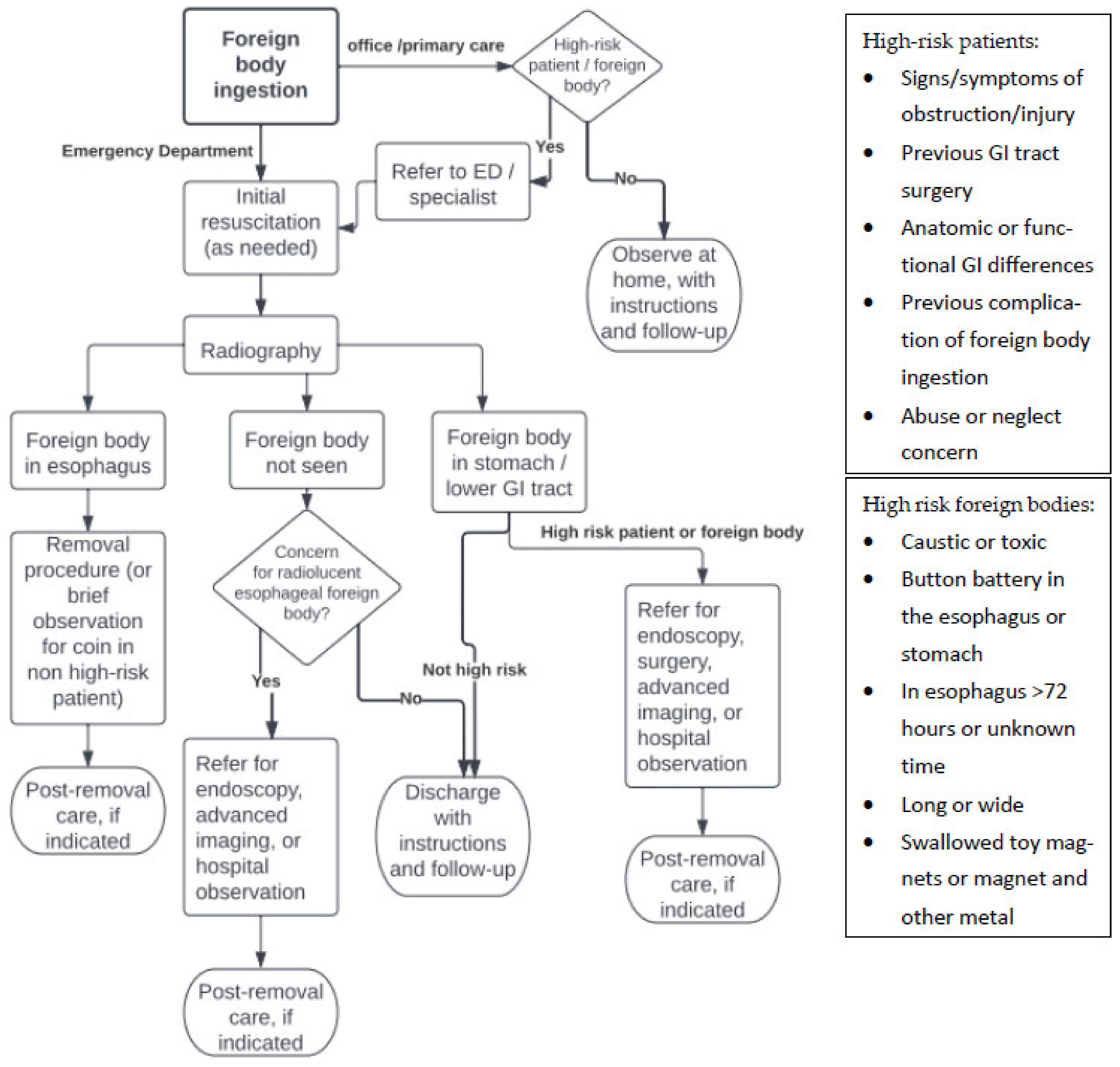
4.1. Emergency Management

The large majority of children with foreign body ingestion will present for care in stable condition. Unstable patients require transfer to an emergency setting, with urgent assessment of airway, breathing, and circulation. Following necessary resuscitation, early imaging may be required to identify the location and nature of swallowed objects. A few scenarios are most likely. A caustic or toxic ingestion may have caused tissue damage or metabolic injury. A lodged button battery may have created an aortic-esophageal fistula, which may have gone on to hemorrhage. A sharply-pointed foreign body, or swallowed magnets, may have led to GI tract perforation and peritonitis. An esophageal coin may have migrated into the mediastinum or airway. Early surgical involvement in the management of unstable patients with complication of foreign body ingestion is often beneficial. Keeping a broad differential diagnosis is important.

4.2. History and Physical Examination

The evaluation of a patient with a reported foreign body ingestion begins with history and physical examination. This should include the circumstances and timing of the event, as well as the nature of the ingested foreign body. Situations suggesting mental health concerns or developmental disabilities, including children with repeated non-accidental foreign body ingestions, may require referral. Potential child abuse or neglect should be reported to the appropriate authorities. The general health of the patient, including history of complications of previous foreign body ingestions or of GI disease or surgery, is important to consider. Signs and symptoms suggestive of esophageal foreign bodies include the presence of foreign body sensation, drooling, pain on swallowing, or refusal to eat or drink [21]. A foreign body that abraded the esophagus while passing into the stomach may also produce these findings. Stridor, shortness of breath, or hoarseness are suggestive of foreign body aspiration, but may also be associated with distention of the esophagus due to a retained foreign body, causing compression of the adjacent airway. Vomiting, hematemesis, or abdominal pain or tenderness may suggest gastric or lower GI tract complications. Examination of the oropharynx may, occasionally, reveal the “swallowed” foreign body [22]. Examination of other body cavities, including the ears and nose, should be performed, as it may reveal additional foreign bodies.

4.3. Imaging

Plain radiography is the mainstay of imaging children after foreign body ingestion. While it is not uncommon for primary care physicians to opt to observe previously healthy, asymptomatic children who have swallowed a low-risk foreign body without imaging [23], acute care/emergency physicians will typically order imaging of all patients. Radiopaque objects such as coins, button batteries, and other metallic objects are easily recognized on plain radiographs. However, even nominally radiolucent foreign bodies, such as meat boluses, may appear as complex, irregular objects when they are in the esophagus. Should there be strong suspicion that a radiolucent object is lodged in the esophagus, administration of a small amount of water-soluble contrast to highlight the foreign body on plain radiography may be a consideration, although endoscopic evaluation and potential object removal may be preferable. Although not widely available, digital tomosynthesis, a non-CT scan form of tomographic imaging, has shown promise as an accurate method for assessing children for potential radiolucent esophageal foreign bodies [24].
Since assessing for esophageal impaction is the usual goal of imaging, a two-view chest radiograph series is often an appropriate initial imaging exam. Care should be taken to include the pharynx. The two-view combination allows better identification, as well as anatomic orientation, of swallowed object(s). For patients specifically presenting with coin ingestion, a single frontal chest radiograph is sufficient [25]. A second, orthogonal view, such as a lateral radiograph, should be performed when an esophageal object is suggested by the first image, or there is uncertainty. It is not uncommon for a child who is reported to have swallowed a coin or similar radiopaque object to not have such an object appear on acutely-performed whole-torso radiographs, suggesting that it was not actually swallowed [21].
Alternative forms of imaging, such as CT, MRI, or ultrasound imaging are rarely necessary, but may offer advantages in specific situations. It has been reported that small, aluminum “pop tabs” and aluminum Japanese coins may be radiolucent on plain radiographs, so other imaging modalities may be required to evaluate these particular ingestions [26,27]. Small fish bones may also be radiolucent. Use of a hand-held metal detector to screen for esophageal coins has been described as sensitive and specific, as well as entertaining to patients and families, but there are limited reports of metal detector use in actual practice [28,29]. As use of point of care ultrasound (POCUS) has grown, there have been reports of its use to successfully identify esophageal foreign bodies [30].
It is widely taught that an esophageal coin, or a similarly shaped object, will appear as a disc on a frontal radiograph, while an aspirated coin will be oriented on-edge. However, case reports of coronally-oriented esophageal coins suggest that this possibility should still be considered when such an image is encountered [31,32]. This may be confirmed when the airway is seen well anterior to the foreign body on a lateral radiograph. Coin aspiration is relatively uncommon, especially at age >2 [33].
Since removal of an esophageal button battery is an emergency, while children with esophageal coins may typically be managed less urgently, distinguishing between the two on radiographs is important [14]. Belief that a disc-shaped esophageal object is a coin, when it was actually a button battery, has been associated with potentially avoidable major morbidity. On frontal radiographs, a button battery has a distinctive circle-within-a-circle appearance, while a coin is more commonly a rather uniform disc [34]. An orthogonal image of a button battery will typically show a distinctive two-tier profile (Figure 4) [34]. In some instances, a magnified, digital image will reveal the distinctive appearance of a particular coin (Figure 1) [35,36]. On rare occasions, two (or more) adherent coins of different sizes may mimic the radiographic image of a button battery; when in doubt, prompt removal of any foreign body that may be a button battery is prudent [37,38].
Imaging of a child whose symptoms suggest abdominal pathology, or who is suspected of ingesting small, powerful magnets [18], should include the abdomen. While dedicated chest (and, if necessary, abdominal) radiographs are generally preferable, a single “babygram” that includes both areas is sufficient to screen for the presence of easily-identified metallic foreign bodies in the chest or abdomen of small children; additional, follow-up imaging may be necessary for those revealing a foreign body.

4.4. Management

Once the nature and location of swallowed foreign bodies are identified, management decisions should be guided by the risk of complication (Figure 5). Children at low risk of future complications may be discharged with instructions to return for re-evaluation should pain, vomiting, bleeding, or other signs or symptoms suggestive of complication arise. Higher-risk patients may require observation, admission, or consultation with a specialist, such as a pediatric surgeon, otolaryngologist, or gastroenterologist.
In most cases, esophageal foreign bodies should be removed. Endoscopy allows simultaneous removal of known foreign bodies, identification of any additional objects, and inspection of the esophageal mucosa for injury. Endoscopic removal of esophageal button batteries should be considered an emergency [14]. Other blunt foreign bodies, such as coins, may be allowed 12–24 h to pass into the stomach from the mid or low esophagus, unless the child has had previous GI tract pathology or surgery that may lead to further complication [21,39]. Previous esophageal scarring, trachea-esophageal fistula repair, pyloromyotomy, or other relevant medical or surgical history may make uneventful transit of the GI tract unlikely; affected patients should be referred for endoscopy [5,7]. Children with otherwise uncomplicated esophageal coin impaction within the past 12–24 h may be considered for coin removal via bougienage or use of a Foley or other balloon catheter in centers with experience with these methods [40,41,42]. Radiographs should be repeated just prior to a procedure to remove a radiopaque object such as a coin, as some will spontaneously pass into the stomach, potentially negating the need for the procedure. [43] Potentially perforating esophageal foreign bodies, such as bones or push pins, should be urgently removed [43]. Management of esophageal food boluses in children generally requires endoscopy; the use of anti-spasmodic drugs such as glucagon or diazepam, or of meat-tenderizing papain, have been generally unsuccessful, although the literature on this is minimal [44,45]. Inducing emesis places the patient at risk of aspiration, so is not recommended.
Foreign bodies in the lower GI tract, having already traversed the stomach and esophagus, are likelier to be eliminated without complication. As noted above, however, certain patients are at greater risk, including those with lower GI tract functional, anatomic, or surgical abnormalities. Not all such abnormalities are previously known; for example, a small foreign body may become lodged in a Meckel’s diverticulum, and present with acute symptomatology [46]. Management of higher-risk patients or those who have swallowed higher-risk foreign bodies (e.g., small magnets, sewing needles) requires early consultation with a subspecialist, such as a pediatric surgeon. Fortunately, button batteries in the lower GI tract rarely lead to complications [14]; patients who become symptomatic, however, require urgent assessment. The use of laxatives to enhance foreign body passage is not supported by the literature. Parental observation of a child’s stool for passage of a foreign body is often ineffective. Patients who are low risk may be safely discharged, with instructions to return should signs or symptoms of obstruction or perforation (e.g., abdominal pain, vomiting, fever) arise.
Gastric foreign bodies are a particular challenge. Most will traverse the remainder of the GI tract, but some will remain in the stomach for long periods of time, with the potential for important complications [43]. Thus, some consultants prefer to endoscopically remove even low-risk foreign bodies such as coins to minimize this risk. Others recommend re-examination, perhaps with follow-up imaging, a few days following the ingestion of low-risk foreign bodies, with endoscopic removal recommended should the object not progress [43]. Prompt removal of gastric foreign bodies is a sensible approach for patients with clinical signs or symptoms, patients with higher-risk foreign bodies, or with lower GI tract functional, anatomic, or surgical abnormalities that would place the patient at risk of complications if the foreign body progressed to the lower GI tract. Children who have undergone previous gastric surgery are at risk of prolonged foreign body retention in the stomach, and should be considered for endoscopy [7]. Management recommendations for children with gastric button batteries previously suggested watchful waiting and consideration of repeat imaging unless the patient was at high risk, symptomatic, or aged less than six with a button battery of 15 mm diameter or greater [14]; more recent studies, however, have led to a recommendation for prompt removal of all gastric button batteries to prevent erosive complications [47,48].

4.5. Postremoval Care

Children undergoing endoscopic esophageal foreign body removal should have their esophageal mucosa re-inspected for injury before completion of the procedure [43]. Patients should be carefully observed for bleeding, airway concerns, esophageal perforation, or other complications after even apparently uncomplicated removal of an esophageal foreign body. The ability to take drink should be assured before discharge. Patients who have undergone endoscopic button battery removal with deeper esophageal injury should be observed in the hospital, as there is also potential for hemorrhage through a late-emerging aortic-esophageal fistula even well after the button battery has been removed [43].

5. Prevention

The prevention of foreign body ingestion is, of course, preferable to evaluation and treatment of affected children. Counseling parents about foreign body ingestion prevention is an important part of pediatric primary care. Prevention includes engineering solutions to limit access to dangerous foreign bodies, the voluntary or mandatory exclusion of dangerous foreign bodies in products for children, and education of children and parents about the dangers of foreign body ingestion. Government restrictions on their distribution has been associated with a reduction of reported ingestion of toy magnets; subsequent restriction removal was associated with a concomitant increase [49]. Children with ADHD are likelier to ingest foreign bodies [50], suggesting that specific counseling of affected children and their parents about reducing risk has potential for benefit. An increase in button battery ingestions during the COVID-19 pandemic has been described, and may also suggest a need for counseling/increased vigilance during lockdown situations [51].

6. Conclusions

While foreign body ingestion is common in children, the large majority of swallowed foreign bodies will pass through the GI tract without complication. Recognition of those patients and foreign bodies that are most highly associated with complications is important. These include patients with histories of medical or surgical abnormalities of the GI tract, those with symptoms, and those with previous complications of foreign body ingestion. Button batteries, magnets, long objects, or those that are sharply pointed or with a sharp edge, are most prone to complication. Appropriate imaging also helps determine which patients are at risk of complication. Involving an appropriate consultant in higher-risk situations is often helpful; some patients will require endoscopy or other removal procedures, while others will require additional monitoring. Serious complications, such as esophageal or bowel wall perforation, may require surgical intervention. Prevention strategies may help reduce foreign body ingestion.

Funding

This research received no external funding.

Informed Consent Statement

Not applicable.

Conflicts of Interest

The author declares no conflict of interest.

References

  1. Conners, G.P.; Chamberlain, J.M.; Weiner, P.R. Pediatric coin ingestions: A home-based survey. Am. J. Emerg. Med. 1995, 13, 638–640. [Google Scholar] [CrossRef]
  2. Paul, R.I.; Christoffel, K.K.; Binns, H.J.; Jaffe, D.M. Foreign bodies in children: Risk of complication varies with site of initial health care contact. Pediatrics 1993, 91, 121–127. [Google Scholar] [CrossRef] [PubMed]
  3. Louie, J.P.; Alpern, E.; Windreich, R.M. Witnessed and unwitnessed esophageal foreign bodies in children. Pediatr. Emerg. Care 2005, 21, 582–585. [Google Scholar] [CrossRef] [PubMed]
  4. Crysdale, W.S.; Sendi, K.S.; Yoo, J. Esophageal foreign bodies in children: 15-year review of 484 cases. Ann. Otol. Rhinol. Laryngol. 1991, 100, 320–324. [Google Scholar] [CrossRef] [PubMed]
  5. Stanley, P.; Law, B.S.; Young, L.W. Down’s syndrome, duodenal stenosis/annular pancreas, and a stack of coins. Am. J. Dis. Child. 1988, 142, 459–460. [Google Scholar]
  6. Shakir, A.K.; Ramji, F.; El Halabi, I. Penny for your thoughts; a coin in the stomach: Why did it get stuck? Hosp. Pediatr. 2017, 7, 294–296. [Google Scholar] [CrossRef] [Green Version]
  7. Stringer, M.D.; Kiely, E.M.; Drake, D.P. Gastric retention of swallowed coins after pyloromyotomy. Br. J. Clin. Pract. 1991, 45, 66–67. [Google Scholar]
  8. Denney, W.; Ahmad, N.; Billard, B.; Nowicki, M. Children will eat the strangest things: A 10—year retrospective analysis of foreign body and caustic ingestions from a single academic center. Pediatr. Emerg. Care 2012, 28, 731–734. [Google Scholar] [CrossRef]
  9. Kaye, E.T.; Sax, C.F.; DiPietro, E.K.; Wong, A.H.C.; Leung, A.K.C. Systemic nickel hypersensitivity from a swallowed coin. Consult. Pediatr. 2012, 52, 275–277. [Google Scholar]
  10. Slim, R.; Geagea, A.; Yaghi, C.; Honein, K.; Sayegh, R.; Zoghbi, A. Unusual way of purging. Emerg. Med. J. 2006, 23, 486. [Google Scholar] [CrossRef]
  11. Paul, R.I.; Jaffe, D.M. Sharp object ingestions in children: Illustrative cases and literature review. Pediatr. Emerg. Care 1988, 4, 245–248. [Google Scholar] [CrossRef] [PubMed]
  12. Pak, M.W.; Lee, W.C.; Fung, H.K.; van Hasselt, C.A. A prospective study of foreign-body ingestion in 311 cases. J. Paediatr. Child Health 2001, 58, 37–45. [Google Scholar]
  13. Hashmonai, M.; Kaufman, T.; Schramek, A. Silent perforations of the stomach and duodenum by needles. Arch. Surg. 1978, 113, 1406–1409. [Google Scholar] [CrossRef] [PubMed]
  14. Litovitz, T.; Whitaker, N.; Clark, L.; White, N.C.; Marsolek, M. Emerging battery-ingestion hazard: Clinical implications. Pediatrics 2010, 125, 1168–1177. [Google Scholar] [CrossRef] [PubMed] [Green Version]
  15. National Capital Poison Center Button Battery Ingestion Triage and Treatment Guideline. Available online: https://www.poison.org/battery/guideline (accessed on 7 April 2020).
  16. Arfang, R.R.; Jatana, K.R.; Linn, R.L.; Rhoades, K.; Fry, J.; Jacobs, I.N. pH-neutralizing esophageal irrigations as a novel mitigation strategy for button battery injury. Laryngoscope 2019, 129, 49–57. [Google Scholar] [CrossRef]
  17. Hussain, S.Z.; Bousvaros, A.; Gilger, M.; Mamula, P.; Gupta, S.; Kramer, R.; Noel, R.A. Management of ingested magnets in children. J. Pediatr. Gastroenterol. Nutr. 2012, 55, 239–242. [Google Scholar] [CrossRef]
  18. Strickland, M.; Diamond, I.R.; Rosenfield, D. Case discussions and radiographic illustration of magnet-related injuries in children. J. Emerg. Med. 2020, 58, 902–909. [Google Scholar] [CrossRef]
  19. Pogorelic, Z.; Boric, M.; Markic, J.; Jukic, M.; Grandic, L. A case of a 2-year-old child with entero-enteric fistula following ingestion of 25 magnets. Acta Med. 2016, 59, 140–142. [Google Scholar] [CrossRef] [Green Version]
  20. Shastri, N.; Leys, C.; Fowler, M.; Conners, G.P. Pediatric button battery and small magnet co-ingestion: Two cases with different outcomes. Pediatr. Emerg. Care 2011, 27, 642–644. [Google Scholar] [CrossRef]
  21. Conners, G.P.; Chamberlain, J.M.; Ochsenschlager, D.W. Symptoms and spontaneous passage of esophageal coins. Arch. Pediatr. Adolesc. Med. 1995, 149, 36–39. [Google Scholar] [CrossRef]
  22. Brayer, A.F.; Sciera, M.; Conners, G.P. Pediatric coin ingestion: An unusual presentation. Int. J. Pediatr. Otorhinolaryngol. 2000, 55, 211–213. [Google Scholar] [CrossRef]
  23. Joseph, P.R. Management of coin ingestion. Arch. Pediatr. Adolesc. Med. 1990, 144, 449–450. [Google Scholar] [CrossRef] [PubMed]
  24. Johansen, A.; Conners, G.P.; Lee, J.; Robinson, A.L.; Chew, W.L.; Chan, S.S. Pediatric esophageal foreign body: Possible role for digital tomosynthesis. Pediatr. Emerg. Care 2021, 37, 208–212. [Google Scholar] [CrossRef] [PubMed]
  25. Shatani, N.; Alshaibani, S.; Potts, J.; Phillips, B.; Bray, H. Chest radiograph alone is sufficient as the foreign body survey for children presenting with coin ingestion. Pediatr. Emerg. Care 2021, 37, e524–e527. [Google Scholar] [CrossRef]
  26. Valente, J.H.; Lemke, T.; Ridlen, M.; Ritter, D.; Clyne, B.; Reinert, S.E. Aluminum foreign bodies: Do they show up on X-ray? Emerg. Radiol. 2006, 12, 30–33. [Google Scholar] [CrossRef]
  27. Takahashi, J.; Shiga, T.; Funakoshi, H. Oesophageal coins invisible on chest radiography: A case report. Int. J. Emerg. Med. 2017, 10, 27. [Google Scholar] [CrossRef]
  28. Conners, G.P. Diagnostic uses of metal detectors: A review. Int. J. Clin. Pract. 2005, 59, 946–949. [Google Scholar] [CrossRef]
  29. Lafferty, M.; Lyttle, M.K.; Mullen, N.; on behalf of PERUKI. Ingestion of metallic foreign bodies: A paediatric emergency research in the United Kingdom and Ireland survey of current practice and hand-held metal detector use. J. Paediatr. Child Health 2021, 57, 867–871. [Google Scholar] [CrossRef]
  30. Mori, T.; Nomura, O.; Hagiwara, Y. Another application of point-of-care ultrasound: Detection of esophageal foreign bodies in pediatric patients. Pediatr. Emerg. Care 2019, 35, 154–156. [Google Scholar] [CrossRef]
  31. Dowd, M.D. Esophageal coins. Arch. Pediatr. Adolesc. Med. 1994, 148, 423–424. [Google Scholar] [CrossRef]
  32. Conners, G.P.; Hadley, J.A. Esophageal coin with an unusual radiographic appearance. Pediatr. Emerg. Care 2005, 21, 667–669. [Google Scholar] [CrossRef] [PubMed]
  33. Chen, X.; Milkovich, S.; Stool, D.; Reilly, J.; Rider, G. Pediatric coin ingestion and aspiration. Int. J. Pediatr. Otorhinolaryngol. 2005, 70, 325–329. [Google Scholar] [CrossRef] [PubMed]
  34. Swischuk, L.E. Swallowed a coin. Well-maybe not. Pediatr. Emerg. Care 2007, 23, 47–48. [Google Scholar] [CrossRef] [PubMed]
  35. Jackson, J.T.; Conners, G.P. Radiographic identification of an esophageal United States one cent coin. Vis. J. Emerg. Med. 2017, 9, 31–32. [Google Scholar] [CrossRef]
  36. Voelker, J.; Voelker, C.; Voelker, J.; Engert, J.; Schendzielorz, P.; Hagen, R.; Rak, K. Button batteries and typical swallowed foreign bodies can be differentiated in high-resolution x-rays. Int. J. Pediatr. Otorhinolaryngol. 2021, 142, 110604. [Google Scholar] [CrossRef]
  37. Silverberg, M.; Tillotson, R. Esophageal foreign body mistaken for impacted button battery. Pediatr. Emerg. Care 2006, 22, 262–265. [Google Scholar] [CrossRef]
  38. Cutajar, J.; Astl, J.; Borg, C. Radiologically aligned triple coin impaction in the upper oesophagus: The value of second-look oesophagoscopy. Int. J. Pediatr. Otorhinolaryngol. Extra 2011, 6, 192–194. [Google Scholar] [CrossRef]
  39. Esparaz, J.R.; Carter, S.R.; Mathis, M.S.; Chen, M.K.; Russell, R.T. Esophageal foreign body management in children: Can it wait? J. Laparoendosc. Adv. Surg. Tech. A 2020, 30, 1286–1288. [Google Scholar] [CrossRef]
  40. Conners, G.P. A literature-based comparison of three methods of pediatric esophageal coin removal. Pediatr. Emerg. Care 1997, 13, 154–157. [Google Scholar] [CrossRef]
  41. Gonzalez, K.W.; Reddy, S.R.; Mundakkal, A.A.; St Peter, S.D. The financial impact of flipping the coin. J. Pediatr. Surg. 2017, 52, 153–155. [Google Scholar] [CrossRef]
  42. Heinzerling, N.P.; Christensen, M.A.; Swedler, R.; Cassidy, L.D.; Calkins, C.M.; Sato, T.T. Safe and effective management of esophageal coins in children with bougienage. Surgery 2015, 158, 1065–2015. [Google Scholar] [CrossRef] [PubMed]
  43. Kramer, R.E.; Lerner, D.G.; Lin, T.; Manfredi, M.; Shah, M.; Stephen, T.C.; Gibbons, T.E.; Pall, H.; Sahn, B.; McOmber, M.; et al. Management of ingested foreign bodies in children: A clinical report of the NASPGHAN endoscopy committee. J. Pediatr. Gastroenterol. Nutr. 2015, 60, 562–574. [Google Scholar] [CrossRef] [PubMed]
  44. Lao, J.; Bostwick, H.E.; Berezin, S.; Halata, M.; Newman, L.J.; Medow, M.S. Esophageal food impaction in children. Pediatr. Emerg. Care 2003, 19, 402–407. [Google Scholar] [CrossRef]
  45. Peksa, G.D.; DeMott, J.M.; Slocum, G.W.; Burkins, J.; Gottlieb, M. Glucagon for relief of acute esophageal foreign bodies and food impactions: A systematic review and meta-analysis. Pharmacotherapy 2019, 39, 463–472. [Google Scholar] [CrossRef] [PubMed]
  46. Halverson, J.M.; Butterman, M.K.; Legier, J.F.; Mann, W.J.; Hoefer, R.A. Perforation of a Meckel’s diverticulum caused by ingestion of a coin. South. Med. J. 1994, 87, 823–824. [Google Scholar] [CrossRef]
  47. Khalaf, R.T.; Ruan, W.; Orkin, S.; Wilsey, M.; Fishman, D.S.; Mallon, D.; Pan, Z.; Hazleton, K.Z.; Kramer, R.E.; Walker, T. Gastric injury secondary to button battery ingestions in children: A retrospective multicenter review. Gastrointest. Endosc. 2020, 92, 276–283. [Google Scholar] [CrossRef]
  48. Rios, G.; Rodriguez, L.; Lucero, Y.; Miquel, I.; Arancibia, M.E.; Alliende, F. Endoscopic findings associated with button battery ingestion in children: Do we need to change the protocol for managing gastric location? Pediatr. Emerg. Care 2020, 36, 523–526. [Google Scholar] [CrossRef]
  49. Middelberg, L.K.; Funk, A.R.; Hays, H.L.; McKenzie, L.B.; Rudolph, B.; Spiller, H.A. Magnet injuries in children: An analysis of the National Poison Data System from 2008 to 2019. J. Pediatr. 2021, 232, 251–256. [Google Scholar] [CrossRef]
  50. Turgut, K.; Poyraz, M.K.; Sekmen, E.; Aydin, I.; Algin, A.; Yavuz, E. Prevalence of attention deficit hyperactivity (ADHD) in children presenting with foreign body ingestion. Am. J. Emerg. Med. 2019, 37, 2121–2124. [Google Scholar] [CrossRef]
  51. Pizzol, A.; Rigazio, C.; Calvo, P.L.; Scottoni, F.; Pane, A.; Genneri, F.; Cisaro, F. Foreign-body ingestions in children during COVID-19 pandemic in a pediatric referral center. JPGN Rep. 2020, 1, e018. [Google Scholar] [CrossRef]
Figure 1. Chest radiograph showing a coin lodged in upper esophagus. Details suggesting it is a United States dime are visible on close inspection (photo courtesy of Timothy Johnson, DO, MMM).
Sci 04 00020 g001
Figure 2. Chest radiograph showing swallowed hardware (washer) lodged in the mid-esophagus.
Sci 04 00020 g002
Figure 3. Chest radiograph showing coin lodged at lower esophageal sphincter (photo courtesy of Marcus Rivera, MD).
Sci 04 00020 g003
Figure 4. Abdominal radiograph of a child showing swallowed button batteries in two different orientations.
Sci 04 00020 g004
Figure 5. Flow diagram of general management of pediatric foreign body ingestion.
Sci 04 00020 g005
Publisher’s Note: MDPI stays neutral with regard to jurisdictional claims in published maps and institutional affiliations.

