1. Introduction
Additive Manufacturing (AM) technologies and three-dimensional printing systems are increasingly integrated into routine activities in maxillofacial surgery [
1,
2,
3,
4]. Their diffusion has enabled several clinical applications, including the fabrication of patient-specific medical devices such as cutting guides for oncological procedures and positioning guides for orthognathic surgery, as well as anatomical replicas and surgical simulation models [
5,
6,
7,
8,
9,
10]. The progressive reduction in equipment costs and the widespread availability of AM systems have facilitated their adoption in hospital environments [
11,
12]. Despite these advantages and the broad innovation potential associated with AM, medical applications remain strongly regulated by international and national standards, including the European Medical Device Regulation (MDR 2017/745). These regulations affect both small in-house laboratories operating within hospitals and larger industrial manufacturers supplying implantable devices [
13]. In most clinical workflows, AM technologies are combined with advanced three-dimensional imaging systems capable of processing data acquired from Computed Tomography (CT) scans or equivalent imaging modalities. Through dedicated computational algorithms, patient imaging data can be transformed into digital three-dimensional reconstructions of craniofacial skeletal structures and soft tissues [
14]. Continuous developments in medical imaging technologies have led to the introduction of software environments that support surgical simulation, preoperative planning, and the fabrication of customized devices [
15,
16,
17]. However, these systems generally provide only limited CAD functionalities and frequently lack the flexibility required for highly customized engineering solutions [
18]. In addition, the design and development of patient-specific surgical devices typically require the integration of advanced engineering tools such as Computer-Aided Engineering (CAE) systems together with the collaboration of multidisciplinary experts beyond the surgical team [
19,
20].
Several limitations can still be identified in the current development process of customized devices for maxillofacial surgery. First, despite the widespread adoption of CAD/CAM technologies and patient-specific surgical guides, accurately transferring the virtual surgical plan to the operative scenario remains a major challenge [
21]. Recent studies have shown that the positioning accuracy of the maxilla is strongly influenced by the design of the surgical guide, the selected anatomical references, and the adopted workflow, while postoperative reproducibility and stability continue to represent active research topics [
22,
23,
24,
25]. Second, the generation of alternative device geometries and innovative configurations tailored to the surgeons’ requirements remains largely experience-driven and difficult to optimize systematically [
26]. Third, evaluating the mechanical behavior of customized devices under realistic surgical loading conditions is often challenging, as structural analyses are rarely integrated into the clinical design workflow [
27]. Finally, the development of patient-specific medical devices requires continuous interaction among surgeons, mechanical engineers, material scientists, and manufacturing experts [
28]. The absence of integrated collaborative environments may reduce the efficiency of multidisciplinary decision-making and increase the number of design iterations required before clinical validation [
29]. With regard to geometric modelling, the development of personalized medical devices strongly depends on patient-specific anatomical data and clinical constraints. Consequently, the process requires advanced expertise in medical image processing and three-dimensional modelling techniques [
30]. Such competencies are not always available within hospital facilities, especially when urgent or rapid-response solutions are required. Mechanical validation also represents a critical aspect of the workflow, since ensuring the reliability and safety of medical devices under surgical conditions is essential for clinical success [
31]. Testing activities may be complicated by limitations related to equipment availability, laboratory accessibility, and variable environmental conditions. Furthermore, continuous interaction among surgeons, CAD specialists, manufacturing engineers, and materials experts can increase both the probability of communication errors and the overall lead time for device development [
32]. In addition, the continuous interaction between surgeons, designers, materials/manufacturing engineers, etc., widens the possibility of mistakes as well as the lead time of the medical device.
The literature review highlighted several open challenges associated with the design and fabrication of patient-specific devices for maxillofacial surgery. A first issue concerns the lack of a structured and integrated workflow capable of coordinating expertise from medicine, engineering, manufacturing, and materials science [
33,
34,
35,
36]. A second limitation is the absence of a unified collaborative software environment supporting communication and data exchange among surgeons and engineers while integrating imaging systems, CAD tools, and AM simulation functionalities for the development of innovative customized surgical devices [
37,
38]. These considerations constitute the main research motivations of the present work.
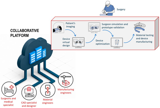
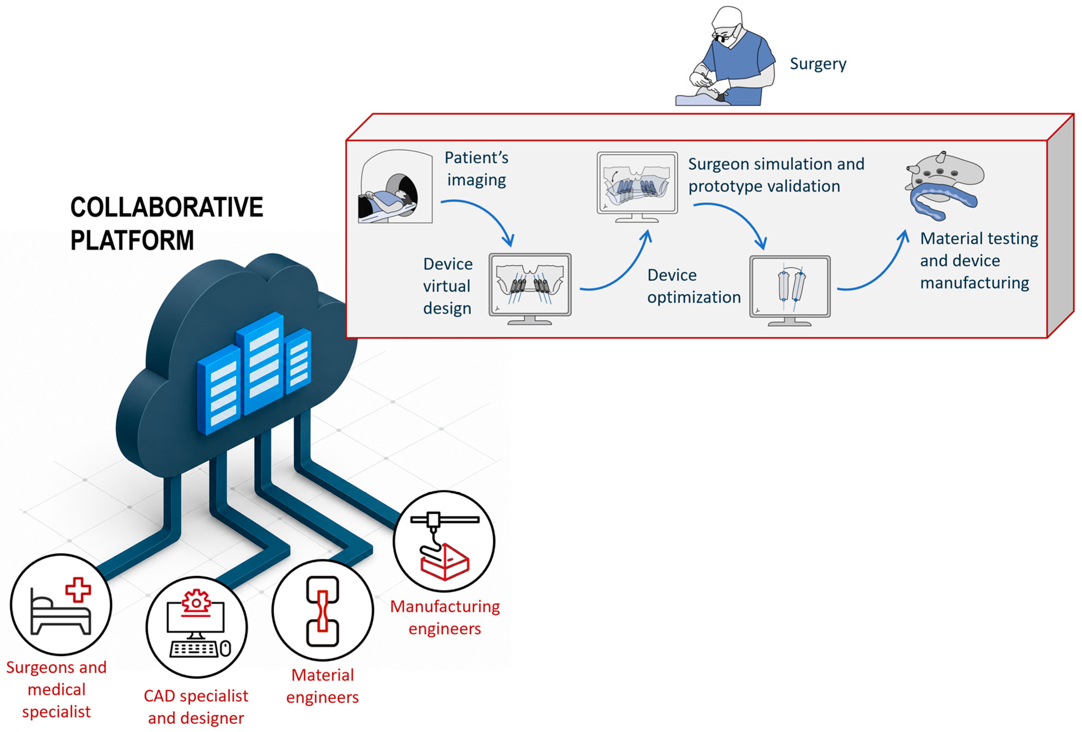
This study proposes an interdisciplinary engineering framework supported by a dedicated collaborative digital platform for the development of patient-specific medical devices in maxillofacial surgery. The proposed methodology aims to overcome the limitations of conventional fragmented workflows by integrating medical imaging, CAD modelling, material characterization, finite element analysis, topology optimization, and additive manufacturing within a unified collaborative environment. The originality of the proposed approach lies in the methodological integration of these engineering and clinical activities, enabling continuous interaction among maxillofacial surgeons, mechanical engineers, materials scientists, and additive manufacturing specialists throughout the entire device development process. The proposed workflow is organized into five interconnected stages, in which the output generated by each activity becomes the input for the subsequent phase, ensuring a structured, traceable, and repeatable development process. In parallel, the collaborative software platform provides a shared digital environment supporting multidisciplinary communication, workflow coordination, document management, and interaction on virtual prototypes, thereby facilitating the translation of clinical requirements into engineering design solutions.
To demonstrate the applicability of the proposed methodology, a novel patient-specific device for orthognathic surgery was developed through the collaboration between the Engineering and Medical Departments of the University of Parma. The framework was applied to five patients presenting dentofacial deformities requiring orthognathic surgery to restore functional occlusion and facial aesthetics. Although the present work focuses on orthognathic surgery, the proposed engineering methodology is sufficiently general to be extended to other patient-specific surgical applications, including oncological, traumatological, reconstructive, and orthopedic procedures.
From a clinical perspective, the proposed workflow has the potential to shorten the development cycle of customized surgical devices by enabling hospitals to exploit point-of-care additive manufacturing while reducing the dependence on external suppliers. Nevertheless, the primary contribution of this work is methodological rather than clinical. Accordingly, the study should be interpreted as a proof-of-concept engineering validation demonstrating the feasibility of an integrated collaborative workflow for the design, optimization, manufacture, and preclinical validation of patient-specific medical devices, rather than as a clinically validated surgical solution. The rest of the paper is organized as follows:
Section 2 presents the approach, workflow, and design of the collaborative web platform describing the expertise needed, the necessary equipment, tools, and outcomes of each step.
Section 3 provides a specific example of a novel customized device for maxillofacial deformities (orthognathic surgery).
Section 4 reports the main outcomes obtained in this field with the adoption of the proposed approach, and
Section 5 summarizes the main findings providing a future research outlook.
2. Methods
This section provides a description of the methodological approach (
Section 2.1) and the design of the collaborative web platform (
Section 2.2) for smart implementation of the proposed approach.
2.1. Methodological Approach
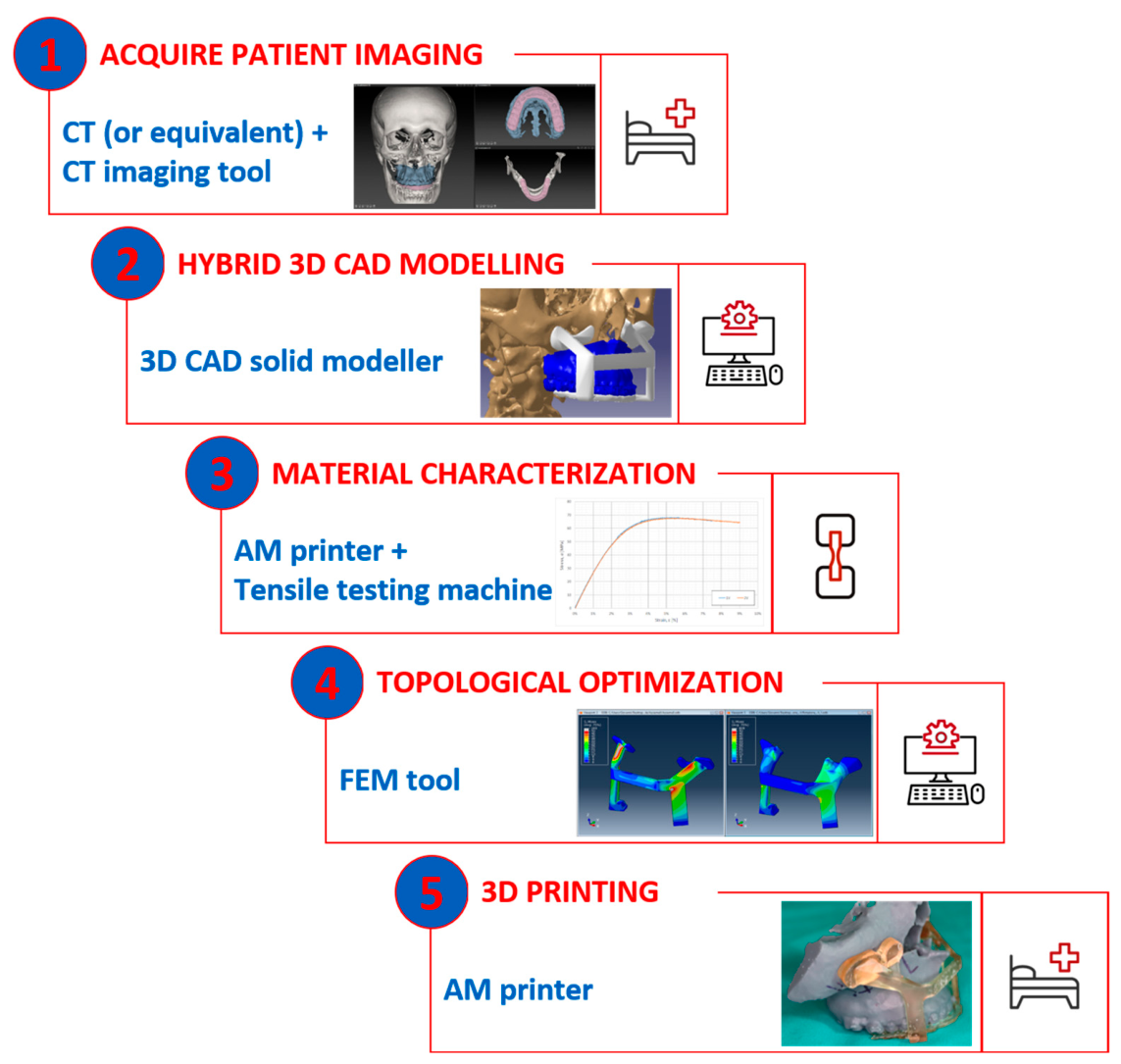
The methodology adopted for the development of customized medical devices for maxillofacial applications is structured into five main phases: (i) acquisition of patient anatomy and surgical simulation, (ii) hybrid three-dimensional CAD modelling, (iii) characterization of material properties, (iv) topology optimization, and (v) AM of the device prototype. The complete workflow is illustrated in
Figure 1.
The
first phase involves acquiring patient anatomical data through Computed Tomography (CT) or Cone Beam Computed Tomography (CBCT) scans. Both imaging techniques provide highly detailed digital representations of anatomical structures, particularly the craniofacial region relevant to maxillofacial surgery. The acquired information is stored in DICOM format, which contains the complete digital description of the patient anatomy. After image acquisition, a post-processing stage is required to accomplish three primary objectives: (i) evaluation of the patient’s dentofacial deformity and definition of the surgical treatment plan, (ii) simulation of skeletal repositioning procedures prior to surgery, and (iii) assessment of postoperative functional and aesthetic outcomes. This activity is generally carried out by the medical specialist, who converts DICOM data into STL files suitable for subsequent digital processing. Dedicated software environments, such as IPS Case Designer
® (version 2.6), are commonly employed during this phase. The DICOM dataset is imported into the surgical planning software to visualize and manipulate the reconstructed anatomical geometry. During segmentation, the anatomical regions of interest, such as the cranial bones, are isolated from surrounding tissues. Image enhancement procedures, including filtering, thresholding, and mesh processing operations, are then applied to improve the quality and clarity of the reconstructed structures. Subsequently, the movements of the maxilla and mandible are simulated to identify the optimal functional and aesthetic surgical configuration. The resulting STL files may undergo further mesh refinement procedures depending on the adopted software environment. Typical operations include mesh smoothing, triangle reduction, and geometry optimization to improve the suitability of the model for CAD and AM applications. A quality verification stage is finally conducted to ensure the accuracy of the anatomical reconstruction and the consistency of the mesh with the intended surgical application. The expected outputs generated during this first phase include: (i) STL models of the patient skull after completion of surgical planning, (ii) STL models of standard surgical devices typically adopted during the intervention, such as wafers or cutting guides [
39,
40,
41], and (iii) a set of surgical and geometrical constraints defined by the surgeon according to the specific clinical application, including dimensions, loading conditions, and accessibility requirements.
The second phase concerns hybrid CAD modelling. This activity can be performed remotely by CAD specialists using the digital outputs generated during the previous stage. Because both free-form surfaces and solid geometries are required, the use of hybrid CAD environments such as CATIA (version 5) is essential for the development of customized devices. Hybrid modelling combines surface-based and solid-based design techniques to accurately reproduce complex anatomical geometries. Starting from the imported mesh model, a closed surface conforming to the patient-specific anatomy is reconstructed and subsequently transformed into a solid model suitable for engineering design activities. Surface modelling is particularly effective in reproducing organic and highly detailed anatomical geometries, whereas solid modelling enables easier manipulation and modification during the CAD development process. At this stage, the customized device is designed according to the requirements defined by the medical specialist. CAD tools allow designers to modify geometries, improve fitting conditions, and introduce specific functional features necessary for the surgical application. Continuous interaction between surgeons and CAD specialists is fundamental to correctly understand clinical requirements and operative needs. The final output of this phase is a STEP-format solid model of the customized medical device, which is necessary for the subsequent CAE and structural analyses.
The third phase focuses on material characterization. This activity is typically carried out by mechanical engineers or materials specialists within dedicated testing laboratories equipped with tensile testing machines and additional characterization equipment. In medical AM applications, only a restricted number of certified materials can usually be adopted because of regulatory and biocompatibility requirements. These materials must therefore be experimentally characterized under the same manufacturing conditions and process parameters used to produce the final device. Material characterization plays a key role in regulatory compliance and patient safety. The experimental evaluation of mechanical, thermal, and biological properties provides essential information for predicting material behavior under surgical operating conditions. Moreover, the characterization phase ensures consistency among production batches and minimizes variability that could compromise the structural reliability of customized medical devices. This activity results in a complete report describing the material mechanical behavior, including properties such as stiffness, strength, elastic modulus, and elongation. The principal outputs consist of stress–strain curves together with static and dynamic mechanical parameters required for the subsequent numerical simulations.
The fourth stage deals with the topology optimization. This phase exploits the outputs generated during the previous activities, namely the material characterization data, the CAD model of the customized device, and the surgical loading constraints, to build a compliant finite element model within an FEM environment such as ABAQUS (version 2025). The aim of this activity is to obtain optimized and mechanically efficient patient-specific devices while minimizing obstructions for the surgeon during the operation. Mechanical engineers perform FEM simulations to improve structural performance by: (i) reducing stress concentrations below the material yield limit and (ii) minimizing structural displacements to guarantee sufficient rigidity and positioning accuracy during surgery. Finite element simulations provide detailed information regarding stress distributions across the device geometry. By optimizing material distribution and geometry, it becomes possible to decrease critical stress concentrations and reduce the risk of structural failure under operative loads. At the same time, minimizing displacements contributes to greater device stability and improved positioning precision during surgical procedures. The final output of this phase is the STL model of the optimized patient-specific device.
The
fifth stage (final step) consists of AM of the prototype within hospital facilities equipped with AM systems. In this stage, the optimized device geometry is physically produced using the selected material and 3D printing technology. The main output is therefore the physical prototype of the patient-specific device intended for surgical use. An overview of the required expertise, equipment, software tools, and outputs associated with each phase of the proposed methodology is summarized in
Table 1.
2.2. Collaborative Environment
To support the multidisciplinary collaboration identified in the previous section, a dedicated collaborative co-design platform was developed according to the specific requirements of patient-specific medical device development. Existing collaborative solutions generally address individual phases of the product development process, such as virtual surgical planning, CAD collaboration, document management, or project management. However, the development of customized medical devices requires continuous interaction among surgeons, mechanical engineers, material scientists, and manufacturing experts, together with the integration of heterogeneous engineering tools throughout the entire design process. Consequently, the proposed platform was conceived as an integrated engineering framework rather than a conventional collaboration environment. Its novelty lies not in the individual software applications employed, but in their methodological integration, enabling multidisciplinary engineering decisions throughout the complete product development workflow [
42]. To better position the proposed framework with respect to the existing literature and commercial solutions,
Table 2 compares its engineering functionalities with the principal categories of collaborative approaches currently adopted for patient-specific medical device development.
As shown in
Table 2, existing collaborative solutions mainly support specific phases of the product development process. Virtual surgical planning systems primarily focus on preoperative planning, collaborative VR/XR environments enhance visualization and communication, whereas PLM-based frameworks mainly address project coordination and document management. Conversely, the proposed framework integrates clinical planning, CAD modelling, finite element analyses, additive manufacturing constraints, documentation management, workflow execution, and multidisciplinary engineering decision-making within a single collaborative environment. This integration establishes direct relationships among clinical requirements, engineering analyses, manufacturing constraints, and project management activities, allowing design decisions to be continuously evaluated from both clinical and engineering perspectives throughout the entire development process.
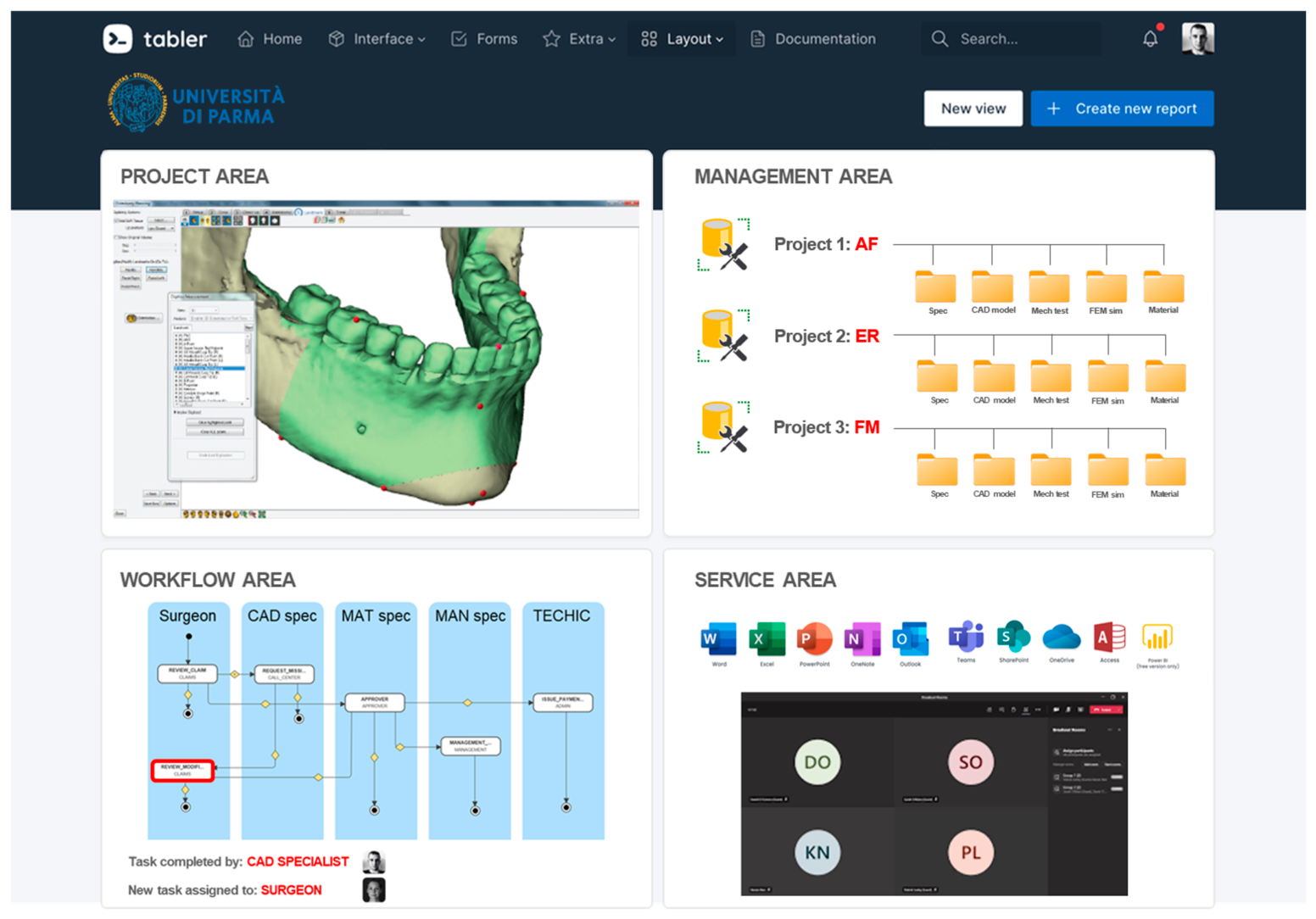
The proposed platform architecture follows a client–server approach and hosts all the software tools required during the development of customized medical devices described in the previous section. Access to the different software applications is managed through role-based authentication, ensuring controlled access according to the responsibilities of each stakeholder. The platform was developed by the ICT Department of the University of Parma following the functional requirements identified during this research. An example of the developed graphical user interface (GUI) is shown in
Figure 2. The portal server enables the management of role-playing and facilitates the organization of project progress.
The platform GUI is characterized by four main windows/environments: (i) project area, (ii) management area, (iii) workflow area, and (iv) service area.
The project area serves as the primary user interface for each participant. The project area represents the engine of the codesign platform, and it consists of a collaborative CAD-based viewer (AutoVue) that allows the demonstration and addition of marks on 3D virtual prototypes in real time and within a common space. In this area virtual models (e.g., STL and STEP files) can be opened and annotations/remarks can be made by different users through a dedicated tool panel. Both synchronous and asynchronous collaborations were conducted. Data can be accessed directly through the project area in the corresponding project space.
The management area plays a crucial role in organizing, tracking, and ensuring the integrity of project-related documents. This window functions as a centralized repository for storing various types of documents related to a specific project. This includes surgery specifications, CAD models, regulatory documents, test reports, and other essential files developed during the device development process. The kernel of the system enables the categorization and organization of documents based on the types and phases of the project or any other relevant criteria. This aids in efficient document retrieval and ensures that the information is structured for easy navigation.
The workflow management system is accessible to the workflow area in the GUI space. It consists of a workflow engine to implement all tasks necessary to properly develop the project. Roles are assigned to all partners at the beginning of the project, and a specific project workflow can be defined to properly manage all tasks. Each member involved in the development of a customized medical device gathers an alert/notification when an action is required, and the list of tasks and activities assigned to each member is available in the collaboration area. These functionalities ensure that the project team remains informed and can take timely actions.
The service area implements all applications and functionalities of the Microsoft 365 package (including TEAMS for videoconferencing) to facilitate communication and meetings among the different actors involved in the device development process. In this area, it is possible to obtain access to different software tools that need to be used during the evolution of the project. (e.g., CAD modeller, segmentation tool, and FEM tool).
The platform supports both collaborative sessions involving multiple stakeholders and asynchronous activities assigned to individual users, providing flexibility throughout the development process. The practical relevance of this integrated environment is demonstrated in the case study presented in the following sections. During the structural optimization of the customized device, finite element analyses identified the mechanically critical regions, whereas the collaborative platform enabled surgeons and engineers to jointly define the admissible location and maximum dimensions of the reinforcing ribs according to both structural requirements and surgical accessibility constraints. This iterative multidisciplinary interaction illustrates the capability of the proposed framework to support engineering decision-making beyond conventional data sharing.
3. Case Studies
A prospective investigation was carried out on five patients presenting different dentofacial deformities requiring orthognathic surgery. All participants provided consent for the use of CT-acquired data within this research activity. The present study did not involve any experimental procedure on human participants. Patient-specific CT data used to generate the anatomical models were retrospectively collected after obtaining Institutional Review Board and Ethics Committee approval (Approval No. 1291/2020/OSS/AOUPR). All data were fully anonymized prior to processing, and written informed consent for the use of clinical imaging data for research purposes was obtained from all participants, in accordance with the Declaration of Helsinki.
The collaborative software platform developed during this study was employed throughout the design and development process of the customized surgical devices for all clinical cases. The project coordinator, namely the surgeon, initialized the workflow by defining tasks, assigning roles, and managing access permissions to project documentation. It should be emphasized that the actual surgical procedures were conducted according to conventional clinical protocols, as described in [
1]. These procedures relied on the use of two surgical wafers (intermediate and final), generated through dedicated surgical planning software (IPSCaseDesigner
® from KLS Martin, Tuttlingen, Germany) and manufactured by certified suppliers. During surgery, these wafers were employed to assist in repositioning osteotomized skeletal structures by using relative anatomical references between the maxilla and the mandible.
Conversely, the new surgical procedure and the new devices were tested by employing a dummy reproduction of the patient’s skull prototype manufactured using 3D printing technology, starting from patient image acquisition (e.g., CT scan). This validation strategy is consistent with the development pathway commonly adopted for patient-specific medical devices, where proof-of-concept evaluation is first performed through virtual planning and physical anatomical models before clinical implementation [
43]. Such an approach enables verification of anatomical fitting, device functionality, and mechanical behavior while avoiding unnecessary risks to patients during the early stages of device development. In addition to demonstrating the technical feasibility of the proposed workflow and device, the proof-of-concept validation provides a reliable basis for the preliminary estimation of expected performance improvements. In particular, it allows comparison with conventional surgical workflows under controlled conditions, enabling the assessment of aspects such as device fitting, surgical accessibility, procedural simplification, and the potential reduction in operative time. Although these findings cannot be considered clinically validated outcomes, they provide a realistic indication of the expected benefits of the proposed approach and support its progression towards future in vivo validation. Furthermore, the adoption of a proof-of-concept validation was motivated by both ethical and practical considerations. Performing a surgical procedure using a newly developed patient-specific device without prior technical validation could expose patients to unnecessary risks. Accordingly, international ethical principles and medical device regulations require that novel devices undergo extensive preclinical assessment before clinical application. In addition, at the time of device development, the University Hospital of Parma was not authorized to manufacture certified Class IIa medical devices in accordance with EU Regulation 2017/745. Therefore, physical testing on patient-specific skull prototypes represented the most appropriate strategy to assess the functionality, anatomical fitting, and mechanical reliability of the proposed device while minimizing patient risk.
The idea underlying this project was to develop a patient-specific device that uses skull bone positioning by a fixed space reference (i.e., the portion of skull bone around the infraorbital foramen) fitting surgeon requirements and developed with an engineering virtual tool (CAD and CAE) and rapid prototyping equipment (e.g., AM). This kind of procedure and, consequently, the new device allows for coping with two main issues raised by conventional practice. Indeed, since conventional practice leaves the choice of the anterior facial height to the surgeon’s sensibility and uses a mobile landmark (inferior jaw), it is time-intensive and lacks accuracy. The patient was intubated through the nose during the surgery, which led to a change in the 3D position of the tissues constituting the upper lip and nose. Thus, one of the main advantages of the proposed patient-specific device is the possibility to reduce surgical time and to be sure to exactly reproduce what we planned, through the pre-operative clinical exam, on the patient’s face.
To support device development, the collaborative software platform was used to define and discuss all technical specifications and geometrical constraints within dedicated project workspaces. This environment facilitated communication among the involved specialists and simplified the interpretation of clinical and engineering requirements. During the first stage of the workflow, CT scans of the patients were acquired. STL models generated from DICOM data were employed by the surgeon to simulate the surgical planning, including osteotomies, repositioning of bone segments, evaluation of possible interferences among skeletal structures, and verification of postoperative outcomes. Two main digital outputs were generated during this phase and transferred to the subsequent design stage: (i) the STL model of the cranium and osteotomized maxilla following surgical planning, and (ii) the STL model of the final surgical wafer. Additional design constraints defined by the surgeon included the positioning of the anchoring plates, the maximum allowable device dimensions to ensure accessibility during surgery, and the forces expected during the operation. All the mentioned documentation was uploaded to the specific repository of the web platform and accessible to the other members following the workflow rules designed by the project owner. Using the platform, a dedicated working session was launched by the surgeon to clearly identify the portion of the skull that could be used as a reference (i.e., the portion of skull bone around the infraorbital foramen) and the maximum allowable dimensions for the anchor plates. CAD specialists and mechanical engineers participated in this study. In the second step, a CAD specialist imported the two files into a 3D CAD tool for hybrid modelling.
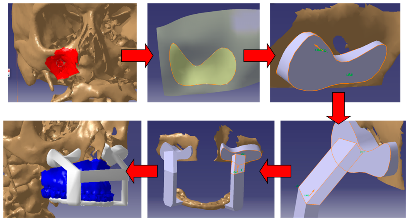
Owing to the characteristics of the acquired models (i.e., mesh) and the need for accurate surface reconstruction, hybrid modelling tools were essential for reproducing the cranial geometry and designing the anchoring system of the customized device. For orthognathic surgery applications, the proposed solution was designed to preserve accessibility from both frontal and lateral directions during surgery. Consequently, the final device geometry was conceived with a C-shaped configuration (
Figure 3).
The C-shaped configuration enabled the connection between the anchoring plates fixed to the cranium and the base of the surgical wafer through two symmetrical supporting brackets. The output generated during this phase consisted of both the solid CAD model in STEP format, required for topology optimization, and the STL mesh employed by the surgeon for surgical simulation activities. Before proceeding to the next stage, an additional collaborative session was organized to review the final design, evaluate fitting accuracy with the patient skull model, and verify the available working space for surgical operations. Following this review, the required design modifications were introduced and the updated files were stored within the platform repository. The third stage focused on characterization of the material selected for device manufacturing. This task was assigned to the material specialist.
The customized device was manufactured using a biocompatible Class I stereolithography photopolymer resin specifically developed for surgical guides. Although the commercial name of the material cannot be disclosed due to confidentiality agreements with the industrial partner, its material class together with the relevant engineering properties are reported to ensure the reproducibility of the proposed methodology. The resin consists of a cross-linkable photocurable polymer network specifically formulated for medical applications and certified for temporary mucosal contact. The adopted SLA photopolymer resin consists of a mixture of methacrylic esters and photoinitiators that polymerize through a light-induced cross-linking reaction during stereolithography. The material complies with the biocompatibility and quality management requirements defined by EN ISO 10993-5 (European Committee for Standardization (CEN),
EN ISO 10993-5:2009+A11:2025, Biological evaluation of medical devices—Part 5: Tests for in vitro
cytotoxicity (ISO 10993-5:2009), CEN, Brussels, Belgium, 2025.) [
44], EN ISO 10993-10 (European Committee for Standardization (CEN),
EN ISO 10993-10:2023, Biological evaluation of medical devices—Part 10: Tests for skin sensitization (ISO 10993-10:2021), CEN, Brussels, Belgium, 2023) [
45], ISO 10993-11 (European Committee for Standardization (CEN),
EN ISO 10993-11:2018, Biological evaluation of medical devices—Part 11: Tests for systemic toxicity (ISO 10993-11:2017), CEN, Brussels, Belgium, 2018.) [
46], ISO 10993-3 (European Committee for Standardization (CEN),
EN ISO 10993-3:2014, Biological evaluation of medical devices—Part 3: Tests for genotoxicity, carcinogenicity and reproductive toxicity (ISO 10993-3:2014), CEN, Brussels, Belgium, 2014) [
47], EN ISO 13485 (European Committee for Standardization (CEN),
EN ISO 13485:2016+A11:2021, Medical devices—Quality management systems—Requirements for regulatory purposes (ISO 13485:2016), CEN, Brussels, Belgium, 2021) [
48], and EN ISO 14971 (European Committee for Standardization (CEN),
EN ISO 14971:2019+A11:2021, Medical devices—Application of risk management to medical devices (ISO 14971:2019), CEN, Brussels, Belgium, 2021) [
49]. Following fabrication, all components underwent the manufacturer’s recommended washing and post-curing procedures prior to mechanical characterization and experimental validation. The principal material properties provided by the manufacturer after post-curing are summarized in
Table 3. These properties were subsequently verified experimentally by the material specialist and used as input parameters for the finite element analyses and topology optimization activities presented in the following sections.
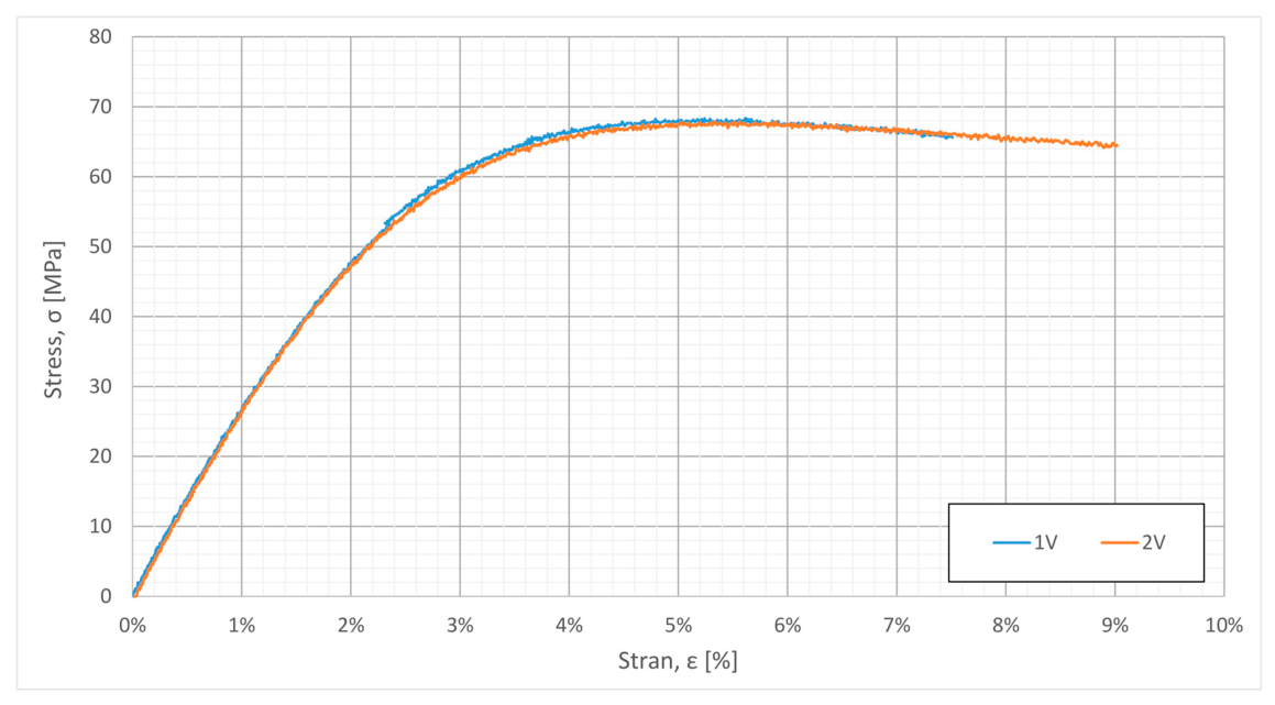
To experimentally validate the mechanical behavior of the adopted resin, dog-bone tensile specimens were manufactured using the same stereolithography process, printer (Form 3B), layer thickness (100 μm), and post-processing protocol employed for the fabrication of the customized surgical devices. Uniaxial tensile tests were then carried out under quasi-static loading conditions. Two independent specimens were tested to evaluate the repeatability of the material response, and the resulting engineering stress–strain curves are reported in
Figure 4.
As shown in
Figure 4, both specimens exhibited an almost identical mechanical response, confirming the excellent repeatability of the adopted additive manufacturing process. The material showed initial linear elastic behavior followed by progressive yielding and limited strain softening after reaching the maximum stress. The experimentally measured ultimate tensile strength was approximately 68 MPa, which is in good agreement with the nominal value of 73 MPa reported by the manufacturer (difference below 7%). This agreement confirms the reliability of the material properties adopted for the numerical simulations while also accounting for the influence of the actual printing process and post-curing conditions used in this study. A comparison between the main nominal material properties provided by the manufacturer and the experimentally measured values is reported in
Table 4.
Consequently, the experimentally determined mechanical properties were adopted as input parameters for the finite element analyses and topology optimization presented in the following sections.
Following material testing, the fourth phase involved topology optimization of the device with the purpose of reducing stress concentration and improving structural robustness. Finite element analyses were performed by a mechanical engineer using Abaqus/CAE (Dassault Systèmes, Vélizy-Villacoublay, France). The device was discretized using three-dimensional 10-node quadratic tetrahedral elements (C3D10) with a characteristic element size of 0.5 mm, resulting in a finite element model composed of approximately 150,000 elements. Considering the high positioning accuracy required during surgery, the material was modelled as homogeneous, isotropic, and linearly elastic using the mechanical properties experimentally determined through the tensile tests described in the previous section. All these features were derived by the previous analyses performed by the material scientist. Accordingly, linear static analyses were performed using the implicit solver Abaqus/Standard. Geometric nonlinearities were neglected because the predicted displacements remained small compared with the characteristic dimensions of the device and the material operated entirely within the linear elastic regime. Prior to the optimization process, a mesh convergence study was carried out to ensure mesh-independent results. The mesh was progressively refined until the variation of the maximum Von Mises stress at the critical stress concentration regions between two successive refinements was below 3%. The adopted mesh therefore represents an optimal compromise between computational efficiency and numerical accuracy. Throughout the optimization process, particular attention was devoted to ensuring that the computed Von Mises stress remained below the experimentally determined yield strength, corresponding to a safety factor greater than one. FEM simulations were conducted by applying boundary conditions consistent with the loading constraints defined by the surgeon during the first phase of the workflow and reported in
Table 5.
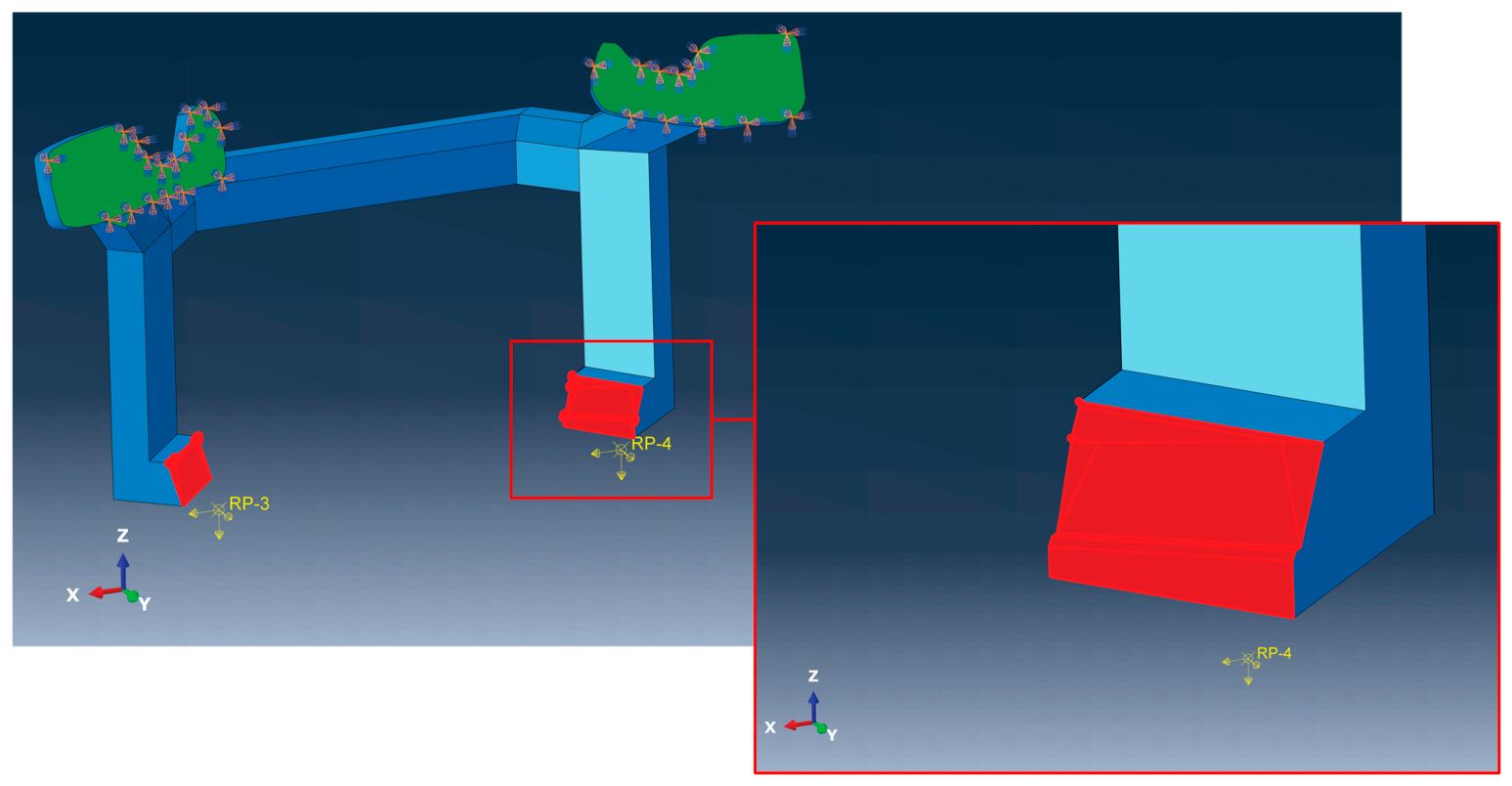
The fixing constraints (green areas,
Figure 5) were applied at the anchoring plate regions, where neither translational nor rotational movements were allowed. Load values were established according to the surgeon’s experience gained from previous orthognathic procedures, considering the weight of the osteotomized maxillary segment together with the elastic reaction generated by the surrounding soft tissues. The highest load component was applied along the Y direction because the patient remains in a supine position during surgery. Two loading regions corresponding to the connection between the C-shaped supporting structure and the dental splint were identified. For each connection region, three orthogonal force components acting along the X, Y and Z directions were applied. For each patient-specific geometry, the numerical analyses were performed by systematically varying the sign of the force components along the X, Y, and Z directions in order to reproduce all possible loading combinations that could occur during the surgical procedure. Among the analyzed configurations, the most critical loading condition was identified and subsequently adopted for the structural assessment and topology optimization of the customized device. The collaborative platform facilitated interaction between surgeons and engineers by enabling visualization of loading conditions and force application areas through a shared three-dimensional environment.
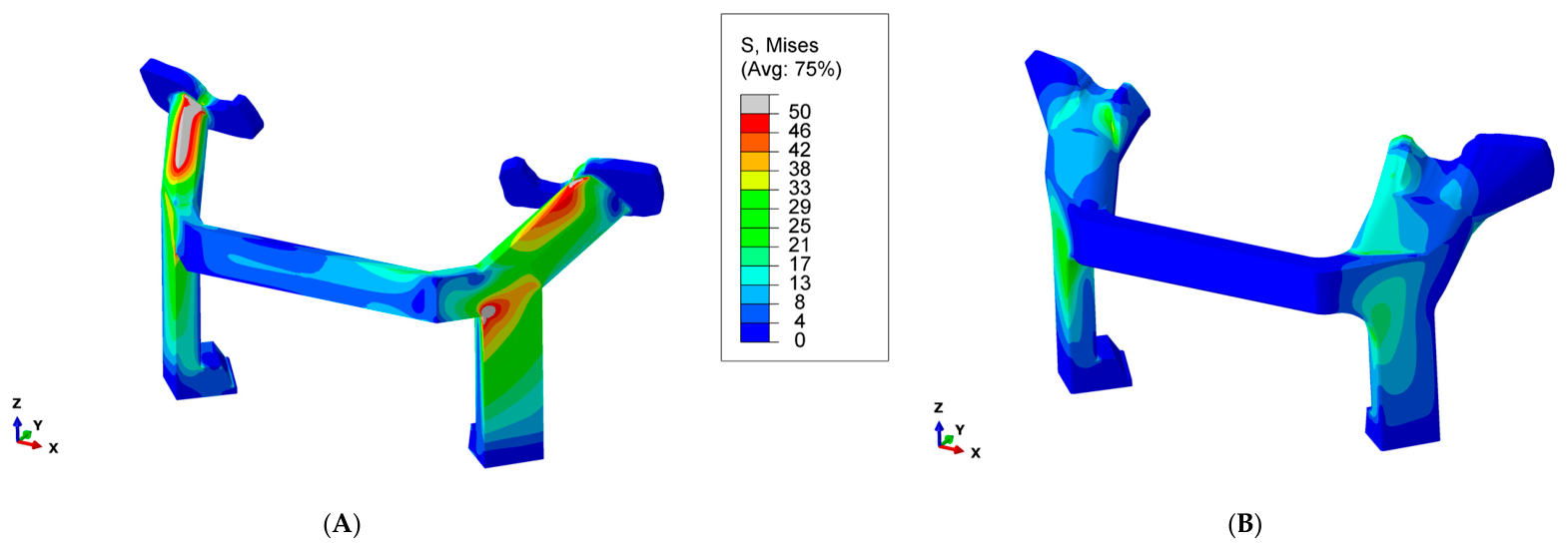
Once the FEM boundary conditions had been defined, numerical simulations were performed and the obtained results are presented in
Figure 6. The optimization process followed an iterative collaborative approach involving both mechanical engineers and maxillofacial surgeons. The FEM results were shared through the collaborative platform, enabling surgeons to directly visualize stress distributions and identify those regions where local geometric modifications could be introduced without interfering with the surgical procedure or reducing accessibility to the operative field. Based on this multidisciplinary interaction, the position, orientation, and maximum allowable dimensions of the reinforcing ribs were jointly defined according to both structural requirements and surgical constraints. Additional optimization activities included the introduction or enlargement of fillets to reduce notch effects and local increases in critical cross-sectional dimensions while preserving the functional and geometrical requirements specified by the surgeons. The Von Mises stress distributions before and after optimization demonstrate a substantial reduction in stress concentrations throughout the device geometry. The reported results refer to the worst-case condition identified considering all five patient-specific case studies and the complete set of analyzed loading combinations. Under this conservative scenario, the maximum Von Mises stress decreased from 130 MPa to 31 MPa, corresponding to an increase in the safety factor from 0.38 to 1.61. Likewise, the maximum displacement decreased from 5.8 mm to 1.7 mm, demonstrating a significant increase in structural stiffness while preserving the functional requirements of the customized device.
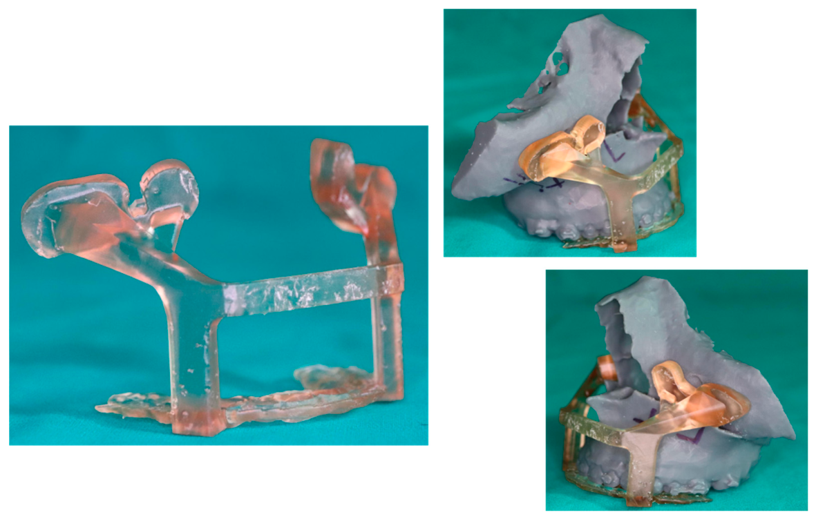
After completion of the optimization stage and final approval of the device geometry, the last phase consisted of prototype fabrication through 3D printing using the Form 3B stereolithography printer and the previously characterized material. The produced devices were attached to the skull prototypes using screws and removed after completion of the surgical simulation procedures, as illustrated
Figure 7.
Although the present work represents a proof-of-concept engineering validation performed on additively manufactured anatomical replicas rather than during clinical procedures, both the material selection and the manufacturing protocol were defined according to the validated manufacturing instructions provided by the resin manufacturer for medical surgical guides. In particular, the customized devices were fabricated using the recommended printing parameters and subsequently subjected to the complete post-processing protocol, including washing in isopropyl alcohol (IPA ≥ 99%) followed by UV post-curing using a dedicated curing unit. These post-processing steps promote the completion of the photopolymerization reaction, increasing the degree of monomer conversion, stabilizing the cross-linked polymer network, and minimizing the presence of residual unreacted monomers. The adopted manufacturing protocol was fully consistent with the validated process specified by the manufacturer for biocompatible surgical guide applications.
From a regulatory perspective, the proposed engineering workflow was conceived considering the manufacturing and quality requirements typically adopted for patient-specific medical devices. Although no clinical validation was performed in the present work, the selected material, manufacturing route, and post-processing protocol comply with the validated processing conditions established by the material manufacturer for surgical guide production, facilitating the future translation of the proposed methodology towards clinical applications.
4. Results and Discussion
This research proposed a preliminary interdisciplinary framework intended to reduce the gap between maxillofacial surgery and mechanical/materials engineering in the development of optimized patient-specific devices for orthognathic surgery. The proposed methodology was specifically conceived to support surgical procedures associated with dentofacial deformities by improving operative reliability and facilitating intraoperative activities through customized solutions. The developed approach is structured into five sequential phases, namely CT-based anatomical acquisition, hybrid CAD modelling, material characterization, topology optimization, and AM of customized prototypes. All these activities were coordinated and supported through the collaborative software platform described in the previous sections. The adopted material underwent tensile characterization in order to experimentally determine its mechanical behavior under the same manufacturing conditions and process parameters used for AM of the customized devices. Furthermore, topology optimization activities were performed to reduce stress concentrations below the material yield limit while simultaneously minimizing structural displacements, thereby ensuring sufficient rigidity and stable positioning during surgical use. At the time this study was conducted, the University Hospital of Parma was not yet authorized to manufacture certified Class IIa medical devices according to EU Regulation 2017/745. Consequently, the effectiveness and fitting accuracy of the developed devices were assessed through two complementary validation approaches.
The first validation strategy consisted of a software-based assessment performed within the IPS Case Designer environment. Since the platform allows the importation of STL geometries, the developed patient-specific device was digitally superimposed onto the reconstructed craniofacial anatomy of each patient. This virtual analysis enabled evaluation of the fitting precision between the anchoring plates and the skull surface, particularly in the region surrounding the infraorbital foramen. The virtual assessment demonstrated complete geometric correspondence between the customized device and the patient-specific craniofacial anatomy, confirming accurate fitting of the anchoring plates without detectable interference or misalignment.
The second validation activity consisted of a proof-of-concept evaluation performed on additively manufactured patient-specific anatomical replicas. Five representative clinical cases with different dentoskeletal deformities were selected to assess the proposed engineering workflow. The demographic and clinical characteristics of the selected patients are summarized in
Table 6.
More specifically, skull prototypes reproducing the upper jaw together with the cranial region up to the orbital and zygomatic structures were fabricated for each of the five investigated clinical cases, together with the corresponding customized devices. The devices were positioned on the physical skull prototypes to verify the correspondence between the anchoring plates and the targeted anatomical regions. The experimental assessment demonstrated accurate adherence of the anchoring plates to the skull geometry without requiring additional force during placement. Threaded fixation elements were subsequently inserted to secure the devices to the skull models and verify their structural stability throughout the simulated surgical procedure. This proof-of-concept validation strategy is consistent with the development pathway commonly adopted for patient-specific medical devices, where physical anatomical models are used to verify device fitting, surgical accessibility, functionality, and workflow feasibility before clinical implementation. Recent studies have demonstrated that patient-specific anatomical replicas produced by additive manufacturing provide high geometric fidelity and represent a reliable platform for preclinical validation, enabling realistic assessment of surgical procedures while avoiding unnecessary risks to patients. Although this validation does not replace in vivo clinical evaluation, it provides a representative environment for estimating the expected performance of the proposed solution, including device fitting, positioning accuracy, procedural feasibility, and potential reductions in operative time compared with the conventional workflow, thereby providing a sound basis for subsequent clinical validation [
53].
To obtain representative estimates of positioning accuracy and operative time, a controlled comparative proof-of-concept assessment was conducted between the conventional wafer-based workflow, and the proposed device-based workflow. Both workflows were executed by the same experienced maxillofacial surgeons on the same patient-specific anatomical replicas under identical simulated surgical conditions, using the same instrumentation and operating protocol. This experimental design ensured a direct like-for-like comparison while minimizing the influence of confounding factors related to patient anatomy, surgical operators, instrumentation, and operating environment. Consequently, the observed differences could be primarily attributed to the proposed device and workflow rather than to external experimental variables. Two key performance indicators were comparatively evaluated: (i) maxillary anchoring interface accuracy and (ii) estimated operative time.
From an anchoring interface accuracy standpoint, one of the principal advantages of the proposed solution is the replacement of moving anatomical references with stable skeletal landmarks during maxillary repositioning. Conventional orthognathic procedures rely on an intermediate wafer and intraoperative assessment of the anterior facial height, both of which may be affected by mandibular repositioning and soft tissue deformation associated with nasal intubation. Conversely, the proposed device is anchored to the infraorbital region, which represents a rigid anatomical reference directly linked to preoperative virtual surgical planning. This design minimizes the dependence on subjective intraoperative assessment and is therefore expected to improve the reproducibility of maxillary positioning. The interface fitting accuracy achieved during the comparative proof-of-concept validation was quantified through a three-dimensional optical inspection process based on reverse engineering. Following fabrication of the patient-specific devices and anatomical skull replicas, and completion of each simulated surgical procedure, the functional surfaces involved in the positioning of the customized device were digitized using a structured-light optical scanner (ATOS Triple Scan, GOM GmbH, Braunschweig, Germany). Specifically, the two anchoring interfaces of the customized device and the corresponding cranial anchoring regions of the skull replica were acquired. The scanned geometries were subsequently registered to the nominal CAD model through best-fit alignment using unchanged cranial anatomical regions as reference surfaces. Interface fitting quality was then assessed by means of a three-dimensional surface deviation analysis between the nominal CAD model and the scanned anchoring interfaces. Two complementary metrics were evaluated: (i) the maximum surface gap, representing the largest local deviation between the nominal and reconstructed geometries, and (ii) the contact area, expressed as the percentage of the anchoring interface exhibiting a surface deviation within a tolerance of ±0.20 mm. This tolerance was selected considering the dimensional accuracy achievable with the SLA manufacturing process and the associated uncertainty of the experimental prototypes. While the maximum surface gap quantifies the worst-case local mismatch, the contact area provides a global assessment of the interface fitting quality between the customized device and the cranial anatomy. The results obtained for the five investigated cases are summarized in
Table 7.
The proposed approach exhibited a mean maximum surface gap of 0.8 ± 0.1 mm, together with a contact area exceeding 97% within the predefined tolerance of ±0.20 mm. The limited variability observed among the five patient-specific cases demonstrates the repeatability of the proposed manufacturing and positioning workflow despite the anatomical differences between patients. This result confirms an excellent fitting quality of the customized device at the anchoring interfaces, supporting its capability to consistently reproduce the planned device-to-bone coupling within the controlled proof-of-concept environment.
From a time saving standpoint, the estimated operative times obtained from the comparative proof-of-concept simulations are summarized in
Table 8. The comparison was made between the conventional wafer-based workflow and the proposed workflow for each of the five patient-specific anatomical replicas.
For this parameter, the proof-of-concept simulations indicated an average estimated reduction in operative time of approximately 33 ± 6 min, corresponding to a relative reduction of 20.4 ± 2.8% compared with the conventional wafer-based workflow. The observed variability (Coefficient of Variation = 13.7%) is limited considering the patient-specific anatomical differences included in the study, indicating that the estimated time reduction is sufficiently repeatable across the investigated cases. The observed differences can be reasonably attributed to the adoption of the proposed device rather than to variations in the surgical setting.
Although this estimate does not represent a clinically validated outcome, it provides a representative preclinical indication of the expected benefits associated with the proposed approach and supports its progression towards future clinical validation. The estimated time saving is primarily attributable to two aspects of the proposed approach. First, the customized device eliminates the need for an intermediate wafer. Second, once the device is fixed in position, the osteotomized skeletal structures become self-supported, enabling the simultaneous fixation of both sides of the maxilla by two surgeons working concurrently, thereby simplifying the surgical workflow.
The obtained results suggest that the proposed workflow may contribute not only to reducing operative time but also to improving reproducibility of surgical planning and facilitating communication between surgeons and engineering specialists during customized device development. In addition, the integration of topology optimization and collaborative digital tools demonstrated the feasibility of combining engineering-driven design methodologies with patient-specific surgical requirements within a unified workflow. Nevertheless, additional investigations involving larger patient cohorts and dedicated in vivo validation campaigns will be necessary to statistically validate the effectiveness, reliability, and clinical benefits of the proposed approach in comparison with conventional surgical practices.
The methodology presented in this work is not limited to orthognathic surgery and may potentially be extended to several additional surgical fields, including oncology, traumatology, cardiology, and bone reconstruction. The proposed framework could contribute to reducing both the time and costs associated with customized surgical device development by enabling complete in-house design and manufacturing workflows while minimizing dependence on external suppliers. Furthermore, the implementation of the collaborative software platform provides real-time access to project information and supports efficient interaction among multidisciplinary actors, thereby streamlining the overall development process and reducing the possibility of communication-related errors. However, proper management of data access, cybersecurity, and patient privacy remains essential, especially when collaboration involves professionals belonging to different organizations or institutions.
5. Conclusions
The present study introduced a preliminary interdisciplinary framework aimed at supporting the development of patient-specific medical devices for maxillofacial surgery through the integration of engineering methodologies, 3D printing technologies, and collaborative digital environments. The obtained results demonstrate the feasibility of combining medical expertise and engineering-driven design processes within a unified workflow capable of supporting customized surgical device development in a structured and repeatable manner through a proof-of-concept engineering validation performed on patient-specific anatomical replicas.
One of the most relevant contributions of this work lies in the establishment of a collaborative environment that enables continuous interaction among surgeons, CAD specialists, material engineers, and manufacturing experts throughout the entire device development process. Current approaches described in the literature are frequently fragmented, with different activities performed using isolated software environments and disconnected workflows. In contrast, the proposed platform provides a centralized environment for data exchange, workflow coordination, and multidisciplinary interaction, potentially reducing communication barriers and improving process traceability.
Another important outcome concerns the integration of engineering validation procedures into the development of customized surgical devices. The adoption of experimentally validated material characterization and FEM-based topology optimization allowed the design process to move beyond purely geometrical customization toward structurally optimized solutions. This aspect is particularly relevant from both engineering and clinical perspectives, since patient-specific devices should not only fit the anatomical structures accurately but also guarantee adequate mechanical reliability during surgical manipulation.
The proposed workflow also demonstrated the potential advantages of combining AM with patient-specific surgical planning. The possibility of producing customized devices directly within hospital facilities could significantly reduce dependence on external suppliers, shorten production lead times, and improve responsiveness in urgent clinical situations. In addition, the capability to rapidly iterate between virtual simulations and physical prototypes represents an important advantage for the optimization of surgical solutions before clinical application.
From a surgical standpoint, the developed device showed promising potential in improving the reproducibility of orthognathic procedures. The use of a stable anatomical reference point, rather than movable skeletal structures, may contribute to reducing intraoperative variability and improving adherence to preoperative planning. Within the proof-of-concept validation performed under identical simulated surgical conditions, the proposed workflow demonstrated sub-millimetric positioning accuracy together with a consistent estimated reduction in operative time when compared with the conventional wafer-based approach. Furthermore, the estimated reduction in operative time should be interpreted as a representative preclinical estimate rather than a clinically validated outcome, although it could represent an important benefit not only for surgical efficiency but also for patient safety by potentially reducing anesthesia exposure and intraoperative complications.
Despite these promising results, several limitations must be acknowledged. First, the proposed approach was validated on five patient-specific cases and mainly through virtual and prototype-based simulations rather than real intraoperative applications. Although the obtained outcomes support the feasibility of the methodology, statistically significant clinical validation will require a larger number of cases together with controlled in vivo studies.
A second limitation concerns the absence of direct comparison with certified clinical devices during real surgical interventions. Owing to regulatory constraints related to medical device certification, the proposed solution could not yet be tested during operative procedures on patients. Consequently, the present study should be interpreted primarily as a proof-of-concept investigation demonstrating the technical viability of the proposed workflow and device design strategy, while the quantitative benefits reported should be considered preliminary estimates requiring confirmation through future clinical investigations.
Additional challenges are associated with the practical implementation of this approach in clinical environments. The proposed workflow requires multidisciplinary expertise spanning medicine, CAD modelling, FEM analysis, materials engineering, and AM. Not all hospitals currently possess the infrastructure, equipment, or technical competencies necessary to independently manage the entire process. Therefore, future adoption may require dedicated training programs and the establishment of specialized collaborative units integrating engineering and medical expertise.
Another relevant aspect concerns regulatory compliance and quality assurance. Although AM technologies provide high flexibility for customized production, the certification of patient-specific devices remains a complex process involving strict validation of materials, manufacturing parameters, traceability, sterilization procedures, and reproducibility. Future research should therefore investigate how the proposed workflow can be aligned with emerging regulatory frameworks governing point-of-care manufacturing within hospitals, including verification and validation procedures, sterilization protocols, quality assurance, and full regulatory compliance for patient-specific medical devices.
Data management and cybersecurity also represent critical aspects for collaborative digital environments involving sensitive patient information. Since the proposed platform enables real-time sharing of medical images, CAD files, and surgical planning data among multiple actors, robust policies for access control, data encryption, and privacy protection are necessary, particularly in interinstitutional collaborations.
Future developments of this research could include the integration of automated optimization algorithms, artificial intelligence tools for anatomical segmentation and surgical planning, and real-time biomechanical simulations to further improve device personalization and reduce development time. In addition, future work will focus on the clinical validation of the proposed workflow through controlled in vivo studies involving a larger patient population in order to confirm the positioning accuracy, operative time reduction, and overall clinical performance observed during the present proof-of-concept validation. Moreover, extending the proposed framework to additional surgical domains such as oncology, traumatology, and reconstructive surgery could broaden the clinical applicability of the methodology.