Article

Digital Workflow for Customized TSME Manufacturing in Interceptive Orthodontics: A Retrospective Clinical Study


1
Fondazione IRCCS Cà Granda Ospedale Maggiore Policlinico, 20122 Milan, Italy
2
Department of Biomedical, Surgical and Dental Sciences, School of Dentistry, University of Milan, 20122 Milan, Italy
*
Author to whom correspondence should be addressed.
Designs 2026, 10(3), 61; https://doi.org/10.3390/designs10030061
Submission received: 19 April 2026 / Revised: 11 May 2026 / Accepted: 19 May 2026 / Published: 1 June 2026

Abstract

Interceptive orthodontics plays a key role in the early management of dento-skeletal discrepancies in growing patients, particularly transverse maxillary deficiency. This retrospective clinical study evaluated the dento-skeletal effects of a digitally manufactured, patient-specific Transversal Sagittal Maxillary Expander (TSME). A sample of 45 pediatric patients (mean age 8.5 years) with transverse and sagittal maxillary deficiency was analyzed. All subjects were treated using a customized titanium TSME designed through a multimodal digital workflow combining intraoral scanning, CBCT imaging, and three-dimensional facial acquisition for diagnostic planning and appliance customization. Quantitative treatment outcome assessment was based on standardized lateral cephalometric analysis between pre-treatment (T0) and post-treatment (T1). Statistically significant changes were observed in sagittal and vertical skeletal parameters, including SNA, SNB, ANB, SN–ANS-PNS, SN–GoGn, N–Me, and APDI. The integration of digital technologies and titanium additive manufacturing may support improved appliance customization and workflow standardization. Within the limitations of this retrospective study, digitally manufactured TSME represents a promising approach for patient-specific appliance customization in interceptive orthodontics.

1. Introduction

Interceptive orthodontics plays a crucial role in the early diagnosis and management of dento-skeletal discrepancies during craniofacial growth of children. Among these, transverse maxillary deficiency is one of the most prevalent conditions in pediatric patients and is frequently associated with posterior crossbite, sagittal discrepancies such as Class II and Class III malocclusions and functional alterations of the stomatognathic system [1,2,3].
Rapid maxillary expansion (RME) has historically been the gold standard for the correction of transverse discrepancies [4,5,6,7,8,9,10,11,12]. Conventional tooth-borne expanders, such as the Hyrax appliance, have demonstrated clinical effectiveness through a biomechanical action characterized by a combination of skeletal and dento-alveolar effects [13,14,15,16].
Farronato et al. introduced the Transversal Sagittal Maxillary Expander (TSME) in 2008 to correct malocclusions in all three planes of space, including sagittal and vertical modifications, thereby expanding its therapeutic potential [17]. The TSME is a fixed versatile device specifically designed for patients with constricted dental arches, and maxillary transverse and anteroposterior deficiency and can also be associated with extraoral devices [18,19,20].
Despite these biomechanical advantages, the fabrication of TSME devices still relies largely on conventional laboratory workflows, which limit customization, introduce variability and reduce reproducibility [21,22,23,24].
In parallel, the advent of digital dentistry and Smart Manufacturing has introduced a paradigm shift, enabling the integration of data acquisition, computational modeling, and additive manufacturing into a unified, patient-specific workflow [17,25,26].
The transition toward digital dentistry changed orthodontic workflows by introducing high-resolution data acquisition systems and advanced computational tools [27,28].
Key enabling technologies include intraoral scanners for precise 3D dental arch acquisition, cone-beam computed tomography (CBCT) for skeletal analysis, 3D facial scanning for soft tissue integration, and CAD software 3.0 for virtual appliance design [29,30]. These technologies support comprehensive digital diagnosis, treatment planning, and patient-specific appliance customization [27,29,31].
While the original TSME has demonstrated clinical effectiveness, its traditional fabrication process remains largely manual and operator-dependent [13,32]. The integration of digital design and additive manufacturing may overcome these limitations, enabling the development of fully customized, patient-specific appliances with improved biomechanical control [33,34].
The aim of this retrospective clinical study was to evaluate the dento-skeletal effects of a digitally manufactured, patient-specific Transversal Sagittal Maxillary Expander (TSME) in growing patients.

2. Materials and Methods

2.1. Appliance Design and Manufacturing

2.1.1. Digital Workflow and Smart Manufacturing in Dentistry: Intraoral Scanning vs. Conventional Impressions

In recent years, the digital workflow replaced conventional impressions with intraoral scanning technology [27]. Compared to traditional elastomeric impressions, intraoral scanning offers higher dimensional accuracy, elimination of material-related distortions, immediate availability of digital models, improved patient comfort, particularly in pediatric subjects. These advantages contributed to a more efficient workflow and reduced the need for intermediate laboratory steps [27,33,35].
A fully integrated Smart Manufacturing TSME was developed to enable the acquisition, processing, and management of patient-specific data within a comprehensive digital orthodontic workflow. The system was structured as a multi-layered information architecture, integrating clinical data acquisition, computational modeling, and advanced manufacturing processes into a unified digital environment.
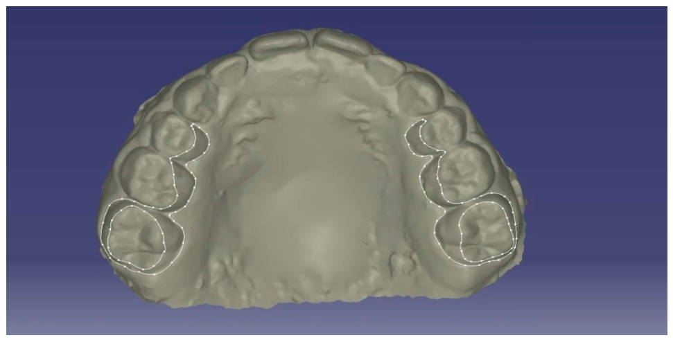
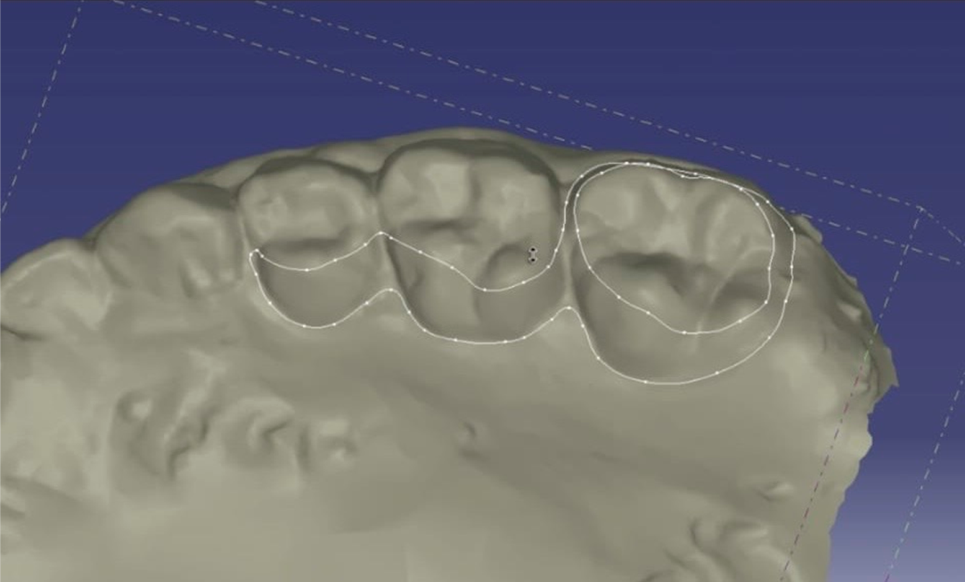
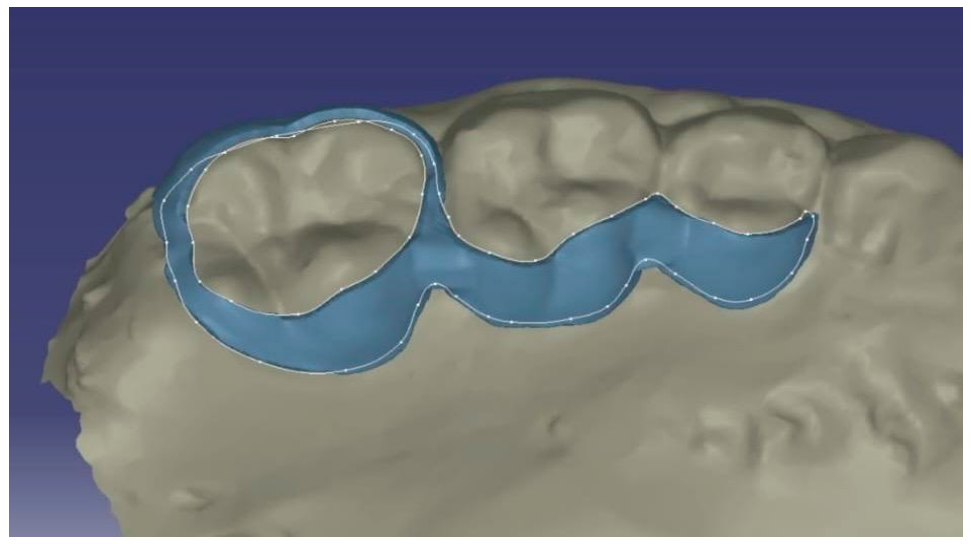
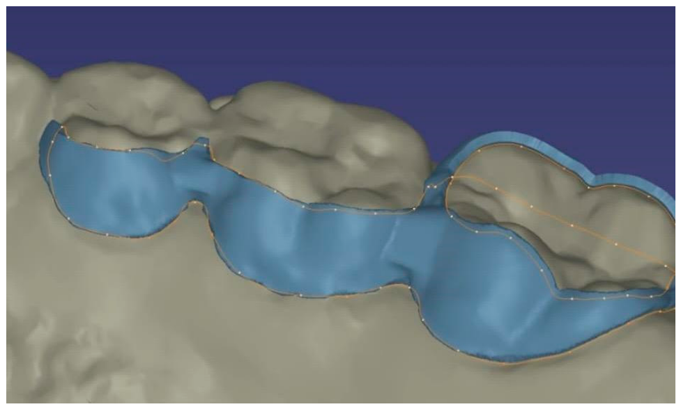
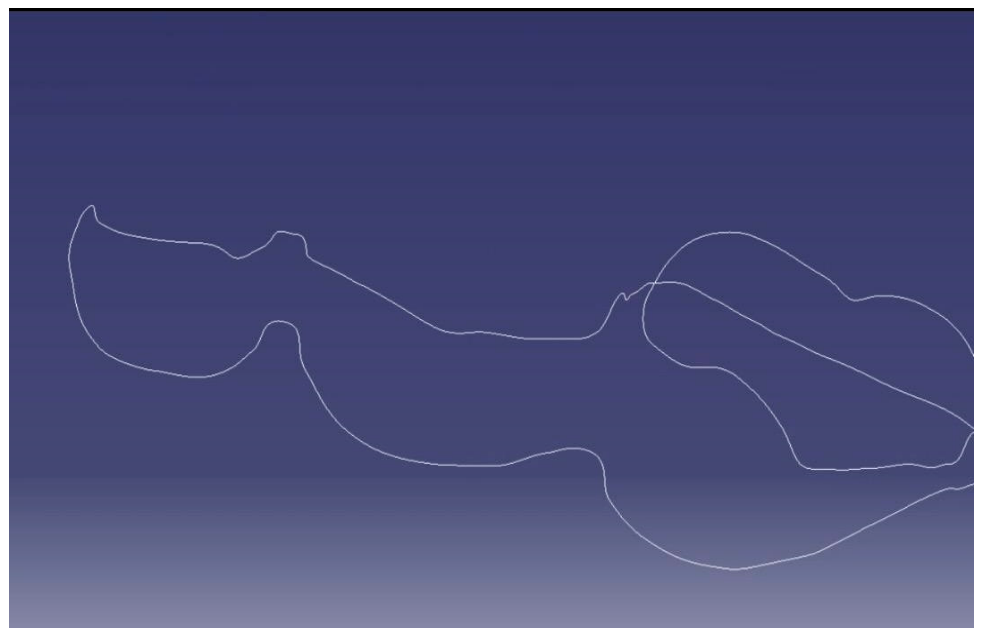
Patient data acquisition was implemented as a multimodal, high-resolution imaging protocol. A comprehensive dataset was generated through the integration of intraoral scanning (IOS), cone-beam computed tomography (CBCT), and three-dimensional facial surface imaging. Intraoral scans were obtained using structured-light or laser-based optical systems, producing high-density point clouds subsequently converted into triangulated surface meshes (STL format) (Figure 1, Figure 2 and Figure 3).
Compared with conventional elastomeric impressions, IOS ensures superior trueness and precision, eliminates material-related distortions, and enables immediate digitization of dental arch morphology.
CBCT imaging provided volumetric datasets with isotropic voxel resolution, allowing accurate reconstruction of craniofacial skeletal structures. Segmentation procedures were performed using semi-automated thresholding and region-growing algorithms to isolate relevant anatomical components, including the maxilla, nasal cavity, and midpalatal suture. These data were essential for biomechanical modeling and three-dimensional assessment of skeletal relationships. Three-dimensional facial scans, acquired via stereophotogrammetry or structured-light systems, generated high-resolution soft tissue meshes, enabling the integration of esthetic parameters into treatment planning.
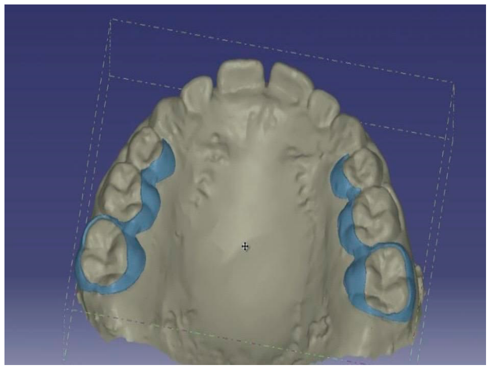
All datasets were digitally integrated through multimodal registration techniques, combining surface-based and voxel-based alignment algorithms (Figure 4, Figure 5 and Figure 6).
CBCT imaging and three-dimensional facial scans were used exclusively during the diagnostic planning and appliance design phases of the digital workflow. Quantitative treatment outcome assessment was based on standardized lateral cephalometric analysis, selected to ensure methodological consistency with previous TSME clinical studies and to limit additional radiological exposure in pediatric patients.
Best-fit surface registration was used to align intraoral scans with CBCT-derived dental structures, while voxel-based registration ensured accurate superimposition of skeletal datasets. Soft tissue alignment was further refined using anatomical landmarks and iterative closest point (ICP) algorithms. To ensure consistency and minimize systematic errors, acquisition protocols were strictly standardized in terms of head positioning, occlusal reference, and scanning pathways.
The resulting integrated digital model supported diagnostic planning, appliance customization, and integration with CAD/CAM systems.
Within this framework, two additive manufacturing technologies were evaluated: stereolithography (SLA), primarily used for the fabrication of diagnostic models and surgical guides, and Direct Metal Laser Sintering (DMLS), employed for the production of the definitive metallic device. DMLS was selected for final manufacturing due to its high dimensional accuracy, capability to produce complex geometries without assembly, and superior mechanical properties of titanium structures.
This approach enables the production of a patient-specific metallic framework, followed by mandatory post-processing procedures including stress-relief treatment, support removal, finishing, polishing, and dimensional verification.
The definitive TSME was manufactured using Direct Metal Laser Sintering (DMLS) technology with Ti-6Al-4V titanium alloy powder. Manufacturing was performed using a metal additive manufacturing system equipped with a ytterbium fiber laser operating at 200–400 W, with a layer thickness ranging from 30 to 60 μm and a laser spot diameter of approximately 70 μm.
The appliance geometry was generated through CAD software and exported in STL format for slicing and toolpath generation. Build orientation and support structures were optimized to minimize thermal distortion and residual stress during fabrication.
Following the printing phase, all devices underwent mandatory post-processing procedures, including thermal stress-relief treatment under controlled atmosphere, removal of support structures, surface finishing, polishing, and dimensional verification.
Thermal post-processing was performed to reduce residual internal stress generated during laser sintering and to improve the mechanical stability of the titanium structure. Surface finishing procedures were applied to reduce surface roughness and improve intraoral suitability of the appliance.
The manufacturing workflow included standard quality control procedures, including dimensional verification and surface inspection.
Overall, the implementation of this Smart Manufacturing framework enhances diagnostic accuracy, reduces operator dependency, and ensures high standards of quality, precision, and reliability in the production of patient-specific orthodontic devices.

2.1.2. Computer-Aided Design (CAD) and Artificial Intelligence

The TSME was designed using advanced CAD software and commercially available digital imaging tools. The digital design workflow enabled precise modeling of the appliance geometry based on patient-specific anatomical data.
Digital software tools incorporating AI-assisted functionalities available within commercially integrated imaging platforms were used to support segmentation, landmark localization, and treatment planning. These tools functioned exclusively as operator-support systems and were not independently validated as autonomous diagnostic algorithms in the present study.
Therefore, AI-assisted functionalities were used only to facilitate the digital workflow and reduce operator-dependent variability during planning. They were not used to generate autonomous diagnoses, independently determine treatment decisions, or provide validated predictive models of craniofacial growth.
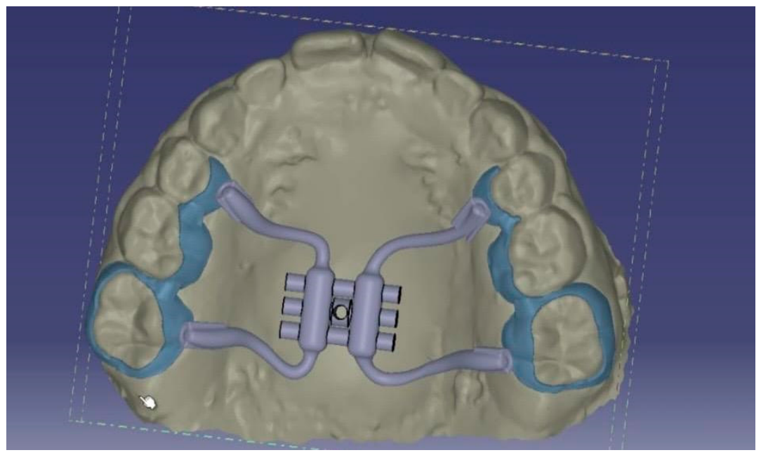
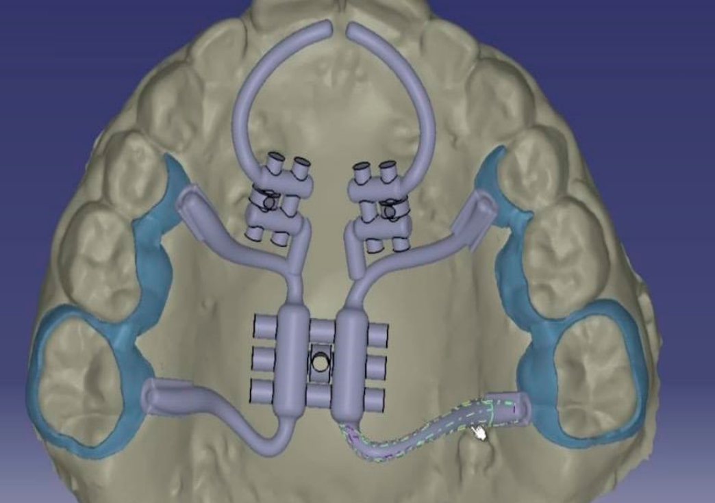
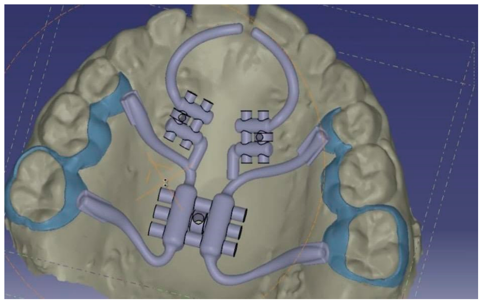
All software-assisted outputs were reviewed and clinically verified by experienced operators before appliance design and manufacturing. (Figure 7, Figure 8, Figure 9 and Figure 10).
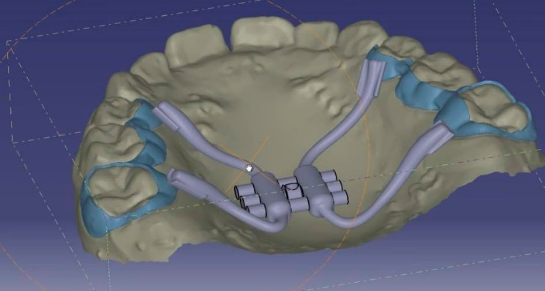
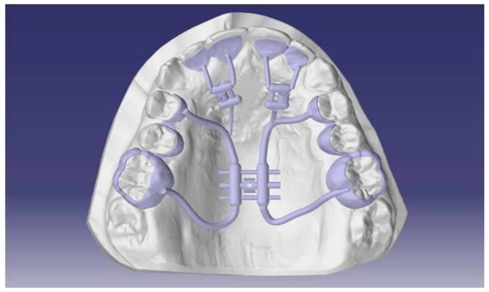
The digital workflow enabled the simultaneous design of palatal expanders and, when indicated, space maintainers, ensuring optimal adaptation to the patient’s anatomy and functional requirements (Figure 11).

2.1.3. Finite Element Analysis (FEA)

Finite element analysis (FEA) principles were considered during the digital design phase to support the biomechanical evaluation of the customized TSME. Within the Smart Manufacturing workflow, computational biomechanical analysis was used as an adjunctive design-support tool to assess theoretical stress distribution and load transfer patterns associated with the appliance configuration.
The digital geometries used for appliance development were derived from the integration of intraoral scans and CBCT-based anatomical datasets, allowing virtual assessment of appliance positioning and structural design. Biomechanical simulation principles were applied to evaluate force transmission patterns and to support design refinement aimed at improving structural balance and reducing excessive stress concentration on anchorage components.
The purpose of this computational approach was to provide biomechanical rationale during appliance development rather than to establish a patient-specific predictive clinical model.
Because the present investigation was designed as a retrospective clinical study focused primarily on dento-skeletal cephalometric outcomes, no direct quantitative correlation between computational simulations and post-treatment three-dimensional skeletal changes was performed.
Therefore, the biomechanical considerations derived from FEA should be interpreted as supportive design information rather than as clinically validated predictive evidence. Future prospective investigations integrating patient-specific computational simulations with quantitative three-dimensional clinical follow-up may help further clarify the translational role of biomechanical digital modeling in customized orthodontic appliance development.

2.1.4. TSME: Components and Activation Protocol

The TSME is a fixed device designed to develop arch form in patients with constricted dental arches. It is specifically designed for transverse and anteroposterior arch development. The TSME consists of two bands cemented to the right and left I maxillary molars, a Hyrax-type transverse expansion screw, two .045′′ stainless steel wires extending to the palatal surfaces of the central incisors, and two 8 mm Hyrax-type screws attached to these wires between the molar bands and the incisors. The appliance was worn for a total duration of 8 to 12 months (mean duration: 10.3 months). The activation protocol consisted of an initial transverse expansion phase, during which the transverse screw was activated twice daily with one-quarter turn per activation until the planned transverse expansion was achieved. This was followed by a sagittal activation phase, during which the sagittal screws were activated one-quarter turn every 15 days until the planned sagittal correction was obtained. After active activation, the appliance was maintained in situ as a passive retainer according to the clinical protocol. No additional orthodontic treatment was performed between T0 and T1 [17,36,37,38].

2.1.5. Titanium TSME

The selection of biomaterials represents an important aspect in the design and manufacturing of orthodontic appliances, particularly in pediatric patients requiring prolonged intraoral appliance wear [39]. Conventional orthodontic appliances frequently include nickel-containing alloys, which have been associated in the literature with hypersensitivity reactions in susceptible individuals [40,41,42,43].
In the present study, the customized TSME was manufactured using Ti-6Al-4V titanium alloy. This material was selected because of its favorable mechanical properties, corrosion resistance, fatigue resistance, and biocompatibility, as reported in previous biomedical and dental literature [40,41,44]. Titanium alloys may represent a suitable material option in patients with known or suspected hypersensitivity to nickel-containing alloys.
The integration of titanium additive manufacturing within the digital workflow allowed the production of a patient-specific metallic framework adapted to individual palatal morphology. This approach supported appliance customization and structural standardization within the CAD/CAM manufacturing process.
However, the present study did not include a dedicated clinical assessment of allergic reactions, mucosal irritation, intraoral adverse events, or patient-reported comfort. Therefore, no direct conclusion can be drawn from the present data regarding the clinical superiority of titanium over conventional nickel-containing orthodontic alloys.
Accordingly, the use of titanium in this study should be interpreted as a material choice supported by previous literature and compatible with the additive manufacturing workflow, rather than as a clinically validated superiority finding of the present investigation.

2.2. Study Design: Population and Sample Size

A retrospective sample of 45 pediatric patients (mean age: 8.5 years) presenting with bilateral posterior crossbite and sagittal and maxillary transverse deficiency was included in the study. All patients were in an active growth phase and had not undergone any previous orthodontic treatment.
Sample selection was based on the availability of complete and high-quality clinical and digital records fulfilling the predefined eligibility criteria. Given the retrospective design, no a priori sample size calculation was performed. However, a post hoc power analysis was carried out using G*Power 3.1 software (Heinrich-Heine-Universität Düsseldorf, Düsseldorf, Germany) to assess the adequacy of the final sample. Assuming a two-tailed t-test, an alpha level of 0.05, and a medium effect size (d = 0.50), the inclusion of 45 subjects provided a statistical power of 0.84, indicating that the sample was adequate to detect clinically meaningful differences.
Inclusion Criteria were:
-
Absence of prior orthodontic treatment;
-
Availability of pre-treatment (T0) and post-treatment (T1) lateral cephalometric radiographs;
-
Presence of transverse maxillary deficiency;
-
Bilateral posterior crossbite.
-
Exclusion criteria were:
-
Congenital anomalies;
-
Previous orthodontic interventions;
-
Craniofacial or dental asymmetries.
All patients were treated with a fully digital, patient-specific Transverse Sagittal Maxillary Expander (TSME), designed through a Smart Manufacturing workflow and manufactured in titanium alloy (Ti-6Al-4V).
Cephalometric changes on lateral radiography between T0 (beginning) and T1 (after the appliance removal) were analyzed. No additional orthodontic treatment was performed between T0 and T1, corresponding to the entire first phase of treatment.
The appliance was worn for a total duration of 8 to 12 months (mean duration: 10.3 months). The activation protocol consisted of an initial transverse expansion phase, during which the transverse screw was activated twice daily with one-quarter turn per activation until the planned transverse expansion was achieved. This was followed by a sagittal activation phase, during which the sagittal screws were activated one-quarter turn every 15 days until the planned sagittal correction was obtained. After active activation, the appliance was maintained in situ as a passive retainer according to the clinical protocol. No additional orthodontic treatment was performed between T0 and T1.
All radiographs were acquired by the same radiographic technician under standardized conditions. Cephalometric tracings were performed by a single experienced operator and subsequently verified by a second examiner. Any discrepancies were resolved by consensus. To assess intra-operator reliability, all measurements were repeated after a 7-day interval. The method error was determined using Dahlberg’s formula ME = √∑d 2/2n, where n is the number of subjects and d is the difference between the two measures. The method error did not exceed 0.1 mm for the linear measurements and 0.2° for angular measurements.
The following angular and linear cephalometric parameters were evaluated: SNA, SNB, ANB, SN–SNP.SNA, SN–Go.Gn, N–Me, S–Go, API+INI+-SN.
No additional orthodontic treatment was performed between T0 and T1.
Statistical analysis was conducted to compare cephalometric changes between T0 and T1 using a paired t-test, with significance set at p < 0.05.

3. Results

Cephalometric changes between T0 and T1 are reported in Table 1.
A statistically significant increase in SNA was observed, indicating a forward change in maxillary position during the observation period. SNB also showed a statistically significant increase. Consequently, ANB demonstrated a small but statistically significant decrease between T0 and T1.
A statistically significant increase in the SN–ANS-PNS angle was observed, suggesting a post-treatment change in palatal plane inclination. Vertical skeletal changes were limited but statistically significant for SN–GoGn and N–Me, indicating a slight increase in mandibular plane angle and anterior facial height. No statistically significant change was observed in S–Go.
APDI showed a statistically significant increase between T0 and T1.
Because the present study did not include an untreated control group or subgroup-specific statistical analysis according to skeletal class, these changes should be interpreted as short-term cephalometric modifications observed during treatment rather than as effects exclusively attributable to the appliance.

4. Discussion

The present study investigated the clinical and dento-skeletal effects of a fully digital, patient-specific Transversal Sagittal Maxillary Expander (TSME) within a Smart Manufacturing framework. The findings should be interpreted considering three key aspects: the transition from analog to digital workflows, the clinical effects observed after TSME treatment, and the use of titanium additive manufacturing within a patient-specific digital workflow.

4.1. From Analog to Digital Orthodontics: Clinical and Technological Implications

The transition from conventional analog workflows to fully digital orthodontic protocols represents a paradigm shift with significant clinical and technological implications [33]. Traditional methods based on elastomeric impressions and manual laboratory procedures are inherently affected by material distortions, operator-dependent variability, and limited reproducibility. In contrast, the integration of intraoral scanning, CBCT imaging, and three-dimensional facial acquisition enables the generation of a high-fidelity digital representation of the patient [44,45,46].
In the present study, the implementation of a multimodal digital workflow supported the integration of dental, skeletal, and soft tissue information for diagnostic planning and appliance customization. This approach may improve standardization of the design process and facilitate communication between clinical and manufacturing phases.
Moreover, the incorporation of CAD/CAM technologies and additive manufacturing facilitated the production of fully customized appliances, overcoming the limitations of traditional laboratory techniques. The ability to directly translate virtual designs into physical devices reduces intermediate steps, minimizes cumulative errors, and enhances standardization [29,47].
Importantly, the incorporation of biomechanical computational principles may support appliance design by providing theoretical information on force distribution and structural balance. However, in the present retrospective study, these considerations were not quantitatively validated against three-dimensional clinical outcomes [31,48].

4.2. Biomechanical and Clinical Advantages of TSME

Previous studies have suggested that TSME may provide clinically relevant effects in the transverse, sagittal, and vertical dimensions. In the present study, the digitally manufactured TSME was associated with statistically significant short-term cephalometric changes. However, because no conventional control group was included, direct superiority over traditional TSME or other expansion appliances cannot be established [25,49].
From a clinical perspective, the TSME has been described in previous studies as an appliance capable of acting in transverse, sagittal, and vertical dimensions. In the present study, statistically significant short-term cephalometric changes were observed after treatment with the digitally manufactured TSME. However, these findings should be interpreted cautiously, as the study design does not allow separation of treatment-related effects from growth-related changes or direct comparison with conventional expanders.
The digital workflow used in this study may support appliance customization by allowing integration of patient-specific anatomical data into CAD-based design and additive manufacturing. Nevertheless, because no conventionally manufactured TSME control group was included, the present data cannot demonstrate improved biomechanical efficiency, reduced dento-alveolar side effects, or superior clinical predictability compared with traditional workflows [13,17,25,38,47,49,50,51].

4.3. Biocompatible Materials and the Role of Titanium

The selection of biomaterials is an important aspect in orthodontic appliance design, especially in pediatric patients requiring prolonged intraoral treatment. Titanium alloys have been widely described in the literature as materials with favorable biocompatibility, corrosion resistance, and mechanical properties. In the present study, Ti-6Al-4V was selected as part of the digital additive manufacturing workflow and allowed the production of a patient-specific metallic framework.
However, this study did not include a dedicated clinical assessment of allergic reactions, mucosal irritation, intraoral adverse events, or patient-reported comfort. Therefore, the present findings cannot be used to demonstrate the clinical superiority of titanium over conventional nickel-containing alloys. The use of titanium should be interpreted as a material choice supported by previous literature and compatible with additive manufacturing, rather than as a superiority outcome of the present study.
Similar patient-specific additive manufacturing strategies have been described in other biomedical fields, where selective laser melting has been used for individualized implant design, process optimization, and manufacturing workflow standardization [40,43,52,53,54,55,56].

4.4. Clinical Implications and Future Perspectives

The present findings suggest that a digitally manufactured, patient-specific TSME can be clinically implemented in growing patients and may support appliance customization and workflow standardization. However, due to the retrospective design and the absence of a conventional control group, the present study cannot establish superiority over traditional manufacturing workflows or other expansion appliances.
Future developments should focus on prospective controlled studies, long-term follow-up, and quantitative three-dimensional outcome assessment to clarify treatment stability and the potential clinical role of digitally manufactured TSME appliances.
Although the digital workflow incorporated CBCT imaging and three-dimensional facial acquisition for diagnostic planning and appliance customization, treatment outcome assessment was intentionally based on standardized two-dimensional cephalometric analysis. This methodological choice was made to maintain consistency with previous clinical TSME investigations and to minimize additional radiation exposure in a pediatric population, in accordance with radiation protection principles. Nevertheless, we acknowledge that this approach does not fully capture the three-dimensional complexity of skeletal and soft tissue adaptations following treatment.

4.5. Limitations of This Study

The present study has several limitations. First, the retrospective design may introduce selection bias, as patient inclusion was based on the availability of complete clinical and digital records rather than randomized allocation.
Second, the absence of a control group treated with conventional TSME or alternative expansion appliances limits the possibility of directly comparing the effectiveness of the digital workflow with traditional approaches. Therefore, the observed changes cannot be attributed exclusively to the appliance or to the digital manufacturing process.
Third, all subjects were actively growing children. Spontaneous craniofacial growth may therefore have contributed to the observed cephalometric changes, limiting the ability to separate treatment-related effects from physiological growth.
Fourth, although CBCT imaging and three-dimensional facial scanning were incorporated into the digital workflow for diagnostic planning and appliance customization, treatment outcome assessment was limited to standardized two-dimensional cephalometric analysis. Consequently, the present study cannot fully characterize three-dimensional skeletal, dento-alveolar, and soft tissue adaptations associated with treatment.
Fifth, although biomechanical computational principles informed the appliance design process, the present study did not include direct validation of FEA-derived predictions against quantitative three-dimensional clinical outcomes.
Sixth, the study did not include dedicated clinical assessment of allergic reactions, mucosal irritation, or patient-reported comfort. Therefore, no direct conclusion can be drawn regarding the clinical superiority of titanium over conventional nickel-containing alloys.
Finally, the observational period was limited to short-term post-treatment assessment. Long-term prospective studies with controlled groups and three-dimensional outcome analysis are required to evaluate treatment stability and to clarify the potential clinical role of digitally manufactured TSME appliances.

5. Conclusions

Within the limitations of this retrospective study, the integration of a digital workflow and additive manufacturing allowed the production of patient-specific TSME appliances for interceptive orthodontic treatment. The treated patients showed statistically significant short-term cephalometric changes after therapy.
The use of intraoral scanning, CBCT-based planning, CAD, and titanium additive manufacturing may support appliance customization and workflow standardization. However, due to the absence of a conventional control group, the retrospective design, the lack of quantitative three-dimensional outcome assessment, and the short-term follow-up, no definitive conclusions can be drawn regarding the superiority of the digital workflow over traditional manufacturing methods.
Further prospective controlled studies with long-term follow-up and three-dimensional outcome evaluation are required to assess treatment stability and to clarify the potential clinical role of digitally manufactured TSME appliances.

Author Contributions

Conceptualization, L.G., A.M. and C.M.; methodology, L.G.; software, A.M.; validation, C.M.; formal analysis, A.M.; investigation, L.G.; data curation, A.M. and L.G.; writing—original draft preparation, L.G. and A.M.; writing—review and editing, C.M.; supervision, C.M.; project administration, C.M. All authors have read and agreed to the published version of the manuscript.

Funding

This research received no external funding.

Institutional Review Board Statement

The study was conducted in accordance with the Declaration of Helsinki. This study was approved by the Research Protocol of the Fondazione IRCCS Ca’Granda, Ospedale Maggiore Policlinico Operative Unit 420, Current Research n 1 year 2022.

Informed Consent Statement

Informed consent was obtained from the parents or legal guardians of all subjects involved in the study.

Data Availability Statement

The original contributions presented in this study are included in the article. Further inquiries can be directed to the corresponding author.

Conflicts of Interest

The authors declare no conflicts of interest.

References

  1. Paglia, L. Interceptive orthodontics: Awareness and prevention is the first cure. Eur. J. Acad. Paediatr. Dent. 2023, 24, 5. [Google Scholar] [CrossRef] [PubMed]
  2. Bistaffa, A.G.I.; Belomo-Yamaguchi, L.; de Castro Ferreira Conti, A.C.; Oltramari, P.V.P.; de Almeida, M.R.; de Almeida-Pedrin, R.R.; Fernandes, T.M.F. Dental arch changes comparison between Expander with Differential Opening, Hyrax-type and Haas-type expanders: A prospective clinical study. Orthod. Craniofac. Res. 2023, 26, 468–475. [Google Scholar] [CrossRef] [PubMed]
  3. Mutinelli, S.; Cozzani, M. Rapid maxillary expansion in early-mixed dentition: Effectiveness of increasing arch dimension with anchorage on deciduous teeth. Eur. J. Paediatr. Dent. 2015, 16, 115–122. [Google Scholar] [PubMed]
  4. Cozza, P.; Giancotti, A.; Petrosino, A. Rapid palatal expansion in mixed dentition using a modified expander: A cephalometric investigation. J. Orthod. 2001, 28, 129–134. [Google Scholar] [CrossRef] [PubMed]
  5. Reichenbach, E.; Taatz, K. Kieferorthopaedische Klinik und Therapie; Johann Ambrosius Barth Verlag: Leipzig, Germany, 1971. [Google Scholar]
  6. Cerritelli, L.; Hatzopoulos, S.; Catalano, A.; Bianchini, C.; Cammaroto, G.; Meccariello, G.; Iannella, G.; Vicini, C.; Pelucchi, S.; Skarzynski, P.H.; et al. Rapid Maxillary Expansion (RME): An Otolaryngologic Perspective. J. Clin. Med. 2022, 11, 5243. [Google Scholar] [CrossRef] [PubMed] [PubMed Central]
  7. Bishara, S.E.; Hoppens, B.J.; Jakobsen, J.R.; Kohout, F.J. Changes in the molar relationship between the deciduous and permanent dentitions: A longitudinal study. Am. J. Orthod. Dentofac. Orthop. 1988, 93, 19–28. [Google Scholar] [CrossRef] [PubMed]
  8. Chung, C.H.; Font, B. Skeletal and dental changes in the sagittal, vertical, and transverse dimensions after rapid palatal expansion. Am. J. Orthod. Dentofac. Orthop. 2004, 126, 569–575. [Google Scholar] [CrossRef] [PubMed]
  9. Kiriakou, J.; Pandis, N.; Madianos, P.; Polychronopoulou, A. Developing evidence-based dentistry skills: How to interpret randomized clinical trials and systematic reviews. Prog. Orthod. 2014, 15, 58. [Google Scholar] [CrossRef] [PubMed]
  10. Lin, L.; Ahn, H.W.; Kim, S.J.; Moon, S.C.; Kim, S.H.; Nelson, G. Tooth-borne vs bone-borne rapid maxillary expanders in late adolescence. Angle Orthod. 2015, 85, 253–262. [Google Scholar] [CrossRef] [PubMed] [PubMed Central]
  11. Volk, T.; Sadowsky, C.; Begole, E.A.; Boice, P. Rapid palatal expansion for spontaneous Class II correction. Am. J. Orthod. Dentofac. Orthop. 2010, 137, 310–315. [Google Scholar] [CrossRef] [PubMed]
  12. MacGinnis, M.; Chu, H.; Youssef, G.; Wu, K.W.; Machado, A.W.; Moon, W. The effects of micro-implant assisted rapid palatal expansion (MARPE) on the nasomaxillary complex—A finite element method (FEM) analysis. Prog. Orthod. 2014, 15, 52. [Google Scholar] [CrossRef]
  13. Farronato, G.; Maspero, C.; Esposito, L.; Briguglio, E.; Farronato, D.; Giannini, L. Rapid maxillary expansion in growing patients. Hyrax versus transverse sagittal maxillary expander: A cephalometric investigation. Eur. J. Orthod. 2011, 33, 185–189. [Google Scholar] [CrossRef][Green Version]
  14. Rosa, M.; Lucchi, P.; Manti, G.; Caprioglio, A. Rapid Palatal Expansion in the absence of posterior cross-bite to intercept maxillary incisor crowding in the mixed dentition: A CBCT evaluation of spontaneous changes of untouched permanent molars. Eur. J. Paediatr. Dent. 2016, 17, 286–294. [Google Scholar] [PubMed]
  15. Cretella Lombardo, E.; Paoloni, V.; Fanelli, S.; Pavoni, C.; Gazzani, F.; Cozza, P. Evaluation of the Upper Arch Morphological Changes after Two Different Protocols of Expansion in Early Mixed Dentition: Rapid Maxillary Expansion and Invisalign® First System. Life 2022, 12, 1323. [Google Scholar] [CrossRef] [PubMed] [PubMed Central]
  16. Rutili, V.; Mrakic, G.; Nieri, M.; Franceschi, D.; Pierleoni, F.; Giuntini, V.; Franchi, L. Dento-skeletal effects produced by rapid versus slow maxillary expansion using fixed jackscrew expanders: A systematic review and meta-analysis. Eur. J. Orthod. 2021, 43, 301–312. [Google Scholar] [CrossRef] [PubMed]
  17. Feng, Z.; Si, M.; Fan, H.; Zhang, Y.; Yuan, R.; Hao, Z. Evolution, current status, and future trends of maxillary skeletal expansion: A bibliometric analysis. Clin. Oral Investig. 2023, 28, 14. [Google Scholar] [CrossRef] [PubMed] [PubMed Central]
  18. Casanova-Sarmiento, J.A.; Arriola-Guillén, L.E.; Ruíz-Mora, G.A.; Rodríguez-Cárdenas, Y.A.; Aliaga-Del Castillo, A. Comparison of anterior mandibular alveolar thickness and height in young adults with different sagittal and vertical skeletal relationships: A CBCT Study. Int. Orthod. 2020, 18, 79–88. [Google Scholar] [CrossRef] [PubMed]
  19. Ballanti, F.; Lione, R.; Viarani, S.; Fanucci, E.; Cozza, P. Post-retention effects of rapid maxillary expansion on nasal cavity and on periodontal structures. Oral Implantol. 2008, 1, 95–103. [Google Scholar] [PubMed] [PubMed Central]
  20. Lo Giudice, A.; Barbato, E.; Cosentino, L.; Ferraro, C.M.; Leonardi, R. Alveolar bone changes after rapid maxillary expansion with tooth-born appliances: A systematic review. Eur. J. Orthod. 2018, 40, 296–303. [Google Scholar] [CrossRef] [PubMed]
  21. Kang, S.H.; Jeon, K.; Kang, S.H.; Lee, S.H. 3D cephalometric landmark detection by multiple stage deep reinforcement learning. Sci. Rep. 2021, 11, 17509. [Google Scholar] [CrossRef] [PubMed] [PubMed Central]
  22. Figueiredo, D.S.; Cardinal, L.; Bartolomeo, F.U.; Palomo, J.M.; Horta, M.C.; Andrade, I., Jr.; Oliveira, D.D. Effects of rapid maxillary expansion in cleft patients resulting from the use of two different expanders. Dent. Press J. Orthod. 2016, 21, 82–90. [Google Scholar] [CrossRef] [PubMed] [PubMed Central]
  23. Ngan, P.; Wilmes, B.; Drescher, D.; Martin, C.; Weaver, B.; Gunel, E. Comparison of two maxillary protraction protocols: Tooth-borne versus bone-anchored protraction facemask treatment. Prog. Orthod. 2015, 16, 26. [Google Scholar] [CrossRef] [PubMed] [PubMed Central]
  24. Perillo, L.; Femiano, A.; Palumbo, S.; Contardo, L.; Perinetti, G. Skeletal and dental effects produced by functional regulator-2 in pre-pubertal class II patients: A controlled study. Prog. Orthod. 2013, 14, 18. [Google Scholar] [CrossRef] [PubMed]
  25. Maspero, C.; Cavagnetto, D.; Fama, A.; Giannini, L.; Galbiati, G.; Farronato, M. Hyrax versus transverse sagittal maxillary expander: An assessment of arch changes on dental casts. A retrospective study. Saudi Dent. J. 2020, 32, 93–100. [Google Scholar] [CrossRef]
  26. Khan, M.K.; Sharma, D.S.; Jindal, M.K. Unusual systemic and nondental effects of maxillary expansion therapy: A comprehensive and updated review of literature. J. Orthod. Sci. 2023, 12, 38. [Google Scholar] [CrossRef] [PubMed] [PubMed Central]
  27. Xepapadeas, A.B.; Weise, C.; Frank, K.; Spintzyk, S.; Poets, C.F.; Wiechers, C.; Arand, J.; Koos, B. Technical note on introducing a digital workflow for newborns with craniofacial anomalies based on intraoral scans—Part I: 3D printed and milled palatal stimulation. BMC Oral Health 2020, 20, 20. [Google Scholar] [CrossRef]
  28. Clark, W.J. Transforce lingual appliances for arch development. J. Clin. Orthod. 2005, 39, 137–142; quiz 153. [Google Scholar] [PubMed]
  29. Dickens, N.; Haider, H.; Lien, W.; Simecek, J.; Stahl, J. Longitudinal Analysis of CAD/CAM Restoration Incorporation Rates into Navy Dentistry. Mil. Med. 2019, 184, e365–e372. [Google Scholar] [CrossRef] [PubMed]
  30. Maspero, C.; Galbiati, G.; Del Rosso, E.; Farronato, M.; Giannini, L. RME: Effects on the nasal septum. A CBCT evaluation. Eur. J. Paediatr. Dent. 2019, 20, 123–126. [Google Scholar] [CrossRef] [PubMed]
  31. Dhopte, A.; Bagde, H. Smart Smile: Revolutionizing Dentistry With Artificial Intelligence. Cureus 2023, 15, e41227. [Google Scholar] [CrossRef] [PubMed] [PubMed Central]
  32. Gandedkar, N.H.; Liou, E.J. The immediate effect of alternate rapid maxillary expansions and constrictions on the alveolus: A retrospective cone beam computed tomography study. Prog. Orthod. 2018, 19, 40. [Google Scholar] [CrossRef] [PubMed] [PubMed Central]
  33. Schwendicke, F.; Samek, W.; Krois, J. Artificial Intelligence in Dentistry: Chances and Challenges. J. Dent. Res. 2020, 99, 769–774. [Google Scholar] [CrossRef] [PubMed] [PubMed Central]
  34. Da Silva Filho, O.G.; Boas, M.C.; Capelozza Filho, L. Rapid maxillary expansion in the primary and mixed dentitions: A cephalometric evaluation. Am. J. Orthod. Dentofac. Orthop. 1991, 100, 171–179. [Google Scholar] [CrossRef]
  35. Maspero, C.; Galbiati, G.; Perillo, L.; Favero, L.; Giannini, L. Orthopaedic treatment efficiency in skeletal Class III malocclusions in young patients: RME-face mask versus TSME. Eur. J. Paediatr. Dent. 2012, 13, 225–230. [Google Scholar] [PubMed]
  36. Wertz, R.A. Skeletal and dental changes accompanying rapid midpalatal suture opening. Am. J. Orthod. 1970, 58, 41–66. [Google Scholar] [CrossRef] [PubMed]
  37. Wertz, R.; Dreskin, M. Midpalatal suture opening: A normative study. Am. J. Orthod. 1977, 71, 367–381. [Google Scholar] [CrossRef] [PubMed]
  38. Maspero, C.; Giannini, L.; Galbiati, G.; Farronato, G. Modified transversal sagittal maxillary expander for correction of upper midline deviation associated with maxillary arch deficiency. Minerva Stomatol. 2015, 64, 53–58. [Google Scholar] [PubMed]
  39. Baker, S.; Brooks, S.C.; Walker, D.M. The release of residual monomeric methyl methacrylate from acrylic appliances in the human mouth: An assay for monomer in saliva. J. Dent. Res. 1988, 67, 1295–1299. [Google Scholar] [CrossRef] [PubMed]
  40. Maspero, C.; Galbiati, G.; Giannini, L.; Guenza, G.; Esposito, L.; Farronato, G. Titanium TSME appliance for patients allergic to nickel. Eur. J. Paediatr. Dent. 2018, 19, 67–69. [Google Scholar] [CrossRef] [PubMed]
  41. Wataha, J.C. Biocompatibility of dental casting alloys: A review. J. Prosthet. Dent. 2000, 83, 223–234. [Google Scholar] [CrossRef] [PubMed]
  42. Noble, J.; Ahing, S.I.; Karaiskos, N.E.; Wiltshire, W.A. Nickel allergy and orthodontics, a review and report of two cases. Br. Dent. J. 2008, 204, 297–300. [Google Scholar] [CrossRef] [PubMed]
  43. Büdinger, L.; Hertl, M. Immunologic mechanisms in hypersensitivity reactions to metal ions: An overview. Allergy 2000, 55, 108–115. [Google Scholar] [CrossRef] [PubMed]
  44. Tallarico, M. Computerization and Digital Workflow in Medicine: Focus on Digital Dentistry. Materials 2020, 13, 2172. [Google Scholar] [CrossRef] [PubMed] [PubMed Central]
  45. Lagravère, M.O.; Ling, C.P.; Woo, J.; Harzer, W.; Major, P.W.; Carey, J.P. Transverse, vertical, and anterior-posterior changes between tooth-anchored versus Dresden bone-anchored rapid maxillary expansion 6 months post-expansion: A CBCT randomized controlled clinical trial. Int. Orthod. 2020, 18, 308–316. [Google Scholar] [CrossRef] [PubMed]
  46. Dindaroğlu, F.; Duran, G.S.; Görgülü, S. Effects of rapid maxillary expansion on facial soft tissues: Deviation analysis on three-dimensional images. J. Orofac. Orthop. 2016, 77, 242–250. [Google Scholar] [CrossRef] [PubMed]
  47. Sendyk, M.; de Paiva, J.B.; Abrão, J.; Rino Neto, J. Correlation between buccolingual tooth inclination and alveolar bone thickness in subjects with Class III dentofacial deformities. Am. J. Orthod. Dentofac. Orthop. 2017, 152, 66–79. [Google Scholar] [CrossRef] [PubMed]
  48. Hicks, E.P. Slow maxillary expansion. A clinical study of the skeletal versus dental response to low-magnitude force. Am. J. Orthod. 1978, 73, 121–141. [Google Scholar] [CrossRef] [PubMed]
  49. Maspero, C.; Galbiati, G.; Giannini, L.; Farronato, G. Sagittal and vertical effects of transverse sagittal maxillary expander (TSME) in three different malocclusion groups. Prog. Orthod. 2015, 16, 6. [Google Scholar] [CrossRef] [PubMed] [PubMed Central]
  50. Perillo, L.; De Rosa, A.; Iaselli, F.; d’Apuzzo, F.; Grassia, V.; Cappabianca, S. Comparison between rapid and mixed maxillary expansion through an assessment of dento-skeletal effects on posteroanterior cephalometry. Prog. Orthod. 2014, 15, 46. [Google Scholar] [CrossRef] [PubMed][Green Version]
  51. Akkaya, S.; Lorenzon, S.; Uçem, T.T. A comparison of sagittal and vertical effects between bonded rapid and slow maxillary expansion procedures. Eur. J. Orthod. 1999, 21, 175–180. [Google Scholar] [CrossRef] [PubMed]
  52. Farronato, G.; Giannini, L.; Galbiati, G.; Maspero, C. Comparison of the dental and skeletal effects of two different rapid palatal expansion appliances for the correction of the maxillary asymmetric transverse discrepancies. Minerva Stomatol. 2012, 61, 45. [Google Scholar]
  53. Ayyıldız, S. The place of direct metal laser sintering (DMLS) in dentistry and the importance of annealing. Mater. Sci. Eng. C Mater. Biol. Appl. 2015, 52, 343. [Google Scholar] [CrossRef] [PubMed]
  54. Sun, J.; Fang, T.; Zhang, Y.; Wang, J.; Han, H.; Chou, T.; Liang, J.; Kalyon, D.M.; Wang, H.; Wang, S. Imaging-Guided Microscale Photothermal Stereolithography Bioprinting. Adv. Sci. 2025, 12, e2500640. [Google Scholar] [CrossRef] [PubMed]
  55. Michelotti, A.; Rongo, R.; Valentino, R.; D’Antò, V.; Bucci, R.; Danzi, G.; Cioffi, I. Evaluation of masticatory muscle activity in patients with unilateral posterior crossbite before and after rapid maxillary expansion. Eur. J. Orthod. 2019, 41, 46–53. [Google Scholar] [CrossRef]
  56. Zhang, G.; Li, J.; Li, J.; Zhou, X.; Xie, J.; Wang, A. Selective laser melting molding of individualized femur implant: Design, process, optimization. J. Biomim. Eng. 2021, 18, 10. [Google Scholar] [CrossRef]
Figure 1. Initial intraoral scan of the maxillary arch showing high-resolution digital acquisition of dental morphology (STL model).
Designs 10 00061 g001
Figure 2. Digital outlining of the palatal surface and preliminary CAD tracing for appliance design.
Designs 10 00061 g002
Figure 3. Virtual design of the palatal coverage area for the TSME framework, adapted to patient-specific anatomy.
Designs 10 00061 g003
Figure 4. Three-dimensional visualization of the designed palatal structure highlighting extension and thickness of the appliance base.
Designs 10 00061 g004
Figure 5. 2D projection of the CAD used for geometric refinement and contour optimization.
Designs 10 00061 g005
Figure 6. Finalized digital model of the maxillary arch with defined anchorage areas for molar bands and palatal support.
Designs 10 00061 g006
Figure 7. Initial CAD positioning of the TSME components, including expansion screws and connecting arms.
Designs 10 00061 g007
Figure 8. Refined virtual assembly of the appliance with optimized geometry and symmetry based on patient-specific data.
Designs 10 00061 g008
Figure 9. Advanced design stage showing integration of transverse and sagittal expansion mechanisms within a unified structure.
Designs 10 00061 g009
Figure 10. Final CAD configuration of the TSME with complete architecture prior to manufacturing.
Designs 10 00061 g010
Figure 11. Definitive patient-specific TSME digitally designed for additive manufacturing, ready for production in titanium alloy (DMLS).
Designs 10 00061 g011
Table 1. Cephalometric evaluation.
VariableT0 Mean ± SDT1 Mean ± SDΔ (T1–T0)p-Value
SNA (°)80.17 ± 1.4180.67 ± 1.440.500.003
SNB (°)78.07 ± 1.5378.70 ± 1.750.630.002
ANB (°)2.10 ± 0.891.97 ± 1.11−0.130.001
SN–ANS-PNS (°)9.73 ± 1.7110.51 ± 1.890.780.001
SN–GoGn (°)32.73 ± 4.1533.37 ± 4.240.630.040
N–Me (mm)110.88 ± 2.89111.35 ± 2.880.470.050
S–Go (mm)64.37 ± 4.1465.03 ± 4.260.660.670
API+INI+-SN (°)99.87 ± 2.72102.41 ± 2.412.550.020
Disclaimer/Publisher’s Note: The statements, opinions and data contained in all publications are solely those of the individual author(s) and contributor(s) and not of MDPI and/or the editor(s). MDPI and/or the editor(s) disclaim responsibility for any injury to people or property resulting from any ideas, methods, instructions or products referred to in the content.