
1. Introduction
Nasopharyngeal (NP) swabs are medical devices used to collect nasal secretions from the back of the nose and throat for diagnosis of diseases of the respiratory system such as COVID-19, SARS, influenza, diphtheria, pertussis, etc. [
1,
2]. Swabs consist of a long and flexible shaft, which enables handling and sampling, and an absorbent head or tip, which collects biological specimens and elutes them in a transport medium for subsequent detection [
2].
During the COVID-19 pandemic, the unprecedented need for testing and care, together with disruptions in manufacturing and supply chains, led to a global shortage of numerous devices, including swabs. Three-dimensional (3D) printing showed great promise to face the healthcare emergency by providing an opportunity for fast, customizable, and on-demand manufacturing of swabs [
3,
4,
5,
6,
7,
8,
9]. The advantages offered by 3D printing over traditional swab manufacturing methods (like molding) are multiple and mostly related to its versatility and customization potential [
2]. First, 3D printing allows for rapid production of large numbers of swab prototypes with various and complex tip geometries [
2,
4,
6], which are meant to enhance or facilitate the uptake and release of biological samples for diagnostic efficiency. Personalized swabs tailored to individual or population-specific (e.g., children) anatomical requirements are also feasible [
9]. Furthermore, the wide availability of commercial printers and printable materials supports the idea of potential on-site production of swabs within hospitals and laboratories [
1,
7]. Lastly, different materials can be printed to produce both the shaft and the tip, potentially enabling the development of swab devices that couple optimal mechanical properties of the shaft (e.g., flexibility and strength) with improved uptake and release efficiency of the tip [
2,
10].
Nonetheless, 3D printing is also affected by several limitations. While swab material selection is crucial for safe and effective sampling, reduced contamination risks, and optimized diagnostic accuracy, not all materials suitable for swab production are 3D-printable (e.g., cotton and rayon fibers); furthermore, not all printable materials are biocompatible and sterilizable for clinical use [
2,
7]. Additionally, achieving the desired mechanical properties of a swab can be difficult, especially for complex shapes and designs [
5,
11]. Moreover, although 3D-printed swabs are compatible with various transport media and testing kits, their plastic heads may be larger and rougher than those of traditional swabs, potentially causing discomfort and mild side effects, such as nasal irritation and headache [
12].
In general, the reliability and diagnostic accuracy of swabs depend on both their mechanical properties and their ability to take up and release a significant quantity of microorganisms [
13]. From a mechanical point of view, NP swabs (with reference to both the shaft and head) should be pliant and robust enough to enable comfortable, pain-free, and safe movements through the nasal cavity [
14]. Sample collection and subsequent release are clearly affected by the swab-head features, as well as by the pressure/load applied by the operator during such phases. In this regard, an interesting study by Melcher et al. [
15] recently evaluated the average force applied by healthcare workers to perform an oropharyngeal swab (about 2.5 N), proposing a force-controlled swab model to standardize sample collection for improved diagnostic reliability. While several studies have investigated the mechanical properties of swabs for clinical applications [
6,
7,
11,
16], to the best of the authors’ knowledge, numerical approaches are not routinely adopted to assess the mechanical performance of swabs. However, due to the growing interest in 3D-printed swabs, a few studies have recently applied computational tools such as the Finite Element Method (FEM) and Computational Fluid Dynamics (CFD) to support swab design and validation. A study by Singh et al. applied ANSYS Fluent with a Volume Of Fluid (VOF) method to simulate dipping tests and optimize swab geometry using Taguchi analysis, demonstrating how design parameters affect liquid retention [
17]. FE models have also been developed to evaluate swab-shaft deformation, buckling resistance, and structural integrity under insertion forces [
12], as well as to analyze the deformation behavior of auxetic swab heads [
11]. Additionally, COMSOL Multiphysics has been employed to simulate mucosal interaction using non-Newtonian fluid models, offering insights into how surface design impacts sample absorption and patient comfort [
18]. These simulation-based approaches provide a reliable framework for testing swab performance, reducing the need for extensive trial-and-error experimentation, and ensuring safety and functionality in clinical use.
Regarding the ability of a swab to take up and release biological specimens, the use of tips made of fibrous materials, such as cotton, rayon, and polyester, generally suffices to achieve good sample uptake, but the yielded elution efficiency is low, as microorganisms can be trapped within the fibers [
13]. Therefore, elution needs to be mechanically enhanced, e.g., by mixing or agitating the swab in the transport medium or by manually pressing and squeezing the swab head against the internal surface of the transport tube. Although flocked (nylon-based) and foam (polyurethane-based) swabs have been shown to achieve superior elution performance compared to fibrous ones [
13,
19], efficient release may still require the use of mechanical forces, which can be operator-sensitive.
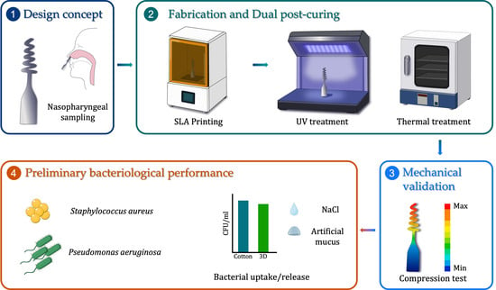
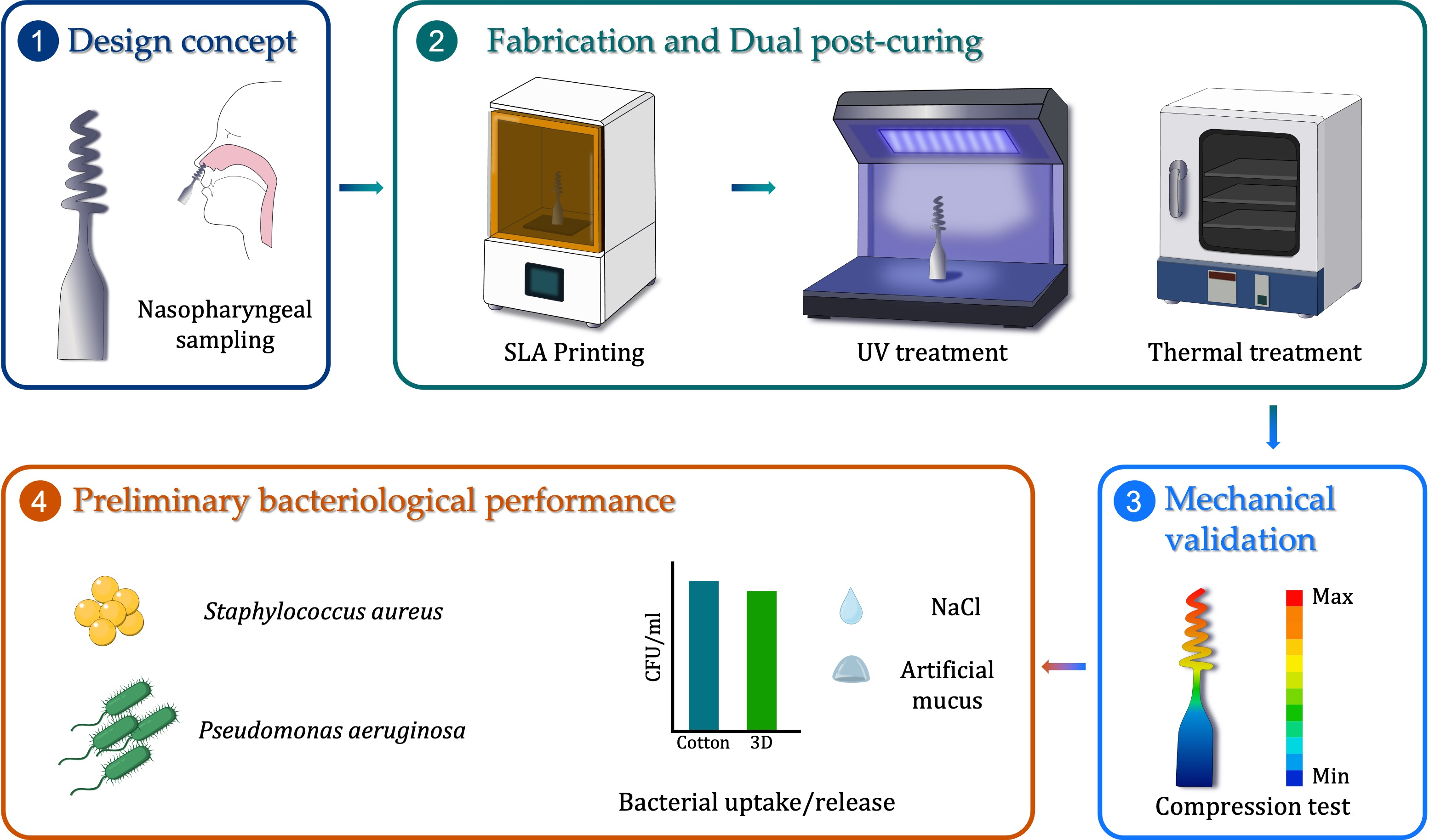
Based on such considerations, the primary objective of this work was to assess the feasibility of a 3D-printed swab prototype intended to facilitate and potentially standardize sample release via controlled elastic deformation. The proposed prototype features a helical head design, which aims to ensure elastic deformation during axial compression, thereby facilitating transfer of the sample into the transport medium without compromising structural integrity. Helical swab prototypes were manufactured using stereolithography (SLA), followed by a dual post-curing process to enhance the mechanical performance of the printed material. Experimental testing was then carried out to determine the material’s elastic modulus and to assess the deformation behavior of the swab’s helical tip under compression loads. A key component of the work included the development of an FE model, which enabled a detailed analysis of the stress distribution and deformation along the swab structure. The goal was to verify that stresses remained below the failure threshold of the material while also comparing simulation results with experimental tracking data. In parallel, the functional performance of the swab prototype was preliminarily evaluated in vitro against that of standard commercial swabs through bacterial uptake and release tests using S. aureus and P. aeruginosa.
2. Materials and Methods
2.1. Materials
The Genesis-Development Resin Base (GDRB), a UV-curable acrylic-based resin produced by Tethon 3D (Omaha, NE, USA) for stereolithography (SLA), was selected to manufacture the swab prototypes. The resin contained photo-initiator (2,4,6-trimethylbenzoyl)diphenylphosphine oxide, which shows a maximum absorption peak at 365 nm. The choice of the GDRB resin was based on previous characterization studies [
20,
21] in which the resin was found to have optimal flow properties and photo-curing kinetics, making it ideal for high-resolution SLA prints.
All other materials and reagents used for the bacteriological tests were purchased from Sigma-Aldrich (Milan, Italy) unless otherwise noted. Polyvinyl alcohol (PVA, Molecular Weight 89,000–98,000 Da) was used to prepare a solution simulating the viscosity of mucus [
12].
2.2. Swab Prototype Design
The effectiveness of an NP swab, in terms of detection sensitivity, depends on its ability to capture a large volume of the cellular mucus matrix and release it into the transport medium. With the aim of facilitating the uptake and release of specimens while considering the dimensions of the nasal passage, a swab tip with a conic–helical shape was designed. In addition to collecting enough material due to its large surface area, this design was intended to allow for the release of the mucus sample trapped between the helical coils by compressing the helix itself, with the aim of acting like a spring.
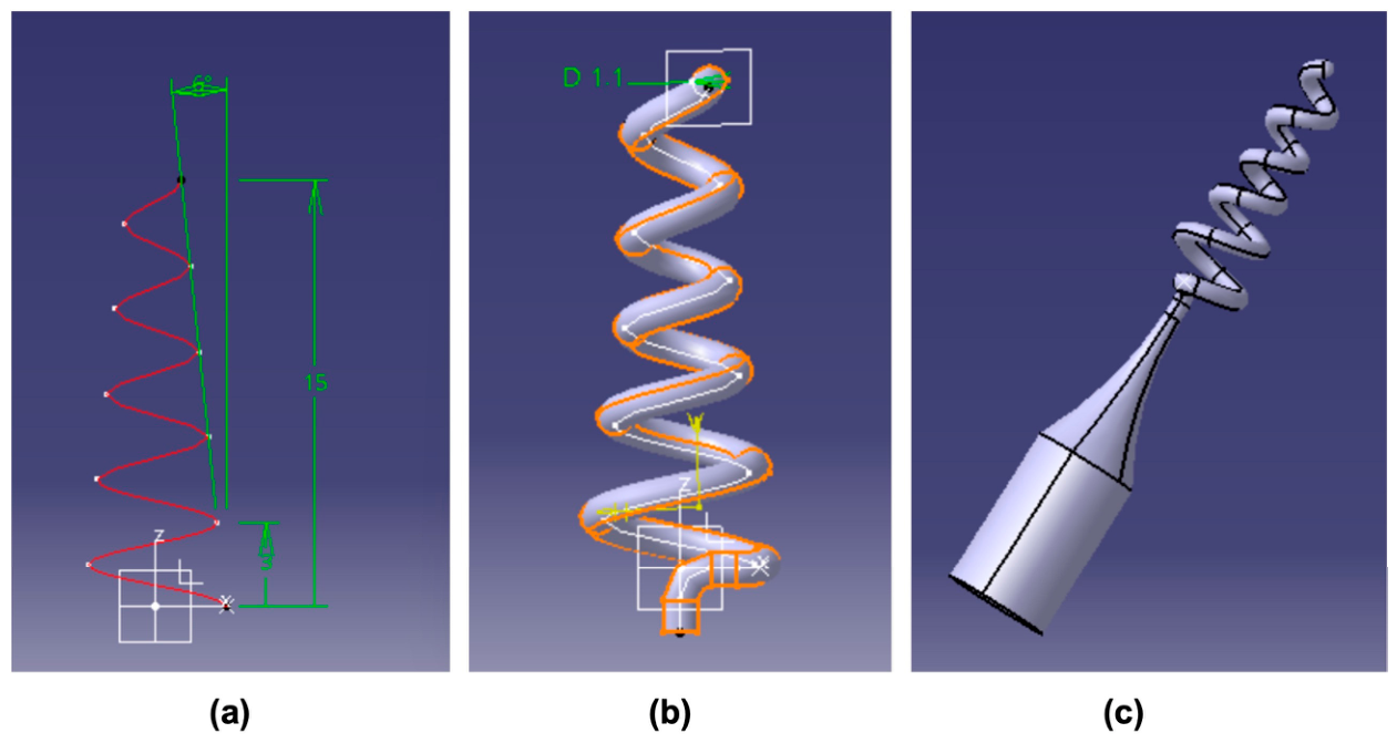
The helical structure was 3D-modeled using Catia V5R21 (Dassault Systemes, Vélizy-Villacoublay, France). The helix consisted of 5 coils along a total length of 15 mm, with a taper angle of 6° (
Figure 1a). The diameter at the tip was 3.6 mm [
4,
7,
10], while the helix section was circular with a diameter of 1.1 mm (
Figure 1b). The final 3D swab prototype model, represented in
Figure 1c, includes a cylindrical base support and has a total length of 40 mm.
2.3. 3D Printing and Post-Curing
The swab model (as shown in
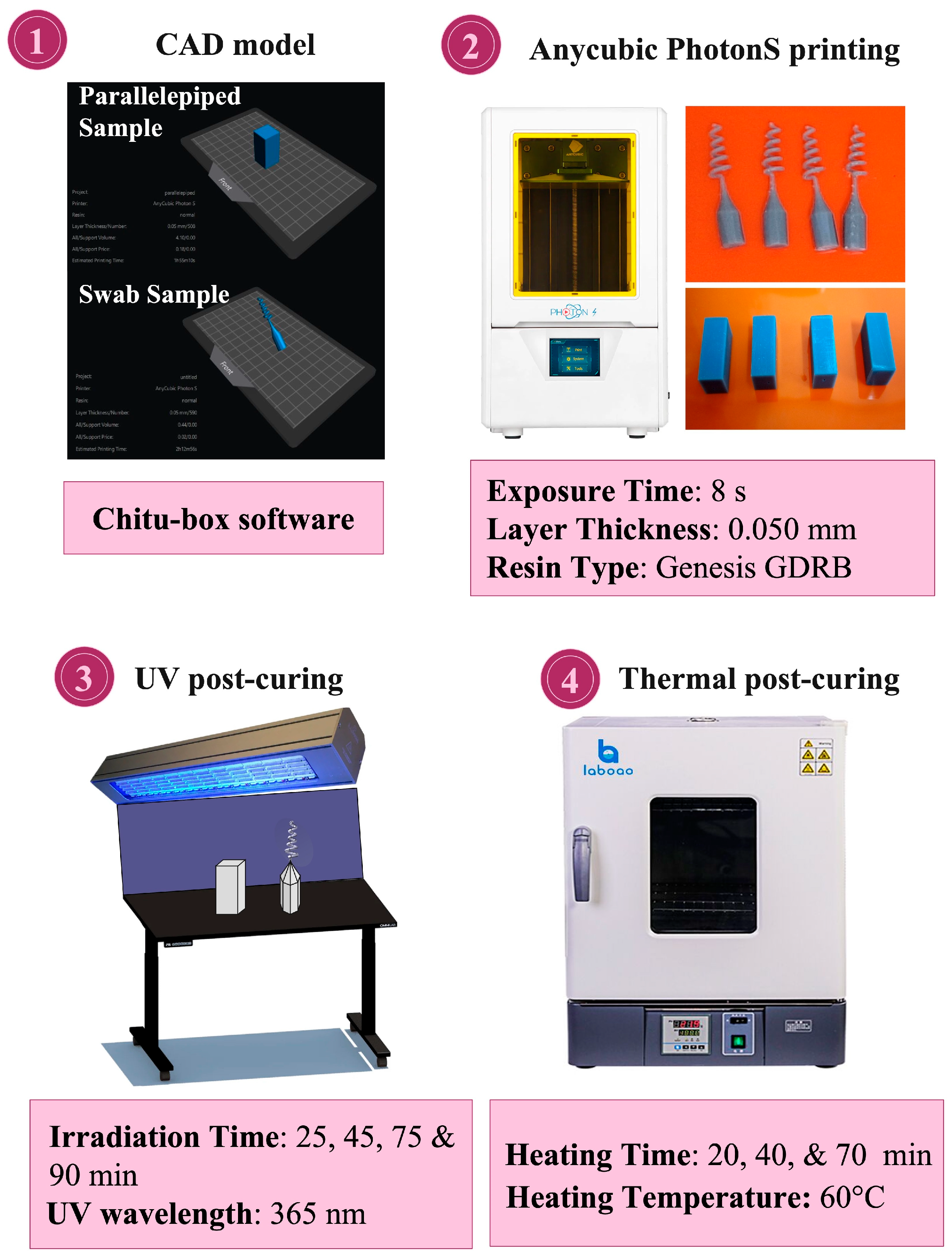
Figure 1) was prepared for 3D printing using Chitu-box software v1.2.0 for slicing and G-code creation in “Draft quality” mode. Swab prototypes were then 3D-printed using an Anycubic PhotonS SLA printer (Anycubic Technology Co., Ltd., Shenzhen, China) and commercial GDRB resin. Printing parameters were as follows: UV exposure time of 80 s for the bottom layer and 20 s for each subsequent layer, layer thickness of 0.050 mm, lift speed of 100 mm/min, and lift distance of 6 mm.
With the aim of evaluating the elastic modulus of the printed resin, additional parallelepiped specimens (12.7 × 12.7 × 25.4 mm3) suitable for compression tests were manufactured, adopting the same printing parameters cited above for the swab prototypes.
Following 3D printing, different post-curing treatments were performed to enhance the mechanical performance of the printed material, as described in the following:
UV-based post-curing via a Spectroline-MODEL ENF-280C/FE UV lamp (Spectronics Corporation, Melville, NY, USA) with a peak wavelength of 365 nm for different durations (0, 25, 45, 75, and 90 min) under ambient atmospheric conditions;
Dual post-curing, achieved by combining 90 min of UV post-curing, as described in point 1, with an additional thermal treatment at 60 °C for 20, 40, and 70 min.
For the sake of clarity, the different phases of 3D printing and the following post-curing treatments are schematized in
Figure 2.
2.4. Microcomputed Tomography
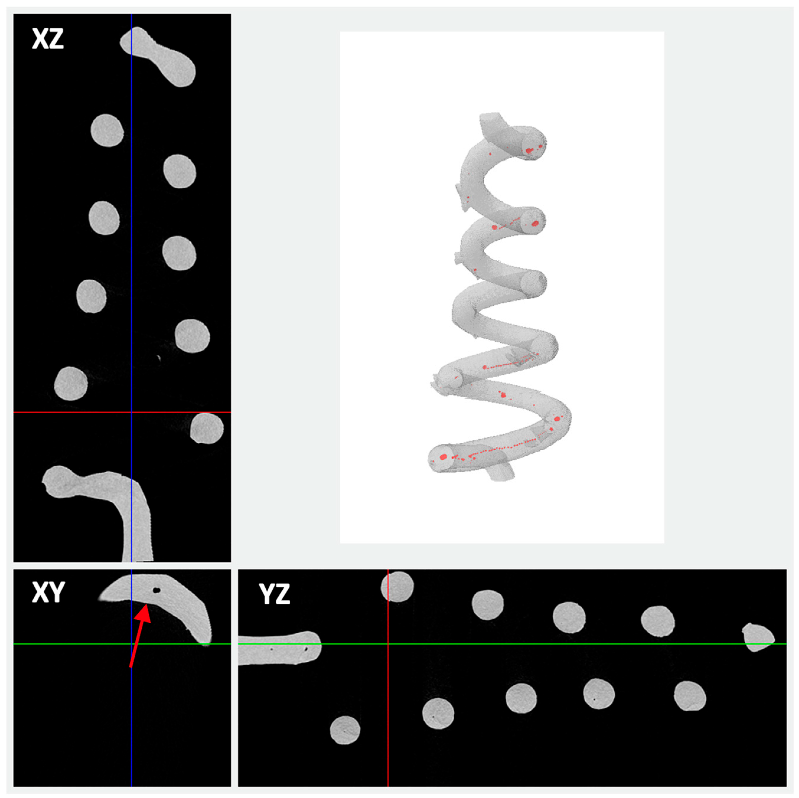
Microcomputed tomography (μCT) (Bruker Skyscan 1172, Kontich, Belgium) was used to analyze the structure of the 3D-printed swab prototypes and assess the accuracy of printing. The settings used for μCT imaging were voltage and anode currents of 25 kV and 140 μA, respectively; a pixel size of 13.4 µm; an exposure time of 1800 ms (with a 2 × 2 binning); and 180° of rotation with a rotation step of 0.8°.
Reconstructed cross-section images (obtained with NRecon, v1.6) were then analyzed by CTAn to evaluate the average helix thickness and quantitate any defects or voids in the 3D-printed helix. DataViewer was also used to visualize the 3D sections of the swab head in the XY, XZ, and YZ planes, while CTVol was used for 3D rendering.
2.5. DSC Analysis
Differential Scanning calorimetry (DSC) (Mettler Toledo, DSC1 STARe System, Greifensee, Switzerland) was used to study the effect of the dual post-curing process on the thermal properties of the 3D-printed resin. Dynamic DSC measurements were performed from 20 °C to 200 °C at a heating rate of 10 °C/min under N2 flow (60 mL/min) on samples of about 10 mg. Three replicates were analyzed for each dual-cured sample.
2.6. Compression Tests
To analyze the mechanical behavior of the 3D-printed samples, compressive tests were carried out by means of a universal testing machine (LR5K, Lloyd Instruments, Bognor Regis, UK) equipped with a 1 kN load cell.
The elastic modulus of the printed resin was preliminarily evaluated as a function of the different post-curing treatments to verify the ability of post-curing to increase the stiffness. Following the indications of ASTM D695-15, 3D-printed and post-cured parallelepiped specimens (12.7 × 12.7 × 25.4 mm3; n = 5) were subjected to compression at room temperature under displacement control with a crosshead speed of 1.3 mm/min; the test was stopped after reaching a maximum load of 800 N. The elastic modulus was then calculated as the slope of the linear portion of the stress–strain curve at low strain values (0–3%), with the exclusion of the ‘toe’ region if present.
Swab prototypes (n = 5) were then tested. The base of each prototype was fixed on the lower plate of the testing machine by means of bi-adhesive tape, while the helical head was compressed down to a maximum deflection of 10 mm by applying a crosshead speed of 1.3 mm/min. Each compression test was video-recorded using a Nikon D5300 camera to monitor and track the displacement of selected marked points on the helical structure.
2.7. Numerical Analysis
To investigate the mechanical behavior of the helical swab under compressive loading, an FE analysis was conducted using ANSYS Mechanical software vR2. The 3D swab geometry, designed in CATIA V5R21 and exported in STEP format, was imported into ANSYS Workbench and used to generate a structural model of the swab under axial compression. The primary objective of the simulation was to replicate the compression conditions applied during the experimental tests and to evaluate the deformation distribution, stress localization, and structural safety of the swab head. A linear elastic material model was adopted, with the elastic modulus derived from the dual-cured samples. The base of the swab was fully constrained to simulate fixation to the testing machine, while an imposed compressive velocity of 1.3 mm/min was applied axially to the upper surface of the helical head.
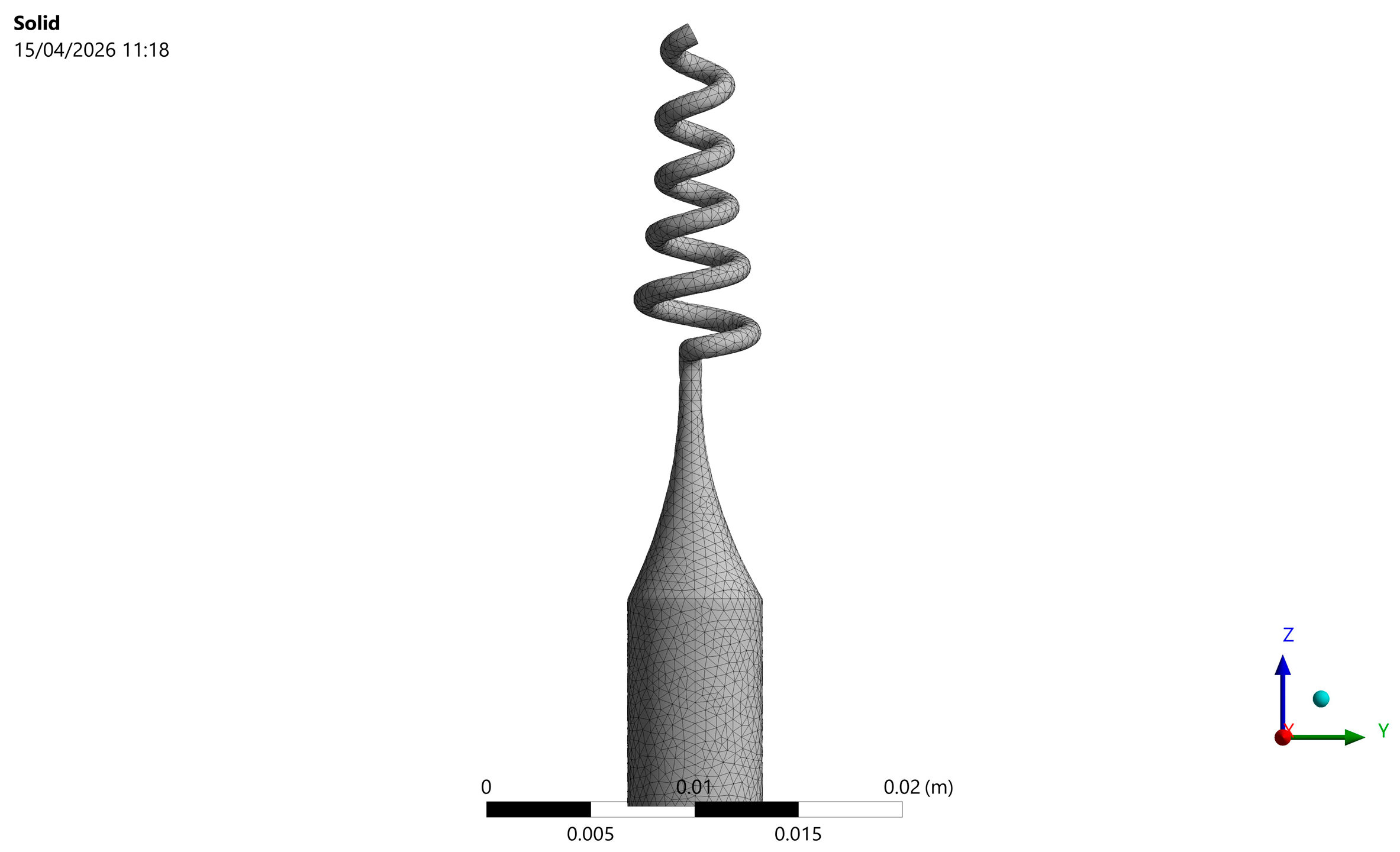
Given the complex geometry, the model was meshed using second-order tetrahedral elements (SOLID186). A mesh convergence study was performed to ensure the reliability and accuracy of the numerical results. Starting from a coarse discretization, the mesh density was progressively refined, particularly in the helical region, where higher stress gradients were expected. For each refinement level, key output parameters such as maximum displacement and von Mises stress were monitored. Convergence was assumed when the variation of these quantities between successive mesh refinements fell below 5%. The selected mesh configuration (
Figure 3), consisting of approximately 44,000 elements and 104,000 nodes, represents an optimal compromise between computational cost and solution accuracy, as further refinement produced negligible changes in the results. This procedure allowed us to confirm that the numerical predictions were not significantly affected by mesh dependency.
Contact interactions between coils and coils with the simulated compressive crossbar were set, assuming large radial deformations, and the entire swab structure was treated as a single continuous solid. Transient structural analysis was performed to capture progressive deformation under the applied load.
2.8. Bacterial Detection
P. aeruginosa PAO1 [
22,
23] and
S. aureus SA01 [
24,
25] were selected to evaluate the efficacy of the 3D-printed prototype as a bacteriological swab. Both bacterial strains were cultured in Luria–Bertani (LB) broth containing 1% sodium chloride (NaCl), 1% tryptone, and 0.5% yeast extract and supplemented with 1.5% Agar for LB solid medium. All media were sterilized by autoclaving at 121 °C for 20 min. Regarding the sterilization of the swab prototypes, autoclaving and UV irradiation were preliminarily tested. While autoclaving was found to compromise the swab geometry, UV irradiation for 20 min successfully sterilized the prototypes and preserved their structural integrity. Effective swab sterilization was verified by streaking the UV-treated prototypes onto solid media, followed by incubation at 37 °C for 24 h; no visible colony growth was observed, confirming sterility.
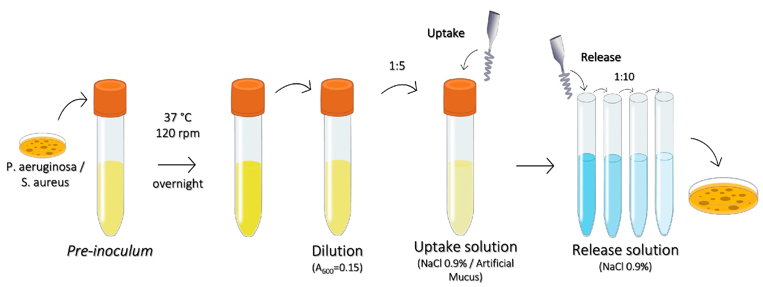
The protocol used to assess the uptake and release capacity of the swab prototypes, sketched in
Figure 4, was adapted from the M40-A2 standard, “Quality Control of Microbiological Transport Systems; Approved Standard—Second Edition” (CLSI. Quality Control of Microbiological Transport Systems; Approved Standard—Second Edition; CLSI Document M40-A2; Clinical and Laboratory Standards Institute: Wayne, PA, USA, 2014) [
26]. Two test solutions were used: i. physiological saline (0.9% NaCl) and ii. artificial mucus prepared as a 15% PVA solution with a viscosity of 200 mPa·s [
12]. Bacterial pre-inoculum was prepared by suspending an isolated colony from a fresh agar plate (24–72 h) in LB broth, followed by incubation overnight at 37 °C with stirring at 120 rpm. The next day, the bacterial suspension was adjusted to an optical density (O.D.) of 0.150 at 600 nm and further diluted to 1:5 in either physiological saline or artificial mucus, obtaining the bacterial inoculum (approximately 10
7 CFU/mL) [
26]. For testing, the swab prototypes were immersed in 500 μL of bacterial inoculum for 15 s (uptake phase), then transferred to 1 mL of 0.9% NaCl solution for 15 s (release phase), followed by the preparation of 10-fold serial dilutions. Two strategies were evaluated: i. static uptake/release and ii. twisting uptake/release. Commercial flocked and cotton swabs served as reference standards. The experiments were conducted in triplicate.
2.9. Statistical Analysis
Data are presented as mean ± Standard Deviation (SD) for the indicated number of experiments. Where appropriate, statistical analysis was conducted using one-way ANOVA. In all comparisons, p < 0.05 was considered statistically significant.
3. Results and Discussion
3.1. 3D Printing of the Swab Prototype
Among 3D printing techniques, SLA was selected due to its well-known ability to manufacture objects with high-resolution, smooth-surfaced structures. Several studies have reported the successful SLA printing of swab devices with various tip designs and flexible shafts for improved diagnostic accuracy [
6,
7,
12]. In this work, by focusing on a helical tip design to potentially improve and control the elution of biological specimens, we chose SLA technology to manufacture proof-of-principle prototypes to be used for further testing.
The accuracy of SLA in reproducing the designed helical head with the adopted printing parameters was assessed by means of
μCT imaging. The quantitative analysis performed on the
μCT images showed that the average thickness of the printed helix was 1.01 ± 0.25 mm, which was very close to the designed helix section diameter (1.1 mm). While this finding suggested the good geometric accuracy of 3D printing, the sections of the swab head and the 3D rendering (
Figure 5) also revealed the presence of a few close, spherical voids inside the helix structure, representative of printing defects. However, this closed porosity accounted for only 0.12% of the total volume of the helix (with most pores located in the lower coils); thus, its effect on the mechanical performance of the swab prototype was negligible. As most pores were found to have a diameter of about 0.050 mm, we also assumed that these defects could be related to possible air inclusions during the movement of the building platform (as the thickness of the printing layer was set to 0.050 mm).
As regards the layer thickness, it is worth noting that the value used in this study is quite a common trade-off value used in SLA printing and lies within the range of 0.050–0.100 mm reported for SLA-printed swabs [
4,
6,
7]. In general, the layer thickness plays a key role in determining the resolution and surface quality of the printed objects while also affecting the printing duration. Smaller layer thicknesses increase the printing time but lead to high-resolution prints, providing objects with smoother, less ‘stepped’ surfaces. However, an insufficient layer thickness may lead to excessive UV exposure times, resulting in object warping. Interestingly, improved mechanical strength has also been reported as the layer thickness is decreased, likely due to enhanced interlayer bonding and a reduced incidence of printing defects [
27]. In this regard, the
μCT analysis performed in this study confirmed that very few printing defects are obtained for a layer thickness as low as 0.050 mm.
3.2. Efficacy of the Dual Post-Curing Treatment
As is well known, post-curing processes are commonly adopted to improve the mechanical properties of SLA-printed materials in order to ensure better performance and durability while also reducing the content of residual unreacted species, which may impact biocompatibility. In this study, UV-based post-curing was first used to enhance the crosslinking density and the extent of polymer network formation. During UV post-curing, additional photon exposure is expected to activate residual photo-initiator and unreacted functional groups, which leads to a progressive increase in the crosslink density. However, due to light attenuation and the limited mobility of reactive species in the partially solidified network, UV curing alone may not ensure complete conversion.
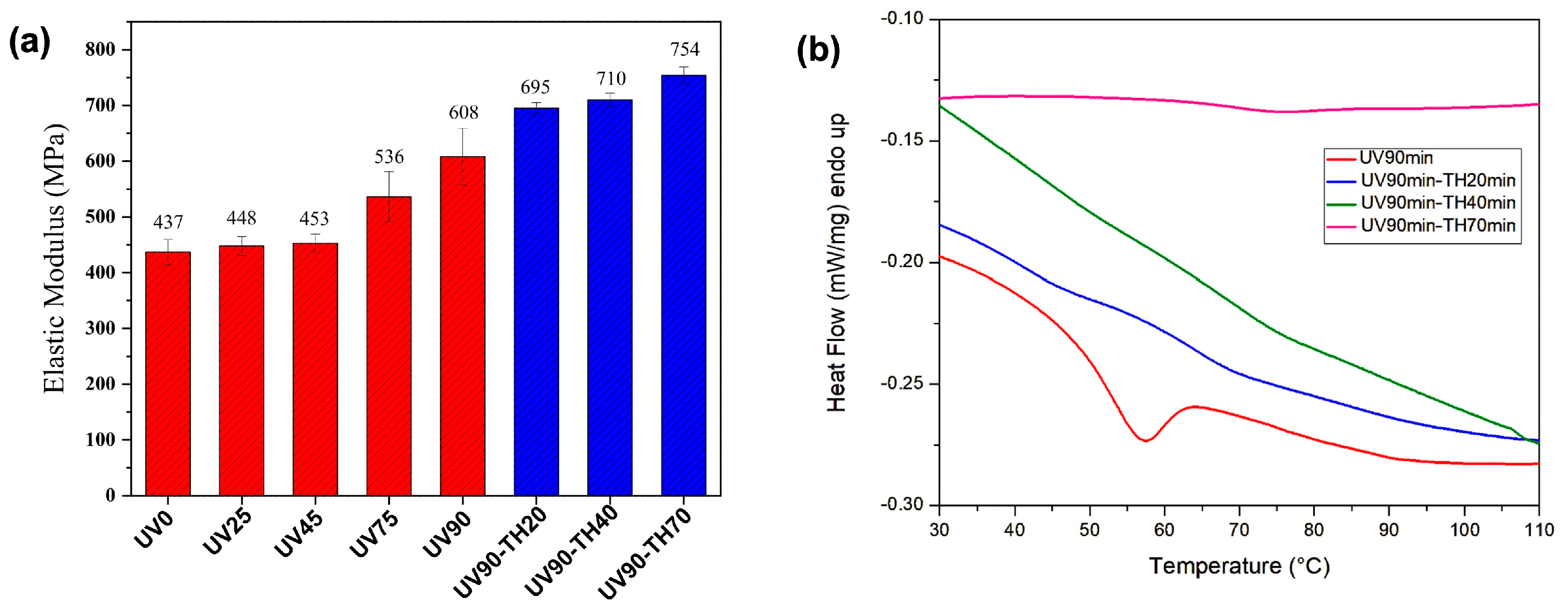
From a structure–property perspective, the increase in crosslink density reduces the chain mobility, thereby leading to higher stiffness values. As expected, compression tests on UV-cured parallelepiped specimens showed a progressive increase in the elastic modulus with increasing UV post-curing times (
Figure 6a, red bars). Samples cured only in the SLA printer, without any additional UV post-curing (UV0), exhibited the lowest modulus of 437 ± 23 MPa, which suggests incomplete crosslinking. Moderate UV exposure (25 and 45 min for UV25 and UV45 samples) showed only minimal improvements in stiffness, while significant increases were observed for UV exposures of 75 min (UV75) and 90 min (UV90), yielding elastic moduli of 536 ± 45 MPa and 608 ± 51 MPa, respectively. This confirmed the benefits of extended UV post-curing in terms of optimizing the mechanical properties of the printed resin.
However, further DSC measurements on UV90 samples showed residual heat from the reaction (
Figure 6b,
Table 1), highlighting that UV post-curing alone was insufficient to achieve complete conversion of the SLA resin.
Based on the glass transition temperature of UV90 samples (52.4 ± 1.2 °C), subsequent thermal post-curing at 60 °C was adopted to enhance the molecular mobility, enabling the further reaction of functional groups to achieve a more densely crosslinked polymer network. The results of both DSC analysis and mechanical tests performed on the dual-cured samples were consistent with what was expected, as longer durations of thermal treatment (20, 40 and 70 min) were found to induce a progressive decrease in the residual heat from the reaction while increasing both the glass transition temperature and the elastic modulus (
Figure 6a, blue bars). In particular, the highest elastic modulus (754 ± 15 MPa) was obtained for a thermal treatment duration of 70 min, which also led to a null value of the residual heat of the reaction. This dual-curing treatment (UV90-TH70) was then selected to increase the mechanical properties of the swab prototypes.
When considering the swab stiffness, a wide range of elastic moduli have been documented in the literature for different swab types, materials, and designs, with a special focus on the shaft. As an example, 3D-printed swabs based on a medical-grade resin have been reported to have a modulus of about 3.5 GPa, which is quite comparable to the modulus of commercial swabs (about 4.4 GPa) [
7]. Conversely, another study showed that polypropylene-based swabs, with proper flexibility and mechanical strength for clinical use, have an elastic modulus of about 400 MPa [
16]. Thus, the stiffness value obtained in this study for the fully dual-cured samples is roughly comparable to that of common plastic-based materials used for swab production.
3.3. Swab Prototype Compression: Correlation Between Numerical and Experimental Data
Compression tests on the helical swab prototypes showed that the upper coils underwent more crushing than the lower ones. However, the specimens were able to fully recover their original shape upon unloading, indicating that the material response remained within the elastic regime and no permanent deformation occurred. The tests were video-recorded by means of a Nikon D5300 camera to track the coil displacement under compression. The acquired videos were elaborated by means of Tracker 6.2.0, a free video analysis and modeling tool built on the Open-Source Physics (OSP) Java framework. This tool enables the following of the positions of selected marked points during the test (
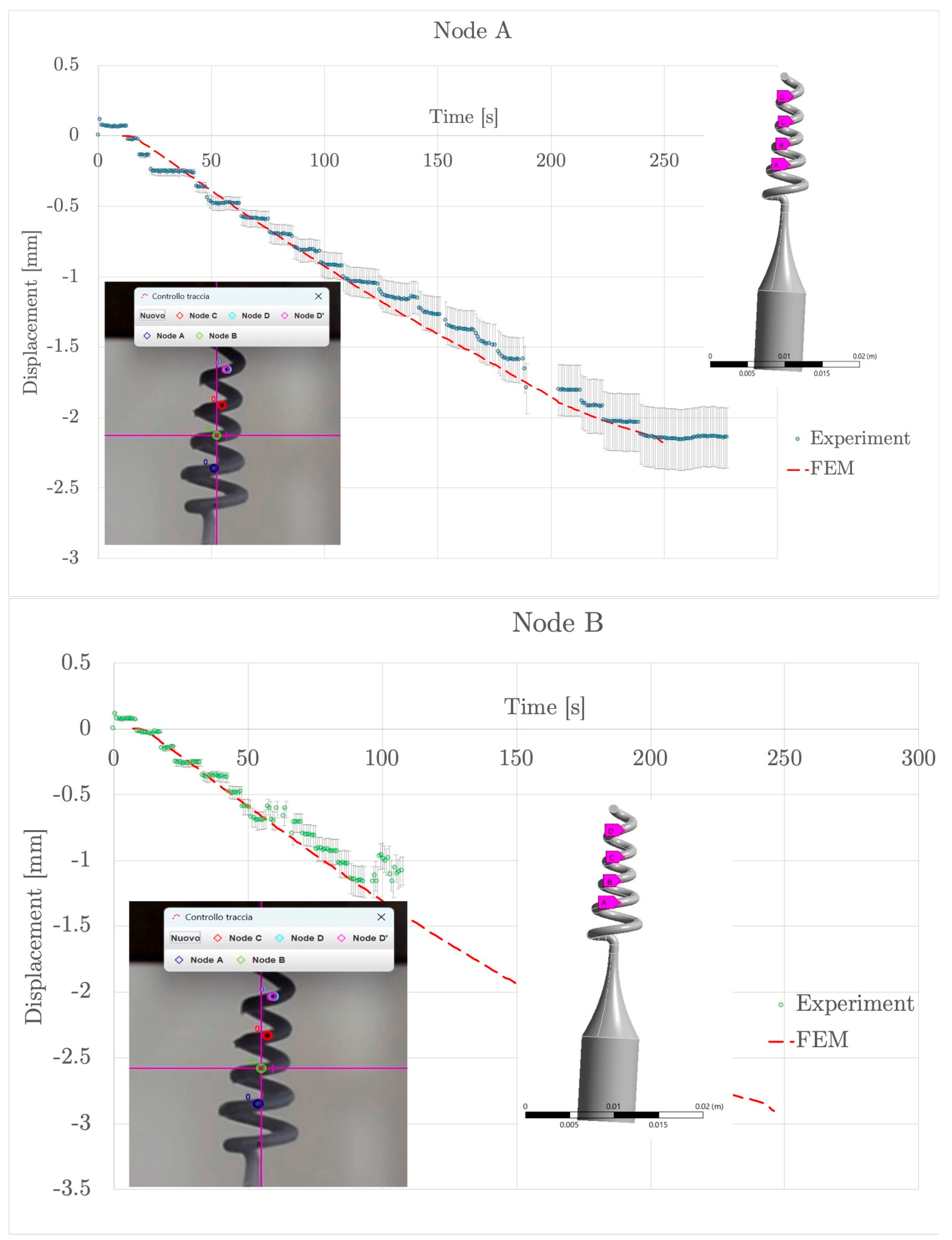
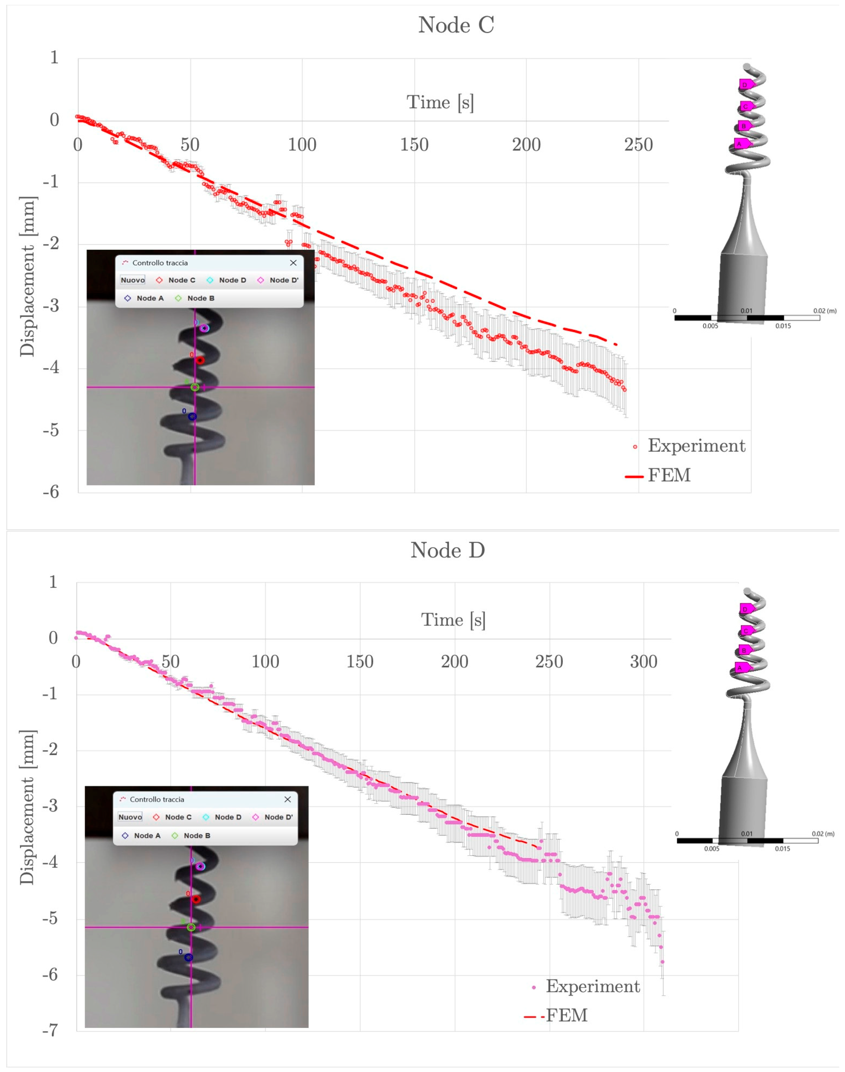
Supplementary Video S1). Selected points, indicated as Nodes A, B, C and D from the bottom to the top of the swab helix, are shown in
Figure 7, together with the plots illustrating their experimental displacement during the axial compression test. Experimental data are denoted with circles and ±10% of error bars, while FE-simulated data are represented by the red dashed lines. In each subfigure, the inset on the left shows the experimental video frame with the tracked nodes (A to D) superimposed. The image on the right offers a 3D model of the swab and tracked nodes, highlighting their spatial distribution.
A quantitative comparison between experimental and numerical results is provided in
Table 2 in terms of the RMSE for each tracked node. The RMSE values were found to progressively increase from Node A to Node D. A similar trend was also observed for the final displacements, which increased from approximately 2 mm for Node A to about 4 mm for Node D. However, all RMSE values remained relatively low, indicating good agreement between the numerical model and the experimental measurements and supporting the reliability of the proposed FE approach in capturing the deformation behavior of the helical swab prototype.
It is also worth noting that, despite some noise and stepwise progression due to frame-sampling resolution, the displacement trend for each node (
Figure 7) indicated stable and continuous deformation, without abrupt discontinuities or structural failure. Only after about 200 s did the trend exhibit non-linear behavior with local fluctuations, likely due to the intrinsic flexibility of the top coils, which could bend and rotate slightly out of the image plane during compression (inducing coil–coil and coil–crossbar contact). Thus, increasing discrepancies between FE predictions and experimental data were ascribed to the progressive onset of geometric nonlinearities. However, the deviation between numerical and experimental displacement remained within approximately ±10–15% for the tracked nodes, in agreement with the experimental uncertainty (including tracking resolution and out-of-plane motion not captured in the 2D analysis). Despite these local deviations, the model could correctly capture the overall deformation pattern and relative displacement distribution along the helix.
The results also confirmed that the lower region of the helical swab could undergo limited deformations during the test, consistent with both the numerical model predictions and the intended design: the upper coils compressed more significantly, while the base region acted as a stable support. This behavior could ensure controlled elastic deformation under load, which is expected to contribute to the safe functionality of the swab upon collection and elution of biological samples.
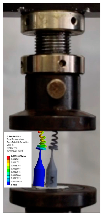
Figure 8 presents a combined visualization of the experimental and numerical analysis for the swab prototype under axial compression (further detailed in
Supplementary Video S2). In the background, the image shows the actual setup of the testing machine, where the swab is positioned between two compression plates. The superimposed simulation result displays the deformation distribution obtained from the FE analysis in ANSYS. The color scale indicates that maximum deformation occurred at the inner curvature of the top coils, where the material underwent the highest bending due to axial deformation. The lower body of the swab remained largely undeformed, confirming the structural isolation of the deformation zone.
The good correlation between the experimentally observed deformation pattern and the numerically predicted one validates the design concept and confirms the effectiveness of the helical geometry in managing applied loads.
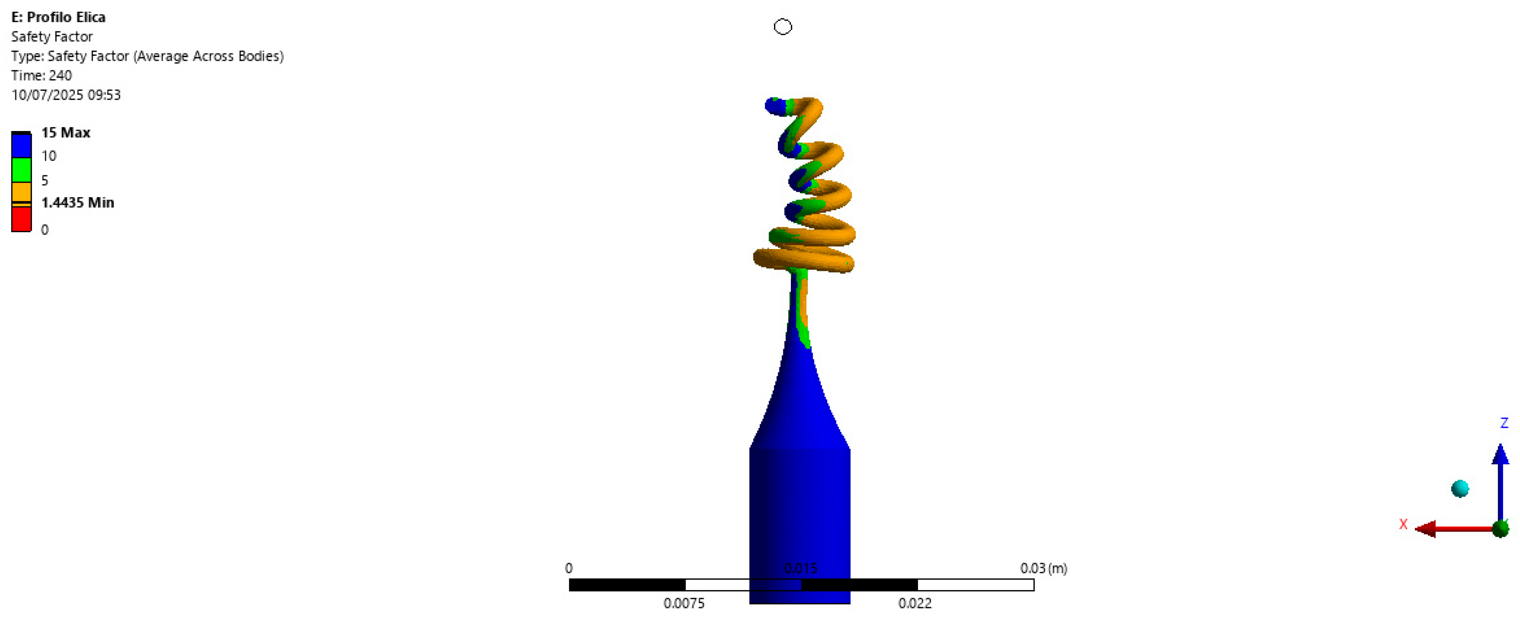
Figure 9 shows the spatial distribution of the safety factor across the swab geometry during the compression simulation. Values are visualized using a color scale, where blue represents highly safe zones (safety factor > 10) while red indicates areas approaching structural limits. For the calculation of the safety factor, defined as the ratio between the material failure strength and the local von Mises stress, the failure strength of the used resin (38 MPa—derived from the material datasheet) was assumed as a reference value.
The results confirmed that the entire swab structure operated within safe stress margins, even under maximum operative deformation. The lowest safety factors were concentrated in the upper coils, with the most significant mechanical stress during compression. This localized reduction was fully expected and intentional, as the swab was designed to deform in this region to enable and facilitate sample release. Notably, no regions fell below the critical safety threshold of 1, indicating no risk of failure or permanent deformation. The base of the swab exhibited very high safety factors, ensuring mechanical stability and secure handling during use. These results, in agreement with the von Mises stress distribution, demonstrated the structural integrity of the prototype under compressive loading. The safety factor analysis confirmed the robustness and reliability of the proposed swab design, further validating the effectiveness of the helical tip geometry for potential clinical applications.
3.4. Functionality of the 3D-Printed Prototype as a Bacteriological Swab
As a preliminary assessment of the functionality of the helical tip, the uptake and release performance of the swab prototypes was evaluated in vitro using
P. aeruginosa (Gram-negative) and
S. aureus (Gram-positive), two pathogenic bacteria commonly associated with respiratory tract diseases and skin infections [
28,
29]. A quantitative protocol was used to compare the prototype’s functionality with that of commercial flocked and cotton swabs. Bacterial uptake was tested using a fixed bacterial load (approximately 10
7 CFU/mL for both bacterial species) in two different solutions: i. physiological saline solution and ii. artificial mucus, simulating the physiological collection in the nasopharynx. Release performance was assessed immediately after inoculation, comparing the prototype with standard flocked and cotton swabs.
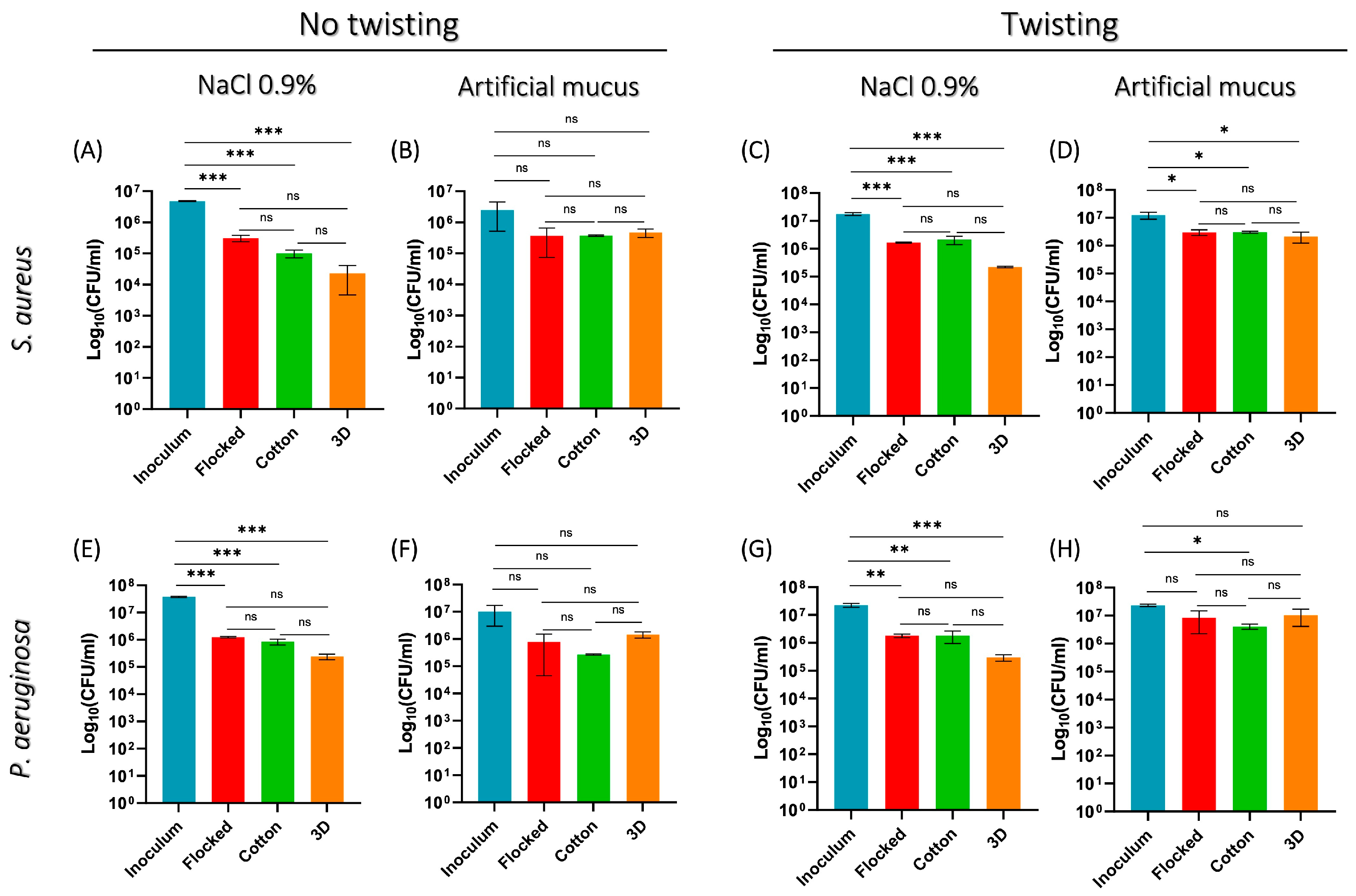
Without twisting in the uptake and release phases, the swab prototype achieved effective collection of both
S. aureus and
P. aeruginosa, showing a 2-log reduction in bacterial load in NaCl solution compared to the inoculum but performing comparably to standard flocked and cotton swabs (
Figure 10A,E). Notably, the best performance was achieved using artificial mucus as uptake solution: in these conditions, the swab prototype collected bacterial loads closely matching the initial inoculum while still performing comparably to commercial swabs (
Figure 10B,F).
Additional tests with twisting during both uptake and release were performed. As reported in
Figure 10, the twisting movement did not improve the swabs’ performance, leading to a trend similar to that observed in the absence of twisting for both physiological saline solution (
Figure 10C,G) and artificial mucus (
Figure 10D,H). The twisting or rotation of a swab is commonly adopted to increase uptake and improve the release of collected bacteria compared to static immersion. Therefore, the absence of a twisting benefit, for all swab types (either fibrous, flocked, or helical) suggests that under the used testing conditions, sample absorption and recovery were mostly due to the physical properties of the swab heads and/or the sampling solutions (e.g., viscosity and bacterial concentration) rather than additional mechanical effects.
While these findings deserve further exploration, it should be noted that the 3D-printed prototypes were tested as manufactured, i.e., without applying any absorbent tip coating to enhance sample uptake or using a compressive load to promote sample release (compression was not applied to allow for a direct comparison with commercial swabs). Thus, the achieved results are encouraging, suggesting the potential of the helical tip design for bacterial capture and detection.
3.5. Limitations of the Study and Future Research
This study has several limitations that should be acknowledged. First, its primary objective was to verify the feasibility of the designed NP swab prototypes by assessing the performance of the helical geometry rather than producing functional swabs directly implementable in clinical practice. Within this scope, the material used to produce the prototypes (i.e., GDBR resin) was chosen based on its ability to achieve high-resolution prints, without addressing the requirements of biocompatibility and sterilizability needed for clinical applications. Several medical-grade or biocompatible resins available for SLA printing (e.g., surgical guide resin) have already been adopted in the literature for 3D-printed swabs [
7,
12]; thus, their future use for the production of helical prototypes is envisaged. Additional strategies that could be explored to ensure the safety of the used material(s) may include the utilization of porous or fibrous biocompatible coatings on the swab-tip surface, which could also contribute to uptake and release efficiency, as well as improved patient comfort.
Second, the linear elastic constitutive law used here for numerical modeling should be considered valid in the initial part of the compression test, up to the onset of significant geometric nonlinearities (up to ~200 s, i.e., before pronounced coil interaction and out-of-plane effects become dominant). While a fully nonlinear approach could further improve the correlation between experimental data and numerical predictions, the adoption of a hyperelastic constitutive law was not expected to significantly affect the results, as the material response was experimentally found to remain within the linear elastic regime. Moreover, the objective of the model was to reproduce the global deformation behavior and stress distribution under load of the prototype rather than achieving exact local kinematic matching under large deformation. Therefore, the adopted modeling strategy appears to be suitable to provide a reasonable and physically consistent approximation.
Third, while the elastic modulus of the printed resin was experimentally evaluated, a direct measurement of the ultimate strength of the dual-cured material was not performed. Thus, the failure strength of the base resin used in this study for safety-factor calculation should be regarded as a conservative assumption, and the resulting safety factor should be interpreted accordingly. In this regard, future work should include the use of medical-grade materials for prototype production, as well as dedicated experimental characterization of the ultimate strength after post-curing to refine the mechanical safety assessment.
Finally, a limited sample size (n = 3) was used to evaluate and compare the bacterial uptake and release ability of the swab prototypes over commercial swabs. While this restricts the statistical significance of the findings, such data provide preliminary evidence of the biological functionality of the helical design, representing the basis for a more accurate biological investigation upon prototype optimization (e.g., by using medical-grade material).