
Physical Exercise and Gut Microbiota: Implications for Alzheimer’s Disease in Experimental Models: A Systematic Review and Meta-Analysis

 , , , ,
  , and
, and
Abstract
1. Introduction
2. Materials and Methods
2.1. Databases and Search Strategy
2.2. Selection Criteria
2.3. Data Extraction
2.4. Risk of Bias and Methodological Quality Assessment
2.5. Statistical Analysis
3. Results
3.1. Studies Characteristics
3.1.1. Risk of Bias Assessment
3.1.2. Methodological Quality Assessment

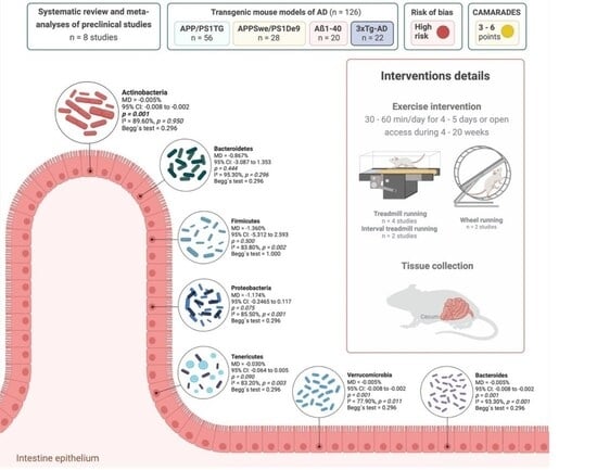
| First Author (Year) | Country | Animal Characteristics | Experimental Groups (n) | PE Protocol | Main Results Related to GM | Main Results Related to AD Biomarkers |
|---|---|---|---|---|---|---|
| Abraham et al. (2019) [56] | Hungary | Sample size: 32 Age: 3 months Sex: 100% male Experimental model: APP/PS1 transgenic mice (B6C3-Tg (APPswe, PSEN1dE9)85Dbo/Mmjax; APP/PS1TG) | EG (n = 8) CG (n = 8) | Modality: Interval treadmill running Frequency: Four times a week Volume: Ten cycles of 6 min (60 min/day) Intensity: Each cycle consisting of four minutes at high intensity (20 m/min) and two minutes at low intensity (10 m/min) Duration: 20 weeks | ↓↓↓ Lactobacillus johnsonii and reuteri ↑↑ Butyrivibrio proteoclasticus and Roseburia spp. ↑ Butyrate producing bacteria ↓ Bacteroides fragilis | ↓↓ Mean number of plaques in hippocampus and mean % of the areas covered by plaques ↑↑ OGG1 levels and occupied area of OGG1 |
| Jin et al. (2023) [32] | Korea | Sample size: 33 Age: 3 months Sex: 100% male Experimental model: 3xTg-AD | EG (n = 11) CG (n = 11) | Modality: Treadmill running Frequency: Five times a week Volume: 30 min/day Intensity: Each session started with a 5 min warm-up at a speed of 5 m/min, increased to 8 m/min for 5 min, and reached a maximum speed of 11 m/min for 20 min. The maximum speed was then increased by 1 m/min every 4 weeks up to a speed of 15 m/min Duration: 18 weeks | ↑↑↑ Akkermansia muciniphila ↑↑ Bacteroides rodentium ↑↑ Claudin and ZO1 ↓↓ Clostridium amazonense, Alistipes putredinis and Turicibacter sanguinis = Simpson index | ↑↑ Time in the target quadrant (MWM testing) ↑↑ ADAM10, hippocampal SOD2 and CAT proteins, Bcl2, hippocampal PSD95, synaptophysin and BDNF ↓↓ Escape latency (MWM testing) ↓↓ Aβ plaque counts in the CA1 and CA3 regions of the hippocampus, APP, BACE1, p-tau205, p-tau231, and total tau in the hippocampus, GFAP-positive cells, TMEM119-positive cells, lower hippocampal caspase 3, caspase 9, and BAX ↑ LRP1 and GLUT1 ↓ IGF1 |
| Rosa et al. (2020) [61] | Brazil | Sample size: 40 Age: 44–55 days Sex: 100% male Experimental model: Swiss mice inducted to be Aβ1–40 model | EG (n = 10) CG (n = 10) | Modality: Treadmill running Frequency: Five times a week Volume: 40 min/day Intensity: The treadmill speed was progressively increased by 2 m/min/week, starting at 6 m/min at the beginning of the protocol Duration: 4 weeks | ↑↑↑ Firmicutes ↓↓↓ Bacteroidetes ↓↓↓ Bacteroidetes/Firmicutes ratio | NR |
| Teglás et al. (2020) [57] | Hungary | Sample size: 30 Age: 3 months Sex: 100% male Experimental model: APP/PS1 transgenic mice (B6C3-Tg (APPswe, PSEN1dE9) 85Dbo/Mmjax; APP/PS1 | EG (n = 6) CG (n = 6) | Modality: Interval treadmill running Frequency: Four times a week Volume: Ten cycles of 6 min (60 min/day) Intensity: four minutes at 20 m/min and two minutes at 10 m/min Duration: 20 weeks | ≠≠ GM composition | NR |
| Wang et al. (2021) [58] | China | Sample size: 14 Age: 9 weeks Sex: 100% male Experimental model: APPSwe/PS1De9 (APP/PS1), (B6C3F1 background, APPswe strain) | EG (n = 7) CG (n = 7) | Modality: Voluntary wheel running Frequency: Undetermined Volume: Undetermined Intensity: Undetermined Duration: 16 weeks | ↑↑ ACE and Chao1 index ↑ Oscillibacter ↑ EU454870_g, Oscillibacter, EU504554_g, EU504066_g, EF097061_g, EU505046_g and EF096172_g ↓ Alistipes | ↓↓ GFAP and Ibα1 in cortex ↓↓ Phosphorylation of tau at ser396 and ser404 (p-Tauser396&404) ↑↑ p-GSK3β _at ser9 ↑ ADAM10, synaptophysin and BDNF protein expression and p-AMPK ↓ GFAP and Ibα1 protein expression in hippocampus |
| Sample size: 14 Age: 24 weeks Sex: 100% male Experimental model: APPSwe/PS1De9 (APP/PS1), (B6C3F1 background, APPswe strain) | EG (n = 7) CG (n = 7) | ↓ Proteobacteria and Tenericutes ↓ ↓ Bacteroides and Faecalibacterium ↑ Allobaculum, ≠≠ β diversity | ↓↓ ADAM10, BACE1 and GFAP ↑ Synaptophysin protein in cortex = p-Tauser396&404, PP2A, p-GSK3β _ser9, CDK5 and p-AMPK | |||
| Wei et al. (2025) [33] | China | Sample size: 48 Age: 3 months Sex: Not specified Experimental model: APP/PS1 | EG (n = 12) CG (n = 12) | Modality: Treadmill running Frequency: Five times a week Volume: 30 min/day Intensity: The protocol started at a speed of 12 m/min for 10 min and progressed to 15 m/min for 20 min. Each session started with a 5 min warm-up at a speed of 5 m/min, increased to 8 m/min for 5 min, and reached a maximum speed of 11 m/min for 20 min. The maximum speed was then increased by 1 m/min every 4 weeks up to a speed of 15 m/min Duration: 20 weeks | ↑↑ Actinomycota ↓↓ Bacteroidetes = Psychrobacter ↑ Ileibacterium and Faecalibaculum | NR |
| Yuan et al. (2022) [59] | China | Sample size: 48 Age: 3 months Sex: 100% male Experimental model: SPF-grade APP/PS1 | EG (n = 12) CG (n = 12) | Modality: Treadmill exercise scheme with gradual increases in exercise intensity Frequency: Five times a week Volume: 45 min/day Intensity: The speed was incrementally increased from 7 to 14 m/min during the first 8 weeks, with a constant speed of 15 m/min for weeks 9–12 with a load intensity of 60% to 73% of the maximum oxygen uptake representing a moderate-intensity aerobic exercise Duration: 12 weeks | ↑ ACE, Chao, Shannon, and Simpson indices ↑ Akkermansia ≠≠ Intestinal microbial composition | ↓↓↓ Aβ aggregation ↑↑ Area of hippocampal and cortical niosomes ↓ LPS |
| Zhang et al. (2023) [60] | China | Sample size: 22 Age: 10 weeks Sex: 100% male Experimental model: APP/PS1 | EG (n = NS) CG (n = NS) | Modality: Voluntary wheel running Frequency: Undetermined Volume: Undetermined Intensity: Undetermined Duration: 12 weeks | ↑↑ ACE and Chao1 index ↑↑ Bacteroidetes, Bacteroidales and Bifidobacteriales = Shannon and Simpson index | NR |
3.2. Quantitative Synthesis
4. Discussion
5. Conclusions
Supplementary Materials
Author Contributions
Funding
Institutional Review Board Statement
Informed Consent Statement
Data Availability Statement
Acknowledgments
Conflicts of Interest
Abbreviations
| Aβ | Amyloid-β |
| AD | Alzheimer’s disease |
| BDNF | Brain-derived neurotrophic factor |
| BBB | Blood–brain barrier |
| CNS | Central nervous system |
| GM | Gut microbiota |
| pCT | Preclinical controlled trials |
| PE | Physical exercise |
| RoB | Risk of bias |
| SCFA | Short-chain fatty acids |
| SYRCLE | SYstematic Review Center for Laboratory Animal Experimentation |
Appendix A
| D1 | D2 | D3 | D4 | D5 | D6 | D7 | D8 | D9 | D10 | Overall | |
|---|---|---|---|---|---|---|---|---|---|---|---|
| Abraham et al. (2019) [56] | UC | UC | H | H | H | H | H | H | L | UC | H |
| Jin et al. (2023) [32] | UC | UC | H | H | H | H | H | H | L | UC | H |
| Rosa et al. (2020) [61] | UC | UC | H | L | H | H | UC | H | L | UC | H |
| Teglás et al. (2020) [57] | UC | UC | H | UC | H | H | H | H | L | UC | H |
| Wang et al. (2021) [58] | UC | UC | H | UC | H | UC | H | H | L | UC | H |
| Wei et al. (2025) [33] | UC | UC | H | L | H | H | H | H | L | UC | H |
| Yuan et al. (2022) [59] | UC | UC | H | L | H | H | L | H | L | UC | H |
| Zhang et al. (2023) [60] | UC | UC | H | L | H | H | H | H | L | UC | H |
References
- Scheltens, P.; De Strooper, B.; Kivipelto, M.; Holstege, H.; Chetelat, G.; Teunissen, C.E.; Cummings, J.; van der Flier, W.M. Alzheimer’s disease. Lancet 2021, 397, 1577–1590. [Google Scholar] [CrossRef] [Scilit] [PubMed]
- Guo, T.; Zhang, D.; Zeng, Y.; Huang, T.Y.; Xu, H.; Zhao, Y. Molecular and cellular mechanisms underlying the pathogenesis of Alzheimer’s disease. Mol. Neurodegener. 2020, 15, 40. [Google Scholar] [CrossRef] [Scilit] [PubMed]
- Long, J.M.; Holtzman, D.M. Alzheimer Disease: An Update on Pathobiology and Treatment Strategies. Cell 2019, 179, 312–339. [Google Scholar] [CrossRef] [Scilit] [PubMed]
- Almohmadi, N.H.; Al-Kuraishy, H.M.; Albuhadily, A.K.; Al-Gareeb, A.I.; Abdelaziz, A.M.; Alexiou, A.; Papadakis, M.; El-Saber Batiha, G. Alzheimer disease: Amyloid peptide controversies and challenges of anti-Abeta immunotherapy. J. Pharmacol. Exp. Ther. 2025, 392, 103639. [Google Scholar] [CrossRef] [Scilit] [PubMed]
- Cable, J.; Holtzman, D.M.; Hyman, B.T.; Tansey, M.G.; Colonna, M.; Kellis, M.; Brinton, R.D.; Albert, M.; Wellington, C.L.; Sisodia, S.S.; et al. Alternatives to amyloid for Alzheimer’s disease therapies-a symposium report. Ann. N. Y. Acad. Sci. 2020, 1475, 3–14. [Google Scholar] [CrossRef] [Scilit] [PubMed]
- Lista, S.; Munafo, A.; Caraci, F.; Imbimbo, C.; Emanuele, E.; Minoretti, P.; Pinto-Fraga, J.; Merino-Pais, M.; Crespo-Escobar, P.; Lopez-Ortiz, S.; et al. Gut microbiota in Alzheimer’s disease: Understanding molecular pathways and potential therapeutic perspectives. Ageing Res. Rev. 2025, 104, 102659. [Google Scholar] [CrossRef] [Scilit] [PubMed]
- Dissanayaka, S.U.; Rainey-Smith, S.R.; Martins, R.N.; Fernando, W.M.A.D.B. The gut microbiome and pathology of Alzheimer’s disease. Alzheimer’s Dement. 2025, 20, e090732. [Google Scholar] [CrossRef] [Scilit]
- Lei, W.; Cheng, Y.; Liu, X.; Gao, J.; Zhu, Z.; Ding, W.; Xu, X.; Li, Y.; Ling, Z.; Jiang, R.; et al. Gut microbiota-driven neuroinflammation in Alzheimer’s disease: From mechanisms to therapeutic opportunities. Front. Immunol. 2025, 16, 1582119. [Google Scholar] [CrossRef] [Scilit] [PubMed]
- Molinero, N.; Anton-Fernandez, A.; Hernandez, F.; Avila, J.; Bartolome, B.; Moreno-Arribas, M.V. Gut Microbiota, an Additional Hallmark of Human Aging and Neurodegeneration. Neuroscience 2023, 518, 141–161. [Google Scholar] [CrossRef] [Scilit] [PubMed]
- Li, H.; Cui, X.; Lin, Y.; Huang, F.; Tian, A.; Zhang, R. Gut microbiota changes in patients with Alzheimer’s disease spectrum based on 16S rRNA sequencing: A systematic review and meta-analysis. Front. Aging Neurosci. 2024, 16, 1422350. [Google Scholar] [CrossRef] [Scilit] [PubMed]
- Kumari, S.; Desai, V.; Sivakumar, S.; Sikri, K.; Kumar, M. Association between gut microbial dysbiosis and Alzheimer’s disease: An umbrella review. npj Dement. 2025, 1, 41. [Google Scholar] [CrossRef] [Scilit]
- Liu, S.; Gao, J.; Zhu, M.; Liu, K.; Zhang, H.L. Gut Microbiota and Dysbiosis in Alzheimer’s Disease: Implications for Pathogenesis and Treatment. Mol. Neurobiol. 2020, 57, 5026–5043. [Google Scholar] [CrossRef] [Scilit] [PubMed]
- Liu, P.; Wu, L.; Peng, G.; Han, Y.; Tang, R.; Ge, J.; Zhang, L.; Jia, L.; Yue, S.; Zhou, K.; et al. Altered microbiomes distinguish Alzheimer’s disease from amnestic mild cognitive impairment and health in a Chinese cohort. Brain Behav. Immun. 2019, 80, 633–643. [Google Scholar] [CrossRef] [Scilit] [PubMed]
- Tarawneh, R.; Penhos, E. The gut microbiome and Alzheimer’s disease: Complex and bidirectional interactions. Neurosci. Biobehav. Rev. 2022, 141, 104814. [Google Scholar] [CrossRef] [Scilit] [PubMed]
- Hung, C.C.; Chang, C.C.; Huang, C.W.; Nouchi, R.; Cheng, C.H. Gut microbiota in patients with Alzheimer’s disease spectrum: A systematic review and meta-analysis. Aging 2022, 14, 477–496. [Google Scholar] [CrossRef] [Scilit] [PubMed]
- Liang, C.; Pereira, R.; Zhang, Y.; Rojas, O.L. Gut Microbiome in Alzheimer’s Disease: From Mice to Humans. Curr. Neuropharmacol. 2024, 22, 2314–2329. [Google Scholar] [CrossRef] [Scilit] [PubMed]
- Yokoyama, M.; Kobayashi, H.; Tatsumi, L.; Tomita, T. Mouse Models of Alzheimer’s Disease. Front. Mol. Neurosci. 2022, 15, 912995. [Google Scholar] [CrossRef] [Scilit] [PubMed]
- Chen, C.; Liao, J.; Xia, Y.; Liu, X.; Jones, R.; Haran, J.; McCormick, B.; Sampson, T.R.; Alam, A.; Ye, K. Gut microbiota regulate Alzheimer’s disease pathologies and cognitive disorders via PUFA-associated neuroinflammation. Gut 2022, 71, 2233–2252. [Google Scholar] [CrossRef] [Scilit] [PubMed]
- Alghadir, A.H.; Gabr, S.A.; Al-Eisa, E.S. Effects of Moderate Aerobic Exercise on Cognitive Abilities and Redox State Biomarkers in Older Adults. Oxid. Med. Cell. Longev. 2016, 2016, 2545168. [Google Scholar] [CrossRef] [Scilit] [PubMed]
- Cerqueira, E.; Marinho, D.A.; Neiva, H.P.; Lourenco, O. Inflammatory Effects of High and Moderate Intensity Exercise-A Systematic Review. Front. Physiol. 2019, 10, 1550. [Google Scholar] [CrossRef] [Scilit] [PubMed]
- Fedewa, M.V.; Hathaway, E.D.; Ward-Ritacco, C.L. Effect of exercise training on C reactive protein: A systematic review and meta-analysis of randomised and non-randomised controlled trials. Br. J. Sports Med. 2017, 51, 670–676. [Google Scholar] [CrossRef] [Scilit] [PubMed]
- Gleeson, M.; Bishop, N.C.; Stensel, D.J.; Lindley, M.R.; Mastana, S.S.; Nimmo, M.A. The anti-inflammatory effects of exercise: Mechanisms and implications for the prevention and treatment of disease. Nat. Rev. Immunol. 2011, 11, 607–615. [Google Scholar] [CrossRef] [Scilit] [PubMed]
- Mathot, E.; Hemadeh, A.; Knoop, V.; Bautmans, I.; Lema-Arranz, C.; Lorenzo-Lopez, L.; Valdiglesias, V.; Laffon, B. The effect of physical interventions in older adults on inflammatory markers (IL-6, IL-10, CRP, TNF-alpha): An umbrella review of systematic reviews and meta-analyses. Innov. Aging 2025, 9, igaf072. [Google Scholar] [CrossRef] [Scilit] [PubMed]
- Lopez-Ortiz, S.; Lista, S.; Valenzuela, P.L.; Pinto-Fraga, J.; Carmona, R.; Caraci, F.; Caruso, G.; Toschi, N.; Emanuele, E.; Gabelle, A.; et al. Effects of physical activity and exercise interventions on Alzheimer’s disease: An umbrella review of existing meta-analyses. J. Neurol. 2023, 270, 711–725. [Google Scholar] [CrossRef] [Scilit] [PubMed]
- Lopez-Ortiz, S.; Pinto-Fraga, J.; Valenzuela, P.L.; Martin-Hernandez, J.; Seisdedos, M.M.; Garcia-Lopez, O.; Toschi, N.; Di Giuliano, F.; Garaci, F.; Mercuri, N.B.; et al. Physical Exercise and Alzheimer’s Disease: Effects on Pathophysiological Molecular Pathways of the Disease. Int. J. Mol. Sci. 2021, 22, 2897. [Google Scholar] [CrossRef] [Scilit] [PubMed]
- Santos-Lozano, A.; Pareja-Galeano, H.; Sanchis-Gomar, F.; Quindos-Rubial, M.; Fiuza-Luces, C.; Cristi-Montero, C.; Emanuele, E.; Garatachea, N.; Lucia, A. Physical Activity and Alzheimer Disease: A Protective Association. Mayo Clin. Proc. 2016, 91, 999–1020. [Google Scholar] [CrossRef] [Scilit] [PubMed]
- Iso-Markku, P.; Kujala, U.M.; Knittle, K.; Polet, J.; Vuoksimaa, E.; Waller, K. Physical activity as a protective factor for dementia and Alzheimer’s disease: Systematic review, meta-analysis and quality assessment of cohort and case-control studies. Br. J. Sports Med. 2022, 56, 701–709. [Google Scholar] [CrossRef] [Scilit] [PubMed]
- Rojas-Valverde, D.; Bonilla, D.A.; Gomez-Miranda, L.M.; Calleja-Nunez, J.J.; Arias, N.; Martinez-Guardado, I. Examining the Interaction between Exercise, Gut Microbiota, and Neurodegeneration: Future Research Directions. Biomedicines 2023, 11, 2267. [Google Scholar] [CrossRef] [Scilit] [PubMed]
- Sanaeifar, F.; Pourranjbar, S.; Pourranjbar, M.; Ramezani, S.; Mehr, S.R.; Wadan, A.S.; Khazeifard, F. Beneficial effects of physical exercise on cognitive-behavioral impairments and brain-derived neurotrophic factor alteration in the limbic system induced by neurodegeneration. Exp. Gerontol. 2024, 195, 112539. [Google Scholar] [CrossRef] [Scilit] [PubMed]
- Cutuli, D.; Decandia, D.; Giacovazzo, G.; Coccurello, R. Physical Exercise as Disease-Modifying Alternative against Alzheimer’s Disease: A Gut-Muscle-Brain Partnership. Int. J. Mol. Sci. 2023, 24, 14686. [Google Scholar] [CrossRef] [Scilit] [PubMed]
- Morella, I.; Negro, M.; Dossena, M.; Brambilla, R.; D’Antona, G. Gut-muscle-brain axis: Molecular mechanisms in neurodegenerative disorders and potential therapeutic efficacy of probiotic supplementation coupled with exercise. Neuropharmacology 2023, 240, 109718. [Google Scholar] [CrossRef] [Scilit] [PubMed]
- Jin, Y.; Kim, T.; Kang, H. Forced treadmill running modifies gut microbiota with alleviations of cognitive impairment and Alzheimer’s disease pathology in 3xTg-AD mice. Physiol. Behav. 2023, 264, 114145. [Google Scholar] [CrossRef] [Scilit] [PubMed]
- Wei, C.; Wu, X.; Li, C.; Zhang, Y.; Yuan, Q.; Huang, R. Aerobic exercise regulates gut microbiota profiles and metabolite in the early stage of Alzheimer’s disease. FASEB J. 2025, 39, e70327. [Google Scholar] [CrossRef] [Scilit] [PubMed]
- Zhao, Z.; Wu, X.; Liu, W.; Zheng, L.; Tang, C. Effects of Treadmill Exercise on Gut Microbiota in Alzheimer’s Disease Model Mice and Wild-Type Mice. Microorganisms 2025, 13, 1765. [Google Scholar] [CrossRef] [Scilit] [PubMed]
- Matsumoto, M.; Inoue, R.; Tsukahara, T.; Ushida, K.; Chiji, H.; Matsubara, N.; Hara, H. Voluntary running exercise alters microbiota composition and increases n-butyrate concentration in the rat cecum. Biosci. Biotechnol. Biochem. 2008, 72, 572–576. [Google Scholar] [CrossRef] [Scilit] [PubMed]
- Clauss, M.; Gerard, P.; Mosca, A.; Leclerc, M. Interplay Between Exercise and Gut Microbiome in the Context of Human Health and Performance. Front. Nutr. 2021, 8, 637010. [Google Scholar] [CrossRef] [Scilit] [PubMed]
- Sleiman, S.F.; Henry, J.; Al-Haddad, R.; El Hayek, L.; Abou Haidar, E.; Stringer, T.; Ulja, D.; Karuppagounder, S.S.; Holson, E.B.; Ratan, R.R.; et al. Exercise promotes the expression of brain derived neurotrophic factor (BDNF) through the action of the ketone body beta-hydroxybutyrate. eLife 2016, 5, e15092. [Google Scholar] [CrossRef] [Scilit] [PubMed]
- Cerna, C.; Vidal-Herrera, N.; Silva-Olivares, F.; Alvarez, D.; Gonzalez-Arancibia, C.; Hidalgo, M.; Aguirre, P.; Gonzalez-Urra, J.; Astudillo-Guerrero, C.; Jara, M.; et al. Fecal Microbiota Transplantation from Young-Trained Donors Improves Cognitive Function in Old Mice Through Modulation of the Gut-Brain Axis. Aging Dis. 2024, 16, 3649–3670. [Google Scholar] [CrossRef] [Scilit] [PubMed]
- Jia, L.; Ke, Y.; Zhao, S.; Liu, J.; Luo, X.; Cao, J.; Liu, Y.; Guo, Q.; Chen, W.H.; Chen, F.; et al. Metagenomic analysis characterizes stage-specific gut microbiota in Alzheimer’s disease. Mol. Psychiatry 2025, 30, 3951–3962. [Google Scholar] [CrossRef] [Scilit] [PubMed]
- Dhahbi, W.; Briki, W.; Heissel, A.; Schega, L.; Dergaa, I.; Guelmami, N.; Omri, A.E.; Chaabene, H. Physical Activity to Counter Age-Related Cognitive Decline: Benefits of Aerobic, Resistance, and Combined Training-A Narrative Review. Sports Med. Open 2025, 11, 56. [Google Scholar] [CrossRef] [Scilit] [PubMed]
- Monda, V.; Villano, I.; Messina, A.; Valenzano, A.; Esposito, T.; Moscatelli, F.; Viggiano, A.; Cibelli, G.; Chieffi, S.; Monda, M.; et al. Exercise Modifies the Gut Microbiota with Positive Health Effects. Oxid. Med. Cell. Longev. 2017, 2017, 3831972. [Google Scholar] [CrossRef] [Scilit] [PubMed]
- Motiani, K.K.; Collado, M.C.; Eskelinen, J.J.; Virtanen, K.A.; Loyttyniemi, E.; Salminen, S.; Nuutila, P.; Kalliokoski, K.K.; Hannukainen, J.C. Exercise Training Modulates Gut Microbiota Profile and Improves Endotoxemia. Med. Sci. Sports Exerc. 2020, 52, 94–104. [Google Scholar] [CrossRef] [Scilit] [PubMed]
- de Sa Leitao, C.V.F.; Moraes, B.F.; Leite, G.; Duarte, A.G.; da Silva, M.V.G.; de Oliveira, G.M.; Andrade, F.A.B.; da Silva, J.A.B.; Dos Santos, R.C.C.; Figueiredo, G.S.; et al. Twelve weeks of exercise training improves cognitive status, physical performance and quality of life in Alzheimer’s disease: A systematic review and meta-analysis. Ageing Res. Rev. 2025, 104, 102655. [Google Scholar] [CrossRef] [Scilit] [PubMed]
- Yang, D.; Hou, N.; Jia, M. Multicomponent exercise interventions for older adults with Alzheimer’s disease: A systematic review and meta-analytical perspective. J. Alzheimer’s Dis. 2025, 106, 876–889. [Google Scholar] [CrossRef] [Scilit] [PubMed]
- Min, L.; Ablitip, A.; Wang, R.; Luciana, T.; Wei, M.; Ma, X. Effects of Exercise on Gut Microbiota of Adults: A Systematic Review and Meta-Analysis. Nutrients 2024, 16, 1070. [Google Scholar] [CrossRef] [Scilit] [PubMed]
- Bonomini-Gnutzmann, R.; Plaza-Diaz, J.; Jorquera-Aguilera, C.; Rodriguez-Rodriguez, A.; Rodriguez-Rodriguez, F. Effect of Intensity and Duration of Exercise on Gut Microbiota in Humans: A Systematic Review. Int. J. Environ. Res. Public Health 2022, 19, 9518. [Google Scholar] [CrossRef] [Scilit] [PubMed]
- Page, M.J.; McKenzie, J.E.; Bossuyt, P.M.; Boutron, I.; Hoffmann, T.C.; Mulrow, C.D.; Shamseer, L.; Tetzlaff, J.M.; Akl, E.A.; Brennan, S.E.; et al. The PRISMA 2020 statement: An updated guideline for reporting systematic reviews. Int. J. Surg. 2021, 88, 105906. [Google Scholar] [CrossRef] [PubMed]
- Lok, K.; Zhao, H.; Shen, H.; Wang, Z.; Gao, X.; Zhao, W.; Yin, M. Characterization of the APP/PS1 mouse model of Alzheimer’s disease in senescence accelerated background. Neurosci. Lett. 2013, 557, 84–89. [Google Scholar] [CrossRef] [Scilit] [PubMed]
- Oddo, S.; Caccamo, A.; Shepherd, J.D.; Murphy, M.P.; Golde, T.E.; Kayed, R.; Metherate, R.; Mattson, M.P.; Akbari, Y.; LaFerla, F.M. Triple-transgenic model of Alzheimer’s disease with plaques and tangles: Intracellular Abeta and synaptic dysfunction. Neuron 2003, 39, 409–421. [Google Scholar] [CrossRef] [Scilit] [PubMed]
- Takeda, S.; Sato, N.; Niisato, K.; Takeuchi, D.; Kurinami, H.; Shinohara, M.; Rakugi, H.; Kano, M.; Morishita, R. Validation of Abeta1-40 administration into mouse cerebroventricles as an animal model for Alzheimer disease. Brain Res. 2009, 1280, 137–147. [Google Scholar] [CrossRef] [Scilit] [PubMed]
- Hooijmans, C.R.; Rovers, M.M.; de Vries, R.B.; Leenaars, M.; Ritskes-Hoitinga, M.; Langendam, M.W. SYRCLE’s risk of bias tool for animal studies. BMC Med. Res. Methodol. 2014, 14, 43. [Google Scholar] [CrossRef] [Scilit] [PubMed]
- Macleod, M.R.; O’Collins, T.; Howells, D.W.; Donnan, G.A. Pooling of animal experimental data reveals influence of study design and publication bias. Stroke 2004, 35, 1203–1208. [Google Scholar] [CrossRef] [Scilit] [PubMed]
- Borenstein, M.; Hedges, L.V.; Higgins, J.P.; Rothstein, H.R. A basic introduction to fixed-effect and random-effects models for meta-analysis. Res. Synth. Methods 2010, 1, 97–111. [Google Scholar] [CrossRef] [Scilit] [PubMed]
- Higgins, J.P.; Thompson, S.G.; Deeks, J.J.; Altman, D.G. Measuring inconsistency in meta-analyses. BMJ 2003, 327, 557–560. [Google Scholar] [CrossRef] [Scilit] [PubMed]
- Bax, L.; Yu, L.M.; Ikeda, N.; Tsuruta, H.; Moons, K.G. Development and validation of MIX: Comprehensive free software for meta-analysis of causal research data. BMC Med. Res. Methodol. 2006, 6, 50. [Google Scholar] [CrossRef] [Scilit] [PubMed]
- Abraham, D.; Feher, J.; Scuderi, G.L.; Szabo, D.; Dobolyi, A.; Cservenak, M.; Juhasz, J.; Ligeti, B.; Pongor, S.; Gomez-Cabrera, M.C.; et al. Exercise and probiotics attenuate the development of Alzheimer’s disease in transgenic mice: Role of microbiome. Exp. Gerontol. 2019, 115, 122–131. [Google Scholar] [CrossRef] [Scilit] [PubMed]
- Teglas, T.; Abraham, D.; Jokai, M.; Kondo, S.; Mohammadi, R.; Feher, J.; Szabo, D.; Wilhelm, M.; Radak, Z. Exercise combined with a probiotics treatment alters the microbiome, but moderately affects signalling pathways in the liver of male APP/PS1 transgenic mice. Biogerontology 2020, 21, 807–815. [Google Scholar] [CrossRef] [Scilit] [PubMed]
- Wang, G.; Zhou, H.H.; Luo, L.; Qin, L.Q.; Yin, J.; Yu, Z.; Zhang, L.; Wan, Z. Voluntary wheel running is capable of improving cognitive function only in the young but not the middle-aged male APPSwe/PS1De9 mice. Neurochem. Int. 2021, 145, 105010. [Google Scholar] [CrossRef] [Scilit] [PubMed]
- Yuan, S.; Yang, J.; Jian, Y.; Lei, Y.; Yao, S.; Hu, Z.; Liu, X.; Tang, C.; Liu, W. Treadmill Exercise Modulates Intestinal Microbes and Suppresses LPS Displacement to Alleviate Neuroinflammation in the Brains of APP/PS1 Mice. Nutrients 2022, 14, 4134. [Google Scholar] [CrossRef] [Scilit] [PubMed]
- Zhang, Y.; Wang, G.; Li, R.; Liu, R.; Yu, Z.; Zhang, Z.; Wan, Z. Trimethylamine N-oxide aggravated cognitive impairment from APP/PS1 mice and protective roles of voluntary exercise. Neurochem. Int. 2023, 162, 105459. [Google Scholar] [CrossRef] [Scilit] [PubMed]
- Rosa, J.M.; Pazini, F.L.; Camargo, A.; Wolin, I.A.V.; Olescowicz, G.; Eslabao, L.B.; Romero, O.B.; Winkelmann-Duarte, E.C.; Rodrigues, A.L.S. Prophylactic effect of physical exercise on Abeta(1-40)-induced depressive-like behavior and gut dysfunction in mice. Behav. Brain Res. 2020, 393, 112791. [Google Scholar] [CrossRef] [Scilit] [PubMed]
- Barka, E.A.; Vatsa, P.; Sanchez, L.; Gaveau-Vaillant, N.; Jacquard, C.; Meier-Kolthoff, J.P.; Klenk, H.P.; Clement, C.; Ouhdouch, Y.; van Wezel, G.P. Taxonomy, Physiology, and Natural Products of Actinobacteria. Microbiol. Mol. Biol. Rev. 2016, 80, 1–43. [Google Scholar] [CrossRef] [Scilit] [PubMed]
- Chen, Y.; Li, Y.; Fan, Y.; Chen, S.; Chen, L.; Chen, Y.; Chen, Y. Gut microbiota-driven metabolic alterations reveal gut-brain communication in Alzheimer’s disease model mice. Gut Microbes 2024, 16, 2302310. [Google Scholar] [CrossRef] [Scilit] [PubMed]
- Hashim, H.M.; Makpol, S. A review of the preclinical and clinical studies on the role of the gut microbiome in aging and neurodegenerative diseases and its modulation. Front. Cell. Neurosci. 2022, 16, 1007166. [Google Scholar] [CrossRef] [Scilit] [PubMed]
- Gubert, C.; Kong, G.; Renoir, T.; Hannan, A.J. Exercise, diet and stress as modulators of gut microbiota: Implications for neurodegenerative diseases. Neurobiol. Dis. 2020, 134, 104621. [Google Scholar] [CrossRef] [Scilit] [PubMed]
- Zou, B.; Li, J.; Ma, R.X.; Cheng, X.Y.; Ma, R.Y.; Zhou, T.Y.; Wu, Z.Q.; Yao, Y.; Li, J. Gut Microbiota is an Impact Factor based on the Brain-Gut Axis to Alzheimer’s Disease: A Systematic Review. Aging Dis. 2023, 14, 964–1678. [Google Scholar] [CrossRef] [Scilit] [PubMed]
- Gubert, C.; Hannan, A.J. Exercise mimetics: Harnessing the therapeutic effects of physical activity. Nat. Rev. Drug Discov. 2021, 20, 862–879. [Google Scholar] [CrossRef] [Scilit] [PubMed]
- Rutsch, A.; Kantsjo, J.B.; Ronchi, F. The Gut-Brain Axis: How Microbiota and Host Inflammasome Influence Brain Physiology and Pathology. Front. Immunol. 2020, 11, 604179. [Google Scholar] [CrossRef] [Scilit] [PubMed]
- Ren, J.; Xiao, H. Exercise Intervention for Alzheimer’s Disease: Unraveling Neurobiological Mechanisms and Assessing Effects. Life 2023, 13, 2285. [Google Scholar] [CrossRef] [Scilit] [PubMed]
- Lee, E.H.; Kim, G.H.; Park, H.K.; Kang, H.J.; Park, Y.K.; Lee, H.A.; Hong, C.H.; Moon, S.Y.; Kang, W.; Oh, H.S.; et al. Effects of the multidomain intervention with nutritional supplements on cognition and gut microbiome in early symptomatic Alzheimer’s disease: A randomized controlled trial. Front. Aging Neurosci. 2023, 15, 1266955. [Google Scholar] [CrossRef] [Scilit] [PubMed]
- Ornish, D.; Madison, C.; Kivipelto, M.; Kemp, C.; McCulloch, C.E.; Galasko, D.; Artz, J.; Rentz, D.; Lin, J.; Norman, K.; et al. Effects of intensive lifestyle changes on the progression of mild cognitive impairment or early dementia due to Alzheimer’s disease: A randomized, controlled clinical trial. Alzheimer’s Res. Ther. 2024, 16, 122. [Google Scholar] [CrossRef] [Scilit] [PubMed]
- Chow, L.S.; Gerszten, R.E.; Taylor, J.M.; Pedersen, B.K.; van Praag, H.; Trappe, S.; Febbraio, M.A.; Galis, Z.S.; Gao, Y.; Haus, J.M.; et al. Exerkines in health, resilience and disease. Nat. Rev. Endocrinol. 2022, 18, 273–289. [Google Scholar] [CrossRef] [Scilit] [PubMed]
- Cui, M.Y.; Lin, Y.; Sheng, J.Y.; Zhang, X.; Cui, R.J. Exercise Intervention Associated with Cognitive Improvement in Alzheimer’s Disease. Neural Plast. 2018, 2018, 9234105. [Google Scholar] [CrossRef] [Scilit] [PubMed]
- Pareja-Galeano, H.; Garatachea, N.; Lucia, A. Exercise as a Polypill for Chronic Diseases. Prog. Mol. Biol. Transl. Sci. 2015, 135, 497–526. [Google Scholar] [CrossRef] [Scilit] [PubMed]
- Wang, R.; Yang, X.; Liu, J.; Zhong, F.; Zhang, C.; Chen, Y.; Sun, T.; Ji, C.; Ma, D. Gut microbiota regulates acute myeloid leukaemia via alteration of intestinal barrier function mediated by butyrate. Nat. Commun. 2022, 13, 2522. [Google Scholar] [CrossRef] [Scilit] [PubMed]
- Rowland, I.; Gibson, G.; Heinken, A.; Scott, K.; Swann, J.; Thiele, I.; Tuohy, K. Gut microbiota functions: Metabolism of nutrients and other food components. Eur. J. Nutr. 2018, 57, 1–24. [Google Scholar] [CrossRef] [Scilit] [PubMed]
- Koblinsky, N.D.; Power, K.A.; Middleton, L.; Ferland, G.; Anderson, N.D. The Role of the Gut Microbiome in Diet and Exercise Effects on Cognition: A Review of the Intervention Literature. J. Gerontol. A Biol. Sci. Med. Sci. 2023, 78, 195–205. [Google Scholar] [CrossRef] [Scilit] [PubMed]
- Liu, X.; Liu, Y.; Liu, J.; Zhang, H.; Shan, C.; Guo, Y.; Gong, X.; Cui, M.; Li, X.; Tang, M. Correlation between the gut microbiome and neurodegenerative diseases: A review of metagenomics evidence. Neural Regen. Res. 2024, 19, 833–845. [Google Scholar] [CrossRef] [Scilit] [PubMed]
- Zhou, J.; Tang, M.; Li, W.; Fang, R.; Tang, C.; Wang, Q. Diet and physical activity influence the composition of gut microbiota, benefit on Alzheimer’s disease. Food Sci. Hum. Wellness 2024, 13, 541–555. [Google Scholar] [CrossRef] [Scilit]
- Bobba-Alves, N.; Juster, R.P.; Picard, M. The energetic cost of allostasis and allostatic load. Psychoneuroendocrinology 2022, 146, 105951. [Google Scholar] [CrossRef] [Scilit] [PubMed]
- Sales, K.M.; Reimer, R.A. Unlocking a novel determinant of athletic performance: The role of the gut microbiota, short-chain fatty acids, and “biotics” in exercise. J. Sport Health Sci. 2023, 12, 36–44. [Google Scholar] [CrossRef] [Scilit] [PubMed]
- Mitchell, C.M.; Davy, B.M.; Hulver, M.W.; Neilson, A.P.; Bennett, B.J.; Davy, K.P. Does Exercise Alter Gut Microbial Composition? A Systematic Review. Med. Sci. Sports Exerc. 2019, 51, 160–167. [Google Scholar] [CrossRef] [Scilit] [PubMed]
- Clark, A.; Mach, N. Exercise-induced stress behavior, gut-microbiota-brain axis and diet: A systematic review for athletes. J. Int. Soc. Sports Nutr. 2016, 13, 43. [Google Scholar] [CrossRef] [Scilit] [PubMed]
- Chen, Y.; Zhou, J.; Wang, L. Role and Mechanism of Gut Microbiota in Human Disease. Front. Cell. Infect. Microbiol. 2021, 11, 625913. [Google Scholar] [CrossRef] [Scilit] [PubMed]
- Ahmadmehrabi, S.; Tang, W.H.W. Gut microbiome and its role in cardiovascular diseases. Curr. Opin. Cardiol. 2017, 32, 761–766. [Google Scholar] [CrossRef] [Scilit] [PubMed]
- Suganya, K.; Son, T.; Kim, K.W.; Koo, B.S. Impact of gut microbiota: How it could play roles beyond the digestive system on development of cardiovascular and renal diseases. Microb. Pathog. 2021, 152, 104583. [Google Scholar] [CrossRef] [Scilit] [PubMed]
- Tang, W.H.; Kitai, T.; Hazen, S.L. Gut Microbiota in Cardiovascular Health and Disease. Circ. Res. 2017, 120, 1183–1196. [Google Scholar] [CrossRef] [Scilit] [PubMed]
- Gao, Y.; Li, W.; Huang, X.; Lyu, Y.; Yue, C. Advances in Gut Microbiota-Targeted Therapeutics for Metabolic Syndrome. Microorganisms 2024, 12, 851. [Google Scholar] [CrossRef] [Scilit] [PubMed]
- Li, H.Y.; Zhou, D.D.; Gan, R.Y.; Huang, S.Y.; Zhao, C.N.; Shang, A.; Xu, X.Y.; Li, H.B. Effects and Mechanisms of Probiotics, Prebiotics, Synbiotics, and Postbiotics on Metabolic Diseases Targeting Gut Microbiota: A Narrative Review. Nutrients 2021, 13, 3211. [Google Scholar] [CrossRef] [Scilit] [PubMed]
- Gill, P.A.; Inniss, S.; Kumagai, T.; Rahman, F.Z.; Smith, A.M. The Role of Diet and Gut Microbiota in Regulating Gastrointestinal and Inflammatory Disease. Front. Immunol. 2022, 13, 866059. [Google Scholar] [CrossRef] [Scilit] [PubMed]
- Simon, E.; Calinoiu, L.F.; Mitrea, L.; Vodnar, D.C. Probiotics, Prebiotics, and Synbiotics: Implications and Beneficial Effects against Irritable Bowel Syndrome. Nutrients 2021, 13, 2112. [Google Scholar] [CrossRef] [Scilit] [PubMed]
- Aljeradat, B.; Kumar, D.; Abdulmuizz, S.; Kundu, M.; Almealawy, Y.F.; Batarseh, D.R.; Atallah, O.; Ennabe, M.; Alsarafandi, M.; Alan, A.; et al. Neuromodulation and the Gut-Brain Axis: Therapeutic Mechanisms and Implications for Gastrointestinal and Neurological Disorders. Pathophysiology 2024, 31, 244–268. [Google Scholar] [CrossRef] [Scilit] [PubMed]
- Socala, K.; Doboszewska, U.; Szopa, A.; Serefko, A.; Wlodarczyk, M.; Zielinska, A.; Poleszak, E.; Fichna, J.; Wlaz, P. The role of microbiota-gut-brain axis in neuropsychiatric and neurological disorders. Pharmacol. Res. 2021, 172, 105840. [Google Scholar] [CrossRef] [Scilit] [PubMed]
- Zacharias, H.U.; Kaleta, C.; Cossais, F.; Schaeffer, E.; Berndt, H.; Best, L.; Dost, T.; Glusing, S.; Groussin, M.; Poyet, M.; et al. Microbiome and Metabolome Insights into the Role of the Gastrointestinal-Brain Axis in Parkinson’s and Alzheimer’s Disease: Unveiling Potential Therapeutic Targets. Metabolites 2022, 12, 1222. [Google Scholar] [CrossRef] [Scilit] [PubMed]
- Atri, A. The Alzheimer’s Disease Clinical Spectrum: Diagnosis and Management. Med. Clin. N. Am. 2019, 103, 263–293. [Google Scholar] [CrossRef] [Scilit] [PubMed]
- Silva, M.V.F.; Loures, C.M.G.; Alves, L.C.V.; de Souza, L.C.; Borges, K.B.G.; Carvalho, M.D.G. Alzheimer’s disease: Risk factors and potentially protective measures. J. Biomed. Sci. 2019, 26, 33. [Google Scholar] [CrossRef] [Scilit] [PubMed]
- Cryan, J.F.; O’Riordan, K.J.; Cowan, C.S.M.; Sandhu, K.V.; Bastiaanssen, T.F.S.; Boehme, M.; Codagnone, M.G.; Cussotto, S.; Fulling, C.; Golubeva, A.V.; et al. The Microbiota-Gut-Brain Axis. Physiol. Rev. 2019, 99, 1877–2013. [Google Scholar] [CrossRef] [Scilit] [PubMed]
- Bonaz, B.; Bazin, T.; Pellissier, S. The Vagus Nerve at the Interface of the Microbiota-Gut-Brain Axis. Front. Neurosci. 2018, 12, 49. [Google Scholar] [CrossRef] [Scilit] [PubMed]
- Agirman, G.; Yu, K.B.; Hsiao, E.Y. Signaling inflammation across the gut-brain axis. Science 2021, 374, 1087–1092. [Google Scholar] [CrossRef] [Scilit] [PubMed]
- Ma, Q.; Xing, C.; Long, W.; Wang, H.Y.; Liu, Q.; Wang, R.F. Impact of microbiota on central nervous system and neurological diseases: The gut-brain axis. J. Neuroinflamm. 2019, 16, 53. [Google Scholar] [CrossRef] [Scilit] [PubMed]
- Madore, C.; Yin, Z.; Leibowitz, J.; Butovsky, O. Microglia, Lifestyle Stress, and Neurodegeneration. Immunity 2020, 52, 222–240. [Google Scholar] [CrossRef] [Scilit] [PubMed]
- Misiak, B.; Loniewski, I.; Marlicz, W.; Frydecka, D.; Szulc, A.; Rudzki, L.; Samochowiec, J. The HPA axis dysregulation in severe mental illness: Can we shift the blame to gut microbiota? Prog. Neuropsychopharmacol. Biol. Psychiatry 2020, 102, 109951. [Google Scholar] [CrossRef] [Scilit] [PubMed]
- Zhou, R.; Qian, S.; Cho, W.C.S.; Zhou, J.; Jin, C.; Zhong, Y.; Wang, J.; Zhang, X.; Xu, Z.; Tian, M.; et al. Microbiota-microglia connections in age-related cognition decline. Aging Cell 2022, 21, e13599. [Google Scholar] [CrossRef] [Scilit] [PubMed]
- Du, Z.; Li, Y.; Li, J.; Zhou, C.; Li, F.; Yang, X. Physical activity can improve cognition in patients with Alzheimer’s disease: A systematic review and meta-analysis of randomized controlled trials. Clin. Interv. Aging 2018, 13, 1593–1603. [Google Scholar] [CrossRef] [Scilit] [PubMed]
- Aczel, D.; Gyorgy, B.; Bakonyi, P.; BukhAri, R.; Pinho, R.; Boldogh, I.; Yaodong, G.; Radak, Z. The Systemic Effects of Exercise on the Systemic Effects of Alzheimer’s Disease. Antioxidants 2022, 11, 1028. [Google Scholar] [CrossRef] [Scilit] [PubMed]
- Chandra, S.; Sisodia, S.S.; Vassar, R.J. The gut microbiome in Alzheimer’s disease: What we know and what remains to be explored. Mol. Neurodegener. 2023, 18, 9. [Google Scholar] [CrossRef] [Scilit] [PubMed]
- Nay, K.; Smiles, W.J.; Kaiser, J.; McAloon, L.M.; Loh, K.; Galic, S.; Oakhill, J.S.; Gundlach, A.L.; Scott, J.W. Molecular Mechanisms Underlying the Beneficial Effects of Exercise on Brain Function and Neurological Disorders. Int. J. Mol. Sci. 2021, 22, 4052. [Google Scholar] [CrossRef] [Scilit] [PubMed]
- Schlegel, P.; Novotny, M.; Klimova, B.; Valis, M. “Muscle-Gut-Brain Axis”: Can Physical Activity Help Patients with Alzheimer’s Disease Due to Microbiome Modulation? J. Alzheimer’s Dis. 2019, 71, 861–878. [Google Scholar] [CrossRef] [Scilit] [PubMed]
- De la Rosa, A.; Olaso-Gonzalez, G.; Arc-Chagnaud, C.; Millan, F.; Salvador-Pascual, A.; Garcia-Lucerga, C.; Blasco-Lafarga, C.; Garcia-Dominguez, E.; Carretero, A.; Correas, A.G.; et al. Physical exercise in the prevention and treatment of Alzheimer’s disease. J. Sport Health Sci. 2020, 9, 394–404. [Google Scholar] [CrossRef] [Scilit] [PubMed]
- Lopez-Ortiz, S.; Valenzuela, P.L.; Seisdedos, M.M.; Morales, J.S.; Vega, T.; Castillo-Garcia, A.; Nistico, R.; Mercuri, N.B.; Lista, S.; Lucia, A.; et al. Exercise interventions in Alzheimer’s disease: A systematic review and meta-analysis of randomized controlled trials. Ageing Res. Rev. 2021, 72, 101479. [Google Scholar] [CrossRef] [Scilit] [PubMed]
- Furuya-Kanamori, L.; Xu, C.; Lin, L.; Doan, T.; Chu, H.; Thalib, L.; Doi, S.A.R. P value-driven methods were underpowered to detect publication bias: Analysis of Cochrane review meta-analyses. J. Clin. Epidemiol. 2020, 118, 86–92. [Google Scholar] [CrossRef] [Scilit] [PubMed]


Disclaimer/Publisher’s Note: The statements, opinions and data contained in all publications are solely those of the individual author(s) and contributor(s) and not of MDPI and/or the editor(s). MDPI and/or the editor(s) disclaim responsibility for any injury to people or property resulting from any ideas, methods, instructions or products referred to in the content. |
© 2026 by the authors. Licensee MDPI, Basel, Switzerland. This article is an open access article distributed under the terms and conditions of the Creative Commons Attribution (CC BY) license.
Share and Cite
Merino-País, M.; López-Ortiz, S.; Emanuele, E.; Imbimbo, C.; Imbimbo, B.P.; Lista, S.; Santos-Lozano, A. Physical Exercise and Gut Microbiota: Implications for Alzheimer’s Disease in Experimental Models: A Systematic Review and Meta-Analysis. J. Funct. Morphol. Kinesiol. 2026, 11, 287. https://doi.org/10.3390/jfmk11030287
Merino-País M, López-Ortiz S, Emanuele E, Imbimbo C, Imbimbo BP, Lista S, Santos-Lozano A. Physical Exercise and Gut Microbiota: Implications for Alzheimer’s Disease in Experimental Models: A Systematic Review and Meta-Analysis. Journal of Functional Morphology and Kinesiology. 2026; 11(3):287. https://doi.org/10.3390/jfmk11030287
Chicago/Turabian StyleMerino-País, María, Susana López-Ortiz, Enzo Emanuele, Camillo Imbimbo, Bruno P. Imbimbo, Simone Lista, and Alejandro Santos-Lozano. 2026. "Physical Exercise and Gut Microbiota: Implications for Alzheimer’s Disease in Experimental Models: A Systematic Review and Meta-Analysis" Journal of Functional Morphology and Kinesiology 11, no. 3: 287. https://doi.org/10.3390/jfmk11030287
APA StyleMerino-País, M., López-Ortiz, S., Emanuele, E., Imbimbo, C., Imbimbo, B. P., Lista, S., & Santos-Lozano, A. (2026). Physical Exercise and Gut Microbiota: Implications for Alzheimer’s Disease in Experimental Models: A Systematic Review and Meta-Analysis. Journal of Functional Morphology and Kinesiology, 11(3), 287. https://doi.org/10.3390/jfmk11030287