
Kinetic Variables as Indicators of Lower Limb Indirect Injury Risk in Professional Soccer: A Systematic Review

 , , , ,
  , , , , and
Abstract
1. Introduction
2. Materials and Methods
2.1. Eligibility and Exclusion Criteria
2.2. Electronic Data Search
2.3. Study Selection
2.4. Data Collection Process
2.5. Risk of Bias
3. Results
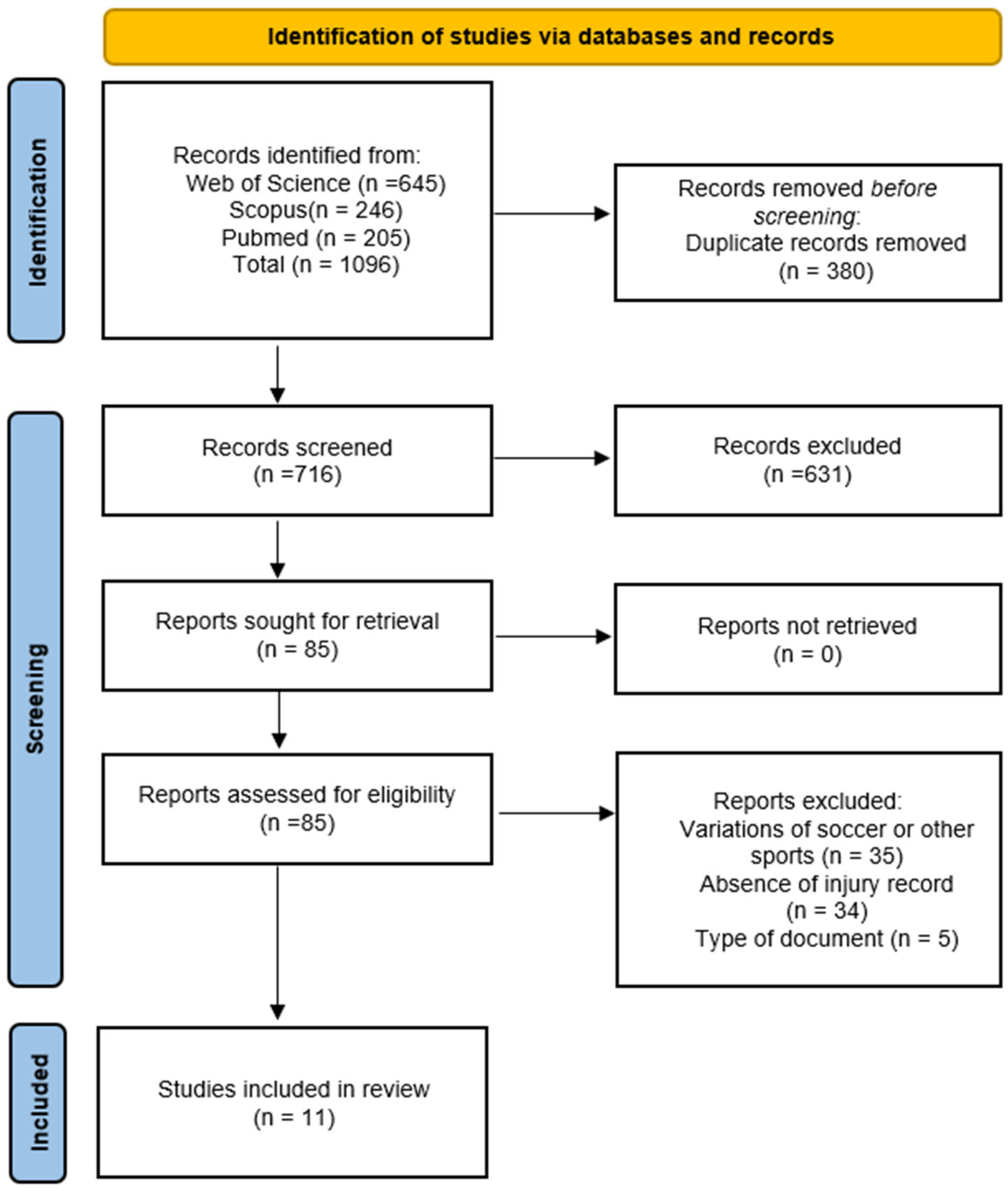
3.1. Search Results
3.2. Characteristics of the Studies
3.3. Results of Risk of Bias
3.4. Strength Tests
3.4.1. Strength Assessment Using Isokinetic Dynamometer
3.4.2. Strength Assessment Using the Nordic Hamstring Curl Test
4. Discussion
4.1. Strength Assessment Using Isokinetic Dynamometer
4.2. Ratios and Asymmetries Derived from Isokinetic Strength Assessments
4.3. Strength Assessment Using the Nordic Hamstring Curl Test
4.4. Limitations of This Study
4.5. Projections
4.6. Practical Applications
5. Conclusions
Author Contributions
Funding
Institutional Review Board Statement
Informed Consent Statement
Data Availability Statement
Conflicts of Interest
References
- Bangsbo, J.; Mohr, M.; Krustrup, P. Physical and metabolic demands of training and match-play in the elite football player. J. Sports Sci. 2006, 24, 665–674. [Google Scholar] [CrossRef] [PubMed]
- Dolci, F.; Hart, N.H.; Kilding, A.E.; Chivers, P.; Piggott, B.; Spiteri, T. Physical and energetic demand of soccer: A brief review. Strength Cond. J. 2020, 42, 70–77. [Google Scholar] [CrossRef]
- Rites, A.; Viana, D.; Merino-Muñoz, P.; Miarka, B.; Aedomuñoz, E.; Perez-Contreras, J.; Salerno, V.P. Do contextual factors, tournament level, and location affect external match load in elite Brazilian youth soccer players? J. Phys. Educ. Sport 2022, 22, 2898–2903. [Google Scholar]
- Bengtsson, H.; Ekstrand, J.; Waldén, M.; Hägglund, M. Muscle injury rate in professional football is higher in matches played within 5 days since the previous match: A 14-year prospective study with more than 130,000 match observations. Br. J. Sports Med. 2018, 52, 1116–1122. [Google Scholar] [CrossRef]
- de Albuquerque Freire, L.; Brito, M.A.; Merino Muñoz, P.; Valenzuela Pérez, D.I.; Cerda Kohler, H.; Aedo-Muñoz, E.A.; Slimani, M.; José Brito, C.; Bragazzi, N.L.; Znazen, H. Match Running Performance of Brazilian Professional Soccer Players according to Tournament Types. Montenegrin J. Sports Sci. Med. 2022, 11, 53–58. [Google Scholar] [CrossRef]
- Silva, J.R.; Rumpf, M.; Hertzog, M.; Castagna, C.; Farooq, A.; Girard, O.; Hader, K. Acute and residual soccer match-related fatigue: A systematic review and meta-analysis. Sports Med. 2018, 48, 539–583. [Google Scholar] [CrossRef]
- Olson, D.; Sikka, R.S.; Labounty, A.; Christensen, T. Injuries in professional football: Current concepts. Curr. Sports Med. Rep. 2013, 12, 381–390. [Google Scholar] [CrossRef]
- Hägglund, M.; Waldén, M.; Magnusson, H.; Kristenson, K.; Bengtsson, H.; Ekstrand, J. Injuries affect team performance negatively in professional football: An 11-year follow-up of the UEFA Champions League injury study. Br. J. Sports Med. 2013, 47, 738–742. [Google Scholar] [CrossRef]
- Ekstrand, J. Preventing injuries in professional football: Thinking bigger and working together. Br. J. Sports Med. 2016, 50, 709. [Google Scholar] [CrossRef]
- Fuller, C.W.; Ekstrand, J.; Junge, A.; Andersen, T.E.; Bahr, R.; Dvorak, J.; Hägglund, M.; McCrory, P.; Meeuwisse, W.H. Consensus statement on injury definitions and data collection procedures in studies of football (soccer) injuries. Br. J. Sports Med. 2006, 40, 193. [Google Scholar] [CrossRef]
- Ekstrand, J. Keeping your top players on the pitch: The key to football medicine at a professional level. Br. J. Sports Med. 2013, 47, 723. [Google Scholar] [CrossRef]
- Huygaerts, S.; Cos, F.; Cohen, D.D.; Calleja-González, J.; Guitart, M.; Blazevich, A.J.; Alcaraz, P.E. Mechanisms of Hamstring Strain Injury: Interactions between Fatigue, Muscle Activation and Function. Sports 2020, 8, 65. [Google Scholar] [CrossRef] [PubMed]
- Mueller-Wohlfahrt, H.-W.; Haensel, L.; Mithoefer, K.; Ekstrand, J.; English, B.; McNally, S.; Orchard, J.; van Dijk, C.N.; Kerkhoffs, G.M.; Schamasch, P.; et al. Terminology and classification of muscle injuries in sport: The Munich consensus statement. Br. J. Sports Med. 2013, 47, 342. [Google Scholar] [CrossRef] [PubMed]
- Klein, C.; Henke, T.; Platen, P. Injuries in football (soccer)—A systematic review of epidemiology and aetiological aspects. Ger. J. Exerc. Sport Res. 2018, 48, 309–322. [Google Scholar] [CrossRef]
- Larruskain, J.; Lekue, J.A.; Diaz, N.; Odriozola, A.; Gil, S.M. A comparison of injuries in elite male and female football players: A five-season prospective study. Scand. J. Med. Sci. Sports 2018, 28, 237–245. [Google Scholar] [CrossRef]
- Ekstrand, J.; Hägglund, M.; Waldén, M. Epidemiology of Muscle Injuries in Professional Football (Soccer). Am. J. Sports Med. 2011, 39, 1226–1232. [Google Scholar] [CrossRef]
- Waldén, M.; Hägglund, M.; Ekstrand, J. The epidemiology of groin injury in senior football: A systematic review of prospective studies. Br. J. Sports Med. 2015, 49, 792. [Google Scholar] [CrossRef]
- Harøy, J.; Clarsen, B.; Thorborg, K.; Hölmich, P.; Bahr, R.; Andersen, T.E. Groin Problems in Male Soccer Players Are More Common Than Previously Reported. Am. J. Sports Med. 2017, 45, 1304–1308. [Google Scholar] [CrossRef]
- Pérez-Contreras, J.; Loro-Ferrer, J.F.; Merino-Muñoz, P.; Hermosilla-Palma, F.; Miranda-Lorca, B.; Bustamante-Garrido, A.; Inostroza-Ríos, F.; Brito, C.J.; Aedo-Muñoz, E. Intra and Inter-Test Reliability of Isometric Hip Adduction Strength Test with Force Plates in Professional Soccer Players. J. Funct. Morphol. Kinesiol. 2024, 9, 270. [Google Scholar] [CrossRef]
- Owoeye, O.B.A.; VanderWey, M.J.; Pike, I. Reducing Injuries in Soccer (Football): An Umbrella Review of Best Evidence Across the Epidemiological Framework for Prevention. Sports Med. Open 2020, 6, 46. [Google Scholar] [CrossRef]
- Delextrat, A.; Piquet, J.; Matthews, M.J.; Cohen, D.D. Strength-Endurance Training Reduces the Hamstrings Strength Decline Following Simulated Football Competition in Female Players. Front. Physiol. 2018, 9, 1059. [Google Scholar] [CrossRef] [PubMed]
- Hughes, S.; Warmenhoven, J.; Haff, G.G.; Chapman, D.W.; Nimphius, S. Countermovement Jump and Squat Jump Force-Time Curve Analysis in Control and Fatigue Conditions. J. Strength Cond. Res. 2022, 36, 2752–2761. [Google Scholar] [CrossRef]
- Lovell, G.A.; Blanch, P.D.; Barnes, C.J. EMG of the hip adductor muscles in six clinical examination tests. Phys. Ther. Sport 2012, 13, 134–140. [Google Scholar] [CrossRef] [PubMed]
- Pedley, J.S.; Lloyd, R.S.; Read, P.J.; Moore, I.S.; De Ste Croix, M.; Myer, G.D.; Oliver, J.L. Utility of Kinetic and Kinematic Jumping and Landing Variables as Predictors of Injury Risk: A Systematic Review. J. Sci. Sport Exerc. 2020, 2, 287–304. [Google Scholar] [CrossRef]
- Fousekis, K.; Tsepis, E.; Poulmedis, P.; Athanasopoulos, S.; Vagenas, G. Intrinsic risk factors of non-contact quadriceps and hamstring strains in soccer: A prospective study of 100 professional players. Br. J. Sports Med. 2011, 45, 709. [Google Scholar] [CrossRef]
- Liporaci, R.F.; Saad, M.; Grossi, D.B.; Riberto, M. Clinical Features and isokinetic Parameters in Assessing Injury Risk in elite Football Players. Int. J. Sports Med. 2019, 40, 903–908. [Google Scholar] [CrossRef]
- Bishop, C.; Turner, A.; Gonzalo-Skok, O.; Read, P. Inter-limb asymmetry during rehabilitation understanding formulas and monitoring the “magnitude” and “direction”. Aspetar Sports Med. J. 2020, 9, 18–22. [Google Scholar]
- Engebretsen, A.H.; Myklebust, G.; Holme, I.; Engebretsen, L.; Bahr, R. Intrinsic Risk Factors for Groin Injuries among Male Soccer Players: A Prospective Cohort Study. Am. J. Sports Med. 2010, 38, 2051–2057. [Google Scholar] [CrossRef]
- Helme, M.; Tee, J.; Emmonds, S.; Low, C. Does lower-limb asymmetry increase injury risk in sport? A systematic review. Phys. Ther. Sport 2021, 49, 204–213. [Google Scholar] [CrossRef]
- Bishop, C.; Lake, J.; Loturco, I.; Papadopoulos, K.; Turner, A.; Read, P. Interlimb Asymmetries: The Need for an Individual Approach to Data Analysis. J. Strength Cond. Res. 2021, 35, 695–701. [Google Scholar] [CrossRef]
- Maloney, S.J. The Relationship Between Asymmetry and Athletic Performance: A Critical Review. J. Strength Cond. Res. 2019, 33, 2579–2593. [Google Scholar] [CrossRef] [PubMed]
- Page, M.J.; McKenzie, J.E.; Bossuyt, P.M.; Boutron, I.; Hoffmann, T.C.; Mulrow, C.D.; Shamseer, L.; Tetzlaff, J.M.; Akl, E.A.; Brennan, S.E.; et al. The PRISMA 2020 statement: An updated guideline for reporting systematic reviews. BMJ 2021, 372, n71. [Google Scholar] [CrossRef]
- McKay, A.K.A.; Stellingwerff, T.; Smith, E.S.; Martin, D.T.; Mujika, I.; Goosey-Tolfrey, V.L.; Sheppard, J.; Burke, L.M. Defining Training and Performance Caliber: A Participant Classification Framework. Int. J. Sports Physiol. Perform. 2022, 17, 317–331. [Google Scholar] [CrossRef] [PubMed]
- Rico-González, M.; Pino-Ortega, J.; Clemente, F.; Los Arcos, A. Guidelines for performing systematic reviews in sports science. Biol. Sport 2022, 39, 463–471. [Google Scholar] [CrossRef]
- Downs, S.H.; Black, N. The feasibility of creating a checklist for the assessment of the methodological quality both of randomised and non-randomised studies of health care interventions. J. Epidemiol. Community Health 1998, 52, 377. [Google Scholar] [CrossRef]
- Hooper, P.; Jutai, J.W.; Strong, G.; Russell-Minda, E. Age-related macular degeneration and low-vision rehabilitation: A systematic review. Can. J. Ophthalmol. 2008, 43, 180–187. [Google Scholar] [CrossRef]
- Landis, J.R.; Koch, G.G. The Measurement of Observer Agreement for Categorical Data. Biometrics 1977, 33, 159–174. [Google Scholar] [CrossRef]
- Dauty, M.; Menu, P.; Fouasson-Chailloux, A. Cutoffs of isokinetic strength ratio and hamstring strain prediction in professional soccer players. Scand. J. Med. Sci. Sports 2018, 28, 276–281. [Google Scholar] [CrossRef]
- Dauty, M.; Menu, P.; Fouasson-Chailloux, A. Hamstring Muscle Injury Prediction by Isokinetic Ratios Depends on the Method Used. Clin. J. Sport Med. 2020, 30, 40–45. [Google Scholar] [CrossRef]
- Grygorowicz, M.; Michałowska, M.; Walczak, T.; Owen, A.; Grabski, J.K.; Pyda, A.; Piontek, T.; Kotwicki, T. Discussion about different cut-off values of conventional hamstring-to-quadriceps ratio used in hamstring injury prediction among professional male football players. PLoS ONE 2017, 12, e0188974. [Google Scholar] [CrossRef]
- Lee, J.W.Y.; Mok, K.-M.; Chan, H.C.K.; Yung, P.S.H.; Chan, K.-M. Eccentric hamstring strength deficit and poor hamstring-to-quadriceps ratio are risk factors for hamstring strain injury in football: A prospective study of 146 professional players. J. Sci. Med. Sport 2018, 21, 789–793. [Google Scholar] [CrossRef] [PubMed]
- Timmins, R.G.; Bourne, M.N.; Shield, A.J.; Williams, M.D.; Lorenzen, C.; Opar, D.A. Short biceps femoris fascicles and eccentric knee flexor weakness increase the risk of hamstring injury in elite football (soccer): A prospective cohort study. Br. J. Sports Med. 2016, 50, 1524. [Google Scholar] [CrossRef] [PubMed]
- van Dyk, N.; Bahr, R.; Burnett, A.F.; Verhagen, E.; von Tiggelen, D.; Witvrouw, E. No association between rate of torque development and onset of muscle activity with increased risk of hamstring injury in elite football. Scand. J. Med. Sci. Sports 2018, 28, 2153–2163. [Google Scholar] [CrossRef] [PubMed]
- van Dyk, N.; Bahr, R.; Burnett, A.F.; Whiteley, R.; Bakken, A.; Mosler, A.; Farooq, A.; Witvrouw, E. A comprehensive strength testing protocol offers no clinical value in predicting risk of hamstring injury: A prospective cohort study of 413 professional football players. Br. J. Sports Med. 2017, 51, 1695. [Google Scholar] [CrossRef]
- van Dyk, N.; Bahr, R.; Whiteley, R.; Tol, J.L.; Kumar, B.D.; Hamilton, B.; Farooq, A.; Witvrouw, E. Hamstring and Quadriceps Isokinetic Strength Deficits Are Weak Risk Factors for Hamstring Strain Injuries: A 4-Year Cohort Study. Am. J. Sports Med. 2016, 44, 1789–1795. [Google Scholar] [CrossRef]
- Shalaj, I.; Gjaka, M.; Bachl, N.; Wessner, B.; Tschan, H.; Tishukaj, F. Potential prognostic factors for hamstring muscle injury in elite male soccer players: A prospective study. PLoS ONE 2020, 15, e0241127. [Google Scholar] [CrossRef]
- Boccia, G.; Brustio, P.R.; Salvaggio, F.; Grossio, L.; Calcagno, E.; Pintore, A.; Rainoldi, A.; Samozino, P. The Rate of Torque Development as a Determinant of the Torque–Velocity Relationship. Scand. J. Med. Sci. Sports 2025, 35, e70035. [Google Scholar] [CrossRef]
- Guan, Y.; Bredin, S.S.D.; Taunton, J.; Jiang, Q.; Wu, N.; Warburton, D.E.R. Association between Inter-Limb Asymmetries in Lower-Limb Functional Performance and Sport Injury: A Systematic Review of Prospective Cohort Studies. J. Clin. Med. 2022, 11, 360. [Google Scholar] [CrossRef]
- Coombs, R.; Garbutt, G. Developments in the use of the hamstring/quadriceps ratio for the assessment of muscle balance. J. Sports Sci. Med. 2002, 1, 56–62. [Google Scholar]
- Hole, C.D.; Smith, G.H.; Hammond, J.; Kumar, A.; Saxton, J.; Cochrane, T. Dynamic control and conventional strength ratios of the quadriceps and hamstrings in subjects with anterior cruciate ligament deficiency. Ergonomics 2000, 43, 1603–1609. [Google Scholar] [CrossRef]
- Mrzygłód, S.; Pietraszewski, P.; Golas, A.; Jarosz, J.; Matusiński, A.; Krzysztofik, M. Changes in Muscle Activity Imbalance of the Lower Limbs Following 3 Weeks of Supplementary Body-Weight Unilateral Training. Appl. Sci. 2021, 11, 1494. [Google Scholar] [CrossRef]
- Bishop, C. Interlimb Asymmetries: Are Thresholds a Usable Concept? Strength Cond. J. 2021, 43, 32–36. [Google Scholar] [CrossRef]
- Read, P.J.; Oliver, J.L.; De Ste Croix, M.B.A.; Myer, G.D.; Lloyd, R.S. A prospective investigation to evaluate risk factors for lower extremity injury risk in male youth soccer players. Scand. J. Med. Sci. Sports 2018, 28, 1244–1251. [Google Scholar] [CrossRef] [PubMed]
- Maher, J.M.; Markey, J.C.; Ebert-May, D. The other half of the story: Effect size analysis in quantitative research. CBE Life Sci. Educ. 2013, 12, 345–351. [Google Scholar] [CrossRef]
- Grindstaff, T.L.; Palimenio, M.R.; Franco, M.; Anderson, D.; Bagwell, J.J.; Katsavelis, D. Optimizing Between-Session Reliability for Quadriceps Peak Torque and Rate of Torque Development Measures. J. Strength Cond. Res. 2019, 33, 1840–1847. [Google Scholar] [CrossRef]
- Aedo-Muñoz, E.; Abarca-Reyes, V.; Torres-Moreno, M.-J.; Bascuñan-Mosqueira, R.; Brito, C.J.; Miarka, B.; Caro-San Juan, J.; Sánchez-Ramírez, C.; Poblete-Gálvez, C. Proposal of a Technical Model of Soccer Kicking: A Systematic Review of Kinematic and Kinetic Variables. MHSalud Rev. Cienc. Mov. Hum. Salud 2020, 17, 1–26. [Google Scholar] [CrossRef]
- Henderson, G.; Barnes, C.A.; Portas, M.D. Factors associated with increased propensity for hamstring injury in English Premier League soccer players. J. Sci. Med. Sport 2010, 13, 397–402. [Google Scholar] [CrossRef]
- Heiderscheit, B.C.; Sherry, M.A.; Silder, A.; Chumanov, E.S.; Thelen, D.G. Hamstring strain injuries: Recommendations for diagnosis, rehabilitation, and injury prevention. J. Orthop. Sports Phys. Ther. 2010, 40, 67–81. [Google Scholar] [CrossRef]
- Liu, H.; Garrett, W.E.; Moorman, C.T.; Yu, B. Injury rate, mechanism, and risk factors of hamstring strain injuries in sports: A review of the literature. J. Sport Health Sci. 2012, 1, 92–101. [Google Scholar] [CrossRef]
- Askling, C.; Karlsson, J.; Thorstensson, A. Hamstring injury occurrence in elite soccer players after preseason strength training with eccentric overload. Scand. J. Med. Sci. Sports 2003, 13, 244–250. [Google Scholar] [CrossRef]
- Majumdar, A.; Bakirov, R.; Hodges, D.; Scott, S.; Rees, T. Machine Learning for Understanding and Predicting Injuries in Football. Sports Med. Open 2022, 8, 73. [Google Scholar] [CrossRef] [PubMed]
- Zebis, M.K.; Andersen, L.L.; Ellingsgaard, H.; Aagaard, P. Rapid hamstring/quadriceps force capacity in male vs. female elite soccer players. J. Strength Cond. Res. 2011, 25, 1989–1993. [Google Scholar] [CrossRef] [PubMed]
- Pataky, T.C. Generalized n-dimensional biomechanical field analysis using statistical parametric mapping. J. Biomech. 2010, 43, 1976–1982. [Google Scholar] [CrossRef] [PubMed]

| Inclusion criteria | Population: Soccer players from both sexes who are categorized as sub-elite (performance at state or national level), elite (international performance), or world-class [33]. |
| Intervention: Epidemiology (follow-up/history) of lower limb musculoskeletal injuries, whether these are functional direct (type 1A and 1B) or structural (type 3A, 3B, and 4) [13]. | |
| Comparison: Dynamometric assessments of lower limb strength that are expressed in kinetic indicators such as force (N), power (W), rate of force development (N/s), impulse (N*s), and torque (N*m). | |
| Outcomes: The presence or absence of a relationship between kinetic indicators and indirect injuries of the lower limb as a risk factor. | |
| Exclusion criteria | Conventional soccer variations such as futsal or beach soccer. |
| Direct musculoskeletal injuries, bone, ligament, and joint injuries. | |
| Sources of information such as books, thesis, reports, and reviews. |
| Reference | Study Design | Population | ||||
|---|---|---|---|---|---|---|
| Sample | Mean Age (SD) Years | Mean Height (SD) m | Mean Body Mass (SD) kg | Competitive Category | ||
| Fousekis et al. [25] | Prospective cohort study | n = 200 16 H 84 HNI 7 Q 93 QNI | NS 22.94 (4.11) 23.00 (3.27) 25.42 (5.28) 23.42 (2.76) | NS 1.80 (4.97) 1.76 (6.19) 1.76 (4.68) 1.80 (2.76) | NS 74.71 (3.64) 71.58 (5.96) 72.35 (4.69) 73.42 (3.36) | 3rd National Soccer League Division |
| Timmins et al. [42] | Prospective cohort study | n = 152 27 H 125 HNI | NS 27.0 (3.8) 24.2 (5.1) | NS 1.80 (0.07) 1.78 (0.06) | NS 76.4 (6.7) 75.3 (6.6) | Professional Australian soccer competition |
| van Dyk et al. [45] | Prospective cohort study | n = 563 167 H 396 HNI | NS 25.3 (4.9) 24.7 (4.7) | NS 1.76 (6.1) 1.76 (6.7) | NS 71.3 (7.8) 71.9 (9.1) | Qatar Stars League |
| van Dyk et al. [44] | Prospective cohort study | n = 413 66 H 347 HNI | NS 27.9 (4.3) 25.9 (4.9) | NS 1.75 (6.7) 1.76 (6.8) | NS 72.2 (7.7) 72.6 (9.2) | Qatar Stars League |
| Dauty et al. [38] | Prospective cohort study | n = 194 36 H 158 HNI | NS 23.4 (4.4) 22.4 (4.1) | NS 1.80 (4.7) 1.80 (5.6) | NS 75.4 (6) 75.1 (6.1) | French Premier and Second League |
| Grygorowicz et al. [40] | Retrospective cohort study | n = 66 11 H 55 HNI | NS | NS | NS | Polish premier league |
| Lee et al. [41] | Prospective cohort study | n = 146 41 H 105 HNI | 24.2 (4.4) NS NS | 1.77 (5.9) NS NS | 72.9 (8.65) NS NS | Top national league |
| Dauty et al. [39] | Prospective cohort study | n = 91 31 H 60 HNI | NS 24.4 (4.4) 23.3 (4.4) | NS 1.80 (4.9) 1.80 (5.8) | NS 75.7 (6.7) 74.8 (5.9) | French Premier League |
| van Dyk et al. [43] | Prospective cohort study | n = 367 62 H 305 HNI | NS 27.8 (4.3) 26.1 (4.8) | NS 176 (7) 177 (6.8) | NS 72.3 (7.8) 72.3 (9.2) | Qatar Stars League |
| Liporaci et al. [26] | Prospective cohort study | n = 68 7 H 2 RF 4 Add | 24.3 (4.6) NS NS NS | 1,81 (0.07) NS NS NS | 78.9 (7.76) NS NS NS | NS |
| Shalaj et al. [46] | Prospective cohort study | n = 143 40 H 103 HNI | 23.2 (4.1) 26.1 (3.4) 22.2 (3.9) | 180.0 (5.3) 180.5 (4.7) 179.8 (5.6) | 74.2 (6.7) 77.0 (6.1) 73.2 (6.6) | Kosovo National Premier Soccer League |
| Reference | Injury Definition | Exposure | |
|---|---|---|---|
| Type of Injury | Follow-Up Durations | ||
| Fousekis et al. [25] | All non-contact muscle strains force players to miss at least one scheduled practice session or game. | Hamstrings Quadriceps strain | 1 season |
| Timmins et al. [42] | Hamstring strain injury defines any acute posterior thigh pain that resulted in the immediate cessation of exercise and is later diagnosed by the club medical staff. | Hamstrings strain | 4 seasons |
| van Dyk et al. [45] | Hamstring strain injury is defined as acute pain in the posterior thigh that occurred during training or match play and resulted in immediate termination of play and inability to participate in the next training session or match. | Hamstrings strain | 4 seasons |
| van Dyk et al. [44] | Hamstring strain injury is defined as acute pain in the posterior thigh that occurred during training or match play and resulted in immediate termination of play and inability to participate in the next training session or match. | Hamstrings strain | 2 seasons |
| Dauty et al. [38] | Physical complaint in the region of the posterior thigh sustained during a soccer match or training, without contact, irrespective of the need for medical attention, with time loss from soccer activities (7 to 28 days). | Hamstrings strain | 1 season |
| Grygorowicz et al. [40] | The player was unable to take part in a match or in a training session due to hamstring strain that happened in a football match or during training, and at least one of the following consequences was present: decrease in the quantity or level of sports activity for at least one day, or need or medical evaluation or non-operative or operative treatment. | Hamstrings strain | 1 Season |
| Lee et al. [41] | Hamstring strain injury is defined as acute pain in the posterior thigh, which causes immediate cessation of match play or training. | Hamstrings strain | 1 season |
| Dauty et al. [39] | Moderate injuries: time loss between 7 and 28 days. Major injuries: time loss > 28 days. | Hamstrings strain | 1 season |
| van Dyk et al. [43] | Hamstring injury is defined as acute pain in the posterior thigh that occurred during training or match play and resulted in immediate termination of play and inability to participate in the next training session or match. | Hamstrings strain | 2 seasons |
| Liporaci et al. [26] | Any physical complaints resulting in a player being unable to take part in at least one subsequent football training session or match. | Hamstrings strain Rectus femoris strain Adductor strain | 1 season |
| Shalaj et al. [46] | Not specified | Hamstrings strain | 2 seasons |
| Authors | Reporting | External Validity | Internal Validity | Selection Bias | Power | Total | Quality | ||||||||||||||||||||||
|---|---|---|---|---|---|---|---|---|---|---|---|---|---|---|---|---|---|---|---|---|---|---|---|---|---|---|---|---|---|
| 1 | 2 | 3 | 4 | 5 | 6 | 7 | 8 | 9 | 10 | 11 | 12 | 13 | 14 | 15 | 16 | 17 | 18 | 19 | 20 | 21 | 22 | 23 | 24 | 25 | 26 | 27 | |||
| Fousekis et al. [25] | 1 | 1 | 1 | 1 | 2 | 1 | 1 | 0 | 1 | 1 | 0 | 0 | 0 | 0 | 0 | 1 | 1 | 1 | 1 | 1 | 1 | 1 | 0 | 0 | 1 | 1 | 1 | 20 | Good |
| Dauty et al. [38] | 1 | 1 | 1 | 1 | 0 | 1 | 1 | 0 | 1 | 1 | 0 | 0 | 0 | 0 | 0 | 1 | 1 | 1 | 1 | 1 | 1 | 1 | 0 | 0 | 0 | 1 | 0 | 16 | Fair |
| Dauty et al. [39] | 1 | 1 | 1 | 1 | 0 | 1 | 1 | 0 | 1 | 1 | 0 | 0 | 0 | 0 | 0 | 1 | 1 | 1 | 1 | 1 | 1 | 1 | 0 | 0 | 0 | 1 | 0 | 16 | Fair |
| Grygorowicz et al. [40] | 1 | 1 | 1 | 0 | 1 | 1 | 0 | 1 | 1 | 0 | 0 | 0 | 0 | 0 | 1 | 1 | 1 | 1 | 1 | 1 | 0 | 0 | 0 | 0 | 1 | 0 | 15 | Fair | |
| Lee et al. [41] | 1 | 1 | 0 | 1 | 1 | 1 | 1 | 0 | 1 | 1 | 0 | 0 | 0 | 0 | 0 | 1 | 1 | 1 | 1 | 1 | 1 | 1 | 0 | 0 | 1 | 1 | 1 | 18 | Fair |
| Liporaci et al. [26] | 1 | 1 | 1 | 1 | 1 | 1 | 1 | 0 | 1 | 1 | 0 | 0 | 0 | 0 | 0 | 1 | 1 | 1 | 1 | 1 | 1 | 1 | 0 | 0 | 0 | 1 | 0 | 17 | Fair |
| Shalaj et al. [46] | 1 | 1 | 1 | 1 | 1 | 1 | 1 | 0 | 1 | 1 | 0 | 0 | 0 | 0 | 0 | 1 | 1 | 1 | 1 | 1 | 1 | 1 | 0 | 0 | 1 | 1 | 1 | 19 | Fair |
| Timmins et al. [42] | 1 | 1 | 1 | 1 | 2 | 1 | 1 | 0 | 1 | 1 | 0 | 0 | 0 | 0 | 0 | 1 | 1 | 1 | 1 | 1 | 1 | 1 | 0 | 0 | 1 | 1 | 1 | 20 | Good |
| van Dyk et al. [45] | 1 | 1 | 1 | 1 | 1 | 1 | 1 | 0 | 0 | 1 | 1 | 0 | 0 | 0 | 0 | 1 | 0 | 1 | 1 | 1 | 1 | 0 | 0 | 0 | 1 | 0 | 0 | 15 | Fair |
| van Dyk et al. [44] | 1 | 1 | 1 | 1 | 1 | 1 | 1 | 0 | 1 | 1 | 1 | 1 | 0 | 0 | 0 | 1 | 1 | 1 | 1 | 1 | 1 | 0 | 0 | 0 | 0 | 0 | 1 | 18 | Fair |
| van Dyk et al. [43] | 1 | 1 | 1 | 1 | 1 | 1 | 1 | 0 | 1 | 1 | 1 | 1 | 0 | 0 | 0 | 1 | 0 | 1 | 1 | 1 | 1 | 0 | 0 | 0 | 0 | 0 | 0 | 16 | Fair |
| Definition | |
|---|---|
| Absolute maximum torque | Maximum torque generated by a joint during a voluntary maximal contraction without considering the individual’s body weight [47]. |
| Relative maximum torque | Maximum torque normalized to body weight (Nm/kg), allowing for comparisons between individuals of different sizes [47]. |
| Asymmetry between limbs | Difference in strength or functional performance between the right and left limbs [48]. |
| H:Q ratio | Ratio between the strength of the hamstrings and quadriceps muscles. Used to assess muscular balance at the knee joint and prevent injury [49]. |
| Dynamic control ratio | Functional comparison between eccentric torque of hamstrings and concentric torque of quadriceps, which is important for dynamic knee stability [50]. |
| Strength imbalance | Significant strength difference between opposing muscles (agonists vs. antagonists) or between limbs, potentially leading to compensatory movement patterns and increased injury risk [51]. |
| Rate of torque development | Speed at which torque is generated from the onset of a muscular contraction. Calculated as the slope (first derivate over time) of the torque–time curve during isometric contraction [47]. |
| References | Kinetics Variables | Outcome of Analysis Univariate | Outcome of Analysis Multivariate |
|---|---|---|---|
| Fousekis et al. [25] | Asymmetry ≥ 15% to Q con strength (yes/no) Asymmetry ≥ 15% to Q ecc strength (yes/no) Asymmetry ≥ 15% to H con strength (yes/no) Asymmetry ≥ 15% to H ecc strength (yes/no) Asymmetry < 1 to ratio H ecc 180°/s:Q con 180°/s (yes/no) | Eccentric hamstring asymmetries (OR = 3.88; CI al 95% 1.13–12.23; p = 0.03) | Not applicable |
| van Dyk et al. [45] | H con at 60°/s and 300°/s absolute (Nm) H con at 60°/s and 300°/s adjusted (Nm/kg) H ecc at 60°/s absolute (Nm) H ecc at 60°/s adjusted (Nm/kg) Q con at 60°/s and 300°/s absolute (Nm) Q con at 60°/s and 300°/s adjusted (Nm/kg) Ratio Q:H con 60°/s (AU) Ratio Q:H con 300°/s (AU) Ratio Q con 300°/s: H ecc 60°/s (AU) | Not applicable | Quadriceps concentric 60°/s adjusted (OR = 1.41; 95% CI 1.03 to 1.92; p = 0.03) Hamstrings eccentric 60°/s adjusted (OR = 1.37; 95% CI 1.01 to 1.85; p = 0.04) |
| van Dyk et al. [44] | H con 60°/s and 300°/s absolute (Nm) H con 60°/s and 300°/s adjusted (Nm/kg) H ecc 60°/s absolute (Nm) H ecc 60°/s adjusted (Nm/kg) Q con 60°/s and 300°/s absolute (Nm) Q con 60°/s and 300°/s adjusted (Nm/kg) Ratio H ecc 60°/s: Q con 300°/s (UA) Dynamic control ratio (UA) Dynamic control ratio con at 30°, 40° and 50° (UA) Dynamic control ratio ecc at 30°, 40° and 50° (UA) Overall H:Q ratio (UA) | Categorical variable (criterion: >1 SD above the mean) Q Concentric at 300°/s adjusted (HR = 2.06; IC 95% 1.21 to 3.51; p = 0.008) | Not applicable |
| Dauty et al. [38] | H:H con 60°/s R/L < 0.9; ≤0.85; <0.87 H:H con 60°/s L/R < 0.9; <0.85; <0.86 H:H ecc 30°/s R/L < 0.9; <0.85; <0.80 H:H ecc 30°/s L/R < 0.9; <0.85; <0.83 H:Q con 60°/s R < 0.6; ≤0.47; <0.55 H:Q con 60°/s L < 0.6; ≤0.47; <0.55 Hecc30°/s/Qcon240°/s R < 1; <0.80; <1.01 Hecc30°/s/Qcon240°/s L < 1; <0.80; <0.99 | No significative results | Not applicable |
| Grygorowicz et al. [40] | Q con 60°/s absolute (Nm) Q con 60°/s relative (Nm/kg) H con 60°/s absolute (Nm) H con 60°/s relative (Nm/kg) Ratio H:Q con 60°/s (AU) Ratio H:Q con 60°/s Cut-off score < 0.47 (AU) Ratio H:Q con 60°/s Cut-off score < 0.6 (AU) Ratio H:Q con 60°/s Cut-off score < 0.658 (AU) | No significative results | Not applicable |
| Lee et al. [41] | H con 60°/s and 240°/s absolute (Nm) H con 60°/s and 240°/s relative (Nm/kg) H ecc 30°/s absolute (Nm) H ecc 30°/s relative (Nm/kg) Q con 60°/s and 240°/s absolute (Nm) Q con 60°/s and 240°/s relative (Nm/kg) Ratio H:Q con 60°/s (%) Ratio H:Q con 240°/s (%) Ratio H ecc 30°/s: Q con 240°/s (%) Strength imbalance H con 60°/s (Nm) Strength imbalance H con 240°/s (Nm) Strength imbalance H con 30°/s (Nm) | H strength absolute value of Con 60°/s (OD = 0.97; 95% CI 0.95 to 0.99; p = 0.002) H strength relative value of Con 60°/s (OD = 0.60; 95% CI 0.14 to 0.26; p < 0.001) H strength absolute value of Con 240°/s (OR = 0.97; 95% CI 0.94 to 1.00; p = 0.03) H strength relative value of Con 240°/s (OR = 0.36; 95% CI 0.00 to 0.30; p = 0.03) H strength absolute value of Ecc 30°/s (OR = 0.98; 95% CI 0.97 to 0.99; p = 0.002) H strength relative value of Ecc 30°/s (OR = 0.15; 95% CI 0.06 to 0.40; p < 0.001) H/Q ratio con 60°/s (OR = 0.92; 95% CI 0.87 to 0.97; p = 0.001) H strength imbalance concentric at 30°/s (OR = 1.05; 95% CI 1.00 to 1.10; p = 0.03) | Preseason hamstring strength measures at 30 deg/s, Nm/kg ≤ 2.40 (adjusted OR = 5.59; 95% CI 2.20 to 12.92; p < 0.001) Preseason hamstring to quadriceps ratios Con 60/Con 60 (%) ≤ 50.5 (adjusted odd ratio = 3.14; 95% CI 1.37 to 7.22; p = 0.01) |
| Dauty et al. [39] | Ratio H:H con 60°/s (AU) Ratio H:H ecc 30°/s (AU) Ratio Q:Q con 60°/s (AU) Ratio Q:Q con 240°/s (AU) Ratio H:Q con 60°/s (AU) Ratio H ecc 30°/s: Q con 240°/s (AU) | H/Hcon60°/s (Ndom/dom): OR = 38; IC 95% 1.06 to 1818; p = 0.04 | Not applicable |
| van Dyk et al. [43] | H RTD 30 ms con 60°/s (Nm/s) H RTD 50 ms con 60°/s (Nm/s) H RTD 100 ms con 60°/s (Nm/s) H RTD 30 ms con 300°/s (Nm/s) H RTD 50 ms con 300°/s (Nm/s) H RTD 100 ms con 300°/s (Nm/s) Q RTD 30 ms ecc 60°/s (Nm/s) Q RTD50 ms ecc 60°/s (Nm/s) Q RTD 100 ms ecc 60°/s (Nm/s) | No significative results | Not applicable |
| Liporaci et al. [26] | Asymmetry > 10% of knee flexion 60°/s (Yes/no). Asymmetry > 10% of knee extension 60°/s (Yes/no). Ratio H:Q 60°/s between 55 and 64% (Yes/no) | Ext PT 10 (OR = 7.49; CI (95%) 1.51–37.26; p = 0.01) Flex PT 10 (OR = 46.94; CI (95 %) 4.16–530; p < 0.01) H:Q ratio con 60°/s (OR = 6.72; CI (95 %) 1.32–34.31; p = 0.02) | Not applicable |
| Shalaj et al. [46] | H dominant con 60°/s and 240°/s absolute (Nm) H non-dominant con 60°/s and 240°/s absolute (Nm) Q dominant con 60°/s and 240°/s absolute (Nm) Q non-dominant con 60°/s and 240°/s absolute (Nm) H dominant ecc 30°/s and 120°/s absolute (Nm) H non-dominant ecc 30°/s and 120°/s absolute (Nm) H dominant con 60°/s and 240°/s relative (Nm/kg) H non-dominant con 60°/s and 240°/s relative (Nm/kg) Q dominant con 60°/s and 240°/s relative (Nm/kg) Q non-dominant con 60°/s and 240°/s relative (Nm/kg) H dominant ecc 30°/s and 120°/s relative (Nm/kg) H non-dominant ecc 30°/s and 120°/s Relative (Nm/kg) Ratio H/Q con 60°/s dominant absolute (%) Ratio H/Q con 60°/s non-dominant absolute (%) Ratio H/Q con 240°/s dominant absolute (%) Ratio H/Q con 240°/s non-dominant absolute (%) | Not applicable | Concentric hamstring 240°/s dominant leg (β = 0.01 ± 0.01; CI 0.00 to 0.01; p = 0.049) |
| References | Kinetics Variables | Outcome of Analysis Univariate | Outcome of Analysis Multivariate |
|---|---|---|---|
| Timmins et al. [42] | Eccentric force (N) Eccentric torque (Nm) Relative eccentric force (N/Kg) Relative eccentric torque (Nm/Kg) Isometric force (N) Isometric torque (N/m) Relative isometric force (N/Kg) Relative isometric torque (Nm/Kg) | Eccentric force ROC-curve determined threshold of 337 N (RR = 4.4; 95% CI 1.1 to 17.6; p = 0.013) Eccentric torque ROC-curve determined threshold of 145 N/m (RR = 3.6; 95% CI 1.2 to 11.4; p = 0.017) Relative eccentric force ROC-curve determined threshold of 4.35 N/kg (RR = 2.5; 95% CI 1.1 to 6.2; p = 0.041) Relative eccentric torque ROC-curve determined threshold of 1.86 Nm/kg (RR = 2.9; 95% CI 1.1 to 7.1; p = 0.011) | Model 3: Mean eccentric strength of both limbs (N) (X2 = 6.33; p = 0.011) Model 4: Mean eccentric strength of both limbs (N) (X2 = 5.05; p = 0.024) Model 5: Mean eccentric strength of both limbs (N) (X2 = 4.29; p = 0.038) |
| van Dyk et al. [44] | Peak force absolute (N) Peak force adjusted (N/kg) Peak force imbalance absolute (N) Peak force imbalance adjusted (N/kg) Average force absolute (N) Average force adjusted (N/kg) | No significative results | Not applicable |
Disclaimer/Publisher’s Note: The statements, opinions and data contained in all publications are solely those of the individual author(s) and contributor(s) and not of MDPI and/or the editor(s). MDPI and/or the editor(s) disclaim responsibility for any injury to people or property resulting from any ideas, methods, instructions or products referred to in the content. |
© 2025 by the authors. Licensee MDPI, Basel, Switzerland. This article is an open access article distributed under the terms and conditions of the Creative Commons Attribution (CC BY) license (https://creativecommons.org/licenses/by/4.0/).
Share and Cite
Pérez-Contreras, J.; Loro-Ferrer, J.F.; Inostroza-Ríos, F.; Merino-Muñoz, P.; Bustamante Garrido, A.; Hermosilla-Palma, F.; Brito, C.J.; Cortés-Roco, G.; Arriagada Tarifeño, D.; Muñoz-Hinrichsen, F.; et al. Kinetic Variables as Indicators of Lower Limb Indirect Injury Risk in Professional Soccer: A Systematic Review. J. Funct. Morphol. Kinesiol. 2025, 10, 228. https://doi.org/10.3390/jfmk10020228
Pérez-Contreras J, Loro-Ferrer JF, Inostroza-Ríos F, Merino-Muñoz P, Bustamante Garrido A, Hermosilla-Palma F, Brito CJ, Cortés-Roco G, Arriagada Tarifeño D, Muñoz-Hinrichsen F, et al. Kinetic Variables as Indicators of Lower Limb Indirect Injury Risk in Professional Soccer: A Systematic Review. Journal of Functional Morphology and Kinesiology. 2025; 10(2):228. https://doi.org/10.3390/jfmk10020228
Chicago/Turabian StylePérez-Contreras, Jorge, Juan Francisco Loro-Ferrer, Felipe Inostroza-Ríos, Pablo Merino-Muñoz, Alejandro Bustamante Garrido, Felipe Hermosilla-Palma, Ciro José Brito, Guillermo Cortés-Roco, David Arriagada Tarifeño, Fernando Muñoz-Hinrichsen, and et al. 2025. "Kinetic Variables as Indicators of Lower Limb Indirect Injury Risk in Professional Soccer: A Systematic Review" Journal of Functional Morphology and Kinesiology 10, no. 2: 228. https://doi.org/10.3390/jfmk10020228
APA StylePérez-Contreras, J., Loro-Ferrer, J. F., Inostroza-Ríos, F., Merino-Muñoz, P., Bustamante Garrido, A., Hermosilla-Palma, F., Brito, C. J., Cortés-Roco, G., Arriagada Tarifeño, D., Muñoz-Hinrichsen, F., & Aedo-Muñoz, E. (2025). Kinetic Variables as Indicators of Lower Limb Indirect Injury Risk in Professional Soccer: A Systematic Review. Journal of Functional Morphology and Kinesiology, 10(2), 228. https://doi.org/10.3390/jfmk10020228