Article

Exploring the Inhibitory Efficacy of Resokaempferol and Tectochrysin on PI3Kα Protein by Combining DFT and Molecular Docking against Wild-Type and H1047R Mutant Forms

by
Cristina Paraschiv
,
Steluța Gosav
,
Catalina Mercedes Burlacu
and
Mirela Praisler
*
Department of Chemistry, Physics and Environment, Faculty of Science and Environment, “Dunarea de Jos” University of Galati, 47 Domneasca Street, 800008 Galati, Romania
*
Author to whom correspondence should be addressed.
Inventions 2024, 9(5), 96; https://doi.org/10.3390/inventions9050096
Submission received: 5 July 2024 / Revised: 21 August 2024 / Accepted: 29 August 2024 / Published: 5 September 2024

Abstract

:
This study explores the inhibitory potential of the flavonoids resokaempferol and tectochrysin against both wild-type and H1047R mutant forms of PI3Kα, aiming to expand the repertoire of targeted cancer therapies. Employing an array of computational techniques, including Density Functional Theory (DFT), calculations of electronic parameters such as the energies of the frontier molecular orbitals, Molecular Electrostatic Potential (MEP) mapping, and Molecular Docking, we investigate in detail the molecular interactions of these compounds with the PI3Kα kinase. Our findings, corroborated by DFT calculations performed based on the B3LYP (Becke, three-parameter, Lee-Yang-Parr) hybrid functional and the 6-311G++(d,p) basis set, align well with experimental benchmarks and indicate substantial inhibitory efficacy. Further analysis of chemical potential and bioavailability confirmed the drug-like attributes of these flavonoids. Binding affinity and selectivity were rigorously assessed through self-docking and cross-docking against the PIK3CA PDB structures 7K71 and 8TS9. The most promising interactions were validated using Pairwise Structure Alignment and MolProbity analysis of all-atom contacts and geometry. Collectively, these results highlight the flavonoids’ potential as PI3Kα inhibitors and exemplify the utility of natural compounds in the development of precise anticancer treatments.

1. Introduction

Flavonoids, a class of polyphenolic compounds, have long captivated the scientific community due to their myriads of biological activities and therapeutic potential. Among these, resokaempferol and tectochrysin stand out as molecules of interest due to their distinct chemical properties and promising pharmacological profiles. Resokaempferol, a synthetic derivative, is known for its bioactive potential despite its non-natural origin. This compound, 3,7-dihydroxy-2-(4-hydroxyphenyl) chromen-4-one, showcases the advancement in biotechnological methods, such as its synthesis via engineered Saccharomyces cerevisiae, which highlights the potential of microbial platforms in drug development [1]. Similarly, tectochrysin, a naturally occurring flavonoid identified as 5-hydroxy-7-methoxy-2-phenylchromen-4-one, has been studied for its anticancer properties, further underscoring the therapeutic relevance of flavonoids [2].
The PI3Kα protein, a critical enzyme in the PI3K/AKT signaling pathway, plays a pivotal role in cell growth and survival, making it a significant target in cancer therapy [3]. Particularly, the H1047R mutation within this protein is associated with enhanced kinase activity and oncogenic transformation, presenting a unique challenge and an opportunity for targeted therapeutic intervention. The H1047R mutation in the PIK3CA gene, which encodes the catalytic subunit of phosphatidylinositol-4,5-bisphosphate 3-kinase (PI3K), is one of the most common activating mutations in various human cancers. This point mutation, involving the substitution of histidine (H) with arginine (R) at position 1047, has significant consequences on the functioning of the PI3K/AKT/mTOR signaling pathway, known for its critical role in regulating cell growth, survival, metabolism, and motility.
The H1047R mutation leads to the constitutive activation of PI3K, independent of extracellular stimuli. This means that the PI3K enzyme becomes permanently activated, resulting in continuous phosphorylation of phosphatidylinositol-4,5-bisphosphate (PIP2) to phosphatidylinositol-3,4,5-trisphosphate (PIP3). PIP3 serves as a signaling molecule, recruiting and activating protein kinase B (AKT), which in turn activates multiple signaling pathways that promote cell survival and proliferation. Due to its ability to constitutively activate the PI3K/AKT/mTOR pathway, the H1047R mutation is considered oncogenic. It contributes to malignant cellular transformation by promoting uncontrolled cell proliferation and inhibiting apoptosis (programmed cell death). Studies have shown that cancer cells expressing the H1047R mutation exhibit accelerated tumor growth and an increased capacity for metastasis. The H1047R mutation can contribute to tumor resistance to certain anticancer therapies, particularly those targeting the PI3K/AKT/mTOR pathway. This necessitates the development of specific therapeutic strategies to counteract the effects of this mutation. The H1047R mutation is commonly found in breast cancer, being reported in approximately 25–40% of breast carcinoma cases. It is particularly associated with the luminal subtype of breast cancer, which is characterized by the expression of hormone receptors. In colorectal cancer, the H1047R mutation is present in about 10–20% of cases.
Studies have shown that this mutation is associated with a poorer prognosis and a weaker response to certain chemotherapeutic treatments. The H1047R mutation is also frequent in endometrial cancer, with a reported prevalence of approximately 20–25%. It is associated with the type I endometrial carcinoma subtype, which is characterized by indolent growth and a better prognosis compared to the type II subtype. The H1047R mutation has been identified in other cancer types, including glioblastoma, bladder cancer, and lung cancer, although with a lower prevalence compared to the aforementioned cancers. The H1047R mutation in the PIK3CA gene has major significance in oncology due to its ability to constitutively activate the PI3K/AKT/mTOR pathway, thereby promoting uncontrolled cell growth and survival. It is commonly found in various cancer types and is associated with variable prognoses and therapeutic responses. Understanding the mechanisms by which the H1047R mutation contributes to oncogenesis and therapy resistance is essential for developing targeted therapies and improving the prognosis of patients affected by cancers harboring this mutation [4].
This study aims to explore the inhibitory potential of resokaempferol and tectochrysin against both the wild-type and mutant H1047R forms of PI3Kα kinase. Notably, our literature review has not identified any published studies that investigate the use of these particular flavonoids as potential inhibitors for these specific forms of PI3Kα. This gap underscores the novelty and significance of our research, which could provide foundational insights into the development of new targeted therapies for cancer treatment.
By employing a suite of complex computational techniques, such as Density Functional Theory (DFT), HOMO-LUMO calculations, and Molecular Electrostatic Potential (MEP) mapping, we delve into the detailed molecular interactions of these compounds with PI3Kα. Supported by DFT calculations based on the B3LYP hybrid functional and the 6-311G++(d,p) basis set, our results closely align with experimental benchmarks, indicating significant inhibitory efficacy [5,6]. Further analysis of chemical potential and bioavailability confirms the drug-likeness of these flavonoids, with their binding affinity and selectivity rigorously assessed through self-docking and cross-docking using AutoDock, Vina, and Schrodinger Glide against PIK3CA PDB structures 7K71 and 8TS9 [7,8,9,10,11,12,13,14,15,16,17,18,19,20,21,22]. Validation through Pairwise Structure Alignment and MolProbity analysis of all-atom contacts and geometry further highlights the potential of resokaempferol and tectochrysin as effective PI3Kα inhibitors [23,24,25,26,27].
This research not only contributes to our understanding of flavonoids as potential anticancer agents but also exemplifies the integration of natural compounds in the design of targeted cancer therapies, offering promising avenues for future therapeutic development.

2. Materials and Methods

Drug discovery is a complex, time-consuming, and costly endeavor. To enhance the efficiency of this process, employing in silico methods to investigate the molecular properties and interactions of potential therapeutic agents is increasingly favored. The molecular geometries investigated in this study include the flavonol 3,7-dihydroxy-2-(4-hydroxyphenyl)chromen-4-one (resokaempferol, ID: HMDB0034004) and the flavone 5-hydroxy-7-methoxy-2-phenylchromen-4-one (tectochrysin, ID: C00003795). These reference potential ligands were sourced from the Human Metabolome Database (HMDB) and the PhytoChemical Interactions Database (PCIDB) [28,29].
Additionally, the modeled three-dimensional crystal structures of the phosphatidylinositol 4,5-bisphosphate 3-kinase catalytic subunit α isoform (PI3Kα wild-type, PDB ID: 7K71) and its H1047R mutant variant (PDB ID: 8TS9) were obtained as reference receptors from the RCSB PDB (Research Collaboratory for Structural Bioinformatics Protein Data Bank).
In our research, we have used a variety of computational tools to examine the ligands of interest, which have promising utility in cancer treatment. These tools include software applications such as Gaussian (v.09W), GaussView (v.6.1.1), alvaMolecule (v.2.0.6), Toxicity Estimation Software Tool (v.5.1.2), Avogadro (v.1.2), SpectraGryph (v.1.2), PyMol (v.2.5.7), Chimera X (v.1.8), Glide-Maestro (v.13.5), AutoDock (v.4.2.6), Vina (v.1.2.5), and Discovery Studio 2024 [30,31,32,33,34,35,36,37]. To obtain supplementary relevant data, we have accessed various online databases and platforms such as ADMETlab 3.0, Pharos Knowledge Management Center (KMC), UniProt (Universal Protein Resource), Pubchem, MolProbity, which provide valuable biochemical data and interaction profiles [38,39,40,41].
Our methodology incorporated Density Functional Theory computations at the B3LYP/6-311G++(d,p) level in order to calculate the geometrical parameters (bond Lenths and angles) and energies. We also explored the molecular electrostatic potential, conducted chemical reactivity analyses, performed molecular docking, and predicted ADMET (Absorption, Distribution, Metabolism, Excretion, and Toxicity) properties. These diverse techniques enabled a multifaceted evaluation of the compound’s potential as a cancer therapeutic agent.

2.1. Evaluation of the Ligands’ Optimized Molecular Structures

The optimized ground state molecular geometries of ligands were calculated by using the DFT theory with the B3LYP hybrid functional and Pople’s 6-311G++(d,p) basis set. This method is particularly valued in computational chemistry due to its balance between computational efficiency and reliable accuracy in predicting molecular properties.

2.2. Assessing the Reactivity Profiles of Compounds

2.2.1. Numerical Analysis of Quantum Chemical Attributes

Quantum chemical parameters play a crucial role in predicting the biological properties of molecules. Through the analysis of these parameters, insights into the global chemical reactivity of a compound can be obtained. We assessed the flavonoids resokaempferol and tectochrysin through theoretical chemical analyses to gauge their potential as drug-like molecules. By utilizing the B3LYP/6-311G++(d,p) computational level, we derived the energies of the frontier molecular orbitals (EHOMO and ELUMO), the dipole moment (DM), and the polarizability [42]. These calculations facilitated the determination of several quantum chemical parameters, which include ionization potential (IP), electron affinity (EA), chemical hardness (η), chemical softness (σ), electronegativity (χ); electrophilic index (ω), whose calculation formulas were presented in a previously published study [42].
The molecular electrostatic potential maps further complement these findings, where the color gradient represents the electrostatic potential from positive to negative [43]. Both flavonoids exhibit regions of varying electrostatic potential, which can influence how they interact with other molecules, particularly in binding to target proteins within biological systems.
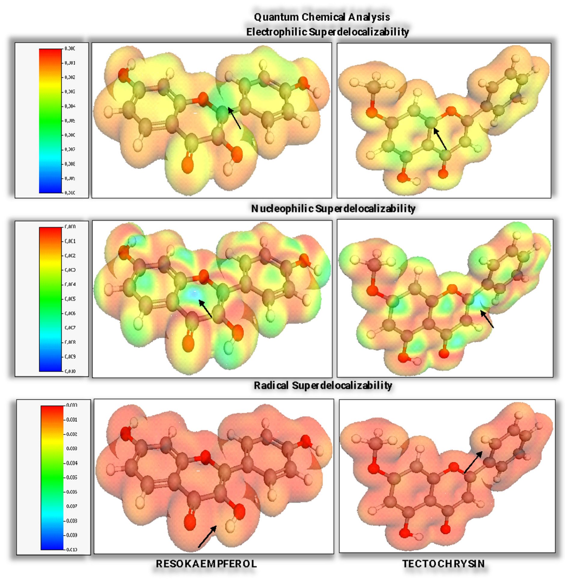
In our comprehensive quantum chemical analysis of resokaempferol and tectochrysin, we have expanded our investigation to include evaluations of electrophilic, nucleophilic, and radical susceptibilities, as well as their corresponding superdelocalizabilities [44,45]. These parameters offer deeper insights into the reactivity of these flavonoids and their potential as ligands in biochemical contexts.
The electrophilic susceptibility measures a molecule’s propensity to be attacked by an electrophile. A higher value indicates a greater likelihood of electrophilic attack at specific sites on the molecule. Nucleophilic susceptibility quantifies the likelihood of a molecule to interact with nucleophiles. A higher value suggests more reactive sites for nucleophilic attack. Radical susceptibility indicates the tendency of a molecule to react with free radicals. Molecules with higher values have regions more prone to radical reactions. This parameter is significant for evaluating the antioxidant potential of flavonoids, as their ability to interact with radicals is associated with their capacity to act as radical scavengers.
Electrophilic superdelocalizability identifies the ability of electron-rich regions within a molecule to delocalize charge towards an electrophilic center. High values can predict the sites where electron density might accumulate in the presence of an electrophile, which is valuable for understanding regioselectivity in electrophilic attacks. Nucleophilic superdelocalizability highlights areas within a molecule that can readily delocalize electrons toward a nucleophilic center. It’s useful for predicting the reactive sites that might attract electron density from a nucleophile, indicating how a molecule can stabilize additional charge or electron pairs. Radical superdelocalizability assesses the ability of a molecule to delocalize electrons in response to radical interactions. This parameter is particularly relevant in predicting how a molecule might engage with radicals, which is essential for the design of molecules with potential radical-inhibiting properties.
The susceptibility (frontier density) and the superdelocalizability share similarities, such as the types of surfaces they produce. However, susceptibility experiments and the resulting surfaces provide more precise results across a broader spectrum of chemical samples.
Superdelocalizability experiments yield more precise outcomes under specific conditions:
  • The HOMO energy of the ligand sample falls below the default thresholds of −8 eV for electrophilic superdelocalizability, −2 eV for nucleophilic superdelocalizability, and −5 eV for radical superdelocalizability.
  • The LUMO energy of the ligand sample exceeds the default levels of −8 eV for electrophilic superdelocalizability, −2 eV for nucleophilic superdelocalizability, and −5 eV for radical superdelocalizability.

2.2.2. Bioavailability, Drug-likeness, and Medicinal Chemistry Attributes

The foundation of modern drug discovery and development is the identification of chemical entities that can effectively and selectively modulate target molecular functions, thereby inducing the desired biological responses. This process underpins our efforts to predict and optimize the physico-chemical and pharmacokinetic properties, bioavailability, drug-likeness, and medicinal chemistry attributes of these compounds. This section details the methodologies and results of our assessments, providing insights into how these compounds might perform in clinical settings.
In this in silico screening, we employed the alvaMolecule2.0.6 software and the Toxicity Estimation Software Tool (T.E.S.T. 5.1.2) by using the Consensus method to average predicted toxicity values derived from various QSAR models [46]. Additionally, we utilized the ADMETlab 3.0 web platform, known for its precision and comprehensive capabilities in predicting ADMET properties. These tools were instrumental in assessing the safety profiles of the compounds under study.
Leveraging these advanced instruments, we undertook a comprehensive analysis encompassing screenings of 29 physicochemical properties, assessments of 6 environmental toxicity endpoints, evaluations of 23 ADMET and bioavailability profiles including radar chart analyses, scrutiny of 13 attributes related to drug-likeness and medicinal chemistry, and 12 molecular similarity-based predictions of the Tox21 pathway endpoints for resokaempferol and tectochrysin. This multifaceted approach provided a robust framework for predicting the pharmacological and environmental impact of these compounds, guiding their potential development as therapeutic agents.
We have implemented toxicophore rules and protocols based on scientifically validated literature guidelines. Additionally, we have updated the property and endpoint descriptions, optimal empirical ranges, explicit classifications, result interpretations, empirical decisions, and labeling definitions to align with the guidelines of the instruments used in the evaluation. These standards and protocols, which impose restrictions on molecular properties such as molecular weight, logP, hydrogen bond acceptors and donors, topological polar surface area, number of rotatable and rigid bonds, and the heteroatom to carbon ratio, were instrumental in determining if our compounds could be orally available, stable, and non-problematic under physiological conditions. Additionally, the rules governed the number of charges and total charge, which further helped predict the compounds’ pharmacokinetic properties, including absorption, distribution, metabolism, and excretion. Adhering to these criteria increased the likelihood of identifying compounds with successful therapeutic applications and directed our research towards optimizing the pharmacological profiles of our targeted compounds [47].
The molecular weight (MW) of a compound, optimally between 100 and 600, plays a crucial role in determining the drug’s solubility and permeability. The Van der Waals volume affects spatial occupancy and steric interactions, while density is important for molecular compactness, a key factor in dosage form design. The number of hydrogen bond acceptors (nHA) and donors (nHD), with optimal ranges of 0 to 12 and 0 to 7, respectively, are essential for forming hydrogen bonds with biological targets, influencing binding affinity and specificity. The heteroatoms (nHet) are indicative of the molecule’s reactivity and its capacity for diverse interactions (optimal: 1~15). The formal charge (fChar) affects solubility and interaction with charged biological molecules (optimal: −4~4), whereas the number of rigid bonds (nRig) denotes structural rigidity, impacting molecular dynamics and binding affinity (optimal: 0~30). The flexibility of the molecule is linked to how conformational changes affect interactions with biological targets, and the number of stereo centers informs on chiral properties and potential for enantioselective interactions (optimal: ≤2, based on “Lead-Like Soft rule”). The rotatable bonds (nRot), ideally between 0 and 11, enhance molecular flexibility, aiding in the molecule’s fit into receptor sites. The number of rings (nRing) and the maximum ring size (MaxRing) significantly influence molecular stability and rigidity. According to the “Drug-Like Soft rule”, the optimal range for the number of rings is between 0 and 6, and for the maximum ring size, it ranges between 0 and 18 [48,49,50].
The topological polar surface area (TPSA) is crucial for absorption and membrane permeability, directly affecting bioavailability. According to the Veber rule, an optimal TPSA varies between 0 and 140. The Ghose-Crippen molar refractivity (AMR) relates to the compound’s electronic properties, which influence its optical activity and interactions. The solubility (LogS) directly impacts a compound’s absorption. The octanol-water partition coefficient (LogP), along with the Wang octanol-water partition coefficients (XLogP, XLogP2, XLogP3), Ghose-Crippen octanol-water coefficients (ALogP, ALogP2), and the logarithm of the n-octanol/water distribution coefficient at pH 7.4 (LogD7.4) are essential for evaluating the balance between lipophilicity and hydrophilicity. These coefficients are pivotal in predicting how a molecule distributes between aqueous and lipid environments. The predicted solubility of a compound is represented by the logarithm of its molar concentration (log mol/L), with appropriate values ranging from −4 to 0.5 log mol/L. The predicted LogP is similarly expressed, with suitable values from 0 to 3 log mol/L, and the predicted LogD7.4, also in logarithmic format, falls within an acceptable range from 1 to 3 log mol/L [51,52].
The unsaturation index (Ui) reflects the degree of unsaturation within a molecule, indicating the presence of multiple bonds and rings that may affect reactivity and stability. pKa (Acid) and pKa (Base) denote the acid and base dissociation constants, respectively, which are essential for understanding the ionization state of a molecule under physiological conditions. The latter affects the solubility, absorption, distribution, and metabolism in different physiological environments and molecular interactions with proteins [53].
The melting point and boiling point in degrees Celsius are fundamental physical properties that can predict the compound’s stability and suitability for various formulations. A boiling point under 25 °C categorizes a substance as a gas. Substances with melting points below 25 °C are classified as liquids, and those with melting points above 25 °C are considered solids.
The hydrophilic factor (Hy) is calculated as follows:
Hy = 1 + N H y log 2 1 + N Hy + nC 1 nSK   log 2 1 nSK + N Hy nSK 2 log 2 1 + nSK
where NHy represents the number of hydrophilic groups (specifically -OH), with 4 for resokaempferol and 3 for tectochrysin [54]. The term nC denotes the number of carbon atoms, totaling 15 for resokaempferol and 16 for tectochrysin. The variable nSK indicates the number of non-hydrogen atoms, which include both carbon and oxygen, i.e., 20 for both resokaempferol and tectochrysin.
The evaluation of environmental toxicity endpoints for compounds is critical for assessing their potential ecological impact and ensuring compliance with environmental safety standards. This analysis incorporates a range of toxicity measures that provide insights into the harmful effects on various organisms and ecosystems.
The T. pyriformis IGC50 (48 h) quantifies the inhibitory growth concentration for the protozoan Tetrahymena pyriformis over 48 h, offering a measure of chemical toxicity at the cellular level in aquatic environments. The oral rat LD50 provides a logarithmic expression of the median lethal dose required to kill 50% of a rat population through oral administration, a standard measure of acute toxicity. Fathead minnow LC50 (96 h) represents the lethal concentration for 50% of fathead minnow populations within 96 h, assessing the acute toxicity in freshwater aquatic settings. Daphnia magna LC50 (48 h) details the concentration lethal to 50% of Daphnia magna populations, commonly used as an indicator species for freshwater ecotoxicology studies due to their sensitivity to pollutants. The bioconcentration factor indicates the extent to which a substance can accumulate in aquatic organisms compared to its concentration in water, highlighting potential risks of biomagnification. Developmental toxicity examines the potential adverse effects on the development of an organism, such as teratogenicity or other developmental abnormalities, which are critical for understanding the broader implications of exposure to the chemical [55].
Together, these toxicity endpoints provide a comprehensive picture of the environmental and health risks associated with the compounds. Each endpoint is meticulously measured to ensure that the toxicological profiles are well understood, thereby aiding in the development of safer chemicals and mitigation strategies for those already in use.
The comprehensive analysis of ADMET properties provides essential insights into the pharmacokinetic and pharmacodynamic characteristics of compounds. By leveraging predicted values and empirical assessments based on defined assumptions, this study reveals the molecular behavior and potential therapeutic efficacy of each analyzed compound. Below is a detailed explanation of the properties assessed and their corresponding scoring interpretations.
The Caco-2 permeability measures the permeability of compounds across the human colon carcinoma cell line. Before an oral drug can enter systemic circulation, it must traverse the intestinal cell membranes. A compound with a predicted permeability greater than −5.15 log cm/s is considered to have satisfactory Caco-2 permeability. Empirical evaluation categorizes a permeability value greater than −5.15 as excellent, while values below this threshold are deemed poor [56].
The P-glycoprotein inhibition (Pgp-inhibitor) assesses the probability of a compound acting as an inhibitor of the P-glycoprotein efflux pump. The Pgp-inhibitors are classified into two categories: non-inhibitors and inhibitors. The probability that a compound is a Pgp inhibitor is quantified on a scale from 0 to 1. P-glycoprotein Substrate (Pgp-substrate) determines whether a compound is likely to be a substrate of P-glycoprotein. Scoring follows the same pattern as for the Pgp-inhibitor classification, i.e., non-substrate and substrate. The output is the probability of being a Pgp-substrate, which ranges between 0 and 1 [57].
The human intestinal absorption (HIA) classifies compounds based on their absorption percentage. Molecules with an absorbance under 30% are poorly absorbed. Those with a HIA above 30% are classified as HIA-, and those below 30% are classified as HIA+. The predicted value is the probability of a molecule being HIA+ and ranges from 0 to 1 [58].
The human oral bioavailability 20% (F20%) categorizes compounds based on whether their bioavailability is above or below 20%. Oral bioavailability is a critical pharmacokinetic parameter for drugs taken orally as it measures the efficiency of drug delivery to the bloodstream. Molecules with a bioavailability of 20% or higher are classified as F20%−, and those below 20% as F20%+. The output indicates the probability of being F20%+ on a scale from 0 to 1. The human oral bioavailability 30% (F30%) is similar to F20%, but the threshold is 30%. Molecules with a bioavailability of 30% or higher are classified as F30%−, and those below 30% as F30%+. The output indicates the probability of being F30%+ on a scale from 0 to 1. Empirical assessments are rated as follows: 0 to 0.3 as excellent, 0.3 to 0.7 as medium, and 0.7 to 1.0 as poor [59].
The MDCK permeability evaluates the passive membrane permeability in Madin-Darby Canine Kidney cells. The predicted MDCK permeability is measured in cm/s. Compounds are classified based on their permeability as high if Papp is greater than 20 × 10−6 cm/s, medium if between 2 and 20 × 10−6 cm/s, and low if less than 2 × 10⁻⁶ cm/s. Empirically, a Papp greater than 2 × 10⁻⁶ cm/s is rated as excellent; anything less is considered poor [60].
The plasma protein binding (PPB) predicts how much of a compound is bound to plasma proteins. A predicted value <90% is ideal and scored as excellent; higher values may indicate a low therapeutic index and are scored as poor. The volume of distribution at steady state (VDss) is a key pharmacokinetic property and reflects a drug’s distribution across the body. It influences its half-life and dosing interval when it is combined with the clearance (CL). VDss is deemed appropriate if its predicted value ranges from 0.04 to 20 L/kg. Empirically, an evaluation within this range is rated as excellent, while values outside this range are considered poor [61].
Blood-brain barrier penetration (BBB) measures the ability of compounds to penetrate the blood-brain barrier. BBB penetration is measured in cm/s. Molecules are categorized as BBB+ if logBBB is greater than −1 and as BBB- if logBBB is less than or equal to −1. The output indicates the probability of a molecule being BBB+, with a scale from 0 to 1. Empirical ratings are assigned as follows: 0 to 0.3 is excellent, 0.3 to 0.7 is medium, and 0.7 to 1.0 is poor [62].
The fraction unbound in plasma (Fu) reflects the equilibrium between a drug’s unbound state and its binding to serum proteins, impacting its efficacy and cellular membrane traversal. Fu is categorized as high if over 20%, medium between 5% and 20%, and low if under 5%. Empirically, a Fu of 5% or more is rated excellent, while less than 5% is considered poor [63].
The cytochrome P450 enzyme interaction (CYP1A2/2C19/2C9/2D6/3A4) assesses the likelihood of a compound being an inhibitor or substrate of crucial CYP450 enzymes. The output value is the probability of being an inhibitor/substrate within the range of 0 to 1. The clearance (CL) determines the rate at which a compound is eliminated from the body. High clearance (>15 mL/min/kg) is excellent; moderate (5–15 mL/min/kg) and low (<5 mL/min/kg) clearances are less desirable. The half-life (T1/2) integrates the clearance and the volume of distribution to estimate the duration a drug remains active in the body. A T1/2 > 3 is considered excellent, while a T1/2 ≤ 3 is poor [64].
The hERG blockers evaluate the potential for compounds to block the human ether-a-go-go-related gene, which can lead to cardiac risks. IC50 values higher than 10 μM are scored as excellent, while lower values indicate significant risk and are scored as poor. The human hepatotoxicity (H-HT) and the drug-induced liver injury (DILI) are used to assess the potential for liver damage. The compounds that are non-toxic are scored as excellent, while those with hepatotoxic potential are scored as poor. Ames test for mutagenicity (AMES Toxicity) detects the mutagenic potential. Non-mutagens are scored as excellent, while mutagens are scored as poor [65].
The maximum recommended daily dose (FDA MDD) evaluates the safety of the maximum daily dose. Values within safe limits are scored as excellent, while those exceeding the limits are poor. The skin sensitization determines the sensitizing potential of dermally applied products. Non-sensitizers are excellent, while sensitizers are poor. The carcinogenicity assesses the cancer-causing potential based on TD50 values. Non-carcinogens are excellent, while carcinogens are poor. The eye irritation/corrosion (EI/EC) and the respiratory toxicity (RT) evaluate the potential for eye and respiratory irritation. Non-irritants/corrosives are scored as excellent, while irritants/corrosives are scored as poor [66].
The comparative analysis of the medicinal chemistry attributes of resokaempferol and tectochrysin provides both predicted values and empirical interpretations across various parameters crucial for meeting industry standards. Each attribute is evaluated in order to obtain comprehensive insights into the drug-likeness and the overall medicinal chemistry properties of these compounds, thereby helping in the optimization of their therapeutic potential.
The drug-likeness, quantified through the quality estimate of drug-likeness (QED), integrates the outputs from desirability functions associated with eight critical properties relevant to drug-likeness. These properties include the molecular weight (MW), logarithm of the partition coefficient (logP), number of hydrogen bond acceptors (nHA), number of hydrogen bond donors (nHD), topological polar surface area (TPSA), number of rotatable bonds (nRot), the number of aromatic rings (NAr), and the count of alerts for undesirable functional groups. QED is computed based on the average weights of these descriptors.
The QED score itself is derived by calculating the geometric mean of these desirability functions, mathematically represented as
QED = 1 n i = 1 n l n d i
where di represents the dth desirability function and n = 8 corresponds to the number of drug-likeness related attributes. The mean QED is 0.67 for the attractive compounds, 0.49 for the unattractive compounds, and 0.34 for the unattractive compounds, which are considered too complex. A score > 0.67 is considered excellent, while a score ≤ 0.67 is poor [67].
The synthetic accessibility score (SAscore) is designed to assess the ease of synthesis for drug-like molecules by using a methodology that combines fragment contributions with a complexity penalty. This score ranges from 1, indicating a straightforward synthesis, to 10, which indicates a high complexity and difficulty. The SA score is computed by subtracting a complexity penalty from the fragment score, formalized as
SAscore = fragmentScore − complexityPenalty,
A high SAscore, which is 6 or greater, denotes a challenging synthesis process, whereas a score below 6 suggests an easier synthesis. A score ≤ 6 is excellent, while one > 6 is poor [68].
Fsp3, which represents the ratio of sp3 hybridized carbons to the total carbon count, is used to evaluate the carbon saturation and to delineate the complexity of the spatial structure of a molecule. Higher saturation levels, as indicated by Fsp3, combined with an increased number of chiral centers, are associated with improved clinical success rates. Fsp3 ≥ 0.42 is considered a suitable value; a score ≥ 0.42 is excellent, while a score < 0.42 is poor [69].
The medicinal chemistry evolution in 2018 MCE-18 is a metric designed to assess the novelty and lead potential of molecular structures based on their cumulative sp3 complexity. This metric offers a more effective way to evaluate structures for novelty compared to the simpler and often misleading sp3 index. The calculation of MCE-18 is given by the formula
MCE 18 = AR + NAR + CHIRAL + SPIRO + sp 3 + C y c Acyc 1 + sp 3 Q 1 ,
where AR denotes the presence of an aromatic or heteroaromatic ring (0 or 1), NAR represents the presence of an aliphatic or a heteroaliphatic ring (0 or 1), CHIRAL indicates the presence of a chiral center (0 or 1), SPIRO signifies the presence of a spiro point (0 or 1), sp3 is the fraction of sp3-hybridized carbon atoms (ranging from 0 to 1), Cyc represents the fraction of cyclic carbons that are sp3 hybridized (from 0 to 1), Acyc is the fraction of acyclic carbon atoms that are sp3 hybridized (from 0 to 1) and Q1 is the normalized quadratic index. This formula integrates various structural features to provide a comprehensive score reflecting the potential of the molecule in medicinal chemistry. Empirical interpretation considers an MCE-18 < 45 as uninteresting, with a low degree of 3D complexity and novelty. If MCE-18 ranges between 45 and 63, then it indicates sufficient novelty, between 63 and 78—a high structural similarity to the compounds, >78—the need to be inspected visually to assess their target profile and drug-likeness, a score ≥ 45 is excellent, while <45 is poor [70].
The natural product-likeness score is a valuable metric that aids in steering the design of new molecules toward the bioactive regions of chemical space, which are identified through natural evolution as likely to exhibit bioactivity. The empirical interpretation considers that the scores typically range from −5 to 5, with higher scores indicating a greater likelihood that the molecule resembles a natural product [71].
Lipinski’s rule of five requests that MW ≤ 500, logP ≤ 5, nHA ≤ 10, and nHD ≤ 5. If two properties are out of range, poor absorption or permeability is possible, while one is acceptable. For a score < 2, violations are excellent, while for a score ≥ 2, violations are poor. The Pfizer Rule states that logP > 3 and TPSA < 75. Compounds with a high logP (>3) and low TPSA (<75) are likely to be toxic. The scores are: two conditions satisfied—poor, otherwise—excellent. The GSK rule is part of a set of guidelines developed by the GlaxoSmithKline pharmaceutical company to predict the drug-like properties of compounds based on their molecular weight (≤400) and logP (≤4) values, which are critical factors in determining a compound’s ADMET profile. Compounds satisfying the GSK rule may have a more favorable ADMET profile. A score of 0 violations is considered excellent; otherwise—it is poor. The Golden Triangle Rule requires that 200 ≤ MW ≤ 50 and −2 ≤ logD ≤ 5. Compounds satisfying this rule may have a more favorable ADMET profile. A score of 0 violations is excellent; otherwise—poor [72,73,74].
The Pan assay interference compounds (PAINS) represent a well-known filter for identifying frequent hitters, consisting of 480 substructures identified through the analysis of high-throughput screening (HTS) assays targeting six specific mechanisms. The use of these filters facilitates the screening process by effectively identifying and flagging false positive hits and suspect compounds within screening databases [75].
The analysis of the molecular similarity-based predictions of the TOX 21 pathway endpoints delves into the predictive interactions of these compounds within specified biological pathways, highlighting the potential impacts on cellular mechanisms. Predictive modeling techniques were employed to estimate the effects of these compounds, complemented by empirical interpretations that enrich our understanding of their pharmacodynamic profiles. The output value, representing the probability of a compound being active, ranges from 0 to 1. The TOX 21 pathway endpoints and their underlying assumptions are the following: androgen receptor (NR-AR), androgen receptor (NR-AR-LBD), aryl hydrocarbon receptor (NR-AhR), aromatase nuclear receptor (NR-Aromatase), estrogen receptor (NR-ER), estrogen receptor (NR-ER-LBD), peroxisome proliferator-activated receptor (NR-PPAR-gamma), antioxidant response element signaling pathway (SR-ARE), ATPase family AAA domain-containing protein 5 (SR-ATAD5), the heat shock factor response element (SR-HSE), mitochondrial membrane potential (SR-MMP) and tumor suppressor protein (SR-p53) [76].

2.3. Molecular Docking Analysis

In the final phase of our study, we performed molecular docking analyses using three software applications: AutoDock, AutoDock Vina, and Glide. We investigated the binding affinity profiles and interactions between the phosphatidylinositol 4,5-bisphosphate 3-kinase catalytic subunit α isoform (PI3Kα, wild-type, PDB ID: 7K71) and its H1047R mutant variant (PDB ID: 8TS9) with the native ligands VYP and UE9, as well as with the targeted novel ligands resokaempferol and tectochrysin.

2.3.1. Preparing Molecular Structures for Docking Simulation

This step involves preparing the receptor and ligand molecules as inputs for docking calculations, which predict the orientations of a ligand within the active site of a receptor. We have used the tools available in Glide and employed its default framework, the Protein Preparation Workflow, which includes preprocessing, optimizing H-bond assignments, and cleanup steps. For the preparation, our protocol involved a detailed examination of the initial target files by using PyMOL to identify and visualize the structures. This step was followed by the removal of extraneous atoms, including alternate conformations, native ligands, ions, solvent molecules, and cofactors. Subsequent steps include the addition of missing atoms, such as hydrogens and incomplete side chains, the assignment of atom types and partial charges, and the creation of a final mol2 file. A refined pdb file is then generated, from which all hydrogens are excluded, ensuring a clean and accurate structure for docking simulations. For the ligands, the preparation follows a generally simpler yet similar approach performed with the Glide LigPrep tools. This streamlined approach ensures that both the ligands and receptors are optimally prepared to yield reliable docking results.

2.3.2. Docking Algorithms and Results Analysis

AutoDock (AD4) uses a computational algorithm known as the Lamarckian Genetic Algorithm (LGA) to simulate and predict how a ligand might bind to a receptor. The process begins by generating a diverse set of random ligand conformations, essentially creating a broad search space of possible shapes and orientations for the ligand as it approaches the receptor. In the context of AD4, this approach helps identify the ligand conformation that has the lowest potential energy and, presumably, the most stable binding to the receptor. The key parameters and values used in the AutoDock genetic algorithm are the number of GA runs (50), population size (300), maximum number of evaluations (25,000,000), maximum number of generations (27,000), maximum number of top individuals that automatically survive (1), rate of gene mutation (0.02) and crossover (0.8), mean and variance of Cauchy distribution for gene mutation (0.0 and 1.0) and the number of generations for picking the worst individual (10).
Vina employs a global optimization algorithm, known as a gradient-based local search genetic algorithm, to predict the binding modes of small molecules to their protein targets. The process initiates with a diverse collection of random ligand conformations placed near the protein’s binding site. These initial conformations serve as the starting point for a detailed exploration aiming to optimize the binding interactions. The algorithm, set with an exhaustiveness of 8 and a verbosity of 1, evaluates these conformations using a scoring function designed to calculate binding energies based on ligand-protein interactions.
The comprehensive array of constraints of Glide allows us to adhere closely to experimental data. Hydrogen bond constraints necessitate the formation of a hydrogen bond with specific functional groups on the receptor, ensuring molecular interactions mirror those observed in experimental settings. The Glide XP docking methodology that we have used incorporates a sophisticated hierarchy of filters that are used to explore the potential docking sites within the receptor’s binding region. The receptor’s shape and properties are encapsulated within a grid, characterized by various fields that progressively enhance the accuracy of scoring the ligand’s pose.
Glide employs the Emodel scoring function to differentiate between various protein-ligand complexes for a given ligand, while using the GlideScore function to rank compounds. This hierarchical approach helps us to identify strongly binding compounds (actives) and distinguish them from weaker binders (inactives). The Emodel scoring function primarily uses the protein-ligand Coulomb-van der Waals (vdW) energy, with a minor integration of GlideScore contributions, focusing on the fundamental interaction energies. GlideScore, an empirical scoring function, is tailored to effectively separate compounds based on their binding affinities.
The docking results were analyzed by focusing on the scoring metrics provided by each docking software. The scoring functions evaluated the binding affinities and stability of the ligand-protein complexes, providing insights into the potential efficacy of the novel compounds as inhibitors of PI3Kα.

3. Results

3.1. Analysis of the Optimized Molecular Configurations

The optimized molecular structures of the resokaempferol and tectochrysin are presented in Figure 1. The molecular optimization data for resokaempferol and tectochrysin highlight different behaviors in their convergence processes. Resokaempferol, characterized by larger displacements and forces, seems to require more iterations to achieve a stable molecular configuration, indicating a more complex or less inherently stable structure. tectochrysin, with its smoother and more consistent convergence metrics, represents a simpler or more stable molecular structure from the outset.
The information provided by these metrics is crucial for refining computational methods and parameters in quantum mechanical calculations. By understanding how different molecules behave under optimization, the outcomes of such processes can be better predicted and controlled, leading to more efficient and accurate molecular designs.

3.2. Quantitative Evaluation of Quantum Chemical Parameters

In quantum chemistry and computational chemistry, the terms HOMO (Highest Occupied Molecular Orbital) and LUMO (Lowest Unoccupied Molecular Orbital) refer to specific molecular orbitals. HOMO represents the highest-energy molecular orbital that contains electrons, while LUMO is the lowest-energy unoccupied molecular orbital, which is the first available to accept electrons. The energy difference between HOMO and LUMO, known as the HOMO-LUMO gap, is an indicator of the chemical stability and reactivity of the molecule. A small gap indicates a more reactive molecule, while a large gap suggests a more stable and less reactive molecule.
The HOMO-LUMO gap influences how electrons can be transferred between the ligand and the macromolecule. The binding of a ligand to a protein receptor often involves electron transfer or electron sharing, which is facilitated by an appropriate HOMO-LUMO gap. If the gap is too large, electron transfer becomes difficult, negatively affecting the binding affinity. A ligand with a small HOMO-LUMO gap is more reactive and can interact more efficiently with the active sites of the macromolecule, such as amino acid groups with catalytic functions. This can lead to the formation of strong and specific bonds essential for inhibiting or activating the function of the macromolecule. The stability of the ligand-macromolecule complex is influenced by electronic interactions between the HOMO orbitals of the ligand and the LUMO orbitals of the macromolecule (or vice versa). An optimal HOMO-LUMO gap ensures favorable energetic stability for complex formation, increasing binding efficiency and, consequently, the therapeutic potential of the ligand. Ligands with appropriately adjusted HOMO-LUMO gaps can exhibit higher selectivity for certain target macromolecules. This is crucial in drug design, where specificity for a particular receptor or enzyme minimizes side effects and increases therapeutic efficacy.
In the molecular design process, the HOMO-LUMO gap can be adjusted to optimize the ligand to have favorable binding properties. Computational methods, such as Density Functional Theory (DFT), are frequently used to calculate and adjust these values, providing valuable insights into the binding potential and reactivity of the ligand. In conclusion, the HOMO-LUMO gap is a critical parameter in determining the efficiency and specificity of ligand-macromolecule binding. Analyzing and adjusting this gap is essential in the rational design of ligands and the development of new drugs, contributing to the creation of stable and therapeutically efficient complexes.
In our study, we have employed advanced quantum chemical calculations to determine the electronic properties of the ligands resokaempferol and tectochrysin. The results presented in Table 1 provide a detailed quantification of the molecular orbital energies, specifically the energies of the highest occupied molecular orbital (HOMO) and the lowest unoccupied molecular orbital (LUMO), as well as the energy gaps (EGAP) between them for both ligands.
The magnitude of the HOMO-LUMO energy gap serves as an indicator of the molecular “hardness” or “softness”. A larger gap typically correlates with higher excitation energies required to reach many excited states, contributing to greater molecular stability and increased chemical hardness. Conversely, molecules with a smaller HOMO-LUMO gap are classified as “soft”. These soft molecules exhibit greater polarizability compared to their harder counterparts, as they require lower excitation energies to reach their excited states. This characteristic makes them more susceptible to deformation under an electric field, influencing their optical and chemical properties.
Table 1 indicates that tectochrysin exhibits a lower HOMO energy as compared to resokaempferol. This lower energy indicates a more stable HOMO, suggesting that tectochrysin is less prone to donating electrons. Similarly, the LUMO energy of tectochrysin is slightly lower than that of resokaempferol, indicating that it also requires slightly more energy to accept electrons.
The energy gap between the HOMO and LUMO for tectochrysin is larger than that for resokaempferol. A larger energy gap generally signifies greater chemical stability and lower reactivity, as a larger amount of energy is required to excite an electron from the HOMO to the LUMO.
Table 2 outlines the dipole moment and polarizability of each ligand. These properties are pivotal for understanding the ligands’ interactions with their environment, be it within a solvent or a biological system.
The results indicate that tectochrysin has a significantly higher dipole moment than resokaempferol. This suggests a more asymmetric distribution of electronic charge within the molecule, which can enhance interactions with other polar substances, potentially affecting solubility and reactivity. Both ligands exhibit similar values of polarizability, which indicates that both molecules have comparable abilities to deform their electron clouds under external electric fields, suggesting similar susceptibilities to van der Waals interactions.
The accompanying visual representation, Figure 2, offers a comparative analysis of the molecular energy gaps for resokaempferol and tectochrysin. The graphical depictions of the molecular orbitals provide a more intuitive understanding of the electronic distributions within each molecule. This image highlights the regions of electron density associated with the HOMO and LUMO orbitals, which are paramount in predicting reactivity and interaction characteristics.
These calculations enabled the identification of several key quantum chemical parameters, as detailed in Table 3.
Based on Koopmans’ theorem, these parameters offer insights into the chemical reactivity. In the evaluation of our studied flavonoid ligands, significant differences in their molecular chemical properties are observed. Tectochrysin exhibits a higher IP than resokaempferol, suggesting that tectochrysin is less prone to donating electrons than resokaempferol. A higher IP indicates a molecule with a more stable electron configuration and greater resistance to losing electrons. Similarly, tectochrysin also shows a slightly higher EA than resokaempferol, which indicates that tectochrysin is more effective at accepting additional electrons, thus enhancing its potential as an electron acceptor in chemical reactions.
Consistent with the trends in IP and EA, tectochrysin is chemically harder than resokaempferol. A higher chemical hardness signifies greater stability against deformations in the electron cloud, indicating that tectochrysin is less reactive and more resistant to change in its electronic structure. Inversely related to hardness, resokaempferol is softer than tectochrysin, implying that resokaempferol is more chemically adaptable and more susceptible to undergo chemical reactions. Tectochrysin also has a higher electronegativity than resokaempferol, which suggests that tectochrysin has a stronger tendency to attract shared electrons in a chemical bond. Reflecting its overall propensity to act as an electrophile, tectochrysin also presents a higher electrophilic index than resokaempferol. This indicates that tectochrysin is more aggressive in seeking electrons from other species, enhancing its reactivity towards nucleophiles.
The comparative analysis reveals that tectochrysin generally displays higher values across most quantum chemical properties, indicative of a molecule with greater chemical stability and a stronger ability to attract and retain electrons. These attributes suggest that tectochrysin might exhibit lower reactivity but higher potential for interacting with other chemical entities in a controlled manner, making it potentially more suitable for applications where stability is crucial. Conversely, the relatively higher softness and lower stability of resokaempferol might make it more reactive, which could be advantageous in dynamic chemical environments. The molecular electrostatic potential (MEP) maps further enhance these findings, with the color gradient illustrating the electrostatic potential from positive (blue) to negative (red). Both flavonoids exhibit regions of varying electrostatic potential, which can influence how they interact with other molecules, particularly in binding to target proteins within biological systems. Figure 3 captures the molecular electrostatic potential (MEP) mapped onto the electron density surfaces of the resokaempferol and tectochrysin molecules. This mapping provides a vivid portrayal of the electron charge distribution and electrostatic potential across different regions of the molecules, which is essential for predicting how these ligands might interact with other charged species, including ions, proteins, and nucleic acids.
The MEP profiles of resokaempferol and of tectochrysin elucidate the charge-related reactivity and interaction potential of these molecules. The visualization of these electrostatic potentials is a powerful tool in medicinal chemistry, assisting in the design and optimization of ligands with desired biological activities. The detailed understanding of these potentials contributes to the exploration of these flavonoids as therapeutic agents, guiding their future applications in drug development and molecular targeting. Combining thermo-chemical data with electronic structure visualizations allows for a multi-faceted understanding of resokaempferol and tectochrysin. The former may engage in more selective and dynamic chemical interactions due to its slightly higher reactivity, whereas tectochrysin is characterized by its stability and broader electron acceptor sites, making it potentially more versatile in forming stable complexes.
Based on the provided HOMO and LUMO energies determined for resokaempferol and tectochrysin, and considering the theoretical benchmarks for superdelocalizability, we may conclude that:
  • The electrophilic, nucleophilic, and radical susceptibilities, as well as their corresponding superdelocalizabilities (see Figure 4), offer deeper insights into the reactivity of these flavonoids and their potential as ligands in biochemical contexts.
  • For resokaempferol, with a HOMO energy of −5.96 eV and a LUMO energy of −2.17 eV, the molecule falls within the energy criteria for nucleophilic and radical superdelocalizability (since the HOMO is less than −2 eV and −5 eV, respectively, and the LUMO is greater than −2 eV and −5 eV, respectively). It does not meet the criteria for electrophilic superdelocalizability, as the HOMO energy is not less than the specified default energy of −8 eV.
  • For tectochrysin, with a HOMO energy of −6.38 eV and a LUMO energy of −2.26 eV, similar to resokaempferol, also qualifies for nucleophilic and radical superdelocalizability for the same reasons. This compound does not meet the threshold for electrophilic superdelocalizability either since the HOMO energy does not surpass the threshold of −8 eV.

3.3. The Physicochemical and Pharmacokinetic Profiles

Concerning the number of rings (nRing) and the maximum ring size (MaxRing), both compounds are within the “Drug-Like Soft rule” recommended ranges (nRing: 0–6, MaxRing: 0–18), which could contribute to the stability and rigidity required for the molecules to interact effectively with the PI3Kα active site. The topological polar surface area (TPSA) of both substances is also within the optimal range according to the Veber rule (0–140 Å2), with resokaempferol having a higher TPSA than tectochrysin (90.900 Å2 vs. 59.670 Å2). This difference suggests that resokaempferol may have better absorption and membrane permeability properties, which is favorable for oral bioavailability. When it comes to solubility (LogS), both compounds are below the optimal range, with resokaempferol being closer to the threshold. This could be a point of concern for bioavailability, as poor solubility can lead to poor absorption.
In summary, resokaempferol seems to have more favorable properties for bioavailability, i.e., a higher number of hydrogen bond acceptors and donors, a higher TPSA, and a better hydrophilic factor. Tectochrysin, while slightly less favorable in these aspects, may have better membrane permeability due to its higher LogD7.4 value. In addition, its increased flexibility could allow a better adaptation to the protein target’s active site. The physico-chemical profiles of both compounds suggest that they have the potential to be good PI3Kα inhibitors, but their efficacy would ultimately need to be validated through biological assays and clinical trials.
The radar charts illustrating the bioavailability profiles are presented in Figure 5. Analyzing the ligand profiles, we can draw several conclusions about these compounds as potential inhibitors of the PI3Kα protein and its mutants, which are targets for cancer therapy.
A series of chemical ADMET (Absorption, Distribution, Metabolism, Excretion, and Toxicity) properties and their associated molecular descriptors for resokaempferol and tectochrysin were evaluated and empirically interpreted based on specific assumptions, as detailed in Table 4. Each property was analyzed based on predicted values, with corresponding empirical decisions providing key insights into the molecular behavior of each compound.
Resokaempferol shows poor absorption, as indicated by its Caco-2 and MDCK values, which may hinder its bioavailability. Conversely, tectochrysin exhibits excellent absorption properties in the same tests, suggesting a higher potential for effective systemic circulation. The permeability characteristics, as influenced by P-glycoprotein (Pgp) substrate and inhibitor assessments, indicate tectochrysin as generally less permeable due to higher inhibition values, which may affect its drug efflux tendencies negatively. Both compounds show poor plasma protein binding (PPB) percentages, indicating a high level of binding, which can limit the free concentration of the drug available for therapeutic action. However, both have excellent volume of distribution (VDss) values, which suggests that once absorbed, they distribute well into body tissues.
To address the discrepancy regarding the absorption and membrane permeability properties of resokaempferol, as reported in Table 4, we must delve into a more nuanced interpretation of the predictions. The ADMET evaluation for resokaempferol shows that while certain descriptors indicate poor absorption, others suggest favorable properties. Specifically, the Caco-2 permeability of resokaempferol is −5.62, which is categorized as poor, suggesting limited absorption through the intestinal membrane. However, other parameters present a contrasting view. For instance, the Pgp-inhibitor probability for resokaempferol is 0.14 (−−), and the Pgp-substrate probability is 0.27 (−−), both indicating that resokaempferol is not likely to be a substrate or inhibitor of P-glycoprotein, which can favorably influence its oral bioavailability by reducing efflux from the cells. Additionally, its Human Intestinal Absorption (HIA) probability is 0.03 (−−−), which classifies it as excellent, further suggesting favorable absorption potential.
The empirical decision values, such as the F20% and F30%, offer mixed insights. For F20%, resokaempferol is classified as medium (0.32), while for F30%, it is classified as poor (0.78). These metrics reflect varying thresholds for absorption efficacy. Therefore, the apparent discrepancy arises from the different facets of absorption and permeability being evaluated. While resokaempferol exhibits poor Caco-2 permeability, its non-involvement with Pgp transporters and its excellent HIA indicate a significant potential for better absorption and bioavailability in a physiological context. This multifactorial nature of absorption properties requires an integrative approach to interpretation, where individual descriptors contribute to an overall understanding. In summary, the mixed results in Table 4 highlight the complex interplay of factors affecting absorption and underscore the need for comprehensive evaluation beyond singular molecular descriptors. Further experimental validation will help reconcile the apparent discrepancies.
Tectochrysin demonstrates a concerning profile with high probabilities of being a poor substrate or inhibitor for multiple cytochrome P450 enzymes (e.g., CYP1A2, CYP2C19, CYP2C9, CYP2D6, CYP3A4), which can lead to significant drug-drug interactions and metabolic stability issues. Resokaempferol, while also showing some poor indicators, has more instances of excellent metabolism characteristics, suggesting a potentially safer metabolic profile. The clearance and half-life values (CLplasma and T1/2) for both molecules are within medium to poor ranges, suggesting a moderate to low rate of elimination, which could impact dosing schedules.
The in silico prediction of the HERG channel blocking potential by the compounds resokaempferol and tectochrysin suggests that these flavonoids might exhibit cardiotoxic activity. This prediction needs a detailed analysis and interpretation, considering several essential aspects. The HERG channels (Human Ether-à-go-go-Related Gene) play a critical role in cardiac action potential repolarization. Blocking these channels can lead to severe arrhythmias, such as Torsade de Pointes. Therefore, accurately assessing the HERG channel-blocking potential of any proposed drug compound is crucial. In the in silico ADMET analysis, resokaempferol and tectochrysin showed HERG channel blocking scores of 0.11 and 0.13, respectively, both rated as “excellent” by the evaluation software. It is important to note that these values do not indicate an absolute block or immediate risk but rather reflect probabilities and potential interactions.
The “excellent” ratings suggest that the compounds have a potential for HERG channel blocking but not necessarily a certainty of cardiac toxicity. In silico data is valuable for initial screening but must be validated through experimental studies. In our opinion, the next steps in evaluating these compounds should include in vitro electrophysiological tests on cardiac cells in order to verify their HERG channel-blocking potential under real physiological conditions.
Toxic effects are often dose-dependent. In preclinical and clinical studies, it is essential to establish the concentrations at which resokaempferol and tectochrysin become toxic. These compounds may be safe at relevant therapeutic doses, even if they have a potential for HERG channel blocking at higher concentrations. Even if there is a potential risk, this must be balanced against the therapeutic benefits of resokaempferol and tectochrysin. Both molecules have demonstrated promising efficacy against PI3Kα proteins, providing a solid foundation for developing innovative cancer treatments. The chemical structure of resokaempferol and tectochrysin may be modified to reduce their affinity for HERG channels while maintaining their therapeutic activity. In drug development, risk assessment is conducted in the overall context of the safety and efficacy profile. Compounds with HERG channel blocking potential can still be developed if their therapeutic benefits outweigh the risks and if appropriate monitoring and risk management measures are implemented.
Both compounds show mixed results in toxicity endpoints. Tectochrysin, however, tends to exhibit higher toxicity across several parameters, including DILI (Drug-Induced Liver Injury), carcinogenicity, and respiratory toxicity, which could limit its use due to safety concerns. Both compounds show potential eye irritation and are poor in eye corrosion assessments, indicating a risk in these specific toxicological areas.
Overall, tectochrysin, despite its better absorption and distribution profiles, presents significant challenges in metabolism and toxicity that could impede its development as a safe therapeutic agent. Resokaempferol, with a more favorable metabolic profile and lower levels of certain toxicities, might offer a safer alternative, though its absorption characteristics are less ideal. The molecular mechanisms of toxicity for tectochrysin and resokaempferol can provide a deeper understanding of the interactions between these chemical compounds and potential biological targets. Understanding these mechanisms is crucial for developing effective strategies to reduce toxicity, such as structural modification of resokaempferol, which could mitigate the compound’s toxicity. Approaches may include modifying functional groups, optimizing lipophilicity and hydrophilicity, and creating structural analogs with improved toxicity profiles. Further in silico molecular modeling studies can predict the effects of these modifications, providing a robust foundation for developing analogs with reduced toxicity and preserved therapeutic efficacy.
Experimental validation of the proposed modifications is essential to confirm the effects on the toxicity and therapeutic efficacy of the modified analogs. In vitro and in vivo tests should evaluate the safety of these analogs in animal models before progressing to clinical studies. This validation phase ensures that the proposed structural modifications effectively reduce toxicity without compromising therapeutic benefits. In drug development, a risk assessment must be balanced with the therapeutic benefits of tectochrysin and resokaempferol. Identifying and developing compounds with reduced toxicity but comparable therapeutic efficacy is crucial for the success of new drug discoveries. By holistically evaluating the safety and efficacy profile, appropriate risk monitoring and management measures can be implemented to ensure the development of effective and safe therapies. Further studies focusing on modifying these compounds to enhance their pharmacokinetic profiles while minimizing toxicity could be vital.
The comparative analysis of resokaempferol and tectochrysin reveals intricate details about their medicinal chemistry attributes, underscoring their potential as ligands, as indicated by the results displayed in Table 5.
Regarding compliance with various drug development rules, both resokaempferol and tectochrysin excellently meet Lipinski’s Rule of Five, indicating good oral bioavailability. However, tectochrysin falls short of Pfizer’s Rule of Three, violating two conditions and suggesting possible pharmacokinetic issues. Both compounds comply excellently with GSK’s Rule, indicating favorable drug-like properties and potential for oral activity. They also meet the criteria of the Golden Triangle, which supports their candidacy as oral drugs. Excellent compliance with PAINS and Bristol-Myers Squibb’s criteria further suggests that neither compound is likely to interfere nonspecifically with biological assays or contain problematic structural features.
Overall, while tectochrysin shows slightly better drug-likeness and potentially meets more medicinal chemistry criteria favorably, it may face challenges due to its pharmacokinetic profile, as indicated by Pfizer’s rule. Resokaempferol, although having a lower drug-likeness score, does not exhibit these pharmacokinetic concerns, possibly making it more advantageous in early-phase drug development. Both compounds, however, would benefit from further optimization to enhance their profiles as potential therapeutic agents targeting cancer proteins and their mutants.

3.4. Molecular Docking and Scoring

Molecular docking and dynamics represent an efficient and cost-effective approach identifying potential therapeutic candidates. Molecular docking is a computational strategy used to predict the binding geometry of compounds within a target protein’s binding site. In this technique, protein structure models are generally kept rigid while ligands are allowed flexibility to explore energetically favorable conformations within the binding cavity. Molecular dynamics (MD), on the other hand, entails the computational analysis of the movements of proteins and their bound ligands over time.
For our research, we have used crystal structures (sourced from the RCSB PDB online database) to conduct re-docking and cross-docking studies. Re-docking, also known as self-docking or cognate docking, involves reproducing the co-crystallized binding geometry and orientation of the ligands within the context of rigid macromolecular frameworks. Cross-docking was applied to examine the interactions of the studied flavonoid ligands, resokaempferol, and tectochrysin with the rigid structures of both the wild-type and the H1047R mutant of the PI3Kα protein.

3.4.1. Self-Docking and Cross-Docking of Native Structures and Ligands of Interest

In our investigation, we conducted a self-docking study to delve into the interaction dynamics and binding affinities of native molecular pairs. We initially isolated the wild-type variant of the Phosphatidylinositol 4,5-bisphosphate 3-kinase catalytic subunit alpha isoform and its H1047R mutant by using the Chimera X software and the crystal structures 7K71 and 8TS9 obtained from the RCSB PDB online database, as depicted in Figure 6.
We further isolated the native inhibitors from these X-ray structures, specifically the VYP ligand 5-(2-morpholin-4-ylpyrimidin-4-yl)pyrimidin-2-amine, from the 7K71 complex and UE9 ligand 5-[3-fluoro-5-(trifluoromethyl)benzamido]-N-methyl-6-(2-methylanilino)pyridine-3-carboxamide, from the 8TS9 complex, by using their name, synonyms or sequence entry number. The first isolated ligand VYP from the 7K71 complex, classified as a non-polymer, comprises 33 atoms, 35 bonds, 12 of which are aromatic, with a formal charge of 0 and no chiral atoms, and characterized by the chemical formula C12H14N6O with a molecular weight of 258.279 g/mol. The second isolated inhibitor UE9, classified as a non-polymer from the 8TS9 complex, is characterized by the chemical formula C22H18F4N4O2, with a molecular weight of 446.398 g/mol and comprises 50 atoms, 52 bonds, 18 of which are aromatic, with a formal charge of 0 and no chiral atoms.
The phosphatidylinositol 4,5-bisphosphate 3-kinase catalytic subunit alpha isoform, designated (7K71 PDB entry), is a vital component in cellular signaling and enzymatic processes, encapsulating key structural and functional attributes. This protein, in its wild-type form without any mutations, consists of 843 amino acid residues and encompasses a total of 5886 atoms. It is classified as a signaling protein and a transferase, originating from the Homo sapiens organism. This protein is a member of the phosphatidylinositol kinase family, specifically within the PI3Kalpha catalytic domain.
In contrast, the mutant variant H1047R of the same protein isoform, identified with the crystal structure 8TS9, displays a modified composition to address specific functional alterations. This mutant contains 1004 amino acid residues and a total of 16,435 atoms. It maintains its classification as both a signaling protein and an inhibitor. Similar to its wild-type counterpart, this variant is derived from Homo sapiens and shares the same family and domain. However, it is distinguished by the presence of the H1047R mutation, a critical alteration that significantly influences its functional dynamics. This mutant also consists of one unique protein chain, designated as chain A, emphasizing its distinct yet related structural framework to the wild-type.
The phosphatidylinositol 4,5-bisphosphate 3-kinase catalytic subunit alpha isoform is associated with a variety of diseases, many of which are related to its signaling functions in cellular pathways. The specific diseases linked to this protein include seborrheic keratosis, squamous cell carcinoma, ovarian cancer, CLOVES syndrome, Cowden disease (Cowden syndrome), colorectal cancer, Cowden syndrome 5, renal cell carcinoma, meglencephaly-capillary malformation-polymicrogyria syndrome (MCAP), epidermal cancer, head and neck squamous cell carcinoma, diffuse large B-cell lymphoma, nasopharyngeal carcinoma, and more. These associations underline the critical role of the PI3Kalpha isoform in various biological processes and disease mechanisms.
The protein data bank (PDB) files often require corrections before they are suitable for molecular docking due to various potential issues, such as missing atoms and the presence of added water molecules, which can significantly impact the accuracy of docking simulations. To prepare these macromolecular receptors, we have used the protein preparation workflow from Glide, which consists of preprocessing to fix structural defects, optimizing hydrogen bond assignments to improve the hydrogen bonding network, and a clean-up process to resolve clashes by adding hydrogens or filling missing side chains. The optimized structures, with all water molecules removed and bond orders corrected by using the OPLS-2005 force field, were subsequently refined for docking studies. In the subsequent step, the two cognate ligands underwent preparation using LigPrep tools in conjunction with Epik from Maestro Suite, which included corrections, conversions, and optimizations of the structures at pH 7.0 ± 2.0. A structure-based drug design approach was used to identify the binding sites on the targeted proteins to inhibit cell progression effectively. These findings were derived from the crystal structures using manual correlation techniques via PyMol (v. 2.5.7) software.
The identified residues at the inhibition active sites of the proteins studied are crucial for understanding the molecular docking mechanisms. For the PI3Kα-VYP844 pair from the 7K71 complex, the active site residues include ILE 800, LYS 802, ASP 805, LEU 807, TYR 836, ILE 848, ALA 851, MET 922, PHE 930, ILE 932, and ASP 933. For the H1047R-UE9 1277 pair from the 8TS9 complex, the active site residues are GLN 809, LEU 812, THR 813, LEU 911, ILE 913, PHE 937, LEU 938, LYS 941, PHE 1002, GLU 1012, LEU 1013, ASP 1018, TYR 1021, and ILE 1022. These residues play a significant role in the binding interactions and are critical targets for therapeutic intervention in the design of inhibitors.
The second phase of our study involved a cross-docking analysis. In this phase, we tested whether the two structures of the PI3Kα protein could accurately accommodate non-native ligands, such as resokaempferol and tectochrysin, which may serve as potential inhibitors for the same protein variants. By using the crystal structures as a reference, we aimed to ascertain whether the binding cavities of the selected protein structures could indeed facilitate the docking of these ligands. The same grid box coordinates used for the previous cognate docking were employed, and the structures of the two ligands were prepared identically to the procedures followed in the preceding step.
The two molecular structures of the PI3Kα protein and their native and new potential inhibitors were subjected to virtual screening through re-docking and cross-docking by using three computational programs, i.e., AutoDock, Vina, and Glide. A comparative analysis of the molecular re-docking, which included binding energy metrics for the PI3Kα receptor variants, is presented in Table 6, Table 7 and Table 8. This analysis provided critical insights into the interaction dynamics and efficacy of the inhibitors across different receptor configurations.
The comparative analysis of molecular docking results presented in Table 7, which were obtained with the Vina program, provides insightful details into the binding energy metrics. VYP demonstrates a strong affinity for the wild-type receptor with a binding energy of −8.0 kcal/mol, suggesting a robust and stable interaction. In contrast, UE9 shows a remarkably strong affinity for the H1047R mutant, recorded at −11.05 kcal/mol. This notably lower energy value indicates an exceptionally stable binding conformation, highlighting the ligand’s enhanced potency and specificity towards the mutant receptor.
The data displayed in Table 8 leverages the Glide software’s ability to provide refined insights into ligand-receptor interactions, which is particularly valuable for drug design and development processes. For VYP, the XP Glide Score for the wild-type receptor is −7.520, indicating a moderately strong interaction, which is consistent with a stable ligand-receptor complex formation. The corresponding Glide Ligand Efficiency of −0.396 further supports the effectiveness of VYP in terms of energy per heavy atom, optimizing its design for potent binding. UE9, on the other hand, demonstrates a significant interaction with the H1047R mutant, reflected by an XP Glide Score of −10.02. This suggests an exceptionally strong and stable binding, and the Glide Ligand Efficiency of −0.31 indicates a relatively efficient use of its molecular structure for binding to the mutant receptor. Resokaempferol shows robust binding to both the wild-type and mutant receptors, with an XP Glide Score of −9.63 for the wild-type and −7.40 for the mutant. This indicates a stronger affinity for the wild-type, complemented by a higher Glide Ligand Efficiency of −0.48 compared to −0.37 for the mutant. This data suggests that resokaempferol is a versatile ligand capable of effectively targeting both receptor variants, although with a preference for the wild-type. Tectochrysin, similarly evaluated for both receptor types, exhibits an XP Glide Score of −8.54 for the wild-type and −6.47 for the mutant. The Glide ligand efficiency scores of −0.43 for the wild-type and −0.32 for the mutant indicate a reasonable use of molecular structure for effective binding, although it shows a better affinity and efficiency for the wild-type. The molecular docking results highlight the differential binding affinities exhibited by each ligand across various computational platforms, assessing their potential as therapeutic agents. VYP exhibits its highest binding affinity towards the wild-type receptor using AutoDock 4, closely followed by Vina, while its least effective interaction was observed with Glide. This variation underscores the differences in how these programs evaluate ligand-receptor interactions.
UE9 displays a notable affinity for the H1047R mutant variant, achieving its strongest binding in the Vina program, with subsequent high-affinity scores in Glide and AutoDock 4. This underlines its potential effectiveness against specific receptor mutations.
For resokaempferol, the strongest binding to the wild-type receptor was obtained with Glide, AutoDock 4 also showing strong affinity. However, its interaction with the wild-type was less favorable in Vina. When considering the mutant variant, AutoDock 4 provided the most favorable binding, closely followed by Vina, while Glide showed the least efficacy.
Tectochrysin performed best with the wild-type receptor in Glide, followed by Vina. Its least effective docking occurred with AutoDock 4. In the case of the mutant variant, Vina exhibited the strongest binding, with subsequent good results in Glide, the weakest performance being indicated by AutoDock 4.
In conclusion, AutoDock 4, AutoDock Vina, and Glide consistently report good values for both cognate-docking and cross-docking. The data especially highlight resokaempferol and tectochrysin as potential inhibitors for both the wild-type and mutant variants of the cancer protein.

3.4.2. Structural Alignment Validation and MolProbity Analysis

The structural alignment validation with the native structure is critically important in the process of in silico re-docking of native ligands and other proposed potential ligands for several fundamental scientific and technical reasons. Ensuring the accuracy of molecular modeling is paramount: aligning the target protein structure with its native conformation guarantees that the molecular model used in simulations is as close as possible to the natural state, thereby yielding precise and relevant results in molecular docking simulations. This validation also preserves the integrity of the binding site, ensuring that the identified binding sites in the modeled protein correspond accurately to those in the native structure. Such alignment is essential for the correct docking of ligands, reflecting the molecular interactions that would occur in vivo.
Moreover, structural alignment validation enhances the comparability of results. It allows for a direct comparison between results obtained for native and proposed ligands, eliminating systematic errors that could arise from conformational differences between the modeled and native structures. Additionally, this process helps identify and correct errors that may have been introduced during the modeling process, such as distortions of side chains, improper hydrogen bond assignments, or the presence of inappropriate residues in the active site. The methodological reliability is significantly improved through structural alignment validation, providing a reference standard for evaluating the robustness of in silico re-docking methodologies. This validation step ensures that the simulation and modeling methods used are both robust and reproducible. Furthermore, it secures the biological relevance of the model; without structural alignment, the model might fail to appropriately reflect the biological context of the target protein. Ensuring that the modeled conformation is biologically relevant allows for a more realistic assessment of the therapeutic potential of the studied ligands.
Accurately modeling the dynamic nature of protein conformations in response to ligand binding represents a challenge in the field of protein-ligand docking. To ascertain the reliability of the docking outcomes, we have applied structural alignment methodologies and analyzed the configurations resulting from both self-docking and cross-docking experiments. These models were superimposed onto native co-crystallized complexes, providing a robust framework for validating the conformational integrity of the predicted interactions. This approach ensures that the docking simulations faithfully represent the structural dynamics observed in biological systems.
To this aim, we have used the Pairwise Structure Alignment method (see Table 9, Table 10, Table 11 and Table 12) by employing the online tools available on the RCSB PDB platform. Structure alignment is a computational approach that seeks to establish correspondence between residues of two or more macromolecular structures by optimally superposing their shapes and three-dimensional conformations. This technique includes options for pairwise structure alignment, where structures are compared in pairs to ascertain their spatial and conformational similarities.
To validate our molecular docking calculations, we have used a suite of algorithms available on the RCSB PDB platform in order to perform pairwise structural alignments on the outputs from our cognate-docking and cross-docking studies, which were saved in .pdb format. More specifically, we conducted structural alignments and comparisons between the VYP-wild-type outputs and the native complex 7K71 and between the UE9-H1047R pairs and the native complex 8TS9. Additionally, the ligand pairs of resokampferol and tectochrysin with both variants of the cancer protein were aligned with both the 7K71 and 8TS9 complexes.
The algorithms used to conduct pairwise structural alignments are jFATCAT-rigid, which performs rigid-body superpositions and maintains the sequence order in the alignments; jFATCAT-flexible, which is used for comparing the proteins that exhibit significant conformational changes, such as those bound to different ligands, crystallized under varying conditions, or those that have undergone mutations; TM-align, which facilitates fast TM-score based protein structure comparisons sensitive to global topology, by employing dynamic programming iterations to generate sequence-independent alignments and hence effectively comparing proteins with similar overall shapes.
Following the Pairwise Structure Alignment, measures describing the extent of structural similarity have been documented in Table 10, Table 11 and Table 12. The results from aligning all referenced structures are quantitatively characterized by several measures. The root means square deviation (RMSD), expressed in angstroms (Å), is calculated between the aligned pairs of backbone C-alpha atoms in the superposed structures. A lower RMSD indicates a better structural alignment between the pair of structures, making it a commonly reported metric in structural comparisons. However, it is sensitive to deviations in local structure, and residues in loops that are not well-aligned are typically excluded from the RMSD calculation, which is then performed only by using the residues that can be effectively aligned.
The template modeling score (TM-score) measures the topological similarity between the template and model structures. This score varies between 0 and 1, where 1 signifies a perfect match, and 0 indicates no match between the two structures. TM-scores below 0.2 usually suggest that the proteins are unrelated, whereas scores above 0.5 generally indicate that the proteins share the same fold, often classified according to databases like Scop or Cath. The identity, or sequence identity percentage, represents the percentage of paired residues in the alignment that are identical in sequence. The equivalent residues refer to the number of residue pairs that are structurally equivalent in the alignment. The sequence length denotes the total number of polymeric residues in the deposited sequence for a given chain, while the modeled residues indicate the number of residues with coordinates that were used for the structure alignment, providing insight into the extent of the model covered by the alignment.
All these findings highlight the successful alignment and identity match between the self-docking and cross-docking pairs with their corresponding native structures. Depending on the docking programs utilized, the identity percentages between the aligned pairs ranged impressively from 67% to 100%. Additionally, the RMSD values were very favorable (all below 2), indicating a high degree of structural congruence. Furthermore, the TM-scores were exceptionally high, all reaching the maximum value of 1, underscoring the precision and effectiveness of our docking approaches in replicating native-like interactions. In conclusion, structural alignment validation with the native structure is an indispensable step in the in silico re-docking process. It ensures the accuracy, comparability, reliability, and biological relevance of the simulations and evaluations conducted. This significantly contributes to the confidence in the obtained results and underpins subsequent decisions in the development of new drug therapies.
Following the completion of the molecular docking processes, we selected the ligand-receptor pairs that exhibited the highest scores from both self-docking and cross-docking phases. These top-performing models were then prioritized for further analysis, and all-atom contacts and geometry were evaluated. This rigorous validation process ensured that our models not only performed well in docking simulations but also adhered closely to biophysical realities, offering a robust basis for subsequent experimental and theoretical investigations.
This assessment was carried out by using the online tools provided by the MolProbity platform, which enabled us to scrutinize the molecular interactions and structural integrity of our docked complexes, ensuring the accuracy and reliability of our docking approach. In the evaluation process of all-atom contacts and geometry, we employed a detailed metric to thoroughly assess the structural integrity of the molecules involved. The analysis included several key metrics, such as clashscore, poor rotamers, favored rotamers, Ramachandran outliers, Ramachandran favored, Ramachandran distribution Z score, MolProbity score, Cβ deviations greater than 0.25 Å, bad bonds, bad angles, CaBLAM outliers, and CA geometry outliers.
The analysis of the best molecular pairs obtained by using the MolProbity platform reveals that the majority of the metrics showed good results, highlighting the structural integrity and accuracy of the modeled complexes. The MolProbity score, which reflects the overall quality of the model, was outstanding. However, there were minor noted discrepancies in the positioning of some beta carbons, suggesting areas for potential refinement. Regarding the bond and angle integrity, the absence of bad bonds indicates perfect covalent connectivity. Yet, a small proportion of angles were flagged as outliers, necessitating a closer inspection of the angle configurations in specific areas of the protein structure in order to ensure structural correctness. Overall, the analysis validates the robustness of the structural models, with most metrics showing excellent conformity to expected standards. The few areas flagged for caution provide valuable insights for targeted refinements, aiming to enhance the precision of our molecular simulations.

3.4.3. Analysis of Molecular Interactions

The Discovery Studio and PyMOL software applications were employed to comprehensively visualize and analyze the interactions between native inhibitors and potential ligands with variants of the PI3Kα protein. In our molecular docking studies, the ligands docked at the inhibition active sites of the receptor proteins exhibit a variety of intricate interactions, which include conventional hydrogen bonds, carbon-hydrogen bonds, unfavorable acceptor-acceptor bonds, Pi-Pi stacked bonds, one Pi-Pi T-shaped bond, alkyl bonds, Pi-alkyl bonds, Pi-sulfur interactions, halogen (fluorine) bonds, and Pi-anion interactions. Each type of interaction plays a critical role in the molecular docking process and can influence the inhibitory activity of the ligands either positively or negatively.
In the molecular pairing of the native inhibitor VYP with the Wild-type PI3Kα protein, the ligand exhibits a range of binding interactions at the inhibition active sites with the receptor. Notably, there are five conventional hydrogen bonds involving the residues VAL 851, LYS 802, ASP 933, ASP 805, and ASH 810. Conventional hydrogen bonds occur when a hydrogen atom is shared between two electronegative atoms, typically oxygen, nitrogen, or fluorine, providing stability to the molecular structure. Additionally, the ligand forms five carbon-hydrogen bonds, two of which are with residue GLU 849 and one each with residues SER 854, VAL 850, and TYR 836. Carbon-hydrogen bonds are a weaker form of hydrogen bond where the hydrogen atom is bonded to a carbon atom instead of a more electronegative atom like oxygen or nitrogen. There is also a significant Pi-Anion bond with residue ASP 933. Pi-Anion interactions occur between the negatively charged region (anion) and the pi-electron cloud of an aromatic ring, contributing to the stabilization of the molecular complex. Furthermore, there are four interactions classified as alkyl and Pi-alkyl bonds with residues MET 922, ILE 800, ILE 932, and ILE 848. Alkyl bonds involve non-covalent interactions with alkyl groups (carbon and hydrogen chains) that contribute to hydrophobic interactions within the molecular structure, while Pi-Alkyl interactions involve the pi-electron cloud of an aromatic ring interacting with an alkyl group, enhancing the molecular binding affinity.
In the complex formed between the ligand UE9 and the mutant H1047R PI3Kα protein, the ligand engages in multiple binding interactions at the inhibition active sites. These interactions include five conventional hydrogen bonds with the residues THR 813, LEU 911, GLU 1012, ASP 1018, and LYS 941. The ligand also forms two carbon-hydrogen bonds with the residue ASP 1018 and two halogen (fluorine) bonds with the residue GLN 809. A halogen (fluorine) bond is a type of non-covalent interaction that occurs between a halogen atom (such as fluorine) and an electronegative atom with a lone pair of electrons (such as nitrogen, oxygen, or sulfur). This interaction is characterized by the attraction between the positively charged region on the halogen (known as the sigma-hole) and the electron-rich region of the electronegative atom. Halogen bonds are similar to hydrogen bonds in terms of their directionality and strength, and they play an important role in the stability and specificity of molecular complexes. Additionally, there are six alkyl and Pi-alkyl bonds, two of which involve the residue PHE 1002 and one each with the residues LEU 812, LEU 911, LEU 938, LEU 1013, and ILE 1022 (see Figure 7).
Maps of hydrogen bonding (acceptor-donor) and hydrophobicity surfaces, which range in values from −3 to 3, were built to help visualize these molecular interactions. These maps provide a detailed representation of the spatial distribution and intensity of the interactions, highlighting the critical areas within the binding site (see Figure 8). In the interaction between resokaempferol and the wild-type PI3Kα protein, the ligand demonstrates a variety of binding interactions at the inhibition active sites. These interactions include three conventional hydrogen bonds with the residues VAL 851, GLU 849, and LYS 802, along with one carbon-hydrogen bond involving the residue VAL 850. There is also a Pi-Sulfur bond with the residue MET 922, defined as a type of interaction where the π-electron cloud of an aromatic ring interacts with the sulfur atom of a thiol group, enhancing the stability and binding affinity of the ligand. Furthermore, a Pi-Pi T-shaped interaction is observed with the residue TRP 780, characterized by the perpendicular orientation of two aromatic rings facilitating strong aromatic interactions. Additionally, there are five Pi-alkyl bonds, two of which are with the residue ILE 800, two with the residue ILE 932, and one with the residue ILE 848, contributing to the hydrophobic interactions that stabilize the ligand within the binding pocket.
In the interaction between resokaempferol and the mutant H1047R PI3Kα protein, the ligand forms several distinct binding interactions at the receptor’s inhibition active sites. These include two conventional hydrogen bonds with the residues ASP 1018 and GLY 912, which are critical for stabilizing the ligand within the binding pocket. Furthermore, the ligand engages in two Pi-Pi stacked bonds with the residue TYR 1021, defined as interactions where two aromatic rings are parallel to each other, enhancing the aromatic interactions that are pivotal for its binding affinity. Additionally, two Pi-alkyl bonds with the residues LEU 911 and LEU 938 contribute to the hydrophobic interactions, further securing the ligand in place (see Figure 9). The associated map displaying the hydrogen bonding and hydrophobicity surfaces is presented in Figure 10.
In the molecular interaction between tectochrysin and the wild-type PI3Kα protein, the ligand demonstrates a complex array of binding interactions at the active inhibition sites. These include three conventional hydrogen bonds with the residue VAL 851, enhancing the stability of the ligand within the binding pocket. Additionally, there is one carbon-hydrogen bond with the residue VAL 850, contributing to the overall binding conformation. The ligand also forms two Pi-Sulfur bonds with the residue MET 922, which are interactions that occur between the π-electrons of an aromatic ring and a sulfur atom. This type of bond is significant in molecular recognition and can influence the binding affinity and specificity of molecules, especially in biological systems. There is also one Pi-Pi T-shaped bond with the residue TRP 780, a type of non-covalent interaction that occurs between the π-electrons of two aromatic rings, where one ring is positioned perpendicular to the other, forming a “T” shape. This orientation allows for interactions between the electron clouds of the rings, contributing to the overall stability and specificity of the molecular complex. Moreover, there are six Pi-alkyl bonds, out of which two involve the residue ILE 932 and one each with the residues ILE 800, ILE 848, TRP 780, and VAL 850, further stabilizing the ligand through hydrophobic interactions.
In the interaction between tectochrysin and the mutant H1047R PI3Kα protein, the ligand engages in various binding interactions at the active inhibition sites. This includes one conventional hydrogen bond with the residue ASP 1018, which is important for stabilizing the ligand within the binding pocket. Additionally, three carbon-hydrogen bonds are formed, two with the residue GLY 912 and one with LYS 941, contributing to the structural integrity of the ligand-receptor complex. The ligand also forms one unfavorable acceptor-acceptor bond with the residue GLU 1012, which requires careful consideration due to its potential to destabilize the interaction. Furthermore, there is one Pi-Pi stacked bond with the residue PHE 1002, which is a non-covalent interaction that occurs between the π-electrons of adjacent aromatic rings. These interactions are crucial in stabilizing the three-dimensional structures of biological molecules, such as DNA and proteins, and play a significant role in molecular recognition processes. Additionally, there is one Pi-Pi T-shaped bond with the residue TYR 1021, which enhances the aromatic interactions essential for the ligand affinity. The binding profile is further complemented by four alkyl and Pi-alkyl bonds with the residues LEU 812, LEU 938, PHE 937, and LYS 941, enhancing the hydrophobic interactions that contribute to the overall binding efficacy (see Figure 11). The map showing the hydrogen bonding and the hydrophobicity surfaces is presented in Figure 12.

4. Conclusions

In our comprehensive investigation into novel cancer treatments, we have scrutinized the inhibitory capabilities of the flavonoids resokaempferol and tectochrysin against both the wild-type and the H1047R mutant forms of the PI3Kα protein. Utilizing a suite of computational modeling techniques such as HOMO-LUMO calculations and molecular electrostatic potential mapping, we delved into the molecular interactions of these compounds with PI3Kα. Our findings, grounded in DFT calculations using the B3LYP hybrid functional and the 6-311G++(d,p) basis set, correlate with other studies in the literature, underscoring the significant inhibitory action of these compounds [2,77,78,79].
Further assessment of the chemical potential and bioavailability affirmed the drug-like properties of these flavonoids. Detailed docking studies, including cognate and cross-docking, were performed with AutoDock, Vina, and Schrodinger Glide software applications against the PIK3CA PDB structures 7K71 and 8TS9. These docking studies have established the binding affinity and selectivity of these flavonoids, highlighting their potential as effective PI3Kα inhibitors. Comparatively, resokaempferol exhibited superior pharmacological properties, including better absorption and membrane permeability, which favor its oral bioavailability. Tectochrysin, while showing slightly higher toxicity, demonstrated robust inhibitory effects. Both compounds showed potential in blocking the PI3Kα pathway, a critical target in cancer therapy, but resokaempferol stands out due to its favorable pharmacokinetic profile.
This study not only demonstrates the promising therapeutic potential of resokaempferol and tectochrysin but also reinforces the value of leveraging natural compounds in the development of targeted anticancer therapies. The pharmacological properties of these compounds suggest their suitability as effective PI3Kα inhibitors, warranting further investigation and development as potential anticancer drugs.

Author Contributions

Conceptualization, C.P., S.G., C.M.B. and M.P.; methodology C.P., S.G., C.M.B. and M.P.; software, C.P., S.G. and C.M.B.; validation, C.P., S.G., C.M.B. and M.P.; formal analysis, C.P., S.G., C.M.B. and M.P.; investigation, C.P., S.G., C.M.B. and M.P.; resources, C.P., S.G. and C.M.B.; data curation, C.P., S.G. and C.M.B.; writing—original draft preparation, C.P., S.G. and C.M.B.; writing—review and editing, C.P., S.G., C.M.B. and M.P.; visualization, C.P., S.G., C.M.B. and M.P.; supervision, S.G. and M.P.; project administration, S.G. and M.P.; funding acquisition, not applicable. All authors have read and agreed to the published version of the manuscript.

Funding

This research received no external funding.

Data Availability Statement

The reference potential ligandsflavonol 3,7-dihydroxy-2-(4-hydroxyphenyl)chromen-4-one (resokaempferol, ID: HMDB0034004) and the flavone 5-hydroxy-7-methoxy-2-phenylchromen-4-one (tectochrysin, ID: C00003795) were sourced from the Human Metabolome Database (HMDB) and the PhytoChemical Interactions Database (PCIDB). The modeled three-dimensional crystal structures of the phosphatidylinositol 4,5-bisphosphate 3-kinase catalytic subunit α isoform (PI3Kα wildtype, PDB ID: 7K71) and its H1047R mutant variant (PDB ID: 8TS9) were obtained as reference receptors from the RCSB PDB (Research Collaboratory for Structural Bioinformatics Protein Data Bank).

Acknowledgments

In order to examine the ligands of interest, we accessed various online databases and platforms such as ADMETlab 3.0, Pharos Knowledge Management Center (KMC), UniProt (Universal Protein Resource), Pubchem, MolProbity, which provided valuable biochemical data and interaction profiles. Additionally, we used the ADMETlab 3.0 web platform to predict the ADMET properties, which were instrumental in assessing the safety profiles of the compounds under study. The authors express their appreciation.

Conflicts of Interest

The authors declare no conflicts of interest.

References

  1. Liu, H.; Tian, Y.; Zhou, Y.; Kan, Y.; Wu, T.; Xiao, W.; Luo, Y. Multi-modular engineering of Saccharomyces cerevisiae for high-titre production of tyrosol and salidroside. Microb. Biotechnol. 2021, 14, 2605–2616. [Google Scholar] [CrossRef] [PubMed]
  2. Park, M.H.; Hong, J.E.; Park, E.S.; Yoon, H.S.; Seo, D.W.; Hyun, B.K.; Han, S.B.; Ham, Y.W.; Hwang, B.Y.; Hong, J.T. Anticancer effect of tectochrysin in colon cancer cell via suppression of NF-kappaB activity and enhancement of death receptor expression. Mol. Cancer 2015, 14, 124. [Google Scholar] [CrossRef] [PubMed]
  3. He, Y.; Sun, M.M.; Zhang, G.G.; Yang, J.; Chen, K.S.; Xu, W.W.; Li, B. Targeting PI3K/Akt signal transduction for cancer therapy. Signal Transduct. Target. Ther. 2021, 6, 425. [Google Scholar] [CrossRef]
  4. Guo, S.; Loibl, S.; von Minckwitz, G.; Darb-Esfahani, S.; Lederer, B.; Denkert, C. PIK3CA H1047R Mutation Associated with a Lower Pathological Complete Response Rate in Triple-Negative Breast Cancer Patients Treated with Anthracycline-Taxane-Based Neoadjuvant Chemotherapy. Cancer Res. Treat. 2020, 52, 689–696. [Google Scholar] [CrossRef] [PubMed]
  5. Gosav, S.; Ion, A.; Praisler, M. DFT Characterization of MDMA Methylene Homologue, a Chemical Compound with Psychoactive Properties. In Proceedings of the 10th Jubilee International Conference of the Balkan Physical Union BPU10, Sofia, Bulgaria, 26–30 August 2018; Volume 2075, p. 170027. [Google Scholar]
  6. Gosav, S.; Paduraru, N.; Maftei, D.; Birsa, M.L.; Praisler, M. Quantum chemical study of a novel derivative of 3-substituted dithiocarbamic flavanone. Spectrochim. Acta-A Mol. Biomol. Spectrosc. 2017, 172, 115–125. [Google Scholar] [CrossRef]
  7. Morris, G.M.; Huey, R.; Lindstrom, W.; Sanner, M.F.; Belew, R.K.; Goodsell, D.S.; Olson, A.J. Autodock4 and AutoDockTools4: Automated docking with selective receptor flexiblity. J. Comput. Chem. 2009, 30, 2785–2791. [Google Scholar] [CrossRef] [PubMed]
  8. Eberhardt, J.; Santos-Martins, D.; Tillack, A.F.; Forli, S. AutoDock Vina 1.2.0: New Docking Methods, Expanded Force Field, and Python Bindings. J. Chem. Inf. Model. 2021, 61, 3891–3898. [Google Scholar] [CrossRef]
  9. Trott, O.; Olson, A.J. AutoDock Vina: Improving the speed and accuracy of docking with a new scoring function, efficient optimization, and multithreading. J. Comput. Chem. 2010, 31, 455–461. [Google Scholar] [CrossRef]
  10. Yang, Y.; Yao, K.; Repasky, M.P.; Leswing, K.; Abel, R.; Shoichet, B.K.; Jerome, S.V. Efficient exploration of chemical space with docking and deep learning. J. Chem. Theory Comput. 2021, 17, 7106–7119. [Google Scholar] [CrossRef]
  11. Friesner, R.A.; Murphy, R.B.; Repasky, M.P.; Frye, L.L.; Greenwood, J.R.; Halgren, T.A.; Sanschagrin, P.C.; Mainz, D.T. Extra precision Glide: Docking and scoring incorporating a model of hydrophobic enclosure for protein-ligand complexes. J. Med. Chem. 2006, 49, 6177–6196. [Google Scholar] [CrossRef]
  12. Johnston, R.C.; Yao, K.; Kaplan, Z.; Chelliah, M.; Leswing, K.; Seekins, S.; Watts, S.; Calkins, D.; Chief Elk, J.; Jerome, S.V.; et al. Epik: pKa and protonation state prediction through machine learning. J. Chem. Theory Comput. 2023, 19, 2380–2388. [Google Scholar] [CrossRef]
  13. Schrödinger. Epik; Release 2024-2; Schrödinger LLC: New York, NY, USA, 2024. [Google Scholar]
  14. Schrödinger. LigPrep; Release 2024-2; Schrödinger LLC: New York, NY, USA, 2024. [Google Scholar]
  15. Schrödinger. Maestro; Release 2024-2; Schrödinger LLC: New York, NY, USA, 2024. [Google Scholar]
  16. Schrödinger. Prime; Release 2024-2; Schrödinger LLC: New York, NY, USA, 2024. [Google Scholar]
  17. Sastry, G.M.; Adzhigirey, M.; Day, T.; Annabhimoju, R.; Sherman, W. Protein and ligand preparation: Parameters, protocols, and influence on virtual screening enrichments. J. Comput. Aid. Mol. Des. 2013, 27, 221–234. [Google Scholar] [CrossRef]
  18. Schrödinger Release 2024-2: Protein Preparation Wizard; Epik, Schrödinger, LLC: New York, NY, USA; Impact, Schrödinger, LLC: New York, NY, USA; Prime, Schrödinger, LLC: New York, NY, USA, 2024.
  19. Murphy, R.B.; Repasky, M.P.; Greenwood, J.R.; Tubert-Brohman, I.; Steven Jerome, S.; Annabhimoju, R.; Boyles, N.A.; Schmitz, C.D.; Abel, R.; Farid, R.; et al. WScore: A flexible and accurate treatment of explicit water molecules in ligand–receptor docking. J. Med. Chem. 2016, 59, 4364–4384. [Google Scholar] [CrossRef]
  20. Berman, H.M.; Westbrook, J.; Feng, Z.; Gilliland, G.; Bhat, T.N.; Weissig, H.; Shindyalov, I.N.; Bourne, P.E. The Protein Data Bank. Nucleic Acids Res. 2000, 28, 235–242. [Google Scholar] [CrossRef]
  21. Cheng, H.; Orr, S.T.M.; Bailey, S.; Brooun, A.; Chen, P.; Deal, J.G.; Deng, Y.L.; Edwards, M.P.; Gallego, G.M.; Grodsky, N.; et al. Structure-Based Drug Design and Synthesis of PI3Kα-Selective Inhibitor (PF-06843195). J. Med. Chem. 2021, 64, 644–661. [Google Scholar] [CrossRef]
  22. Varkaris, A.; Pazolli, E.; Gunaydin, H.; Wang, Q.; Pierce, L.; Boezio, A.A.; Bulku, A.; DiPietro, L.; Fridrich, C.; Frost, A.; et al. Discovery and Clinical Proof-of-Concept of RLY-2608, a First-in-Class Mutant-Selective Allosteric PI3K alpha Inhibitor That Decouples Antitumor Activity from Hyperinsulinemia. Cancer Discov. 2024, 14, 240–257. [Google Scholar] [CrossRef] [PubMed]
  23. Bliven, S.E.; Bourne, P.E.; Prlić, A. Detection of circular permutations within protein structures using CE-CP. Bioinformatics 2015, 31, 1316–1318. [Google Scholar] [CrossRef] [PubMed]
  24. Li, Z.; Jaroszewski, L.; Iyer, M.; Sedova, M.; Godzik, A. FATCAT 2.0: Towards a better understanding of the structural diversity of proteins. Nucleic Acids Res. 2020, 48, W60–W64. [Google Scholar] [CrossRef]
  25. Ma, J.; Wang, S. Algorithms, Applications, and Challenges of Protein Structure Alignment. Adv. Protein Chem. Struct. Biol. 2014, 94, 121–175. [Google Scholar] [PubMed]
  26. Zhang, Y.; Skolnick, J. TM-align: A protein structure alignment algorithm based on the TM-score. Nucleic Acids Res. 2005, 33, 2302–2309. [Google Scholar] [CrossRef]
  27. Williams, C.J.; Headd, J.J.; Moriarty, N.W.; Prisant, M.G.; Videau, L.L.; Deis, L.N.; Verma, V.; Keedy, D.A.; Hintze, B.J.; Chen, V.B.; et al. MolProbity: More and better reference data for improved all-atom structure validation. Protein Sci. 2018, 27, 293–315. [Google Scholar] [CrossRef]
  28. Wishart, D.S.; Guo, A.C.; Oler, E.; Wang, F.; Anjum, A.; Peters, H.; Dizon, R.; Sayeeda, Z.; Tian, S.; Lee, B.L.; et al. HMDB 5.0: The Human Metabolome Database for 2022. Nucleic Acids Res. 2022, 50, D622–D631. [Google Scholar] [CrossRef]
  29. PCIDB: PhytoChemical Interactions DB. Available online: https://www.genome.jp/db/pcidb (accessed on 15 May 2024).
  30. Frisch, M.J.; Trucks, G.W.; Schlegel, H.B.; Scuseria, G.E.; Robb, M.A.; Cheeseman, J.R.; Scalmani, G.; Barone, V.; Petersson, G.A.; Nakatsuji, H.; et al. Gaussian 09; Revision D.01; Gaussian, Inc.: Wallingford, CT, USA, 2016. [Google Scholar]
  31. Dennington, R.; Keith, T.; Millam, J. GaussView; Version 6.1.1.; Semichem Inc.: Shawnee Mission, KS, USA, 2019. [Google Scholar]
  32. Mauri, A.; Bertola, M. Alvascience: A New Software Suite for the QSAR Workflow Applied to the Blood–Brain Barrier Permeability. Int. J. Mol. Sci. 2022, 23, 12882. [Google Scholar] [CrossRef]
  33. Sushko, I.; Novotarskyi1, S.; Körner, R.; Pandey, A.K.; Cherkasov, A.; Li, J.; Gramatica, P.; Hansen, K.; Schroeter, T.; Müller, K.-R.; et al. Applicability domains for classification problems: Benchmarking of distance to models for AMES mutagenicity set. J. Chem. Inf. Model. 2010, 50, 2094–2111. [Google Scholar] [CrossRef]
  34. Cassano, A.; Manganaro, A.; Martin, T.; Young, D.; Piclin, N.; Pintore, M.; Bigoni, D.; Benfenati, E. The CAESAR models for developmental toxicity. Chem. Cent. J. 2010, 4 (Suppl. S1), S4. [Google Scholar] [CrossRef]
  35. Hanwell, M.D.; Curtis, D.E.; Lonie, D.C.; Vandermeersch, T.; Zurek, E.; Hutchison, R. Avogadro: An advanced semantic chemical editor, visualization, and analysis platform. J. Cheminformatics 2012, 4, 17. [Google Scholar] [CrossRef]
  36. Meng, E.C.; Goddard, T.D.; Pettersen, E.F.; Couch, G.S.; Pearson, Z.J.; Morris, J.H.; Ferrin, T.E. UCSF ChimeraX: Tools for structure building and analysis. Protein Sci. 2023, 32, e4792. [Google Scholar] [CrossRef]
  37. BIOVIA, Dassault Systèmes. Discovery Studio, version 24.1.0.23298; Dassault Systèmes: San Diego, CA, USA, 2024. [Google Scholar]
  38. Fu, L.; Shi, S.; Yi, J.; Wang, N.; He, Y.; Wu, Z.; Peng, J.; Deng, Y.; Wang, W.; Wu, C.; et al. ADMETlab 3.0: An updated comprehensive online ADMET prediction platform enhanced with broader coverage, improved performance, API functionality and decision support. Nucleic Acids Res. 2024, 52, W422–W431. [Google Scholar] [CrossRef]
  39. Pharos Knowledge Management Center (KMC). Available online: https://pharos.nih.gov (accessed on 15 May 2024).
  40. UniProt (Universal Protein Resource). Available online: https://www.uniprot.org (accessed on 15 May 2024).
  41. Pubchem. Available online: https://pubchem.ncbi.nlm.nih.gov (accessed on 15 May 2024).
  42. Paduraru, N.; Gosav, S.; Praisler, M. Chemometric Characterization of Some Flavonoids Active Against HT-29 Human Cancer Cells. In Proceedings of the 2015 E-Health and Bioengineering Conference (EHB 2015), Iasi, Romania, 19–21 November 2015; p. 7391429. [Google Scholar]
  43. Murray, J.; Politzer, P. Molecular electrostatic potentials and noncovalent interactions. Wiley Interdiscip. Rev. Comput. Mol. Sci. 2017, 7, e1326. [Google Scholar] [CrossRef]
  44. Martí Gimeno, T.; Marset, X.; Guillem, C.; Ramón, D.; Guillena, G. Electrophilic aromatic substitution in eutectic-type mixtures: From an old concept to new sustainable horizons. RSC Sustain. 2024, 2, 1215–1223. [Google Scholar] [CrossRef]
  45. Smith, M.B. March’s Advanced Organic Chemistry: Reactions, Mechanisms, and Structure, 8th ed.; Wiley: Hoboken, NJ, USA, 2020; pp. 3–1605. [Google Scholar]
  46. Martin, T. User’s Guide for T.E.S.T. (Toxicity Estimation Software Tool) A Java Application to Estimate Toxicities and Physical Properties from Molecular Structure; version 5.1; U.S. Environmental Protection Agency: Washington, DC, USA, 2020.
  47. Lagorce, D.; Bouslama, L.; Becot, J.; Miteva, M.A.; Villoutreix, B.O. FAF-Drugs4: Free ADME-tox filtering computations for chemical biology and early stages drug discovery. Bioinformatics 2017, 33, 3658–3660. [Google Scholar] [CrossRef]
  48. Baell, J.B. Broad coverage of commercially available lead-like screening space with fewer than 350,000 compounds. J. Chem. Inf. Model. 2013, 53, 39–55. [Google Scholar] [CrossRef]
  49. Pihan, E.; Colliandre, L.; Guichou, J.F.; Douguet, D. e-Drug3D: 3D structure collections dedicated to drug repurposing and fragment-based drug design. Bioinformatics 2012, 28, 1540–1541. [Google Scholar] [CrossRef]
  50. Kralj, S.; Jukič, M.; Bren, U. Comparative Analyses of Medicinal Chemistry and Cheminformatics Filters with Accessible Implementation in Konstanz Information Miner (KNIME). Int. J. Mol. Sci. 2022, 23, 5727. [Google Scholar] [CrossRef]
  51. Shityakov, S.; Neuhaus, W.; Dandekar, T.; Förster, C. Analysing molecular polar surface descriptors to predict blood-brain barrier permeation. Int. J. Comput. Biol. Drug Des. 2013, 6, 146–156. [Google Scholar] [CrossRef]
  52. Ertl, P.; Rohde, B.; Selzer, P. Fast calculation of molecular polar surface area as a sum of fragment-based contributions and its application to the prediction of drug transport properties. J. Med. Chem. 2000, 43, 3714–3717. [Google Scholar] [CrossRef]
  53. Labuda, J.; Bowater, R.P.; Fojta, M.; Gauglitz, G.; Glatz, Z.; Hapala, I.; Havliš, J.; Kilar, F.; Kilar, A.; Malinovská, L.; et al. Terminology of bioanalytical methods (IUPAC Recommendations 2018). Pure Appl. Chem. 2018, 90, 1121–1198. [Google Scholar] [CrossRef]
  54. Todeschini, R.; Vighi, M.; Finizio, A.; Gramatica, P. SAR & QSAR. Environ. Res. 1997, 7, 173–193. [Google Scholar]
  55. Wani, K.A.; Mamta (Eds.) Handbook of Research on the Adverse Effects of Pesticide Pollution in Aquatic Ecosystems, 1st ed.; Engineering Science Reference (IGI Global): Hershey, PA, USA, 2018. [Google Scholar]
  56. Lea, T. Caco-2 Cell Line. In The Impact of Food Bioactives on Health; Verhoeckx, K., Ed.; Springer: Cham, Switzerland, 2015; pp. 103–111. [Google Scholar]
  57. Kim, Y.; Chen, J. Molecular structure of human P-glycoprotein in the ATP-bound, outward-facing conformation. Science 2018, 359, 915–919. [Google Scholar] [CrossRef]
  58. Basant, N.; Gupta, S.; Singh, K.P. Predicting human intestinal absorption of diverse chemicals using ensemble learning based QSAR modeling approaches. Comput. Biol. Chem. 2016, 61, 178–196. [Google Scholar] [CrossRef]
  59. Kim, M.T.; Sedykh, A.; Chakravarti, S.K.; Saiakhov, R.D.; Zhu, H. Critical evaluation of human oral bioavailability for pharmaceutical drugs by using various cheminformatics approaches. Pharm. Res. 2014, 31, 1002–1014. [Google Scholar] [CrossRef] [PubMed]
  60. Ye, D.; Harder, A.; Fang, Z.; Weinheimer, M.; Laplanche, L.; Mezler, M. Characterization and validation of canine P-glycoprotein-deficient MDCK II cell lines for efflux substrate screening. Pharm. Res. 2020, 37, 194. [Google Scholar] [CrossRef]
  61. Zhivkova, Z.D. Quantitative structure–pharmacokinetics relationships for plasma protein binding of basic drugs. Int. J. Pharm. Pharm. Sci. 2017, 20, 349–359. [Google Scholar] [CrossRef] [PubMed]
  62. Mo, F.; Pellerino, A.; Soffietti, R.; Rudà, R. Blood–brain barrier in brain tumors: Biology and clinical relevance. Int. J. Mol. Sci. 2021, 22, 12654. [Google Scholar] [CrossRef]
  63. Watanabe, R.; Esaki, T.; Kawashima, H.; Natsume-Kitatani, Y.; Nagao, C.; Ohashi, R.; Mizuguchi, K. Predicting Fraction Unbound in Human Plasma from Chemical Structure: Improved Accuracy in the Low Value Ranges. Mol. Pharm. 2018, 15, 5302–5311. [Google Scholar] [CrossRef] [PubMed]
  64. Kiani, Y.S.; Jabeen, I. Exploring the Chemical Space of Cytochrome P450 Inhibitors Using Integrated Physicochemical Parameters, Drug Efficiency Metrics and Decision Tree Models. Computation 2019, 7, 26. [Google Scholar] [CrossRef]
  65. Kalyaanamoorthy, S.; Barakat, K.H. Binding modes of hERG blockers: An unsolved mystery in the drug design arena. Expert Opin. Drug Discov. 2018, 13, 207–210. [Google Scholar] [CrossRef]
  66. Ta, G.H.; Weng, C.F.; Leong, M.K. In silico prediction of skin sensitization: Quo vadis? Front. Pharmacol. 2021, 12, 655771. [Google Scholar] [CrossRef]
  67. Bickerton, G.R.; Paolini, G.V.; Besnard, J.; Muresan, S.; Hopkins, A.L. Quantifying the chemical beauty of drugs. Nat. Chem. 2012, 4, 90–98. [Google Scholar] [CrossRef]
  68. Skoraczyński, G.; Kitlas, M.; Miasojedow, B.; Gambin, A. Critical assessment of synthetic accessibility scores in computer-assisted synthesis planning. J. Cheminform. 2023, 15, 6. [Google Scholar] [CrossRef]
  69. Wei, W.; Cherukupalli, S.; Jing, L.; Liu, X.; Zhan, P. Fsp3: A new parameter for drug-likeness. Drug Discov. Today 2020, 25, 1839–1845. [Google Scholar] [CrossRef] [PubMed]
  70. Ivanenkov, Y.A.; Zagribelnyy, B.A.; Aladinskiy, V.A. Are We Opening the Door to a New Era of Medicinal Chemistry or Being Collapsed to a Chemical Singularity? J. Med. Chem. 2019, 62, 10026–10043. [Google Scholar] [CrossRef] [PubMed]
  71. Ertl, P.; Roggo, S.; Schuffenhauer, A. Natural product-likeness score and its application for prioritization of compound libraries. J. Chem. Inf. Model. 2007, 48, 68–74. [Google Scholar] [CrossRef]
  72. Lipinski, C.A.; Lombardo, F.; Dominy, B.W.; Feeney, P.J. Experimental and computational approaches to estimate solubility and permeability in drug discovery and development settings. Adv. Drug Deliv. Rev. 2001, 46, 3–26. [Google Scholar] [CrossRef]
  73. Gleeson, M.P. Generation of a set of simple, interpretable ADMET rules of thumb. J. Med. Chem. 2008, 51, 817–834. [Google Scholar] [CrossRef] [PubMed]
  74. Johnson, T.; Dress, K.; Edwards, M. Using the Golden Triangle to optimize clearance and oral absorption. Bioorg. Med. Chem. Lett. 2009, 19, 5560–5564. [Google Scholar] [CrossRef]
  75. Baell, J.B.; Holloway, G.A. New Substructure Filters for Removal of Pan Assay Interference Compounds (PAINS) from Screening Libraries and for Their Exclusion in Bioassays. J. Med. Chem. 2010, 53, 2719–2740. [Google Scholar] [CrossRef]
  76. Tice, R.R.; Austin, C.P.; Kavlock, R.J.; Bucher, J.R. Improving the human hazard characterization of chemicals: A Tox21 update. Environ. Health Perspect. 2013, 121, 756–765. [Google Scholar] [CrossRef] [PubMed]
  77. Yu, Q.; Zeng, K.; Ma, X.; Song, F.; Jiang, Y.; Tu, P.; Wang, X. Resokaempferol-mediated anti-inflammatory effects on activated macrophages via the inhibition of JAK2/STAT3, NF-κB and JNK/p38 MAPK signaling pathways. Int. Immunopharmacol. 2016, 38, 104–114. [Google Scholar] [CrossRef]
  78. Saponara, S.; Carosati, E.; Mugnai, P.; Sgaragli, G.; Fusi, F. The flavonoid scaffold as a template for the design of modulators of the vascular Ca(v) 1.2 channels. Br. J. Pharmacol. 2011, 164, 1684–1697. [Google Scholar] [CrossRef]
  79. Park, M.H.; Hong, J.E.; Hwang, C.J.; Choi, M.; Choi, J.S.; An, Y.J.; Son, D.J.; Hong, J.T. Synergistic inhibitory effect of cetuximab and tectochrysin on human colon cancer cell growth via inhibition of EGFR signal. Arch. Pharm. Res. 2016, 39, 721–729. [Google Scholar] [CrossRef] [PubMed]
Figure 1. Optimized 2D and 3D molecular structures of the resokaempferol and tectochrysin ligands. The numbers in the 3D structure denote the specific atoms within each molecule. These numerical labels are used to identify and differentiate between the various atoms for clarity in chemical structure analysis. Specifically, each number corresponds to a unique position within the molecule, aiding in the precise understanding of their spatial arrangement and bonding interactions.
Inventions 09 00096 g001
Figure 2. Comparative analysis of the molecular energy gaps for (a) resokaempferol and (b) tectochrysin.
Inventions 09 00096 g002
Figure 3. Molecular electrostatic potential on electron density (MEP) for resokaempferol and tectochrysin.
Inventions 09 00096 g003
Figure 4. Mapping of superdelocalizability for electrophilic, nucleophilic, and radical reactions in the resokaempferol and tectochrysin ligands. The color scale utilized across these surfaces employs a gradient where increased reactivity is indicated by a transition from red (at 0.001) to blue (at 0.010). This gradient facilitates the identification of the most probable sites for chemical activity. Additionally, arrows are used to highlight regions of heightened reactivity.
Inventions 09 00096 g004
Figure 5. Bioavailability evaluation radar chart for resokaempferol and tectochrysin. The chart delineates the optimal physico-chemical space for oral bioavailability, represented in different colors: upper limit (green), lower limit (blue), and properties of the compounds (yellow). The upper and lower limits of the chart are the same for both compounds as follows: UPPER LIMIT: MW: 600 g/mol; LogP: 3; LogS: 0.5; LogD: 3; nHA: 12; nHD: 7; TPSA: 140 Å2; nRot: 11; nRing: 6; MaxRing: 18; nHet: 15; fChar: 4; nRig: 30. LOWER LIMIT: MW: 100 g/mol; LogP: 0; LogS: −4; LogD: 1; nHA: 0; nHD: 0; TPSA: 0 Å2; nRot: 0; nRing: 0; MaxRing: 0; nHet: 1; fChar: −4; nRig: 0.
Inventions 09 00096 g005
Figure 6. Comparative visualization of PI3Kα receptor-native ligand pairs: (a) wild-type with 2-(morpholin-4-yl)[4,5′-bipyrimidin]-2′-amine and (b) H1047R mutant with 5-[3-fluoro-5-(trifluoromethyl)benzamido]-N-methyl-6-(2-methylanilino)pyridine-3-carboxamide.
Inventions 09 00096 g006
Figure 7. Two-dimensional interaction diagram of the native inhibitor and PI3Kα protein: VYP with the (wild-type) PI3Kα and UE9 with the (mutant H1047R) PI3Kα.
Inventions 09 00096 g007
Figure 8. Three-dimensional visualization of the interactions between the native inhibitors and PI3Kα protein: VYP with the wild-type PI3Kα and UE9 with the H1047R mutant PI3Kα, highlighting (a) hydrogen bonding and (b) hydrophobic surfaces.
Inventions 09 00096 g008
Figure 9. Two-dimensional interaction diagram of the potential ligand resokaempferol with the (wild-type) PI3Kα and the (mutant H1047R) PI3Kα protein.
Inventions 09 00096 g009
Figure 10. Three-dimensional visualization of the potential ligand resokaempferol interacting with the (wild-type) PI3Kα and (mutant H1047R) PI3Kα protein highlighting (a) hydrogen bonding and (b) hydrophobic surfaces.
Inventions 09 00096 g010
Figure 11. Two-dimensional interaction diagram of the potential ligand tectochrysin with the (wild-type) PI3Kα and the (mutant H1047R) PI3Kα protein.
Inventions 09 00096 g011
Figure 12. Three-dimensional visualization of the potential ligand tectochrysin interacting with the (wild-type) PI3Kα and (mutant H1047R) PI3Kα protein highlighting (a) hydrogen bonding and (b) hydrophobic surfaces.
Inventions 09 00096 g012
Table 1. Molecular orbital energies and energy gaps for the resokaempferol and tectochrysin ligands.
LigandEHOMO
(Hartree/eV)
ELUMO
(Hartree/eV)
EGAP 1
(Hartree/eV)
Resokaempferol−0.22/−5.96−0.08/−2.170.14/3.79
Tectochrysin−0.23/−6.38−0.08/−2.260.15/4.13
1: Energy Gap: ΔE = ELUMO − EHOMO (1 Hartree = 27.21 eV/219474.63 cm−1/627.51 kcal/mol).
Table 2. Dipole moment (DM) and polarizability of the resokaempferol and tectochrysin ligands.
LigandDipole Moment
(Debye)
Polarizability
(ų)
Resokaempferol3.4732.44
Tectochrysin6.6932.21
Table 3. Molecular chemical properties of the resokaempferol and tectochrysin ligands.
Quantum Chemical
Properties *
Resokaempferol
(eV)
Tectochrysin
(eV)
IP5.966.38
EA2.172.26
η1.892.06
σ0.530.48
χ4.064.32
ω4.354.52
* IP: Ionization Potential (the energy required to remove an electron from a molecule). EA: Electron Affinity (the energy changes when an electron is added to a molecule). η: Chemical Hardness (a measure of a molecule’s resistance to change in electron distribution). σ: Chemical Softness (the inverse of chemical hardness, indicating the molecule’s ability to adapt its electron distribution). χ: Electronegativity (the tendency of a molecule to attract electrons). ω: Electrophilicity Index (a measure of a molecule’s ability to accept electrons).
Table 4. ADMET properties and molecular descriptors data sheet for resokaempferol and tectochrysin.
ADMET
Property
Molecular Descriptor *ResokaempferolTectochrysin
Predicted
Value/Probability **
Empirical
Decision
Predicted
Value/Probability **
Empirical
Decision
AbsorptionCaco-2−5.62poor−4.78excellent
Pgp-inhibitor0.14 (−−)excellent0.96 (+++)poor
Pgp-substrate0.27 (−−)excellent0.07 (−−−)excellent
HIA0.03 (−−−)excellent0.01 (−−−)excellent
F20%0.32 (−)medium0.05 (−−−)excellent
F30%0.78 (++)poor0.45 (−)medium
MDCK−4.85poor−4.68excellent
DistributionPPB96.66poor98.51poor
VDss0.20excellent0.77excellent
BBB Penetration0.01 (−−−)excellent0.2 (−−)excellent
Fu3.33poor0.81poor
MetabolismCYP1A2 inhibitor0.75 (++)poor1.00 (+++)poor
CYP1A2 substrate0.12 (−−)excellent0.79 (++)poor
CYP2C19 inhibitor0.25 (−−)excellent0.98 (+++)poor
CYP2C19 substrate0.001 (−−−)excellent0.02 (−−−)excellent
CYP2C9 inhibitor0.89 (++)poor0.038 (−−−)excellent
CYP2C9 substrate0.32 (−)medium0.98 (+++)poor
CYP2D6 inhibitor0.01 (−−−)excellent0.81 (++)poor
CYP2D6 substrate0.75 (++)poor0.98 (+++)poor
CYP3A4 inhibitor0.92 (+++)poor0.98 (+++)poor
CYP3A4 substrate0.01 (−−−)excellent0.01 (−−−)excellent
ExcretionCLplasma7.14medium5.28medium
T1/21.49medium0.75poor
ToxicityhERG Blockers0.11excellent0.13excellent
H-HT0.40medium0.46medium
DILI0.67medium0.94poor
AMES Mutagenicity0.55medium0.64medium
FDAMDD0.74poor0.73poor
Skin Sensitization0.63medium0.42medium
Carcinogenicity0.79poor0.81poor
Eye Corrosion0.77poor0.32medium
Eye Irritation0.99poor0.99poor
Respiratory Toxicity0.64medium0.77poor
* Caco-2: Human Colorectal Adenocarcinoma Cells’; Pgp-inhibitor: P-glycoprotein Inhibitor; HIA: Human Intestinal Absorption; F20%: Oral Bioavailability at 20%; F30%: Oral Bioavailability at 30%; MDCK: Madin-Darby Canine Kidney; PPB: Plasma Protein Binding; VDss: Volume of Distribution at Steady-State; BBB Penetration: Blood-Brain Barrier Penetration; Fu: Fraction Unbound; CYP inhibitor: Cytochrome P450 Inhibitor; CLplasma: Plasma Clearance; T1/2: Half-Life; hERG Blockers: Human Ether-à-go-go-Related Gene Blockers; H-HT: Human Hepatotoxicity; DILI: Drug-Induced Liver Injury; AMES Mutagenicity: Ames Test Mutagenicity; FDAMDD: FDA (Food and Drug Administration) Maximum Recommended Daily Dose. ** The symbols “+++” or “++” are used to classify the predicted probabilities. They indicate a higher likelihood that the molecule is toxic or defective, while “−−−” or “−−” suggest the molecule is non-toxic or suitable. Scores in the 0–0.3 range are categorized as “excellent”, indicating low toxicity. Scores between 0.3 and 0.7, indicated by “−”, are considered “medium”, corresponding to moderate toxicity. The predictive results in this domain are not very reliable. Hence, this type of compounds should be subjected to further analysis. Scores between 0.7 and 1.0 are classified as “poor” and characterize substances with high toxicity.
Table 5. Comparative analysis of medicinal chemistry attributes and toxicophore rules for resokaempferol and tectochrysin.
Medicinal
Chemistry 1
ResokaempferolTectochrysin
Predicted
Value
Empirical
Decision
Predicted
Value
Empirical
Decision
Drug-likeness0.63poor0.78excellent
SAscore3.08excellent3.01excellent
Fsp30poor0.06poor
MCE-1817poor16poor
NPscore1.04medium0.95medium
Lipinski Rule0excellent0excellent
Pfizer Rule0excellent2poor
(2 conditions satisfied) 2
GSK Rule0excellent0excellent
Golden Triangle0excellent0excellent
PAINS0excellent0excellent
BMS0excellent0excellent
NonBiodegradable0excellent0excellent
SureChEMBL Rule0excellent0excellent
1: SAscore: Synthetic Accessibility Score; Fsp3: Fraction of sp3-hybridized carbons; MCE-18: Molecular Complexity Evaluation index; NPscore: Natural Product-likeness Score; GSK Rule: Guidelines used by GlaxoSmithKline for evaluating drug-likeness; PAINS: Pan-Assay Interference Compounds; BMS: Bristol-Myers Squibb; SureChEMBL Rule: Guidelines used by SureChEMBL, a database of bioactive compounds. 2: Two conditions satisfied: logP > 3; TPSA < 75.
Table 6. Comparative analysis of molecular docking by using the AutoDock 4 (AD4) software application: binding energy metrics for the PI3Kα receptor variants.
Ligand 1Best
Docking Conformation 2
Receptor
Wild-TypeH1047R 3
VYPFree Energy of Binding (kcal/mol)−8.44-
Inhibition Constant, Ki (nM)653.08-
Ligand Efficiency (docking energy) (kcal/mol)−0.44-
Intermolecular energy (kcal/mol)−9.33-
Total Internal Energy (kcal/mol)−0.77-
Electrostatic Energy (kcal/mol)0.00-
van der Waals + Hydrogen bonds + Desolations. Energy (kcal/mol)−9.33-
Torsional Free Energy (kcal/mol)0.89-
Unbound System’s Energy (kcal/mol)−0.77-
RMSD from reference structure (Å)0.631-
UE9Free Energy of Binding (kcal/mol)-−8.34
Inhibition Constant, Ki (nM)-772.54
Ligand Efficiency (docking energy) (kcal/mol)-−0.26
Intermolecular energy (kcal/mol)-−10.13
Total Internal Energy (kcal/mol)-−1.82
Electrostatic Energy (kcal/mol)-−0.15
van der Waals + Hydrogen bonds + DE solvation. Energy (kcal/mol)-−9.97
Torsional Free Energy (kcal/mol)-1.79
Unbound System’s Energy (kcal/mol)-−1.82
RMSD from reference structure (Å)-12.96
ResokaempferolFree Energy of Binding (kcal/mol)−8.73−9.22
Inhibition Constant, Ki (nM)395.76175.41
Ligand Efficiency (docking energy) (kcal/mol)−0.44−0.46
Intermolecular energy (kcal/mol)−9.93−10.41
Total Internal Energy (kcal/mol)−1.11−1.09
Electrostatic Energy (kcal/mol)0.000.00
van der Waals + Hydrogen bonds + desolations. Energy (kcal/mol)−9.93−10.41
Torsional Free Energy (kcal/mol)1.191.19
Unbound System’s Energy (kcal/mol)−1.11−1.09
RMSD from reference structure (Å)31.3933.29
TectochrysinFree Energy of Binding (kcal/mol)−6.45−6.16
Inhibition Constant, Ki (nM)18.6030.48
Ligand Efficiency (docking energy) (kcal/mol)−0.32−0.31
Intermolecular energy (kcal/mol)−7.35−7.06
Total Internal Energy (kcal/mol)−1.03−1.03
Electrostatic Energy (kcal/mol)−0.1−0.06
van der Waals + Hydrogen bonds + DE solvation. Energy (kcal/mol)−7.25−6.99
Torsional Free Energy (kcal/mol)0.890.89
Unbound System’s Energy (kcal/mol)−1.03−1.03
RMSD from reference structure (Å)32.3834.39
1: VYP, UE9: Native ligands of Phosphatidylinositol 4,5-bisphosphate 3-kinase catalytic subunit alpha isoform (PI3K\alpha) protein from 7K71 and 8TS9 crystal structures; 2: RMSD: Root Mean Square Deviation; 3: H1047R: A specific point mutation in the PIK3CA gene, where histidine (H) at position 1047 is replaced by arginine (R).
Table 7. Comparative analysis of molecular docking by using the Vina software application: binding energy metrics for the PI3Kα receptor variants.
Best
Docking Conformation
Ligand 1Receptor
Wild-TypeH1047R 2
Affinity (kcal/mol)VYP−8.0-
UE9-−11.05
Resokaempferol−8.23−8.05
Tectochrysin−8.25−7.84
1: VYP, UE9: Native ligands of Phosphatidylinositol 4,5-bisphosphate 3-kinase catalytic subunit alpha isoform (PI3K α ) protein from 7K71 and 8TS9 crystal structures. 2: H1047R: A specific point mutation in the PIK3CA gene, where histidine (H) at position 1047 is replaced by arginine (R).
Table 8. Comparative analysis of molecular docking by using the Glide software application: binding energy metrics for PI3Kα receptor variants.
Ligand 1Best
Docking Conformation 2
Receptor
Wild-TypeH1047R 3
VYPXP Glide Score−7.52-
Glide Ligand Efficiency−0.39-
UE9XP Glide Score-−10.02
Glide Ligand Efficiency-−0.31
ResokaempferolXP Glide Score−9.63−7.40
Glide Ligand Efficiency−0.48−0.37
TectochrysinXP Glide Score−8.54−6.47
Glide Ligand Efficiency−0.43−0.32
1: VYP, & UE9: Native ligands of Phosphatidylinositol 4,5-bisphosphate 3-kinase catalytic subunit alpha isoform (PI3K α ) protein from 7K71 and 8TS9 crystal structures. 2: XP Glide Score: The XP (Extra Precision) Glide Score is part of the Glide (Grid-based Ligand Docking with Energetics) software, developed by Schrödinger; it is designed to provide more accurate docking results by using a more comprehensive and sophisticated scoring algorithm compared to the standard Glide Score. Glide Ligand Efficiency: Metric used to assess the efficiency of a ligand’s binding to a protein relative to its size. 3: H1047R: A specific point mutation in the PIK3CA gene, where histidine (H) at position 1047 is replaced by arginine (R).
Table 9. Comparative structural alignment validation of the 7K71 crystal structure with Glide, AutoDock 4 (AD4), and Vina self-docking outputs for the Wild-type (WT) PI3Kα protein complexed with the native VYP inhibitor.
Pairwise Structure
Alignment 1
7K71
RCSB PDB 2
Glide
Re-Docking
AD 4
Re-Docking 3
Vina
Re-Docking
ChainAAAA
RMSD-0.1700
TM-score-111
Identity-99%100%67%
Aligned Residues-843843843
Sequence Length946843843843
Modeled Residues843843843843
1: Note: Structural alignment was conducted by using three computational algorithms: TM-Align Score (Template Modeling) is used as a measure of the quality of the alignment, providing an indication of how well two protein structures match each other; JFATCAT-rigid (Java Flexible Alignment Tool for Comprehensive Alignment of Three-dimensional structures) is used to compare protein structures by superimposing them in a way that minimizes the Root Mean Square Deviation (RMSD) between corresponding atoms, assuming the proteins are rigid entities (RMSD cutoff of 2, AFP distance of 1600, and fragment length of 8); JFATCAT-flexible additionally adjusts for a maximum of 8 twists along with the same parameters as JFATCAT-rigid. Since the first structure in the table is the reference molecule, the values for RMSD, TM-score, identity, and equivalent residues are not reported. 2: PDB crystal structure; 3: AutoDock 4 (v. 4.2.6) software.
Table 10. Comparative structural alignment validation of the 8TS9 crystal structure with Glide, AutoDock 4 (AD4), and Vina self-docking outputs for the mutant H1047R PI3Kα protein complexed with the native UE9 inhibitor.
Pairwise Structure
Alignment 1
8TS9
RCSB PDB 2
Glide
Re-Docking
AD 4
Re-Docking 3
Vina
Re-Docking
ChainAAAA
RMSD-0.1700
TM-score-111
Identity-99%100%100%
Aligned Residues-100410041004
Sequence Length1060100410041004
Modeled Residues1004100410041004
1: Note: Structural alignment was conducted using three methods: TM-Align (Template Modeling); JFATCAT-rigid (Java Flexible Alignment Tool for Comprehensive Alignment of Three-dimensional structures), which uses parameters including an RMSD (Root Mean Square Deviation) cutoff of 2, AFP (Aligned Fragment Pair metric used in algorithms for protein structure alignment) distance of 1600, and fragment length of 8; JFATCAT-flexible additionally adjusts for a maximum of 8 twists along with the same parameters as JFATCAT-rigid. Since the first structure in the table is the reference molecule, the values for RMSD, TM-score, identity, and equivalent residues are not reported. 2: PDB crystal structure; 3: AutoDock 4 software.
Table 11. Comparative structural alignment validation of the 7K71 crystal structure with Glide, AutoDock 4 (AD4), and Vina cross-docking outputs for the Wild-type (WT) PI3Kα protein complexed with the potential ligands resokaempferol and tectochrysin.
Pairwise Structure
Alignment 1
7K71
RCSB PDB 2
Glide
Molecular
Docking
AD 4
Molecular
Docking 3
Vina
Molecular
Docking
ChainAAAA
RMSD-0.2900
TM-score-111
Identity-99%100%67%
Aligned Residues-843843843
Sequence Length946843843843
Modeled Residues843843843843
1: Note: Structural alignment was conducted using three methods: TM-Align (Template Modeling); JFATCAT-rigid (Java Flexible Alignment Tool for Comprehensive Alignment of Three-dimensional structures), which uses parameters including an RMSD (Root Mean Square Deviation) cutoff of 2, AFP (Aligned Fragment Pair metric used in algorithms for protein structure alignment) distance of 1600, and fragment length of 8; JFATCAT-flexible additionally adjusts for a maximum of 8 twists along with the same parameters as JFATCAT-rigid. Since the first structure in the table is the reference molecule, the values for RMSD, TM-score, identity, and equivalent residues are not reported. 2: PDB crystal structure; 3: AutoDock 4 software.
Table 12. Comparative structural alignment validation of the 8TS9 crystal structure with Glide, AutoDock 4 (AD4), and Vina cross-docking outputs for the mutant H1047R PI3Kα protein complexed with the potential ligands resokaempferol and tectochrysin.
Pairwise Structure
Alignment 1
8TS9
RCSB PDB 2
Glide
Molecular
Docking
AD 4
Molecular
Docking 3
Vina
Molecular
Docking
ChainAAAA
RMSD-0.1700
TM-score-111
Identity-99%100%100%
Aligned Residues-100410041004
Sequence Length1060100410041004
Modeled Residues1004100410041004
1: Note: Structural alignment was conducted using three methods: TM-Align (Template Modeling); JFATCAT-rigid (Java Flexible Alignment Tool for Comprehensive Alignment of Three-dimensional structures), which uses parameters including an RMSD (Root Mean Square Deviation) cutoff of 2, AFP (Aligned Fragment Pair metric used in algorithms for protein structure alignment) distance of 1600, and fragment length of 8; JFATCAT-flexible additionally adjusts for a maximum of 8 twists along with the same parameters as JFATCAT-rigid. Since the first structure in the table is the reference molecule, the values for RMSD, TM-score, identity, and equivalent residues are not reported; 2: PDB crystal structure; 3: AutoDock 4 software.
Disclaimer/Publisher’s Note: The statements, opinions and data contained in all publications are solely those of the individual author(s) and contributor(s) and not of MDPI and/or the editor(s). MDPI and/or the editor(s) disclaim responsibility for any injury to people or property resulting from any ideas, methods, instructions or products referred to in the content.