Six Concepts in Search of an Author

European Organization for Nuclear Research, 1211 Geneva, Switzerland
Instruments 2019, 3(3), 51; https://doi.org/10.3390/instruments3030051
Submission received: 15 August 2019
/
Revised: 5 September 2019
/
Accepted: 9 September 2019
/
Published: 12 September 2019
(This article belongs to the Collection Selected Papers from Instruments’ Editorial Board Members)
Abstract
:This paper describes several seemingly interesting detector research projects that, after their initial development, seem not to have been pursued.
1. Introduction
In a captivating play, Italian 1934 Nobel laureate Luigi Pirandello portrays the struggles of six fictional characters in search of an author, abetting their coming into existence (Sei personaggi in cerca d’autore, 1921). Mimicking the original plot, this article describes six projects, fostered by the author in the course of his years-long research on detectors, which are seemingly interesting but have apparently not been pursued. Hopefully, some readers will be sufficiently interested to bring some of them closer to existence, or to oblivion.
The undertakings are not always original, and some may have been developed further without the author’s awareness; I apologize in advance for involuntary plagiarisms and omissions.
2. Active Impurities Filter
When exposed to large radiation fluxes, gaseous detectors are affected by the presence of minute amounts of pollutants. Numerous phenomena occur during the electron–ion avalanche process in high electric fields, and can originate damages and deposits on the electrodes, generally resulting in a gradual (and often fast) degradation of performances. Devices with thin anodes, such as multiwire proportional chambers (MWPCs) [1] and micro-strip gas counters (MSGCs) [2] are acutely affected by this degeneracy. This so-named aging process has been studied extensively in the course of device development (for a review, see for example [3]).
Aging is studied in the laboratory by irradiating the detector under scrutiny with an intense ionizing radiation, X-rays, or radioactive source; the proportional gain is measured at intervals, and when possible, the damaged electrodes are inspected under a microscope.
While the use of organic quenchers largely enhances the process, due to the formation of solid and liquid polymers, aging is also observed when using inorganic fillings, pure noble gases and carbon dioxide, with the gradual growth of deposits on the anodes. The proceedings of the aging workshop held at the Lawrence Berkeley Laboratory in 1986 report an impressive collection of pictures of monstrous concretions observed on anode wires and cathodes [4].
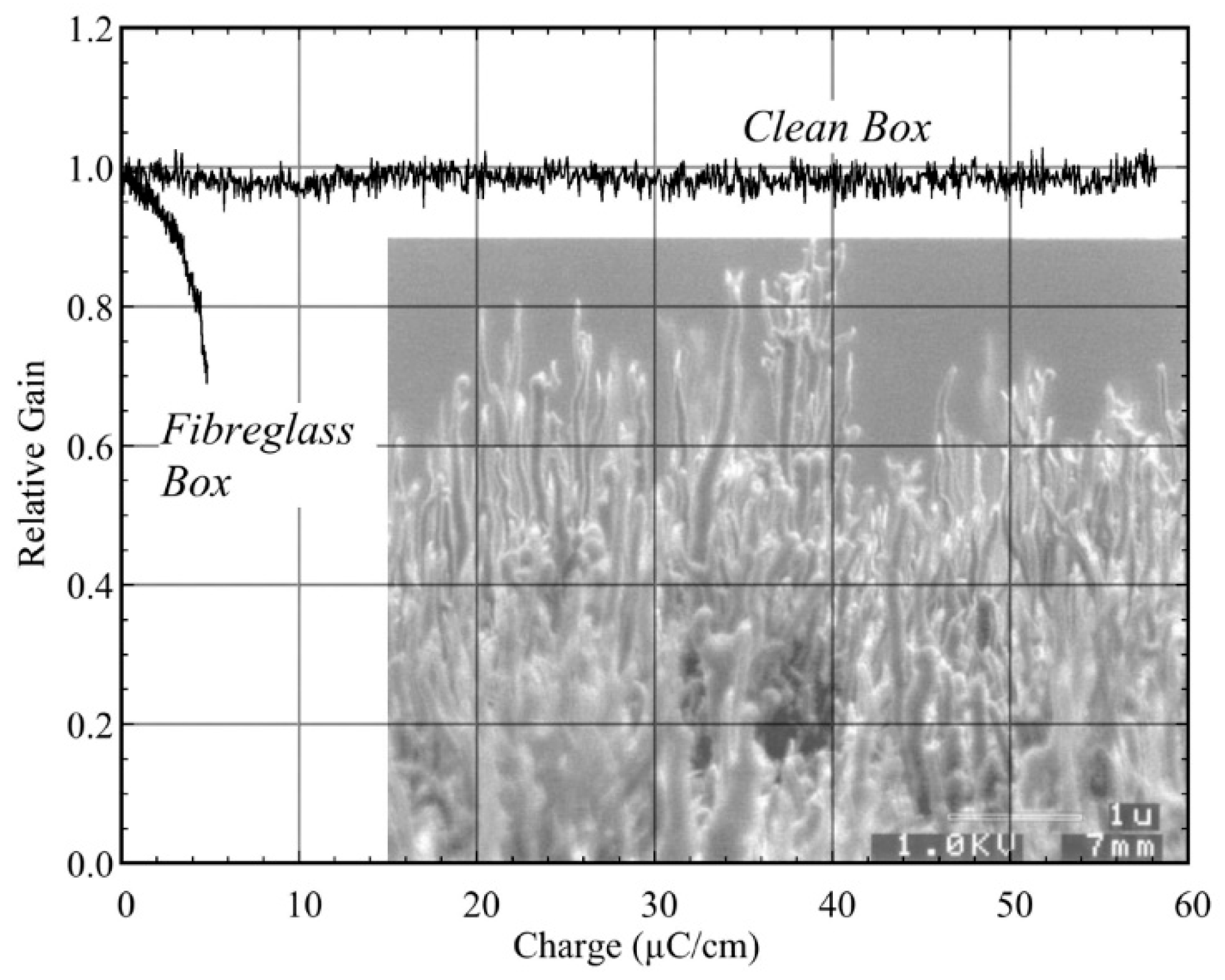
When operating in the cleanest conditions, the most frequent observation is the growth of a forest of sub-micron filaments, identified by several analytic methods to be mostly composed of silicones. Shown in the overlay of Figure 1 is a representative example of gain evolution at increasing collected charge, measured with MSGC plates assembled in a “clean” container or using epoxy-based fibreglass frames [5]. In the inset, an electron microscope picture shows an aged MWPC anode: the filaments appear to be mostly composed of silicone materials [6].
The common explanation for these observations is the presence in the gas flow of traces of silicone-based molecules, released by frames, gas pipes, lubricants and other components. As the vapour pressure of these pollutants is extremely low, the deposition process under avalanche conditions appears to be very efficient.
A small counter with a large linear anode development, inserted in the gas flow, could help in removing pollutants before they enter the main counter; a structure related to the MSGC, which is easy to manufacture with standard photolithographic technologies, could be used to this extent. The active filter can then be cleaned regularly or replaced.
This naïve concept needs to be supported by quantitative measurements and verifications; if effective, it could be extended to other industrial processes sensitive to silicone contamination.
3. Enhanced Secondary Electron Emission
A basic limit in the achievable time resolution of gaseous counters is set by the statistics of ionization energy loss. For fast charged particles, the mean distance between primary ionization clusters is around 300 µm at standard temperature and pressure (STP); with a typical value of mobility in high fields, this entails a dispersion in the arrival time of electrons at the anodes of about 5 ns FWHM (see for example [7]).
A possible way to overcome the limit was investigated long ago: it consists of coating the cathode of a gas counter with materials having large electron emissivity when traversed by charged particles. As these secondary electrons travel through equal distance to the anode, the statistical time dispersion is reduced.
The detection of fast electrons through secondary emission of a thin CsI layer was demonstrated in a pioneering work at the Weizmann Institute using a low-pressure electron multiplier [8]; the work has been extended to the detection of soft X-rays using CsI and other thin-layer deposits [9]. A group at the Lawrence Berkeley Laboratory (LBL) succeeded in detecting the signal produced from a ~200-µm-thick cesium iodide layer with a special columnar structure to enhance emissivity (Figure 2) [10]. Deposited on the cathode of a micro-strip counter detector, the layer can be subjected to a strong extraction field with the help of a grid place in contact with its inner surface. The figure shows the enhancement on the collected charge on application of the extraction potential, recorded exposing the counter to an electron source.
Further work seemed very promising [11]; however, the search seems to not have been pursued further.
Considering the substantial progress made with the deposition of thin layers, motivated by the development of the gaseous photon detectors used in Cherenkov ring imaging (see for example [12]), it would seem that this promising line of research should be pursued and might result in the development of sub-nanosecond timing devices.
4. Endoscopic X-Ray Generator
The gas electron multiplier (GEM) detector introduced by the author consists of a thin polymer foil metal-coated on both faces and pierced by a high density of holes, typically 50 to 100 mm−2 [13]. With a suitable gas filling and applied electrical potentials, it acts as an amplifier of the electrons released by ionizing radiation. GEM devices are basic constituents of large detector arrays used in particle physics experiments, and are used in many other applied fields [14].
It was suggested to the author by Ugo Amaldi, forefather of the TERA (i.e., the nickname for “TERApia adronica”) foundation dedicated to medical applications, that by operating a GEM structure in vacuum one could reach accelerating potentials large enough to generate keV X-rays hitting a metal target. A narrow tubular GEM structure could then be used to manufacture a thin, needle-like X-ray generator, possibly to be used for the endoscopic irradiation of delicate internal structures such as the prostate.
A conceptual design of the device is shown in Figure 3. A thin cylindrical metallic container encloses a hollow GEM-like electrode with a high density of holes, insulated from the external hose; a central thin filament constitutes the electron emitter (insert a). The structure could be realized using a metallized ceramic tube, pierced by mechanical or laser drilling. After manufacturing the internal components, the pipe is evacuated and sealed. With appropriate high voltages applied to the GEM electrodes and to the wire, the outer jacket being grounded, electrons emitted from the filament are transported into the holes and accelerated by the applied field (insert b). Hitting the outer jacket, they generate multi-keV X-ray emission spectra characteristic of the metal used. To enhance the electron field emission, one could imagine a micro-needle structure growth on the wire. Insert (c) shows an example, observed in a gaseous counter subjected to aging; another example is shown in the inset in Figure 1 [4].
The operation of the concept relies on two basic assumptions:
- -
- A GEM-like structure could hold in vacuum the required potential difference to generate X-rays of therapeutic interest—ten to fifteen kV;
- -
- A thin wire can generate a sufficient electron current by spontaneous field emission, without heating—possible, but a significant complication to the design. One way to enhance the spontaneous electron emission might be to coat the wire with a high density of sub-micron needles or filaments.
Initial tests by the author with a small prototype provided promising results; however, requiring a substantial capital investment for further developments, a realistic device was never realized.
5. Squaring the Circle
In argon-based gaseous detectors operated at STP, the mean absorption path for 10-keV photons is ~8 cm; to achieve the efficient detection of X-rays, the counters require a relatively thick conversion gap. For non-parallel photon fields, this introduces a parallax error in the localization, largely exceeding the intrinsic sub-mm localization properties of most detectors.
The author devised a way to overcome this limitation using the so-called planispherical chamber, making use of a specially designed GEM foil with the electrodes shaped as concentric rings, electrically insulated and powered with graded potentials to create a quasi-spherical electric field in the conversion gap [15]. Photons emitted at the focus of the structure convert along quasi-radial field lines, and provide a detected coordinate independent of the penetration (Figure 4). The structure can include one or more additional GEM foils to permit the attainment of the charge gain needed for detection. The position of the conversion can be recorded with a projective strip electronic readout [16], or with a simpler albeit rate-limited optical recording, as described for example in [17]. Promising results have been reported with a prototype device [18]; see the inserted recorded patterns in the figure.
The discrete size of the field-shaping electrodes, with insulating gaps, results in non-linearities and local efficiency losses, visible in the picture. This problem could be resolved using a continuous resistive electrode, with a polarizing current from centre to edges creating the required radially shaped electric field; to ensure linearity of response, the electrode resistivity should be well controlled and tailored with a value increasing from centre to periphery—a challenging but achievable technological accomplishment. Help in this direction may be provided by the development of diamond-like coatings, motivated by the requirements of gaseous micro-pattern devices (see for example [19]).
Coupled to an optical readout of the images, a planispherical GEM device could be used in many fields requiring portable, efficient and fast mapping of X-ray fields, such as fluorescence materials analysis, cultural heritage investigations, and crystal diffraction studies.
6. Prompt Gamma Imager
The treatment of deep tumours with charged particle beams is a widespread hospital practice, with more than thirty major therapy centres worldwide [20]. The abovementioned TERA foundation has markedly contributed to the development of hadrontherapy, which exploits the large increase in the differential energy loss of particles near the end of their penetration in matter (the so-called Bragg peak) to target the neoplasms while sparing the surrounding tissues. While thousands of patients have been successfully cured following elaborated treatment plans, no realistic tools exist to proactively assess the position and penetration of the beams during the exposures.
Of all outcomes of the beam-target interaction that can be exploited to this extent, the copious production of MeV photons (the so-called prompt gammas) is the most appealing for several reasons: photons are not scattered by the target body, they are generated with a reasonable rate until the end of the range, and their positional spectrum rather faithfully reproduces the shape of the Bragg peak [21].
Pixelized, crystal-based detectors, with good positional accuracy and detection efficiency have been successfully used to map the prompt gammas activity [22]; however, the devices require a rather complex electronic readout and data handling system, which is unappealing for wide use in hospital environments.
An alternative approach could be to use a monolithic scintillating crystal slab, where the photon conversion takes place, followed by a gaseous device capable of detecting and localizing the scintillation photons. A method was developed long ago that coupled barium fluoride crystals with a detector filled with a photosensitive gas mixture containing TMAE (tetrakis dimethyl amino ethylene: C2[(CH3)2N]4), a vapor with the exceptionally low photoionization threshold of 5.4 eV, adequate to detect part of the fast scintillation component of BaF2 centred at 6 eV. The device was successfully tested for charged particle calorimetry [23].
TMAE is not suitable for medical applications because it is rather chemically aggressive and difficult to handle. The development of low-threshold solid photosensitive materials capable of operating in a gaseous environment (e.g., cesium iodide) permits the revival of this approach. Taking into account the yield of the BaF2 fast scintillation component and of the CsI quantum efficiency, the photoelectron yield can be estimated to be of about two electrons per MeV; for 5 MeV gammas (i.e., the most abundant prompt emission), this results in a charge that is easily detected by a cascade of GEM electrodes.
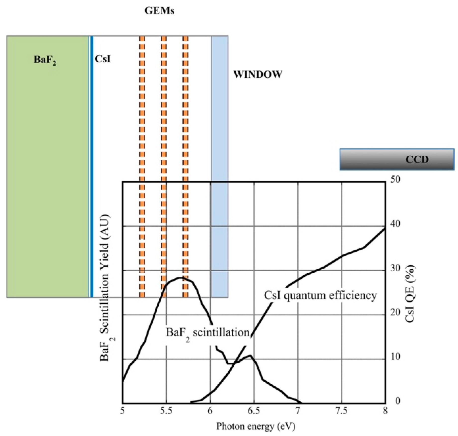
Figure 5 shows a schematic of the detector: a BaF2 crystal, coated on one side with the photosensitive CsI layer, is mounted on the cathode side of the multi-GEM system; photoelectrons emitted by the layer drift to the multiplier and are amplified.
While a system of electronic detection could be used (e.g., those implemented in Cherenkov ring imaging), the development of a GEM-based optical recording system mentioned above would permit great simplification of the detector. For the optical readout version of the device, a window separates the active detector from a solid-state sensor. The insert shows the BaF2 scintillation yield and the CsI quantum efficiency as a function of photon energy. The gas scintillation yield is particularly abundant in the visible wavelengths, and is easy to detect for a gas mixture including carbon tetrafluoride [24].
Proposed by the author some time ago, this approach seems not to have been pursued further. The concurrent development of alternative photosensitive layers, better protected from damages due to positive ions and impurities in the gas, may help the realization of prompt gamma imagers suited for clinical use.
7. Porous Resistive Dielectrics
The statistics of ionization set a limit to the achievable time resolution in the detection of charged particles with a gaseous device. A possible solution—the deposit on the cathode of thin layers with large enough secondary electron emission—was discussed in Section 2. An interesting alternative is the use of a thick porous dielectric sheet as sensitive medium (Figure 6a) [25]. Electrons released at the surface of the pores are accelerated (in vacuum) by an applied electric field and hit the pores’ wall, creating secondary electrons; the process continues until the amplified charge emerges from the dielectric, and is collected by strips or wires. With a porous CsI layer about 1 mm thick, embedded in a multiwire chamber, detection efficiencies close to 100% have been demonstrated for soft X-rays and alpha particles. However, the device suffers from severe charging-up problems, even at moderate rates, due to the accumulation of charges on the dielectric [26]. Added to the need of operating at near vacuum, in practice this difficulty has prevented the wide use of the detector.
A technology for manufacturing large-volume, low-density materials has been developed to act as radiators for Cherenkov counters; named silica aerogels, the materials are transparent, highly porous, open cell foams with a structure that can be tailored to the needs; Figure 6b shows an example of a silica aerogel slab (for a review of aerogel properties and manufacturing methods, see [27]).
Owing to their constitution, aerogels seem suitable for use as radiation detector, following the approach described above. This has indeed been attempted, with promising results, replacing the gas filling in the ionization chamber with an argon-saturated aerogel block. Under application of a suitable electric field, exposing the device to an X-ray generator, a current enhancement by almost two orders of magnitude has been observed with an aerogel compared to a gas filling [28]. However, the device suffers from instabilities and non-linearities and of operation are imputed to trapping–detrapping of charges by the insulating silica matrix.
Similar instabilities due to the surface charging of the glass support were observed in the early development of the micro-strip gaseous counters; they were solved to a large extent by the development of custom-made glass supports having low resistivity. Called “Pestov glasses”, from the name of one of the original developers [29], the materials have resistivities between 109 and 1012 Ω cm [30]. A value of resistivity that is stable in time and a linear dependence of current on voltage indicate the ohmic nature of conductivity, due to electrons, as against the ionic conductivity of borosilicate glass.
One can then speculate on the possibility of developing an aerogel-like material having as constituents the components of an electron-conducting glass as described above. A porous resistive glass detector could then have the structure shown in (Figure 6c: sandwiched between two thin glass or ceramic plates with the voltage distribution and readout strips, the detector should be able to withstand the large difference of potential required for the collection and multiplication of charges, and could operate either in near-vacuum or with a noble gas filling. Anodes and cathodes could be realized with resistive diamond-like carbon (DLC) coatings; this technology, developed for use with micro-pattern detectors, can provide thin layers with resistivities that are tunable in a wide range (i.e., MΩ/square to GΩ/square—see for example [31]). One millimetre thick and with an area of 10 × 10 cm2, if realized with a glass with ~1010 Ω cm, such a detector would have a resistance between electrodes of ~100 MΩ—possibly acceptable for operation (assuming that the bulk resistivity of aerogel scales with its density). Signals are capacitively picked up through the resistive electrodes by external strips at ground potential along perpendicular directions.
This approach is highly hypothetical, and would permit the achievement of good efficiencies and time resolutions in the detection of fast charged particles, while being cheap and light enough to be affordable in experimental setups.
8. Conclusions
This note describes six research projects on advanced radiation detectors, seemingly interesting but for various reasons not pursued by the original proponents (including the author). In case of positive completion, the projects may have appealing applications for particle physics experimentation and other fields, namely:
- -
- An active filter for impurities, possibly offering a way to remove traces of silicon compounds from a gas flow;
- -
- A fast, position-sensitive charged-particle detector exploiting the enhanced secondary electron emission from the cathodes of gaseous counters;
- -
- A soft X-rays generator with a needle-like device, suitable for endoscopic irradiation;
- -
- A method to remove the parallax error intrinsic in the use of thick conversion volume in X-ray imagers, largely increasing the detection efficiency for fluorescence analysis and X-ray diffraction studies;
- -
- A position-sensitive imager for gamma rays in the MeV region, providing real-time monitoring of the irradiation profile in hadrontherapy;
- -
- The development of a porous, high-resistivity material suitable to replace the gas as sensitive medium in counters, providing improved efficiency and time resolutions.
The likelihood of these approaches varies from realistic to rather unreasonable, but not impossible; hopefully, this note will encourage further research to assess their interest.
Funding
This research received no external funding.
Conflicts of Interest
The author declares no conflict of interest.
References
- Charpak, G.; Bouclier, R.; Bressani, T.; Favier, J.; Zupancic, C. The use of multiwire proportional counters to select and localize charged particles. Nucl. Instr. Meth. 1968, 62, 262. [Google Scholar] [CrossRef]
- Oed, A. Position-sensitive detector with microstrip anode for electron multiplication with gases. Nucl. Instr. Meth. Phys. Res. A 1988, 263, 351–359. [Google Scholar] [CrossRef]
- Va’vra, J. Physics and chemistry of aging-early developments. Nucl. Instr. Meth. Phys. Res. A 2003, 515, 1–14. [Google Scholar] [CrossRef]
- Kadyk, J.A. Proceedings of the Workshop on Radiation Damage to Wire Chambers; Lawrence Berkeley National Laboratory: Berkeley, CA, USA, 1986. [Google Scholar]
- Bouclier, R.; Garabatos, C.; Manzin, G.; Million, G.; Sauli, F.; Temmel, T.; Shekhtman, L. Ageing studies with microstrip gas chambers. Nucl. Instr. Meth. Phys. Res. A 1994, 348, 109–118. [Google Scholar] [CrossRef] [Green Version]
- Binkley, M.; Wagner, R.L.; Mukherjee, A.; Ambrose, D.; Bauer, G.; Khazins, D.M.; Atac, M. Aging in large CDF tracking chambers. Nucl. Instr. Meth. Phys. Res. A 2003, 515, 53–59. [Google Scholar] [CrossRef] [Green Version]
- Sauli, F. Gaseous Radiation Detectors: Fundamentals and Applications; Cambridge University Press: Cambridge, UK, 2014. [Google Scholar]
- Chechik, R.; Breskin, A.; Aclander’, H.; Comforti, E.; Gibrekhterman, A. Secondary electron emission from thin CsI films induced by relativistic electrons. Nucl. Instr. Meth. Phys. Res. A 1994, 342, 458–465. [Google Scholar] [CrossRef]
- Bieskin, A.; Chechik, R.; Gibrekhterman, A.; Levinson, L.; Notea, A.; Weingarten, B. Secondary electron emission gaseous detectors for fast X-ray imaging. Nucl. Instr. Meth. Phys. Res. A 1994, 353, 302–306. [Google Scholar] [CrossRef]
- Cho, H.S.; Park, I.J.; Hong, W.S.; Perez-Mendez, V.; Kadyk, J. Utilization of a thin columnar cesium iodide (CsI) layer in gas avalanche microdetector. Nucl. Instr. Meth. Phys. Res. A 1999, 422, 269–272. [Google Scholar] [CrossRef]
- Fonte, P.; Peskov, V. Micro-gap parallel-plate chambers with porous secondary electron emitters. Nucl. Instr. Meth. Phys. Res. A 2000, 454, 260–266. [Google Scholar] [CrossRef]
- Di Mauro, A. Status and perspectives of gaseous photon detectors. Nucl. Instr. Meth. Phys. Res. A 2014, 766, 126–132. [Google Scholar] [CrossRef] [Green Version]
- Sauli, F. GEM: A new concept for electron amplification in gas detectors. Nucl. Instr. Meth. Phys. Res. A 1997, 386, 531–534. [Google Scholar] [CrossRef]
- Sauli, F. The gas electron multiplier (GEM): Operating principles and applications. Nucl. Instr. Meth. Phys. Res. A 2016, 805, 2–24. [Google Scholar] [CrossRef]
- Sauli, F. Radiation Detector of Very High Performance and Planispherical Parallax-Free X-ray Imager Comprising such Radiation Detector. Patent WO/99/21211, 24 April 1999. [Google Scholar]
- Bressan, A.; de Oliveira, R.; Gandi, A.; Labbé, J.C.; Ropelewski, L.; Sauli, F.; Mörmann, D.; Müller, T.; Simonis, H.J. Two-dimensional readout of GEM detectors. Nucl. Instr. Meth. Phys. Res. A 1999, 425, 254–261. [Google Scholar] [CrossRef] [Green Version]
- Sauli, F. Radiation imaging with gaseous detectors. Nucl. Instr. Meth. Phys. Res. A 2018, 878, 1–9. [Google Scholar] [CrossRef]
- Brunbauer, F.; Ropelewski, L.; Sauli, F. The planispherical chamber: A parallax-free X-ray gaseous detector for imaging applications. Nucl. Instr. Meth. Phys. Res. A 2017, 875, 16–20. [Google Scholar] [CrossRef]
- Abbrescia, M.; Peskov, V.; Fonte, P. Resistive Gaseous Detectors: Designs, Performance, and Perspectives; John Wiley & Sons: Hoboken, NJ, USA, 2018. [Google Scholar]
- Amaldi, U.; Bonomi, R.; Braccini, S.; Crescenti, M.; Degiovanni, A.; Garlasch, M.; Garonna, A.; Magrin, G.; Mellace, C.; Pearce, P.; et al. Accelerators for hadrontherapy: From Lawrence cyclotrons to linacs. Nucl. Instr. Meth. Phys. Res. A 2010, 620, 563–577. [Google Scholar] [CrossRef]
- Krimmer, J.; Dauvergne, D.; Létang, J.M.; Testa, É. Prompt-gamma monitoring in hadrontherapy: A review. Nucl. Instr. Meth. Phys. Res. A 2018, 878, 58–73. [Google Scholar] [CrossRef]
- Perali, I.; Celani, A.; Busca, P.; Fiorini, C.; Marone, A.; Basilavecchia, M.; Frizzi, T.; Roellinghoff, F.; Smeets, J.; Prieels, D.; et al. Prompt gamma imaging with a slit camera for real-time range control in proton therapy: Experimental validation up to 230 MeV with HICAM and development of a new prototype. IEEE Nucl. Sci. Symp. Conf. Rec. 2012, 3371, 3883–3886. [Google Scholar]
- Anderson, D.F.; Bouclier, R.; Charpak, G.; Majewski, S.; Kneller, G. Coupling of a BaF2 scintillator to a TMAE photocathode and a low- pressure wire chamber. Nucl. Instr. Meth. 1983, 217, 217. [Google Scholar] [CrossRef]
- Fraga, M.; Fraga, F.A.F.; Fetal, S.T.G.; Margato, L.M.S.; Marques, R.F.; Policarpo, A. The GEM scintillation in He-CF4, Ar-CF4, Ar-TEA and Xe-TEA mixtures. Nucl. Instr. Meth. Phys. Res. A 2003, 504, 88–92. [Google Scholar] [CrossRef]
- Gavalian, V.G.; Lorikyan, M.P.; Markarian, K.J. Multiwire particle detectors based on porous dielectric layers. Nucl. Instr. Meth. Phys. Res. A 1994, 350, 244–249. [Google Scholar] [CrossRef]
- Lorikyan, M.P.; Asryan, G.A.; Gary, C.K. Investigation of porous dielectric detectors at high intensity particles. Nucl. Instrum. Methods Phys. Res. A 2007, 570, 475–478. [Google Scholar] [CrossRef]
- Fricke, J.; Tillotson, T. Aerogels: Production, characterization, and applications. Thin Solid Film. 1997, 297, 212–223. [Google Scholar] [CrossRef]
- Caresana, M.; Zorloni, G. Preliminary study of silica aerogel as a gas-equivalent material in ionization chambers. Nucl. Instr. Meth. Phys. Res. A 2017, 874, 35–42. [Google Scholar] [CrossRef]
- Frolov, A.R.; Pestov, Y.N.; Primachek, V.V. Position resolution of the spark counter with a localized discharge. Nucl. Instr. Meth. 1991, A307, 497. [Google Scholar] [CrossRef]
- Bouclier, R.; Florent, J.J.; Gaudaen, J.; Millon, G.; Pasta, A.; Ropelewski, L.; Sauli, F.; Shekhtman, L.I. High flux operation of microstrip gas chambers on glass and plastic supports. Nucl. Instr. Meth. Phys. Res. A 1992, 323, 240–246. [Google Scholar] [CrossRef] [Green Version]
- Zhou, Y.; Lv, Y.; Shang, L.; Hong, D.; Song, G.; Liu, J.; Feng, J.; Shao, M.; Wang, X.; Zhang, Z. Fabrication and performance of a μRWELL detector with Diamond-Like Carbon resistive electrode and two-dimensional readout. Nucl. Instr. Meth. Phys. Res. A 2019, 927, 31–36. [Google Scholar] [CrossRef]
Figure 1.
An example of the fast aging of micro-strip gas counter (MSGC) plates in clean or fibreglass assemblies, and of silicone filaments growth on a multiwire proportional chamber (MWPC) anode.
Figure 1.
An example of the fast aging of micro-strip gas counter (MSGC) plates in clean or fibreglass assemblies, and of silicone filaments growth on a multiwire proportional chamber (MWPC) anode.

Figure 2.
Detected signal enhancement on application of an extraction field on the CsI layer (full curve) compared to the ionization signal directly released by fast electrons (dashed curve). The inset shows a microscope view of a section through the columnar CsI layer [10].
Figure 2.
Detected signal enhancement on application of an extraction field on the CsI layer (full curve) compared to the ionization signal directly released by fast electrons (dashed curve). The inset shows a microscope view of a section through the columnar CsI layer [10].

Figure 3.
(a) Schematic of the gas electron multiplier (GEM)-based endoscopic X-ray generator (not to scale); (b) close-up of two GEM holes with the electric field lines; (c) example of sub-micron discharge-grown spikes on a thin wire.
Figure 3.
(a) Schematic of the gas electron multiplier (GEM)-based endoscopic X-ray generator (not to scale); (b) close-up of two GEM holes with the electric field lines; (c) example of sub-micron discharge-grown spikes on a thin wire.

Figure 4.
Schematics and field lines in the planispherical detector; the GEM electrode is patterned with rings powered at appropriate potentials to create a quasi-radial field. In the insert, the image of a square reticle placed in front of the detector with planar (a) and spherical field (b) demonstrate the correction of the parallax error [18]. The black rings correspond to the separations between the various rings.

Figure 5.
Schematic of the prompt gamma imager, barium fluoride scintillation yield, and cesium iodide quantum efficiency.
Figure 5.
Schematic of the prompt gamma imager, barium fluoride scintillation yield, and cesium iodide quantum efficiency.

Figure 6.
(a) Schematic of the porous dielectric detector; (b) an example of a silica aerogel block; (c) the proposed detector with a resistive aerogel sensitive volume. Internally diamond-like carbon (DLC)-coated glass plates supply the operating voltage; external readout strips provide two-dimensional localization.

© 2019 by the author. Licensee MDPI, Basel, Switzerland. This article is an open access article distributed under the terms and conditions of the Creative Commons Attribution (CC BY) license (http://creativecommons.org/licenses/by/4.0/).
Share and Cite
MDPI and ACS Style
Sauli, F. Six Concepts in Search of an Author. Instruments 2019, 3, 51. https://doi.org/10.3390/instruments3030051
AMA Style
Sauli F. Six Concepts in Search of an Author. Instruments. 2019; 3(3):51. https://doi.org/10.3390/instruments3030051
Chicago/Turabian StyleSauli, Fabio. 2019. "Six Concepts in Search of an Author" Instruments 3, no. 3: 51. https://doi.org/10.3390/instruments3030051