Review

MXene-Based Terahertz Metamaterial Biosensors: From Laboratory Simulation to Clinical Application


1
School of Physics, University of Electronic Science and Technology of China, Chengdu 611731, China
2
State Key Laboratory of Electronic Thin Film and Integrated Devices, University of Electronic Science and Technology of China, Chengdu 611731, China
*
Authors to whom correspondence should be addressed.
These authors contributed equally to this work.
Condens. Matter 2026, 11(2), 21; https://doi.org/10.3390/condmat11020021
Submission received: 16 April 2026 / Revised: 23 May 2026 / Accepted: 25 May 2026 / Published: 28 May 2026
(This article belongs to the Special Issue Flexible Matter for Electronics, Photonics, and Energy Conversion)

Abstract

Terahertz (THz) metamaterial biosensors have emerged as a powerful platform for label-free, non-ionizing biodetection, yet their clinical translation is severely hindered by limited sensitivity, poor anti-interference capability, and a fragmented research chain that rarely extends beyond simulation. Two-dimensional transition metal carbides/nitrides (MXenes) offer a transformative alternative to conventional gold-based metamaterials, providing metal-like high conductivity, abundant surface functional groups for specific biomolecular capture, excellent biocompatibility, and mechanical flexibility. This review systematically examines the recent progress of MXene-based THz metamaterial biosensors, covering structural design strategies, material synergistic system, machine learning-assisted optimization, and performance evaluation metrics. While most studies remain in the simulation stage, a landmark in vivo validation by Yang et al. achieved real-time thrombus monitoring with 94.7% sensitivity and 92.3% specificity, bridging the gap between simulation and clinical application. We identified key bottlenecks hindering clinical translation and propose future directions toward clinically adaptive, full-chain development. This review provides a roadmap for transitioning MXene-based THz biosensors from laboratory simulation to practical point-of-care diagnostics.

1. Introduction

The rapid advancement of precision medicine, point-of-care testing (POCT), and wearable health monitoring has imposed unprecedented demands on biodetection technologies. These technologies must not only possess high sensitivity, rapid response, and minimally invasive or even non-invasive characteristics but also be capable of operating reliably in complex physiological environments to enable continuous, real-time monitoring of physiological signals [1]. While traditional laboratory methods, such as polymerase chain reaction and enzyme-linked immunosorbent assay, guarantee accuracy, they suffer from several limitations, including complex operational procedures, reliance on bulky equipment, and rigid structural designs. MXene-based THz metamaterial biosensors, the focus of this review, address some of these limitations—specifically, they offer label-free detection, eliminate the need for complex sample processing, and enable miniaturization potential through chip-scale integration. However, it must be acknowledged that MXene-THz sensors still face certain challenges, such as limited sensitivity in complex biological matrices, susceptibility to environmental interference, and long-term stability concerns under physiological conditions. Nevertheless, these obstacles are by no means fatal; through rational material engineering, optimized metamaterial designs, surface functionalization, and encapsulation strategies, the field has successfully overcome many of these issues, demonstrating the strong potential of MXene-based platforms for practical biosensing applications. Consequently, the development of novel biosensors that combine high sensitivity with rapid, label-free detection has become a central research focus in the field of biophotonics [2].
Terahertz (THz) waves (0.1–10 THz) are regarded as highly promising biophotonic probes due to their unique physical properties [2,3]. Firstly, THz waves possess low photon energy (on the order of millielectronvolts), which does not cause ionizing damage to biological tissues, offering inherent safety [4,5,6]. Secondly, the rotational and vibrational energy levels of many biological macromolecules (e.g., proteins, DNA) fall within the THz frequency range, creating characteristic “fingerprint spectra” that enable label-free detection [7]. This “fingerprint spectra” originates from the resonant absorption of collective molecular vibration modes, which are highly specific to molecular conformation and intermolecular interactions, such as hydrogen bonding networks and torsional motions of the molecular backbone [8]. Different molecules, owing to their distinct structural configurations, exhibit unique absorption peaks within the terahertz (THz) range. This characteristic spectral signature enables the label-free and highly specific identification of biological macromolecules, such as proteins and DNA, based on their intrinsic physical properties [9]. Furthermore, THz waves are highly sensitive to water molecules, allowing indirect assessment of the physiological state of biological tissues by detecting changes in their hydration status [10,11,12]. However, the practical application of traditional THz biosensing systems faces a core bottleneck: limited intrinsic sensitivity, making it difficult to detect trace amounts of biomarkers (typically requiring millimolar concentrations) in complex biological matrices [13]. This limitation has significantly hindered the widespread adoption of THz technology in clinical biomedical applications [14].
In recent years, metamaterials have emerged as the core solution to break through the sensitivity bottleneck of THz biosensing. Through the design of subwavelength artificial structures, metamaterials can confine electromagnetic fields to subwavelength scales, generating significant localized field enhancement effects that amplify sensing signals by 102–106 times. This characteristic fundamentally solves the problem of weak light-matter interaction in traditional THz biosensing and has become the mainstream structural platform for current high-sensitivity THz biosensors. There were two common THz metamaterial sensing configurations: (a) a planar metasurface and (b) a metamaterial absorber with a dielectric spacer. At present, most THz metamaterial biosensors use gold (Au) as the core functional material, which has excellent electrical conductivity and chemical stability [15]. However, gold-based metamaterials have inherent limitations in clinical biomedical applications: first, the chemically inert surface of gold makes it difficult to covalently immobilize biological recognition elements (antibodies, aptamers, etc.), resulting in poor specific binding ability to target biomarkers; second, the non-specific adsorption of high-abundance proteins in biological fluids on the gold surface is severe, which will overwhelm the target signal in real clinical samples [16,17,18].
The emergence of the two-dimensional transition metal carbides/nitrides (MXenes) offers a perfect solution to the above limitations of gold-based THz metamaterials [19]. MXenes, such as Ti3C2Tx, possess three core advantages for THz biosensing applications: (1) metal-like high electrical conductivity (up to ∼104 S/cm), which can optimize the impedance matching of metamaterials, enhance localized field enhancement, and maintain excellent electromagnetic response in the THz band [20]; (2) abundant surface functional groups (-F, -OH, =O), which enable easy covalent functionalization of biological recognition elements, enhance specific binding to target biomarkers, and inhibit non-specific adsorption; (3) excellent biocompatibility and biosafety, which have been widely verified in in vitro cell experiments and in vivo animal models, meeting the basic requirements of clinical biomedical applications. (4) excellent mechanical flexibility, laying the material foundation for the development of wearable devices. To elaborate, the metallic-like conductivity of MXenes arises from their unique layered structure and high carrier concentration (~1021 cm−3), enabling low-dispersion electromagnetic response across the entire 0.5–10 THz spectrum—a crucial feature for achieving broadband impedance matching in metamaterial designs [21]. The abundant surface terminations not only facilitate covalent immobilization of antibodies and aptamers but also render the MXene surface hydrophilic, which effectively suppresses non-specific adsorption of high-abundance proteins in complex biological fluids such as whole blood and serum—a long-standing challenge for conventional gold surfaces [22,23]. Moreover, MXenes are widely recognized for their excellent biocompatibility and biosafety, which are essential prerequisites for biomedical and clinical applications. These intrinsic properties collectively position MXene as a transformative material platform that not only matches but, in many aspects, surpasses conventional noble metals for THz biosensing applications. These unique properties make MXene an ideal alternative to gold for the next generation of THz metamaterial biosensors [24].
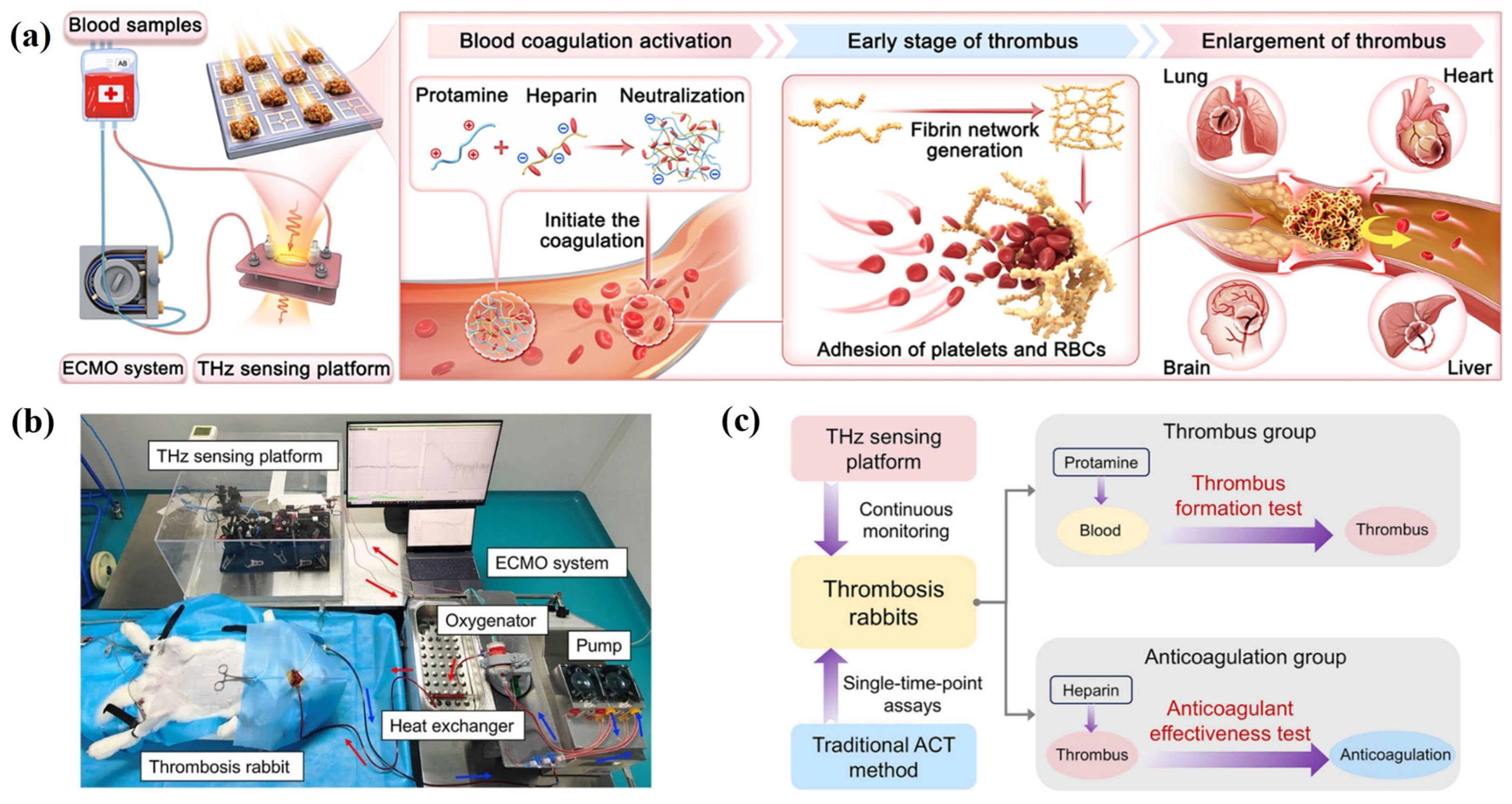
Research on MXene-based THz metamaterial biosensors has been flourishing in recent years [25]. By optimizing material synergy systems (e.g., MXene/graphene, MXene/black phosphorus, graphene/metal, etc.) and structural topologies (e.g., ring-shaped, H-shaped, T-shaped, M-shaped, and multi-layer stacks), researchers have achieved impressive performance metrics in simulations and in vitro experiments: sensitivities up to 10,000 GHz/RIU, detection limits as low as 0.014 RIU, and expanded detection targets ranging from viruses and cancer biomarkers to environmental toxins [26]. These studies not only validate the theoretical feasibility of the MXene-based metamaterial platform, but also accumulate a substantial database of foundational data for subsequent structural optimization and performance enhancement [27]. However, almost all of these studies relied on electromagnetic simulations or idealized in vitro solution experiments, leaving the sensor’s performance in real, dynamic, and interference-rich “in vivo” biological environments unresolved. Until very recently, Yang et al. [28,29] reported the first application of an MXene-THz metamaterial sensor in an in vivo animal model. They integrated this sensor into an extracorporeal membrane oxygenation (ECMO) circuit, achieving real-time, continuous monitoring of the entire thrombus formation process in rabbits. They validated its diagnostic performance using 78 clinical blood samples, achieving a sensitivity of 94.7% and specificity of 92.3%. Furthermore, density functional theory calculations revealed the charge transfer mechanism at the MXene-thrombus interface [28]. This groundbreaking work marks a significant transition for the field of MXene-based THz biosensing from a “simulation-driven” phase into a new stage of “preclinical validation” [30].
While several excellent reviews have separately summarized the progress of MXene-based biosensors or THz metamaterial biosensors, none have systematically bridged these two fields with a specific focus on the translational pathway from laboratory simulation to clinical application. Previous reviews have primarily concentrated on material synthesis, general biosensing mechanisms, or THz metamaterial design principles, largely overlooking the fundamental disconnection between simulation-driven optimization and real-world clinical validation. More critically, the field still faces three unresolved clinical challenges that pure simulation studies cannot address: the lack of quantitative correlation between simulation metrics and clinical diagnostic outcomes, the complete neglect of physiological variables in ideal models, and a fragmented research chain that rarely extends beyond in vitro validation. The landmark in vivo work by Yang et al. has fundamentally shifted the field toward preclinical validation, calling for a timely re-evaluation from a clinical perspective. In contrast to existing overviews that remain either materials- or simulation-focused, this review provides a systematic side-by-side comparison of conventional gold-based and MXene-based platforms, a critical analysis of clinical translation bottlenecks, a clear distinction between simulation-oriented and application-oriented performance metrics, and a full-chain roadmap from material design to preclinical validation, thereby establishing its unique importance in the field.
The field of MXene-based THz metamaterial biosensors still faces several critical challenges that must be addressed to enable clinical translation. First, the vast majority of studies remain at the pure simulation stage, with optimized sensitivity and Q-factor achieved in ideal electromagnetic models that bear little resemblance to real-world physiological environments. Second, the performance of these sensors in complex biological matrices such as whole blood, serum, and tissue fluid remains largely unverified, leaving critical issues, including non-specific protein adsorption, material stability at 37 °C, and dynamic flow interference unresolved. Third, the lack of standardized evaluation frameworks that bridge simulation-oriented metrics and clinical diagnostic outcomes makes it difficult to compare different studies or assess their true translational potential. Fourth, scalable fabrication of high-precision MXene metamaterial arrays at low cost remains a significant manufacturing hurdle. Without systematic efforts to overcome these challenges, the remarkable performance demonstrated in simulations will likely never reach clinical practice.
This review systematically summarizes the research progress of MXene-based THz metamaterial biosensors, with a focus on bridging the critical gap between simulation-oriented design and clinical translation. It covers the fundamental principles of THz metamaterial biosensing, the inherent limitations of conventional gold-based devices, the material advantages of MXene and its composite synergistic systems, structural design strategies tailored for MXene, and the landmark preclinical validation that has shifted the field toward clinic-oriented research. By providing a clear roadmap from “laboratory simulation” to “clinical application,” this review aims to guide researchers in developing next-generation THz biosensors that are not only high-performing in ideal environments but also clinically viable, robust, and translation-ready, ultimately accelerating the adoption of MXene-based THz technologies in precision medicine and point-of-care diagnostics.

2. Fundamental Concepts of THz Metamaterial Biosensing

2.1. Basic Principles of THz Metamaterial Biosensing

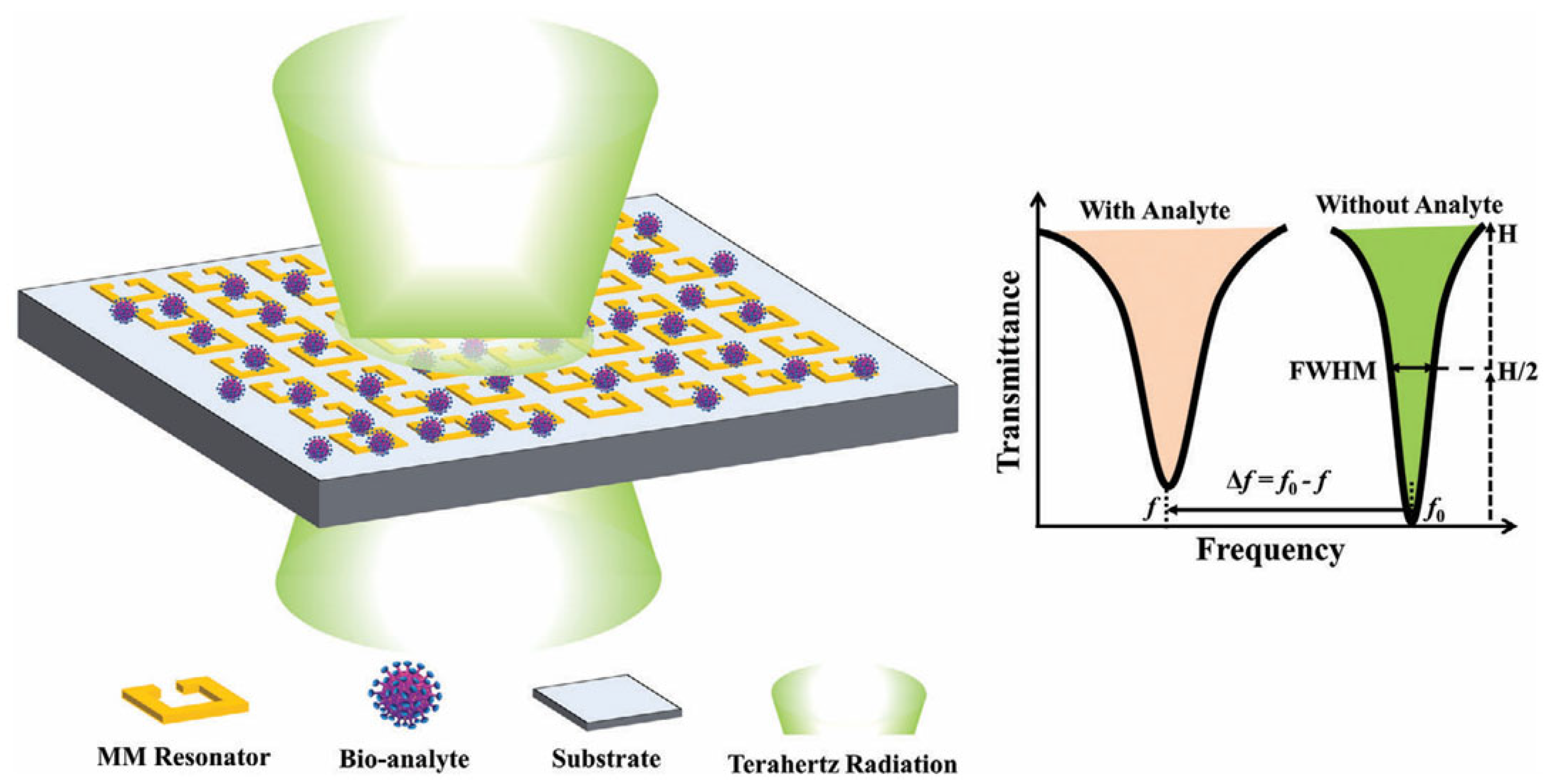
The core physical mechanism of THz metamaterial biosensing is the interaction between THz electromagnetic waves and metamaterial resonant structures, as well as the high sensitivity of this interaction to changes in the surrounding dielectric environment. When THz waves are incident on the metamaterial array, if the frequency of the incident wave matches the intrinsic resonant frequency of the metamaterial unit cell, strong electromagnetic resonance occurs, and the electromagnetic field is highly confined in the subwavelength gap of the resonant structure, forming a significant localized field enhancement effect (usually 102–106 times the electric field intensity of the incident wave). The resonant frequency and transmittance/absorbance of the metamaterial are extremely sensitive to the refractive index change in the medium in the field enhancement region. When the target biomolecule binds to the sensor surface, it causes a tiny change in the local refractive index, which in turn leads to a measurable shift in the resonant frequency or a change in the transmittance amplitude. This is the core working principle of THz metamaterial biosensors [31]. Figure 1 schematically depicts the redshift of the resonance peak in transmission, reflection, or absorption spectra upon analyte binding.
Compared with traditional optical biosensing technologies, THz biosensing has three unique advantages for biomedical applications: first, the low photon energy of THz waves will not cause ionizing damage to biological samples, enabling non-destructive and label-free detection; second, the vibrational and rotational energy levels of biological macromolecules are in the THz band, which can realize specific fingerprint recognition of molecules; third, THz waves are highly sensitive to the hydration state of biological tissues and molecules, which can reflect the physiological and pathological changes in organisms at the molecular level [32]. The combination of MXene materials and metamaterial structures further amplifies these advantages: MXene’s high conductivity optimizes the resonance characteristics of metamaterials, enhances the localized field effect, and its abundant surface functional groups provide abundant binding sites for biomolecules, laying a solid foundation for high-sensitivity and high-specificity biosensing [22,24].

2.2. Design of THz Metamaterial Biosensors

2.2.1. THz Metamaterial Resonant Structures

Metamaterials constitute a class of artificially engineered sub-wavelength periodic structures whose electromagnetic response is primarily determined by the precise geometry and spatial arrangement of their unit cells rather than the intrinsic properties of the constituent materials. By meticulously tailoring the dimensions, morphology, and configuration of these unit cells, metamaterials can achieve effective permittivity and permeability that are not naturally occurring or are difficult to realize, enabling unique phenomena such as negative refraction, perfect lensing, and electromagnetic cloaking [33]. In the terahertz (THz) frequency band, where the response of natural materials to THz radiation is generally weak, the strategic design of metamaterials becomes critically important. The ability of metamaterials to manipulate electromagnetic waves originates from electromagnetic resonances within the unit cells: when the frequency of the incident wave matches the intrinsic resonant frequency of the structure, energy is efficiently coupled and confined, generating a pronounced enhancement of the local electromagnetic field in specific regions [34]. This localized field enhancement is exquisitely sensitive to changes in the surrounding dielectric environment, constituting the core physical mechanism that enables highly sensitive biosensing [20,35].
Metamaterials enable precise control over electromagnetic waves through subwavelength periodic structures, with the core principle being the manipulation of effective permittivity and permeability via geometric design, thereby generating localized field enhancement effects. In the field of THz biosensing, the design of metamaterial structures directly determines the sensor’s sensitivity, quality factor, and operational bandwidth [36]. Based on their geometric configuration, metamaterials can be broadly categorized into electric resonators (e.g., split-ring resonators, dipole antennas) and magnetic resonators (e.g., split-ring resonators, double split-ring resonators), which are primarily responsive to the electric and magnetic field components of incident electromagnetic waves, respectively. This sensitivity arises from the distinct current distributions and near-field patterns excited within each resonator type. By strategically combining these electric and magnetic resonant elements within a unit cell or array, it is possible to finely manipulate the effective impedance of the metamaterial. This allows for precise control over the amplitude, phase, and polarization state of the transmitted or reflected wave, enabling the design of structures that meet specific impedance-matching conditions for enhanced wave-matter interaction or tailored electromagnetic functionality. This section summarizes four main categories of metamaterial structural design approaches widely adopted in this field.
(1)
Ring Resonators and Disk Resonators
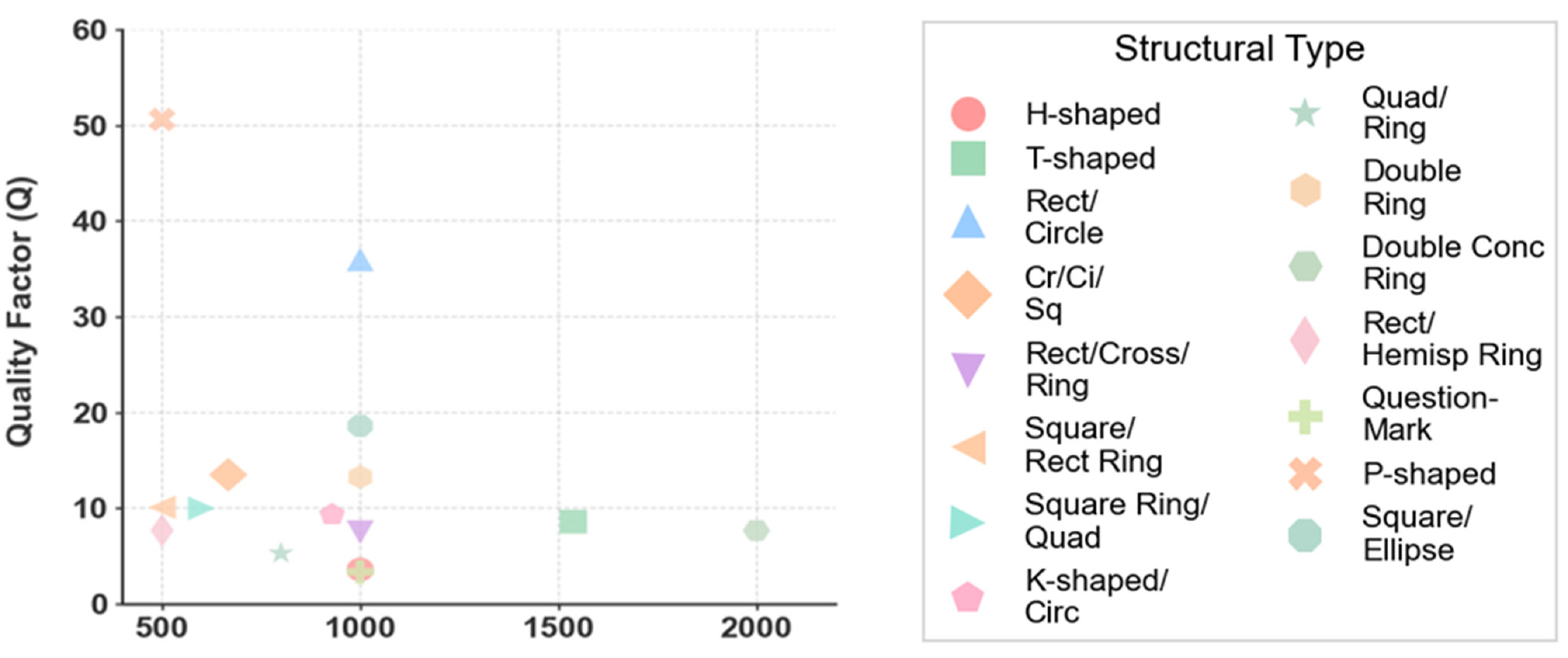
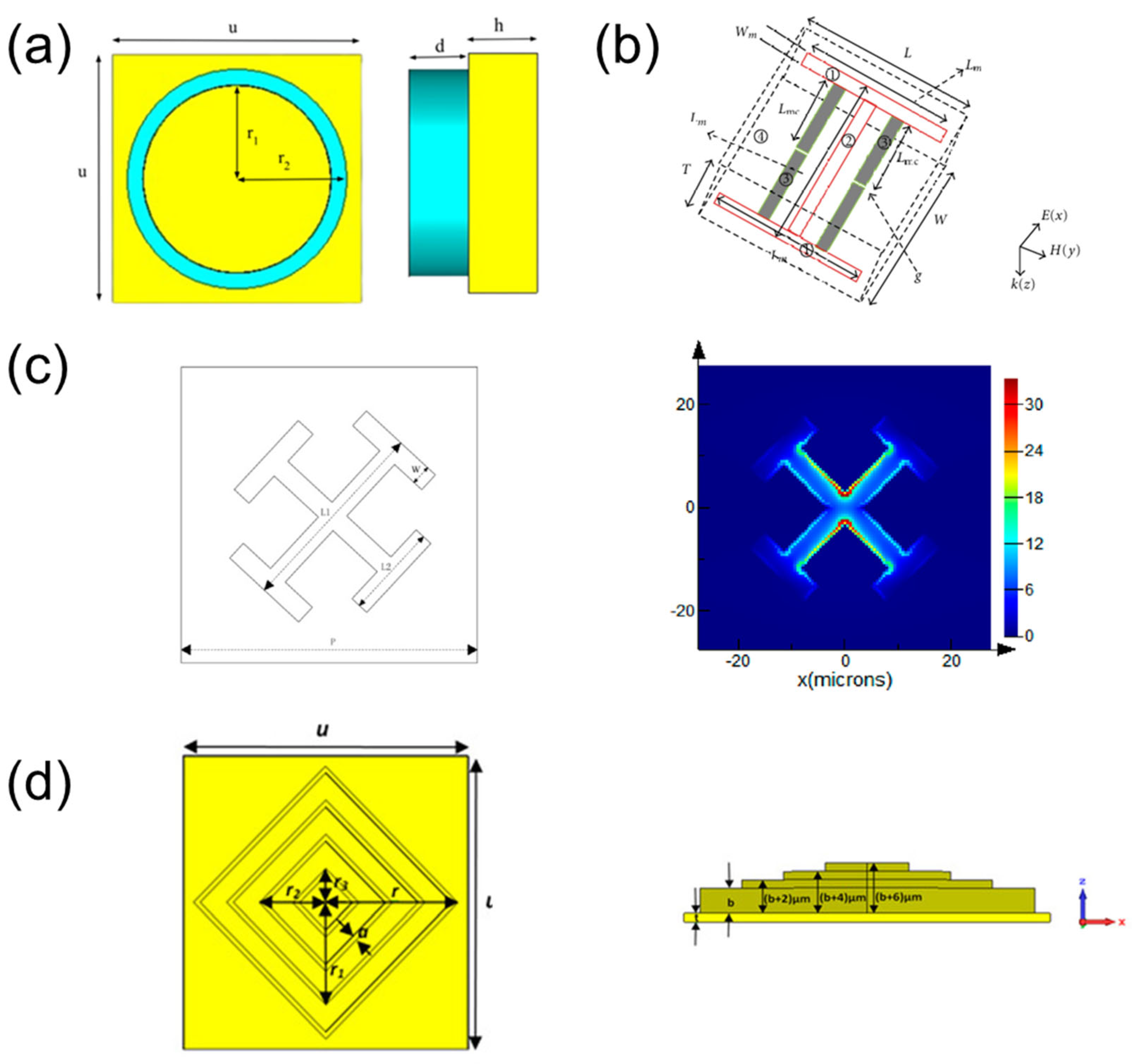
Ring and disk shapes represent the most fundamental metamaterial resonant elements, operating on the principle of LC resonance: the ring structure provides inductance, while the ring gap provides capacitance. When THz waves are incident, a strong localized electric field enhancement forms at the ring gap, making it extremely sensitive to changes in the external dielectric environment. Key design parameters primarily include the ring’s radius, linewidth, gap width, and periodicity constant. By adjusting these geometric parameters, the resonance frequency can be tuned to cover the desired THz frequency band. The ring resonator also exhibits multiple resonant modes, including the fundamental mode (λ/2) and higher-order modes (λ, 3λ/2, etc.). Each resonant mode corresponds to a distinct spatial distribution of the localized electromagnetic field. Crucially, these different modes possess varying degrees of sensitivity to changes in the external dielectric environment. This characteristic provides the physical foundation for the implementation of multi-band or multi-parameter sensing functionalities on a single device platform. The symmetrical structure of ring resonators makes them insensitive to the polarization direction of incident waves, which is advantageous for simplifying the sensing system design [29]. For example, Ponlatha et al. [37] employed a square/elliptical ring structure for glucose detection, achieving a sensitivity of 1000 GHz/RIU and a high Q-factor of 18.8 [37]. In Figure 2a, the indium antimonide (InSb) cylindrical structure is depicted [38].
(2)
Multimode Composite Resonator
To achieve multiple resonant modes or multi-band responses within a single device, researchers have developed multi-modal composite resonators. The design principle of multi-mode composite resonators involves integrating multiple unit cells with distinct resonant frequencies within a single periodic element, or alternatively, introducing asymmetric features into a single unit cell to excite multiple resonant modes. The former approach is exemplified by symmetric geometries such as H-shaped, U-shaped, and L-shaped structures, which combine different resonant elements. The latter strategy employs asymmetric configurations, including P-shaped, T-shaped, M-shaped, and K-shaped resonators, where structural asymmetry breaks symmetry and enables the excitation of multiple, closely spaced resonant modes. These structures combine resonant elements of different shapes (such as H-shaped, U-shaped, L-shaped, P-shaped, T-shaped, M-shaped, K-shaped, etc.) or introduce asymmetric elements within a single unit to excite multiple resonant modes, thereby expanding the operational frequency band or enabling multi-target detection. Furthermore, the coupling between multiple resonant modes can give rise to interference effects. Consequently, this leads to a significant enhancement of the quality factor (Q-factor), a critical parameter defined as the ratio of the resonant frequency to its full width at half maximum. A higher Q-factor, indicative of a narrower resonance and lower energy loss, is highly desirable for sensing applications as it translates to improved resolution and a lower limit of detection. For instance, Birunda et al. [19] proposed a P-shaped resonator for malaria detection, achieving an ultra-high Q-factor of 50.8; Sarankumar R et al. [42] designed a T-shaped structure for brain tumor detection, achieving a sensitivity of 1538 GHz/RIU; Wekalao et al. [43] used an H-shaped structure combined with multiple materials to achieve multi-cancer detection. Multi-modal composite resonators offer higher design freedom, but electromagnetic simulations are necessary to optimize and avoid mode crosstalk. Figure 2b shows an “I” shape structure of the proposed metamaterial that produces a high refractive index at each frequency band [39]. Figure 3 shows an X-shaped asymmetric resonator (designed using FDTD Solutions software) with intersecting arms that enable multiple current pathways [40].
(3)
Multi-layer Stacking and Heterogeneous Integration
By stacking multiple functional layers in the vertical direction, a three-dimensional electromagnetic environment can be constructed, further enhancing light-matter interaction [44,45]. Compared with single-layer structures, multi-layer stacked structures exhibit distinct advantages in the design of MXene-based terahertz metamaterial biosensors: (1) They can generate new resonant modes through interlayer plasmonic coupling, which effectively expands the operating frequency band (typically covering 0.1–2 THz) to accommodate multi-target detection scenarios such as simultaneous sensing of multiple biomarkers; (2) The electromagnetic response (including resonance frequency, field confinement, and transmittance dip depth) can be finely tuned by adjusting the interlayer spacing and the thickness of dielectric layers (e.g., SiO2 substrate), enabling precise optimization of sensor sensitivity and figure of merit (FOM); (3) Different functional materials (e.g., MXene for high conductivity, graphene for tunability, and black phosphorus for anisotropic optical properties) can be integrated into separate layers, realizing synergistic complementarity to enhance biocompatibility, signal transduction efficiency, and target selectivity. Multilayer stacked structures are typically composed of metal-dielectric-metal layers, where each layer can be designed with different resonant frequencies. Interlayer coupling can produce broadband responses or enhance absorption in specific frequency bands [46]. For example, Wekalao et al. designed a composite multilayer structure incorporating cross-shaped, circular, and square resonators for formalin detection, achieving a stable Q-factor of 13.6 [47]. Similarly, Figure 2d presents a compact multi-layer absorber configuration in which four concentric square resonators are vertically stacked above a metallic ground plane—a geometry that exemplifies the interlayer plasmonic coupling and design flexibility characteristic of this class of metastructures [41]. Heterogeneous integration refers to combining different materials (such as MXene, graphene, metals) within the same structure, leveraging their complementary properties for performance optimization. The formation of heterogeneous interfaces (e.g., between MXene and graphene, black phosphorus and gold) may also induce effects such as interfacial charge transfer and energy band bending, which further modulate the electromagnetic response of the terahertz biosensor by regulating plasmonic coupling efficiency and local electromagnetic field distribution. These interfacial effects enhance the interaction between the sensor and target analytes, thereby improving detection sensitivity and spectral selectivity for biomolecules like disease biomarkers and pathogens [23,45]. For instance, combining highly conductive MXene with high carrier mobility graphene can achieve tunability while maintaining field enhancement.
(4)
Coding and Tunable Design
In recent years, encoded metamaterials have garnered extensive attention, with their core idea lying in representing distinct phase responses through digital encoding (e.g., “0” and “1”), thereby enabling dynamic manipulation of electromagnetic waves. Each unit of the encoded metamaterial can be programmed as a “bit” with a specific phase response (typically covering a phase range of 0 to 2π), and through the arrangement and combination of these units, dynamic control over beam direction, polarization state, focusing position, and other electromagnetic properties can be achieved [48,49]. In MXene-based metamaterials, phase encoding can be realized by designing graphene regions with different chemical potentials, endowing the sensor with information transmission capabilities. This is attributed to the synergistic effect of MXene’s excellent conductivity and graphene’s tunable plasmonic properties, which jointly regulate the electromagnetic response of the metamaterial. The chemical potential of graphene can be real-time adjusted by applying an external bias voltage, which in turn modifies its electrical conductivity and phase response. This reversible regulation mechanism facilitates the realization of reconfigurable encoded metamaterials, making them promising for applications such as intelligent sensing, dynamic beam steering, and high-speed information transmission in terahertz regimes. For example, Wekalao et al. [50] introduced graphene chemical potential coding into quadrant and ring resonator structures, achieving 2-bit encoding functionality. Figure 2d illustrates the top-down view and 3D model of a sensor featuring 2-bit encoding capability via graphene chemical potential tuning. This encoding strategy can not only be applied to information transmission but also achieve differentiated responses to multiple targets through the optimization of encoding sequences, laying a solid foundation for the development of intelligent sensing systems. Furthermore, by incorporating active components or tunable materials, real-time tuning of the resonance frequency can be realized, offering new avenues for developing multifunctional, reconfigurable sensors.
Existing structural design strategies have achieved remarkable breakthroughs in the optimization of Q-factor and sensitivity in simulation environments, but most of the complex multi-layer and asymmetric structures proposed in current studies rely on high-precision electron beam lithography (EBL) for fabrication, which has low processing efficiency and high cost, making it difficult to achieve large-scale mass production. The structural fidelity and processing deviation in large-area array fabrication, which are directly related to the consistency of actual device performance, are almost completely ignored in ideal simulation models, which is a core challenge for the translation of these optimized structural designs from simulation to practical clinical devices.

2.2.2. Machine Learning-Assisted Optimization for THz Metamaterial Biosensors

The growing complexity of THz metamaterial structures and parameter space has rendered traditional trial-and-error design inefficient. Machine learning (ML) offers a powerful alternative by learning complex relationships between structural parameters and sensing performance. ML has thus become a valuable tool for THz metamaterial optimization.
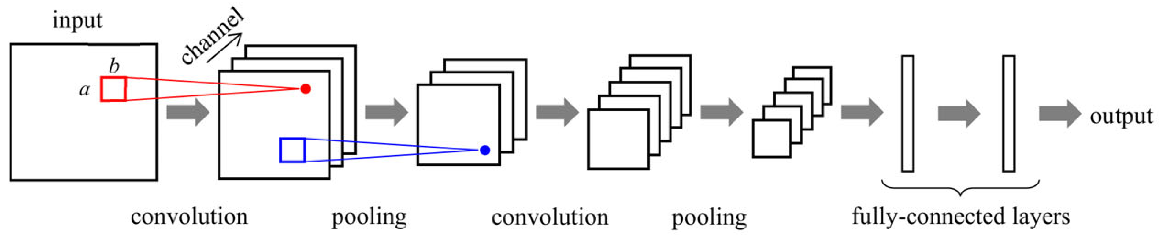
Current ML applications fall into three categories. Regression algorithms, such as WKNN and polynomial regression, work well with small datasets and offer strong interpretability. Neural networks, particularly 1D-CNN, excel at processing sequential THz spectra and can automatically extract local features, achieving prediction accuracy with R2 up to 1.00. Ensemble learning methods, including Random Forest, XGBoost, and Stacking Ensemble, combine multiple base learners to improve prediction stability and are widely used for multi-objective optimization [51]. Figure 3 outlines the architecture of a convolutional neural network (CNN) used for feature extraction from THz spectra.
Figure 3. Structure and operation of convolutional neural network (CNN). Reproduced with permission from ref. [51].
Condensedmatter 11 00021 g003
Nearly all ML models applied in this field are trained and validated based on ideal simulation datasets, without incorporating real-world variables such as micro-nano processing deviations, environmental noise, and biological sample multi-component interference. This leads to the high prediction accuracy of ML models in simulation environments often being difficult to replicate in actual experiments, which is an important limitation of current ML-assisted optimization strategies.

2.3. Core Performance Evaluation Metrics for THz Metamaterial Biosensors

To establish a unified, standardized evaluation benchmark for the subsequent systematic sorting and comparative analysis of research progress in the field of THz biosensing, this section clearly defines two sets of independent but logically related performance evaluation systems, which are, respectively, applicable to the theoretical simulation stage and clinical application validation stage of biosensor development.

2.3.1. Intrinsic Sensing Performance Metrics (For Theoretical Simulation and In Vitro Basic Characterization)

This set of metrics reflects the intrinsic physical sensing capability of the THz biosensor and is the core evaluation standard for the theoretical design, electromagnetic simulation, and basic in vitro characterization of the sensor. It is universally used in almost all simulation studies and basic experimental research in the field of THz biosensing. The four core indicators are defined as follows:
(1)
Sensitivity, S
Sensitivity is defined as the ratio of the resonance frequency shift Δ f to the refractive index change Δ n of the analyte:
S = Δ f Δ n
The unit is GHz/RIU or nm/RIU. Sensitivity reflects the response intensity of the sensor to changes in the refractive index and is the most fundamental indicator for measuring detection capability. Higher sensitivity means a greater frequency shift caused by per unit refractive index change, and the sensor has a stronger detection capability for small concentration changes.
(2)
Quality Factor, Q
Quality Factor describes the sharpness of the resonance peak and is defined as the ratio of the resonance frequency fres to the full width at half maximum (FWHM):
Q = f r e s F W H M
The higher the Q-factor, the sharper the resonance peak, and the stronger the ability to resolve minute frequency shifts. A high-quality factor (Q) indicates a narrow resonance peak width and high precision of frequency reading, which is conducive to distinguishing similar refractive index changes, but may also result in limited dynamic range.
(3)
Figure of Merit, FOM
Figure of Merit combines sensitivity and linewidth, defined as the ratio of sensitivity to the full width at half maximum:
F O M = S F W H M
The unit is RIU−1. FOM considers both the response intensity and the spectral resolution, making it a key indicator for evaluating the comprehensive performance of a sensor. A sensor with a high FOM must have sufficiently high sensitivity and a sufficiently narrow resonance peak; neither can be dispensed with.
(4)
Detection Limit, DL
Detection Limit is defined as the minimum change in refractive index that the sensor can identify, typically calculated using the following formula:
D L = Δ n 1.5 × F W H M Δ f 1.25
This empirical formula comprehensively considers noise level and spectral linewidth. A smaller DL value indicates a stronger detection capability of the sensor. The detection limit is positively correlated with FWHM and negatively correlated with Δf. Therefore, it is necessary to trade off sensitivity and linewidth when designing the sensor to achieve optimal detection capability.

2.3.2. Clinical Diagnostic Performance Metrics (For In Vivo and Clinical Sample Validation) [52]

This set of metrics is the universally recognized gold standard for evaluating the clinical application value of in vitro diagnostic devices in the medical field, which directly determines whether the sensor can meet the requirements of clinical disease diagnosis and be approved for clinical application. It is the core evaluation standard for the final clinical translation of the sensor and is universally used in clinical validation studies of biosensors. The four core indicators are defined as follows:
(1)
Clinical Sensitivity
Clinical sensitivity refers to the probability that the sensor correctly identifies patients with the target disease, which is calculated as:
Clinical   Sensitivity = T r u e   P o s i t i v e T P T r u e   P o s i t i v e T P + F a l s e   N e g a t i v e F N
It reflects the ability of the sensor to avoid missed diagnosis, which is the core requirement for early disease screening and critical disease early warning.
(2)
Clinical Specificity
Clinical specificity refers to the probability that the sensor correctly identifies healthy people without the target disease, which is calculated as:
C l i n i c a l S p e c i f i c i t y = T r u e N e g a t i v e T N T r u e N e g a t i v e T N + F a l s e P o s i t i v e F P
It reflects the ability of the sensor to avoid misdiagnosis, which is the key indicator to ensure the reliability of the sensor in clinical population screening.
(3)
Area Under the Curve (AUC)
AUC is the area under the Receiver Operating Characteristic (ROC) curve, with a value range of 0.5 to 1. It comprehensively evaluates the overall diagnostic performance of the sensor, balancing clinical sensitivity and specificity. An AUC value closer to 1 means better overall diagnostic performance, and an AUC > 0.9 indicates that the sensor has excellent clinical application value.
(4)
Response Time and Early Warning Time
Response time refers to the time required for the sensor to output a stable and accurate detection signal after contact with the analyte; early warning time refers to the time that the sensor can detect the disease or pathological changes earlier than the clinical gold standard detection method. These two indicators reflect the real-time monitoring capability of the sensor in dynamic clinical scenarios, which is critical for critical care scenarios such as intraoperative thrombus monitoring and vital sign real-time monitoring.

2.3.3. Logical Correlation Between the Two Sets of Metrics

The two sets of evaluation systems have an inherent progressive logical correlation, which corresponds to different stages of the entire development chain of the biosensor from laboratory design to clinical translation:
(1) The intrinsic sensing performance metrics (simulation-oriented) are the theoretical prerequisite and necessary basis for achieving excellent clinical diagnostic performance. Only when the sensor has sufficient sensitivity, high Q-factor and low detection limit in the theoretical simulation and basic in vitro characterization, can it have the potential to achieve high-precision detection of trace biomarkers in complex clinical environments [53,54].
(2) The clinical diagnostic performance metrics (clinical-oriented) are the final evaluation standard and ultimate goal of sensor development. The optimization of intrinsic sensing performance in the simulation stage is ultimately to achieve accurate, reliable and safe disease diagnosis in real clinical scenarios [54,55].
With the theoretical framework, design methodologies, and performance evaluation metrics firmly established, it becomes possible to critically examine the actual progress and limitations of existing THz metamaterial biosensors. Although the field has achieved remarkable sensitivity and quality factors in simulation environments, the translation of these promising results into clinical practice remains elusive. The next chapter systematically reviews the development of conventional (primarily gold-based) THz metamaterial biosensors, identifies the core bottlenecks that hinder their clinical translation, and ultimately makes the case for why a fundamentally new material platform is urgently needed.

3. Progress and Bottlenecks of Non-MXene (Conventional) THz Metamaterial Biosensors

3.1. Research Status of Conventional THz Biosensing

Terahertz (THz) biosensing has emerged as a promising non-invasive analytical tool for biomedical applications, owing to its unique advantages of non-ionizing photon energy, label-free detection capability, and intrinsic sensitivity to the molecular vibrational and rotational modes that form the characteristic “fingerprint spectra” of biological macromolecules [56,57]. As the foundational technical route of THz biomedical detection, conventional THz biosensing technologies have laid a complete theoretical and application framework for the subsequent development of metamaterial-enhanced sensing platforms, while their inherent limitations also directly drive the innovation of metamaterial-based THz biosensing systems [58].
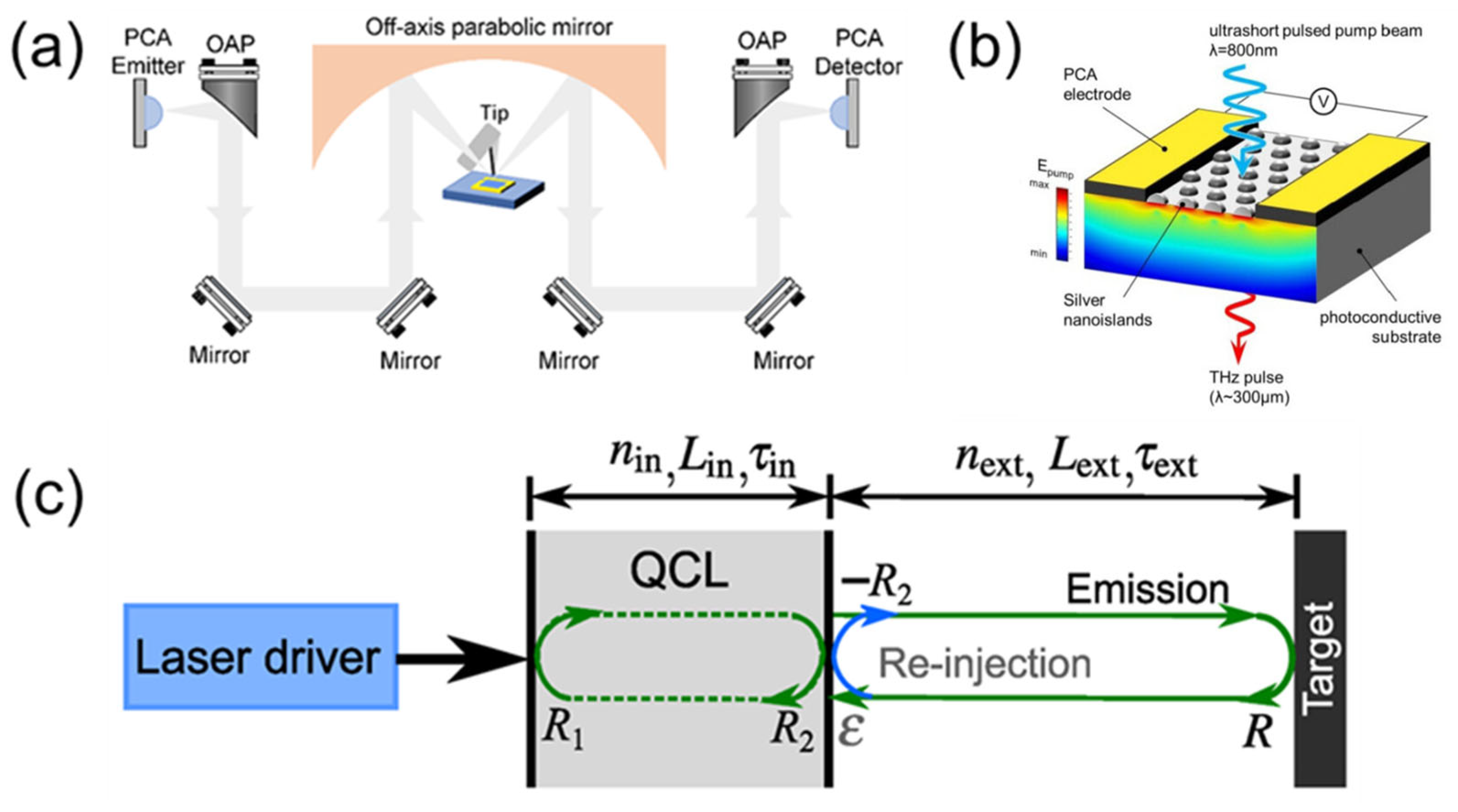
The most mainstream and well-established conventional modality is terahertz time-domain spectroscopy (THz-TDS), which realizes label-free molecular recognition by measuring the amplitude and phase changes in THz pulses interacting with biological samples, and has been applied in clinical-relevant scenarios, including tumor biomarker identification, malignant tissue differentiation, and thrombosis-related molecular detection [59,60]. However, its intrinsic limit of detection (LOD) only reaches the millimolar (mM) level, which is far insufficient for the trace biomarker detection at the picogram per milliliter (pg/mL) level required by clinical scenarios [61,62]. To break the far-field optical diffraction limit of conventional THz-TDS, THz near-field microscopy has been developed to achieve nanoscale spatial resolution for single-cell and single-molecule analysis, but its strict requirements for sample preparation and complex instrumentation make it unsuitable for high-throughput clinical point-of-care testing [63]. Figure 4a presents the experimental optical path diagram of a scattering-type scanning near-field optical microscope based on a terahertz time-domain spectrometer (THz-TDS s-SNOM) [64]. For system miniaturization, integrated photoconductive antenna (PCA) modules [65,66] and portable quantum cascade laser (QCL) systems [67] have promoted the development of benchtop THz systems towards miniaturization, but most miniaturized devices still cannot balance system volume and ultra-sensitive detection performance in complex biological matrices [65,68]. Figure 4b illustrates a nanoplasmonic photoconductive antenna (PCA) with metal nanoislands for enhanced THz emission. Figure 4c illustrates a THz quantum cascade laser (QCL) operating under optical feedback [67,69].
In summary, the clinical translation of all conventional THz biosensing technologies is severely restricted by three inherent bottlenecks, which cannot be fully solved by single technology optimization alone. First, insufficient intrinsic sensitivity: the extremely short effective light-matter interaction length of free-space THz sensing leads to a LOD that cannot meet clinical trace detection requirements. Second, poor anti-interference capability: the strong THz absorption of water in complex biological samples (whole blood, serum, tissue fluid) completely masks the weak fingerprint signal of target biomarkers [32,54]. Third, unbalanced miniaturization and detection performance: most miniaturized THz systems cannot achieve high-sensitivity detection while reducing the system volume, failing to realize true point-of-care testing. The localized field enhancement effect of THz metamaterials provides a fundamental solution to the core problem of insufficient sensitivity in conventional THz biosensing, which has become the mainstream research direction of high-performance THz biosensing.

3.2. Development Status of THz Metamaterial Biosensing

THz metamaterial biosensing technology breaks through the sensitivity limit of conventional THz sensing through the localized field enhancement effect of artificial subwavelength structures and has become the mainstream research direction of high-sensitivity THz biosensing. At present, the research in this field is mainly divided into two directions: structural design optimization and clinical translation exploration.
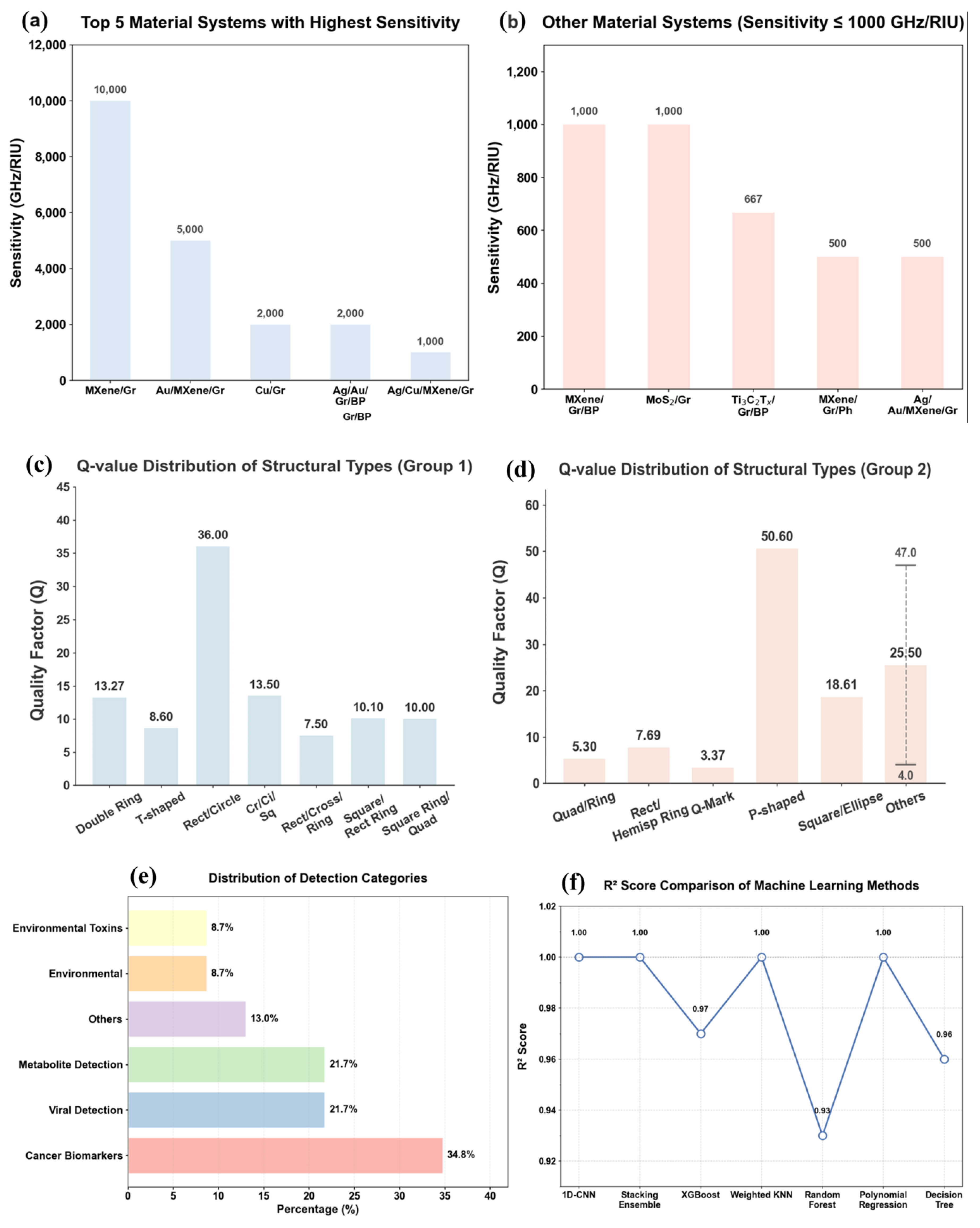
In terms of structural design and performance optimization, researchers have developed a variety of metamaterial resonant structures, including ring resonators, multimode composite resonators, multi-layer stacked structures, etc. By optimizing the geometric parameters of the unit cell, the sensitivity and Q-factor of the sensor have been continuously improved [15,18]. At present, the maximum sensitivity of THz metamaterial biosensors in simulation has exceeded 10,000 GHz/RIU, and the maximum Q-factor has exceeded 100, which fully meets the theoretical requirements of trace biomarker detection [26]. The key performance metrics (sensitivity and Q-factor) of typical metamaterial resonant structures reported in the field are compared in Figure 5.
In terms of clinical translation, researchers have carried out exploratory validation of THz metamaterial biosensors in biomedical scenarios in recent years and initially verified the clinical application potential of this technology, laying a preliminary foundation for the subsequent translation from laboratory research to practical diagnostic devices [8]. As summarized in the authoritative review by Wei et al., conventional gold-based THz metamaterial biosensors, as the most mature technical route in this field, have completed preliminary application exploration in multiple clinical-related scenarios, including circulating tumor cell and tumor biomarker detection for malignant tumors, pathogen nucleic acid and protein detection for infectious diseases, and chromosome aneuploidy detection for non-invasive prenatal diagnosis, forming a relatively complete preliminary research framework for clinical application [32]. These exploratory studies have verified that THz metamaterial biosensors can achieve specific detection of target biomarkers in complex biological matrices such as serum, plasma and saliva, providing a theoretical and experimental basis for subsequent large-sample clinical validation.
For infectious disease detection, which is one of the most widely explored application directions, Ahmadivand et al. constructed a functionalized graphene-based THz plasmonic metasensor, which achieved label-free and highly sensitive detection of the SARS-CoV-2 spike protein, the core biomarker of COVID-19 [70]. By modifying the metasurface with angiotensin-converting enzyme 2 (ACE2) as the biological recognition element, this work realized specific capture of the target spike protein and achieved an ultra-low limit of detection down to 1 fM in buffer solution, which is far lower than the concentration range of clinical detection requirements, fully verifying the ultra-high sensitivity of THz metamaterial biosensors for trace biomarker detection [70]. This work has established a representative technical paradigm for the application of THz metamaterial biosensors in infectious disease early warnings and provided a standardized technical route for the subsequent clinical sample validation of such sensors.
For the clinical translation of THz metamaterial biosensors, the existing studies have generally adopted clinical sensitivity, specificity, and area under the curve (AUC) as the core evaluation indicators for diagnostic performance, which are the universally recognized gold standards for the clinical translation of in vitro diagnostic devices in the medical field [5]. The establishment of this standardized evaluation system has unified the research benchmark for clinical validation in this field, and laid a normative foundation for the subsequent full-chain research from in vitro performance optimization to in vivo animal experiments and multi-center clinical sample validation [5]. However, it is worth noting that most of the current clinical-related exploratory studies still stay in the stage of mimicking clinical samples in vitro, and there are few studies that have completed large-sample validation in real clinical cohorts, which is also the core gap restricting the final clinical translation of this technology [54].

3.3. Core Bottlenecks Hindering Clinical Translation

However, it is worth noting that more than 90% of THz metamaterial biosensor studies still stay in the pure simulation stage, and only the above-mentioned few studies have completed clinical sample validation. There is a serious association fracture between the simulation optimization system and clinical translation demand in this field, which is the core bottleneck restricting the clinical translation of THz metamaterial biosensors, mainly reflected in three aspects:
(1) No quantitative correspondence between simulation indicators and clinical effects has been established in the field: At present, the vast majority of simulation studies only pursue the limit of intrinsic performance indicators such as sensitivity and Q-factor in ideal environments, but no research has established a clear quantitative correspondence between these simulation indicators and the final clinical diagnostic sensitivity, specificity and AUC. Researchers do not know how much the sensitivity in the simulation needs to be improved to bring about a substantial improvement in clinical diagnostic effect, which leads to the optimization of simulation indicators becoming a “self-contained closed loop” completely decoupled from clinical needs [54].
(2) Simulation models completely ignore the key factors that determine clinical performance: The simulation models of existing studies are all based on ideal assumptions such as defect-free materials, perfect structure, single analyte, and static room temperature environment, but completely ignore the key factors that will directly affect the final clinical diagnostic effect in real clinical scenarios, such as material oxidation in 37 °C physiological environment, non-specific adsorption of high-abundance proteins in blood, processing deviation of large-area arrays, and dynamic flow interference. Even if the sensor has ultra-high sensitivity in the simulation, it will completely fail in the real clinical environment because these factors are not considered [54].
(3) The research chain from simulation to clinical validation is seriously broken: Most studies only stay in the pure simulation stage and stop after obtaining excellent intrinsic performance indicators in the simulation, without carrying out subsequent in vitro experimental verification, in vivo animal experiments and clinical sample validation. The lack of full-chain research leads to problems in the clinical application of the technology that cannot be found and solved in time, and a large number of simulation research results cannot provide effective guidance for clinical translation [1].
These three industry-wide bottlenecks cannot be fundamentally resolved solely through geometric optimization of gold-based metamaterial structures. The inherent limitations of noble metal metamaterials in surface functionalization, anti-nonspecific adsorption, and long-term physiological stability can be effectively addressed by the emerging two-dimensional MXene materials, whose well-verified advantages in biosensing provide a brand-new paradigm to bridge the gap between simulation optimization and clinical translation [70].
In summary, gold-based THz metamaterial biosensors have demonstrated impressive simulation performance, yet they suffer from three interconnected bottlenecks: lack of quantitative correlation between simulation indicators and clinical outcomes, neglect of real-world physiological variables in simulation models, and a fragmented research chain that rarely extends beyond pure simulation. These intrinsic limitations of noble metals—chemically inert surfaces, severe non-specific adsorption, and poor long-term stability in biological fluids—call for a paradigm shift in material selection.

4. MXene-Based THz Metamaterial Biosensors: From Material Foundations to Clinical Breakthroughs

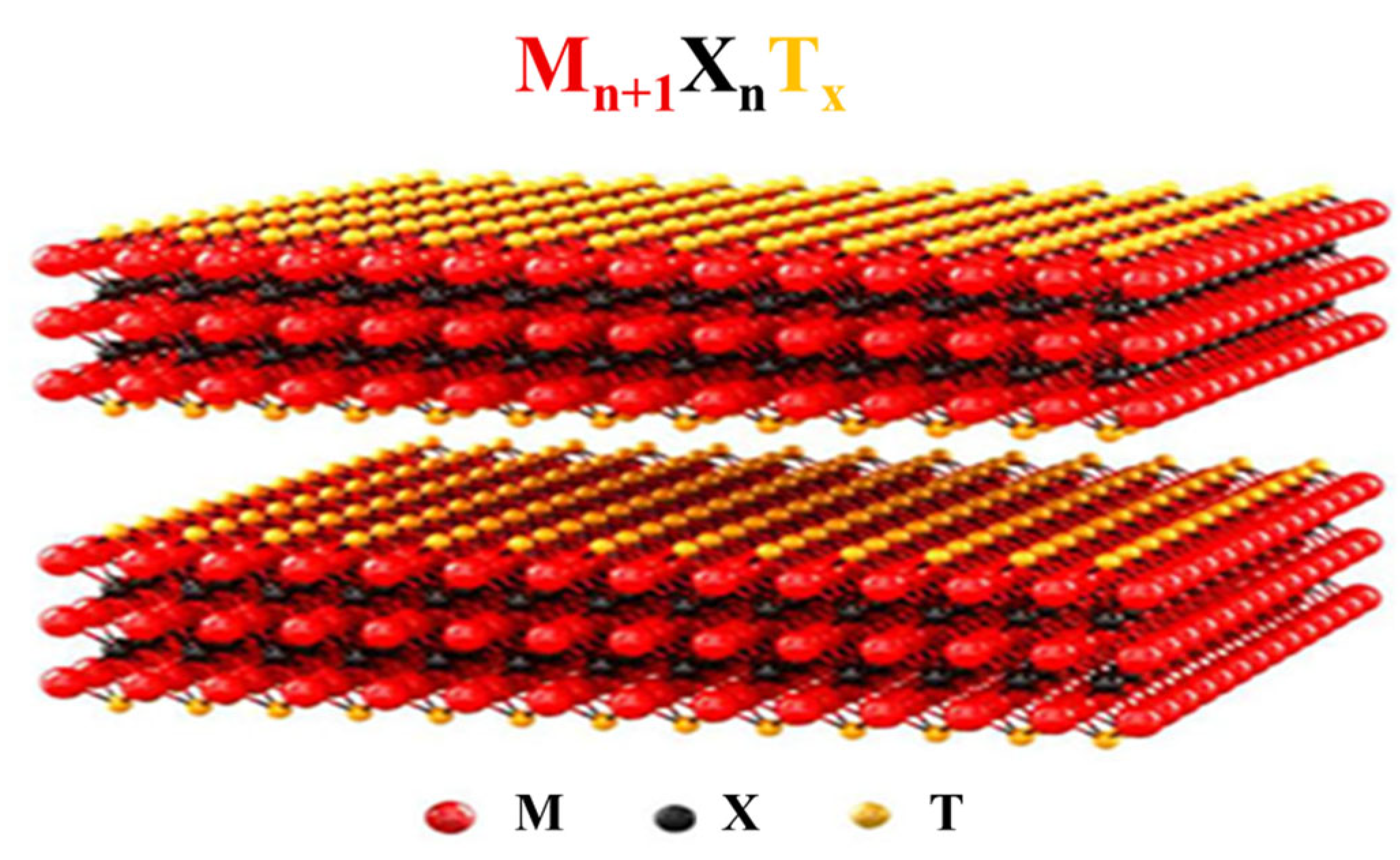
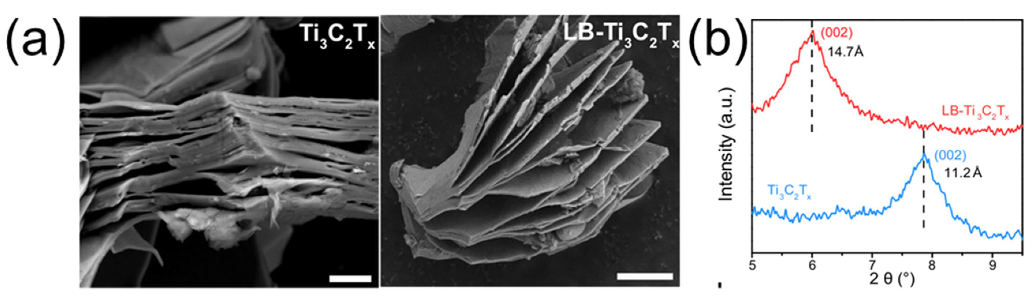
Two-dimensional transition metal carbides/nitrides (MXenes) have recently emerged as a transformative alternative, offering metal-like conductivity, abundant surface functional groups, excellent biocompatibility, and mechanical flexibility. Figure 6 presents the general formula Mn+1XnTx of MXenes and illustrates structural examples for n = 1 to 4. The following chapter provides a comprehensive account of MXene-based THz metamaterial biosensors, starting from the fundamental properties of MXene, progressing through conventional MXene biosensing platforms that serve as a technical foundation, and culminating in the design, performance, and landmark preclinical validation of MXene-integrated THz metamaterial devices [24,71]. Figure 7a displays SEM images of multilayer and LB-deposited Ti3C2Tx MXene, and Figure 7b shows XRD patterns confirming the expanded interlayer spacing.

4.1. Intrinsic Properties of MXene

This section systematically elaborates on the three core intrinsic properties of MXene that make it an ideal material for THz metamaterial biosensors, which are also the core advantages over traditional gold-based metamaterials.

4.1.1. Electronic Properties and THz Compatibility

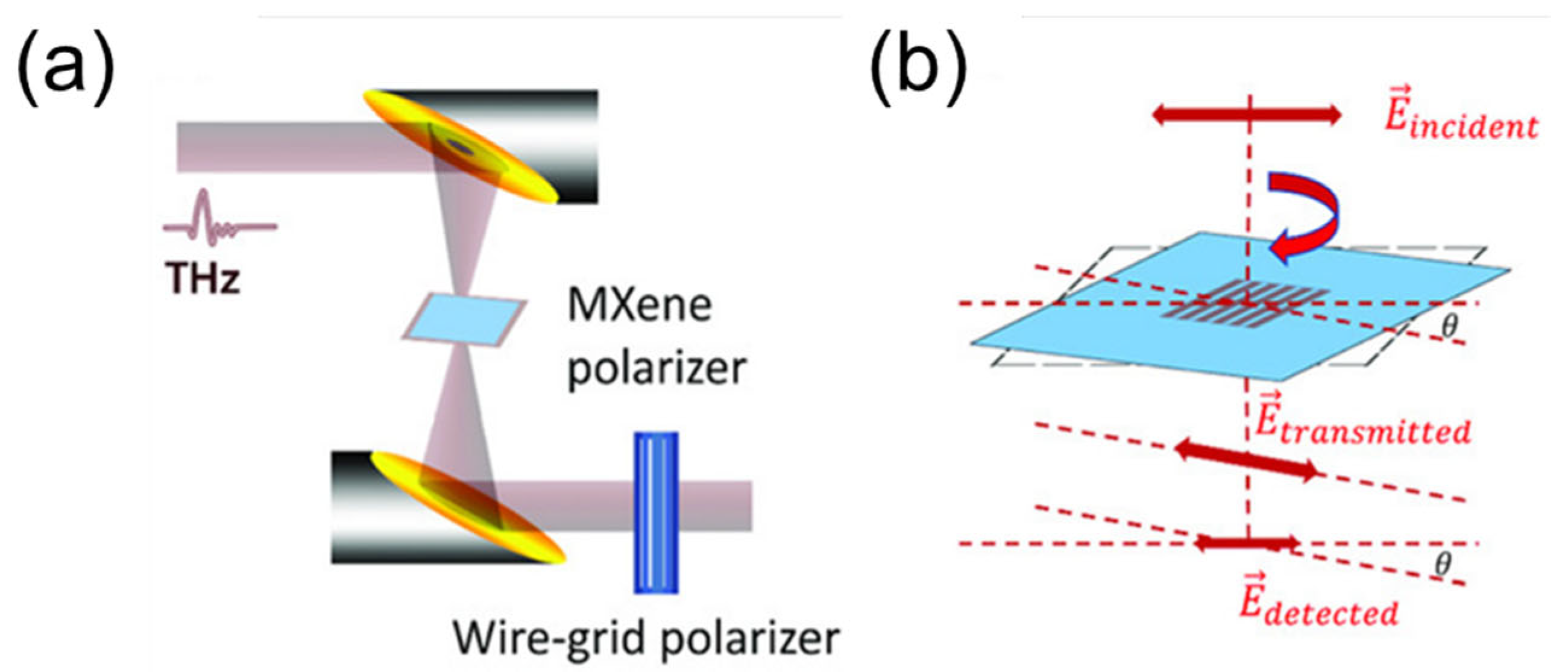
MXenes are a class of two-dimensional transition metal carbides and nitrides with the general chemical formula Mn+1XnTx (n = 1–4). In this formula, M represents an early transition metal element (such as Ti, Nb, Mo, V, W, etc.), X is carbon and/or nitrogen, and Tx represents surface terminations (e.g., -F, -O, -OH). The MXene family encompasses more than 30 distinct members to date. This material diversity stems from the versatile combinations of early transition metal elements (M) and the layer number (n) in their chemical formula Mn+1XnTx. Such compositional and structural variations endow the MXene family with a rich and wide-ranging tunability, which is fundamental for tailoring their electronic, optical, and chemical properties for specific applications, such as in tunable terahertz metamaterials [72,73]. MXenes possess a metallic-like electronic band structure, characterized by a high density of states near the Fermi level and room-temperature electrical conductivities reaching up to ~104 S/cm [21]. This characteristic implies that the conductivity of MXene within the THz frequency band remains nearly constant, independent of frequency. This provides a fundamental physical basis for achieving broadband impedance matching. This high conductivity originates from their unique layered structure and high carrier concentration (~1021 cm−3). Concurrently, their extremely short relaxation time(~10 fs)ensures the condition ωτ << 1 in the THz frequency range, resulting in low-dispersion conductivity across the entire 0.5–10 THz spectrum [21]. These properties provide the physical foundation for achieving strong localized field enhancement within metamaterial structures, which is comparable to or even better than traditional gold materials. The intrinsic THz response of MXene can be probed using polarization-resolved transmission measurements, as illustrated in Figure 8: a linearly polarized THz pulse is normally incident on a MXene-based polarizer, which can be rotated around the normal axis, and only the field component parallel to the incident polarization is detected [74].

4.1.2. Surface Functional Groups and Interface Tunability

The surface functional groups of MXenes arise from the selective etching process of MAX phases, including -F, -OH, =O and other terminations, which can be precisely regulated by adjusting the etching method and post-treatment process. This is the core advantage of MXene over chemically inert gold materials [19,35,75].
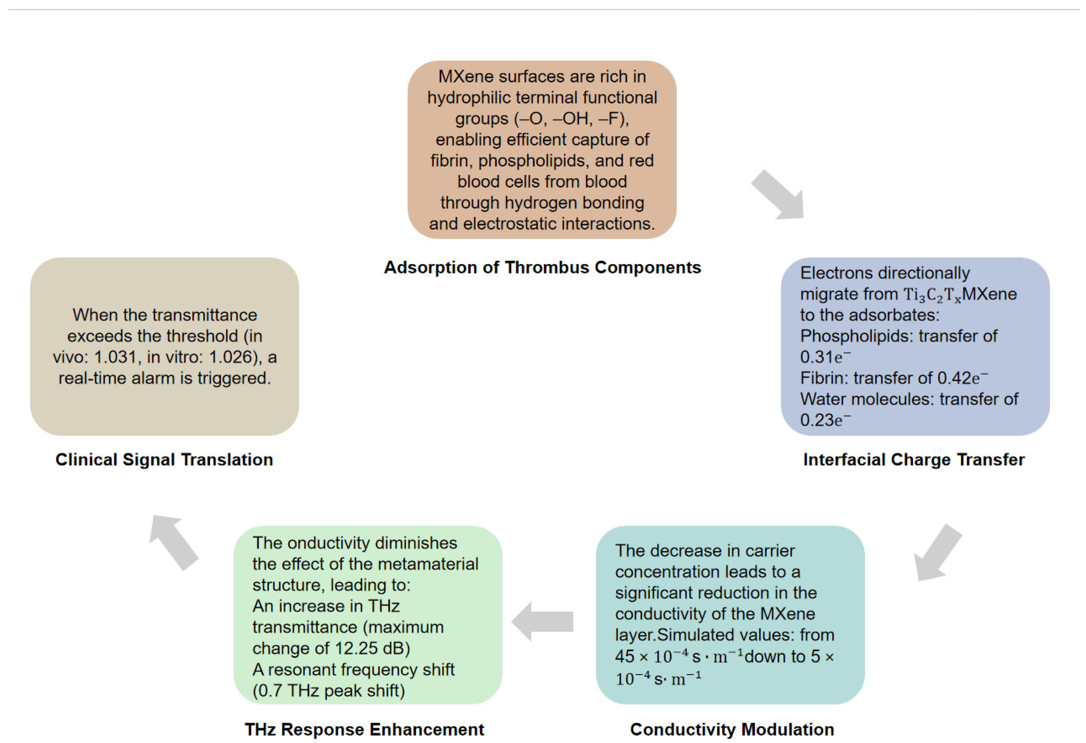
The type of surface functional group directly influences the interfacial interaction between MXene and biomolecules: abundant oxygen-containing and hydroxyl functional groups enable MXene to easily realize covalent immobilization of antibodies, aptamers and other biological recognition elements through simple chemical modification, which significantly enhances the specific binding ability to target biomarkers [36]. Meanwhile, the hydrophilic surface formed by these functional groups can effectively inhibit the non-specific adsorption of high-abundance proteins in biological fluids, which is a key challenge that is difficult to solve for traditional gold-based metamaterials [46,76,77]. In addition, by precisely tuning the type and proportion of surface functional groups, the conductivity and dielectric properties of MXene can be adjusted, thereby realizing the fine regulation of the electromagnetic response of THz metamaterials [29]. The charge transfer between thrombus and MXene (Δq = 0.31 e/molecule) revealed by Yang et al. [28] in their Advanced Materials paper relies precisely on the differences in electron affinity stemming from these surface groups [37,43]. The interfacial charge transfer mechanism between MXene and thrombus molecules is schematically illustrated in Figure 9.
The primary synthesis methods for MXenes currently include the following categories: (1) Etching with hydrofluoric acid (HF) or HF-containing solutions; (2) the in situ HF method (e.g., using LiF+HCl mixtures); (3) molten salt etching; (4) electrochemical etching [44,45]. MXenes obtained via these different methods exhibit variations in the type and ratio of surface functional groups, flake lateral size, and defect density, which critically determine the ultimate properties of the resulting MXene material [47,78,79]. Zhang et al. [71] developed a low-temperature Lewis base halide treatment method, achieving substitution of -F terminations with Br/I, while simultaneously expanding the interlayer spacing from 11.2 Å to 14.7 Å [48,49]. This method enables precise regulation of MXene surface functional groups under low-temperature conditions, avoiding material oxidation caused by high-temperature treatment, and has become the mainstream strategy for MXene interface engineering for biosensing applications [24].

4.1.3. Biocompatibility and Biosafety

Excellent biocompatibility and biosafety are core prerequisites for the clinical translation of MXene-based THz biosensors, especially for long-term in vivo dynamic monitoring scenarios such as thrombus detection [25]. The biocompatibility of MXene is mainly regulated by four key parameters: flake lateral size, surface termination type, exposure concentration, and action time, among which surface termination is the most critical regulatory factor, consistent with our previous discussion on MXene surface interface chemistry [76,80,81].
The Lewis-basic halide treatment introduced earlier can precisely replace the -F terminal groups of MXene with Br/I while expanding the interlayer spacing, which not only optimizes the THz electromagnetic response of MXene, but also effectively reduces the potential cytotoxicity caused by fluorine-containing terminals [78]. Conventional working concentrations (10–100 μg/mL) of MXene have been widely verified to have no significant toxic effects on mammalian cells and in vivo models, fully meeting the concentration requirements for THz biosensor surface functionalization [82].
In terms of in vitro biocompatibility, standardized cytotoxicity assays confirmed that the cell viability remained above 90% after 72 h co-incubation with 50 μg/mL pristine Ti3C2Tx MXene, and the viability of Lewis-basic halide-modified Br-terminated MXene still exceeded 85% even at 200 μg/mL [22,71]. For blood-contact THz sensing scenarios, the hemolysis rate of the MXene film in the landmark in vivo study by Yang et al. was only 0.32% at 100 μg/mL, far below the 5% safety threshold for clinical biomedical materials, and its hydrophilic surface can effectively inhibit non-specific protein adsorption in blood [28].
For in vivo biosafety, comprehensive evaluations in small and medium animal models (mice, rats, rabbits) confirmed that MXene has no acute or chronic systemic toxicity at conventional doses [83]. Blood biochemical analysis showed no abnormal changes in liver and kidney function indexes after MXene exposure, and histopathological analysis found no significant organ damage or inflammatory infiltration [26]. MXene can be gradually metabolized and cleared through the liver and kidney without long-term in vivo accumulation. Meanwhile, the Ti3C2Tx/extracted bentonite composite interface can effectively inhibit MXene oxidation in physiological environments, further improving its long-term in vivo biosafety [23].
Compared with traditional gold-based THz metamaterial materials, MXene exhibits more significant advantages in clinical biocompatibility and long-term in vivo application safety [84]. Gold nanomaterials, despite their good chemical stability, face potential clinical risks, including long-term in vivo accumulation of gold nanoparticles, chronic inflammatory response caused by metal ion shedding, and significantly reduced biocompatibility after surface modification with biological recognition elements [85,86]. In contrast, MXene can be gradually metabolized and cleared through the liver and kidney without long-term in vivo accumulation, and its abundant surface functional groups enable stable bioconjugation without compromising its inherent biocompatibility [87]. These advantages make MXene a more suitable material candidate for long-term in vivo dynamic monitoring and clinical translation of THz biosensors [25,88,89].

4.2. MXene-Based Conventional Biosensing Platforms as Technical Foundation

As a novel family of two-dimensional (2D) transition metal carbides/nitrides, MXenes (e.g., Ti3C2Tx) have attracted extensive attention in the field of biosensing due to their metal-like high electrical conductivity, abundant surface functional groups, excellent biocompatibility, and large specific surface area. Conventional MXene-based biosensing technologies have been widely validated in clinical and preclinical studies, which fully demonstrate the application potential of MXene materials in biomedical detection and provide a solid material and technical foundation for their subsequent integration with THz metamaterial biosensing platforms [24].

4.2.1. MXene-Based Electrochemical Biosensing

Electrochemical biosensing is the most mature and widely explored application direction of MXene materials in the biosensing field. The core advantages of MXene in electrochemical sensing stem from its ultra-high electrical conductivity (up to ~104 S/cm), which accelerates electron transfer at the electrode interface, and abundant surface terminal groups (-F, -OH, =O), which enable facile covalent immobilization of biological recognition elements (e.g., antibodies, aptamers, and enzymes) [24].
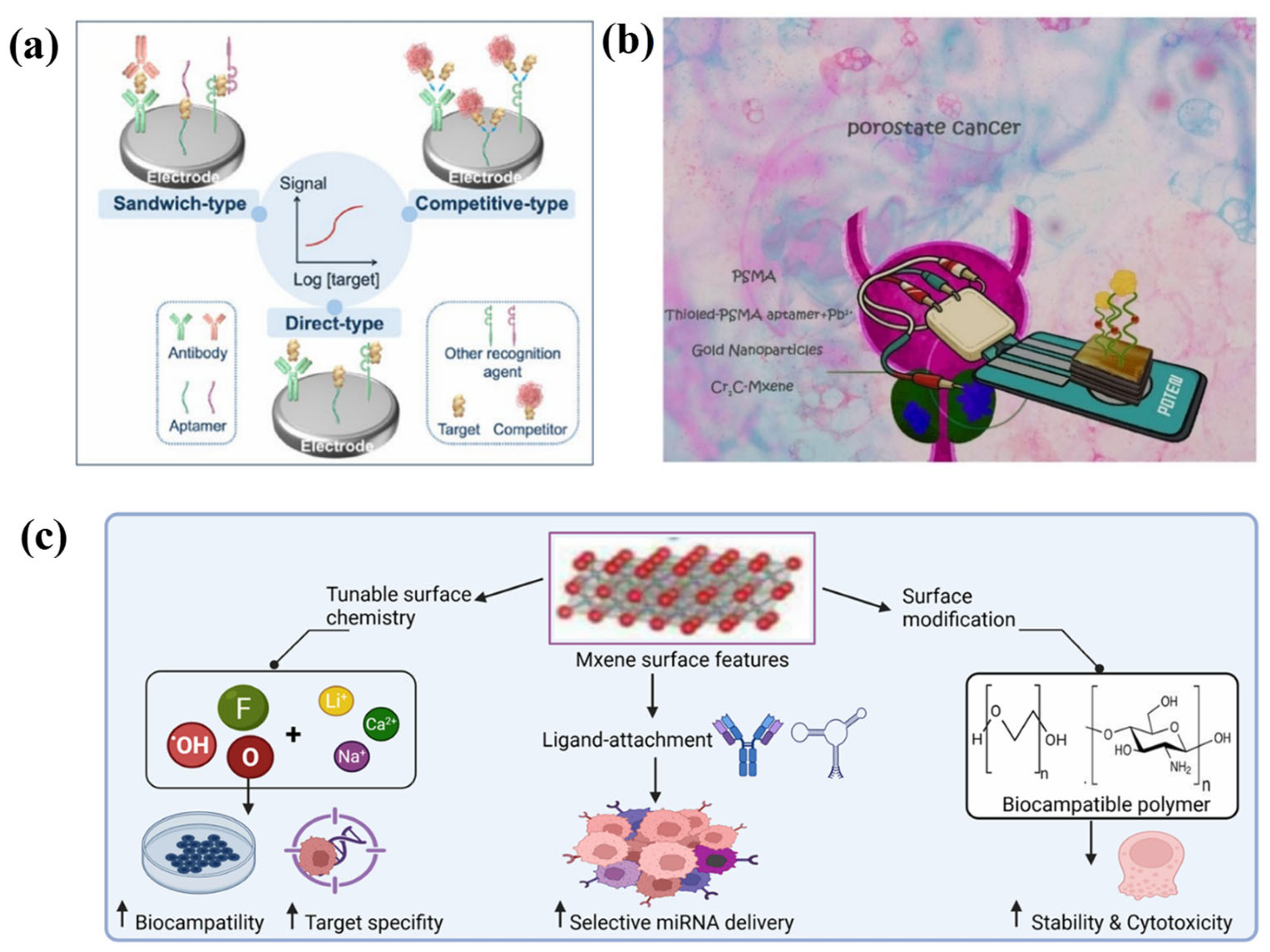
In practical biomedical applications, MXene-based electrochemical biosensors have achieved breakthroughs in multiple clinical detection scenarios. For cancer biomarker detection, these sensors have realized ultra-sensitive detection of tumor markers including carcinoembryonic antigen (CEA), prostate-specific membrane antigen (PSA), carbohydrate antigen 125 (CA125), and microRNAs, with a LOD down to the pg/mL level and even fg/mL level in optimized systems [90,91,92]. Figure 10a schematically depicts an electrochemical sensor for tumor marker detection using MXene-modified electrodes. Figure 10b illustrates the fabrication steps of an aptamer-based biosensor for PSMA detection. Figure 10c summarizes the versatile surface chemistry of MXene, including tunable terminations, ion intercalation, and bioconjugation strategies. For cardiovascular disease monitoring, MXene-based electrochemical sensors have achieved highly sensitive detection of cardiac troponin T (cTnT), myoglobin, and other myocardial injury markers, with rapid response and high specificity in clinical serum sample validation. For pathogen detection, MXene-modified electrochemical biosensors have been developed for the rapid detection of bacteria (e.g., Escherichia coli, Mycobacterium tuberculosis), viruses (e.g., SARS-CoV-2), and antibiotic residues, with excellent anti-interference performance in complex biological matrices [93]. A large number of studies have validated the clinical reliability of these sensors in hundreds of clinical samples, with diagnostic sensitivity and specificity exceeding 90% in most cases, fully demonstrating the clinical application potential of MXene-based biosensing platforms [24]. However, it is important to acknowledge a major practical limitation: the cost of MXene-based electrochemical sensors remains substantially higher than that of conventional disposable electrodes such as screen-printed carbon or graphene oxide electrodes. The synthesis of high-quality MXene nanosheets involves expensive precursors like MAX phases, hazardous etching agents such as HF, and multi-step delamination processes, which are difficult to scale up for cost-effective production. Furthermore, the long-term storage stability of MXene-modified electrodes under ambient conditions is still suboptimal, requiring strict handling and storage protocols that add to operational costs. Without significant advances in low-cost, green synthesis routes and scalable, robust electrode fabrication methods, the translation of these high-performance MXene electrochemical sensors into resource-limited or point-of-care clinical settings will remain challenging.

4.2.2. MXene-Based Fluorescence and Raman Biosensing

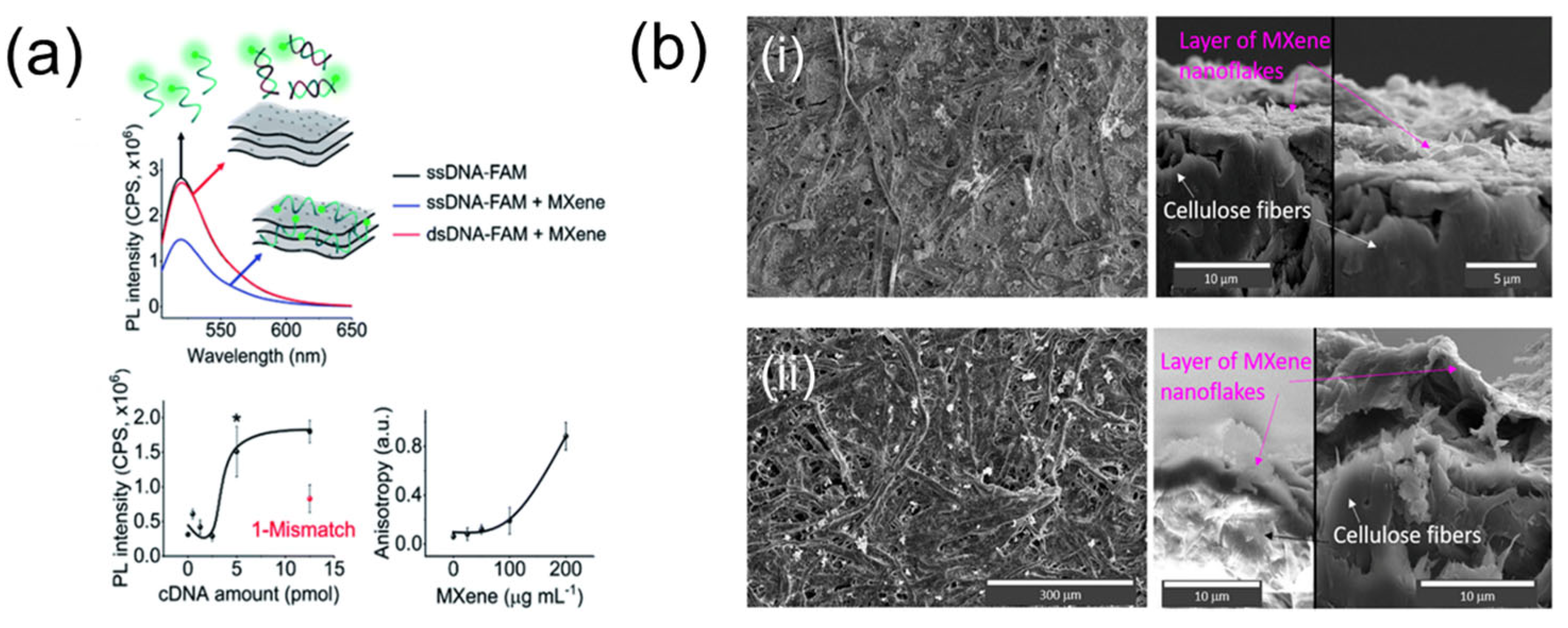
MXene materials exhibit unique optical properties that make them ideal functional materials for fluorescence and surface-enhanced Raman scattering (SERS) biosensing. For fluorescence sensing, MXene acts as a highly efficient fluorescence quencher via fluorescence resonance energy transfer (FRET), which enables the construction of label-free or “turn-on” fluorescent biosensors based on the specific binding between target analytes and nucleic acid probes/antibodies immobilized on the MXene surface [94,95]. These sensors have been widely applied in the detection of microRNAs, cancer biomarkers, and pathogens, with ultra-high sensitivity and a LOD down to the fM level [91]. In addition, MXene-based fluorescent probes have been successfully applied in intracellular imaging and in vivo small animal imaging, enabling real-time monitoring of biomarker expression in living cells and tumor tissues. As demonstrated in Figure 11a, fluorescence spectroscopy and anisotropy measurements reveal the strong interaction between DNA and MXene, underpinning the FRET-based quenching mechanism that enables highly sensitive label-free detection of nucleic acids and other biomolecules [96].
For SERS biosensing, MXene serves as an excellent SERS substrate with dual enhancement mechanisms: electromagnetic enhancement from the localized surface plasmon resonance of MXene nanosheets, and chemical enhancement from interfacial charge transfer between MXene and target molecules. These SERS platforms have been validated in clinical applications such as circulating tumor cell detection, bacterial identification, and intraoperative tumor margin assessment, with high specificity and spatial resolution for tissue imaging [98]. As shown in Figure 11b, vacuum-assisted filtration (VAF) yields a dense MXene nanoflake layer with a smooth surface morphology, providing an ideal SERS substrate that leverages both electromagnetic enhancement from localized surface plasmon resonance and chemical enhancement from interfacial charge transfer [97].

4.2.3. Other MXene-Based Optical Biosensing Modalities

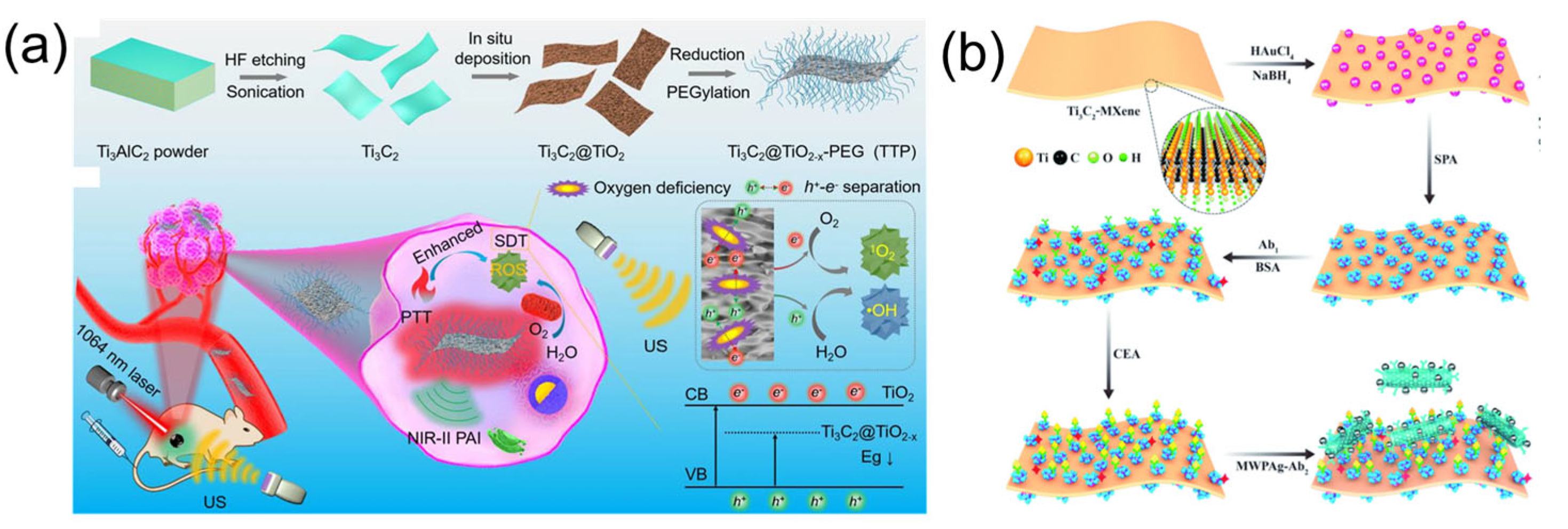
Beyond fluorescence and SERS sensing, MXene materials have also shown promising application prospects in other optical biosensing modalities, including photoacoustic (PA) [99] sensing and surface plasmon resonance (SPR) sensing [100,101]. For PA sensing, MXene exhibits excellent photothermal conversion efficiency in the near-infrared and THz bands, which enables the construction of high-sensitivity PA biosensors and imaging probes. MXene-based PA sensors have achieved non-invasive in vivo detection of tumor biomarkers and real-time monitoring of thrombus formation in small animal models, with deep tissue penetration and high spatial resolution [99,102]. As illustrated in Figure 12a, the Ti3C2@TiO2−x nanohybrid platform exemplifies MXene-based photoacoustic theranostics, leveraging the excellent photothermal conversion efficiency of MXene in the NIR-II window for deep-tissue PA imaging and high-precision tumor ablation [103].
For SPR sensing, MXene nanosheets can be used to modify the gold film surface of traditional SPR sensors, which not only enhances the SPR signal by optimizing the interfacial refractive index matching, but also provides abundant binding sites for biological recognition elements, significantly improving the sensitivity and specificity of the sensor [100]. Figure 12b shows a Kretschmann-configured SPR sensor employing a Ti3C2-MXene/AuNPs platform for enhanced signal amplification. More importantly, the excellent biocompatibility of MXene has been widely verified in in vitro cytotoxicity assays and in vivo animal experiments, with negligible hemolysis and no significant systemic toxicity at conventional working concentrations [101]. The facile surface functionalization capability of MXene also enables the construction of anti-fouling and anti-non-specific adsorption sensing interfaces, which is critical for reliable detection in complex clinical samples. These unique advantages of MXene fully support its application as an ideal functional material for next-generation high-performance biosensing platforms [100].
Beyond these specific platforms, the broader utility of MXenes in analytical and biomedical sensing has been well recognized in the literature. For instance, systematic investigations into the tunable work function and layer-dependent electronic structures of MXenes have established a physicochemical basis for their integration into high-performance optical devices [104], while comprehensive reviews have highlighted the versatility of MXene-based optical sensors in detecting a wide range of analytes from environmental contaminants to early-stage disease biomarkers [105]. Collectively, these studies reinforce that MXene is not merely a passive conductive scaffold but an active functional material capable of addressing multiple challenges across different sensing modalities, thereby positioning it as an ideal candidate for next-generation THz metamaterial biosensors.

4.2.4. Comparative Superiority of MXene-Based Biosensors over Conventional Methods

To quantitatively demonstrate the advantages of MXene-based biosensors, we compare their performance against electrochemical, optical, immunoassay, and magnetic methods using the HER2 breast cancer biomarker as a unified target. As shown in Table 1, the MXene-based electrochemical immunosensor achieves a limit of detection as low as 0.26 fg/mL for HER2 with an assay time of only 20 min. This sensitivity is 50 to 100 times better than non-MXene electrochemical sensors, which typically achieve 13 to 33 fg/mL within 60 min, and nearly one million times better than conventional ELISA, which detects HER2 at 0.1 to 1 ng/mL and requires 4 to 6 h. When compared with optical methods, the MXene sensor at 0.26 fg/mL surpasses the liquid crystal sensor at 1 fg/mL and the fiber-optic immunosensor at 0.001 nM, approximately 0.138 pg/mL. Although these optical methods approach similar sensitivity, they rely on costly spectrometers and laser sources, whereas the MXene platform operates with a simple low-cost potentiostat, making it far more suitable for point-of-care testing. The magnetic exosome method detects HER2 at 28 to 1232 particles per microliter, but its particle-based unit prevents direct comparison with protein-level sensors. The superior performance of the MXene-based sensor originates from two intrinsic material properties: its metal-like electrical conductivity reaching approximately 104 S/cm facilitates efficient electron transfer at the electrode interface, and its abundant surface functional groups, including fluorine, hydroxyl, and oxygen terminations, enable high-density covalent immobilization of antibodies without requiring additional crosslinkers. As summarized in Table 1, these synergistic advantages position MXene as an ideal platform material bridging laboratory sensor design and clinical diagnostic requirements [106,107,108,109,110].

4.3. MXene Composite Synergistic Systems for Enhanced THz Sensing

To further optimize the sensing performance of MXene-based THz metamaterials, researchers have developed a variety of MXene-based composite synergistic systems, which leverage the complementary advantages of different materials to achieve synergistic enhancement of sensing performance. This section systematically elaborates on the mainstream composite systems and their synergistic mechanisms [24]. The detailed information of all included studies is systematically summarized in Table 2.

4.3.1. MXene-Graphene and MXene-Black Phosphorus Synergistic Systems

Graphene is renowned for its exceptionally high carrier mobility and gate-tunability. When combined with MXene, the resulting composite leverages the complementary advantages of both materials: graphene provides a rapid charge transport channel, while MXene contributes high conductivity and abundant surface-active sites [14,116]. Furthermore, an interfacial heterojunction can form between the two materials, which generates a synergistic enhancement of the localized electromagnetic field. In THz sensors, this synergy manifests as enhanced localized fields and tunable resonance characteristics [117]. For instance, Birunda et al. [19,118] utilized a P-shaped resonator to achieve malaria detection, attaining a sensitivity of 500 GHz/RIU and a quality factor (Q) as high as 50.8 [19].
Black phosphorus (BP), characterized by its layered structure, tunable bandgap, and in-plane anisotropy [119], can further enhance sensor selectivity when composited with MXene. The bandgap of BP varies with the number of layers, ranging from approximately 2 eV in monolayers to about 0.3 eV in bulk form. This property enables BP to exhibit a broad spectral responsivity spanning from the visible to the mid-infrared region. The narrow bandgap inherent in its few-layer or bulk forms facilitates efficient photogeneration and separation of electron-hole pairs. The narrow bandgap of BP facilitates charge transfer, while its anisotropy enables polarization-sensitive responses. This anisotropy stems from the puckered honeycomb lattice structure of BP. The differing effective masses of charge carriers along the armchair and zigzag crystallographic directions result in direction-dependent electrical conductivity and optical responses. A representative study by Arun Kumar et al. [16,78] employed a ternary composite of MXene, BP, and graphene for the dual-target detection of cancer and malaria, achieving a sensitivity of 1000 GHz/RIU for cancer. Such synergistic systems not only broaden the detection range but also open possibilities for multimodal sensing.

4.3.2. MXene-Metal and Other Materials Synergistic Systems

Noble metals (Au, Ag, Cu) exhibit strong localized surface plasmon resonance (LSPR) effects; however, achieving a broadband response with a single metal is challenging. The localized surface plasmon resonance (LSPR) exhibited by noble metals arises from the collective oscillation of their free electrons. The resonance frequency of this phenomenon is determined by the intrinsic properties of the metal itself, as well as the physical dimensions and morphology of the nanostructures. Consequently, the LSPR response is typically characterized by a narrow bandwidth, often less than 1 THz. Compositing MXene with metallic nanostructures combines the strong field enhancement of metals with the high conductivity and flexibility of MXene [17]. The advantages of this composite structure lie in the following aspects: Firstly, the metallic components provide localized field enhancement “hot spots”, while the MXene matrix serves as a long-range charge transport network. Secondly, the high carrier concentration of MXene facilitates the modulation of the dielectric environment surrounding the metal nanostructures, thereby further optimizing the Localized Surface Plasmon Resonance (LSPR) characteristics. For example, Sarankumar R et al. [42] integrated graphene, MXene, and Au using a T-shaped resonator for brain tumor detection, achieving a sensitivity of 1538 GHz/RIU [42]. The incorporation of Cu helps reduce costs and improve biocompatibility. For instance, in the H-shaped structure reported by Wekalao et al. [84], a combination of graphene, MXene, Ag, and Cu enabled multi-cancer detection with a sensitivity of 1000 GHz/RIU [84]. Copper (Cu) is abundant in the Earth’s crust, resulting in significantly lower costs compared to precious metals like gold (Au) and silver (Ag). Moreover, it possesses inherent antibacterial properties, which provide distinct advantages for biomedical applications. However, its susceptibility to oxidation poses a significant challenge. This issue necessitates mitigation strategies, such as surface passivation or composite formation with MXenes, to enhance its environmental stability and long-term performance. Furthermore, other materials such as perovskites, MoS2, and borophene are also being explored for compositing with MXene to expand functional diversity. The introduction of these novel two-dimensional materials provides greater design freedom for the functional engineering of MXene-based composite systems. This enhanced versatility holds promise for the development of next-generation THz biosensors that integrate multimodal sensing, multi-target detection, and multifunctional capabilities. These synergistic strategies fully demonstrate the inclusivity of MXene as a platform material, providing a rich material library for constructing high-performance THz sensors.

4.3.3. Synergistic Mechanism of Hybrid 2D Materials and Underlying Electromagnetic Physics

The excellent sensing performance of MXene-based THz metamaterial biosensors is rooted in the synergistic effect of multi-dimensional hybrid material systems, which address the inherent limitations of single-material designs through complementary electromagnetic and plasmonic properties.
For the widely reported MXene-graphene hybrid system, graphene provides ultra-high carrier mobility and a fast charge transport channel, while MXene contributes metal-like high conductivity and abundant surface-active sites. The interfacial heterojunction formed between the two materials synergistically enhances the localized electromagnetic field; the surface conductivity of graphene can be precisely modulated by adjusting its chemical potential (0.1–0.9 eV), endowing the sensor with dynamic tunability without structural reconfiguration.
For MXene-noble metal hybrid systems, noble metal nanostructures provide strong localized surface plasmon resonance (LSPR) “hot spots”, while the MXene matrix acts as a long-range charge transport network. The high carrier concentration of MXene optimizes the LSPR characteristics of metal nanostructures, further amplifying light-matter interaction. Meanwhile, MXene has better hydrophilicity and chemical stability in aqueous biological environments than conventional noble metals, which is critical for maintaining stable sensing performance in biofluid detection.
For multi-component systems (MXene with other 2D materials with dielectric materials), the high dielectric constant of dielectrics further enhances the local electric field in the sensing region, optimizes the mode overlap between incident THz waves and surface plasmon polaritons (SPPs) of the heterostructure, and amplifies the resonance frequency shift caused by tiny changes in the analyte’s refractive index.
From the perspective of electromagnetic perturbation theory, sensor sensitivity is directly proportional to the electric field energy localized in the analyte volume. The hybrid material system maximizes this field localization through multi-material synergy, which is the core physical mechanism for achieving high sensitivity and low detection limits.
The above-mentioned material systems have all been verified to achieve excellent sensing performance in electromagnetic simulation environments, but most studies have not further validated the stability and actual sensing effect of these composite systems in complex physiological environments (such as 37 °C whole blood, dynamic flow conditions). The material synergy strategies proposed in existing research focus on the optimization of electromagnetic performance in ideal environments, while the biocompatibility, long-term stability, and non-specific adsorption resistance of composite materials in clinical scenarios have not been systematically evaluated, which is a key gap between current simulation research and clinical translation.

4.4. MXene-Based THz Metamaterial Biosensing Technology

4.4.1. Research Progress in Material System, Structural Design and Application Expansion

Benefiting from the unique advantages of MXene materials, MXene-based THz metamaterial biosensors have developed rapidly in recent years, and a large number of research results have been achieved in material system optimization, structural design, and application expansion. The sensitivities of various MXene-based material systems are statistically compared in Figure 13a,b.
In terms of material synergy systems, the sensitivity of MXene/graphene systems covers a range of 500–10,000 GHz/RIU [26], which is the most widely used composite system; MXene/black phosphorus systems exhibit unique advantages in polarization-sensitive sensing and multi-target detection; multi-material composite systems (MXene-graphene-black phosphorus) achieve the highest sensitivity of more than 2000 GHz/RIU [109]; MXene-metal composite systems combine the LSPR effect of noble metals and the high conductivity of MXene, achieving a sensitivity of up to 5000 GHz/RIU [86]. All material systems have achieved excellent performance in ideal simulation environments, but most studies have not been validated in real biological matrices or in vivo environments.
In terms of metamaterial structure design, ring and disk structures have a simple design and good process compatibility, with a Q-factor concentrated between 10 and 20; multimode composite structures (P-shaped, T-shaped, etc.) achieve a maximum Q-factor of 50.8 and a maximum sensitivity of 1538 GHz/RIU [19,42], which is the mainstream design scheme for pursuing high performance; multi-layer stacked structures achieve a stable Q-factor around 13.6 [47], with good stability. It is worth noting that while complex multi-modal and multi-layer structures achieve higher sensitivity and Q-factor in simulations, their fabrication difficulty and cost increase significantly, and their performance is more sensitive to processing deviations, which is not conducive to subsequent clinical translation and large-scale production. The Q-factor distribution of different metamaterial resonant structures is statistically presented in Figure 13c,d.
In terms of application expansion, cancer biomarker detection accounts for the highest proportion at 34.8%, followed by virus detection and metabolite detection, each accounting for 21.7%, covering almost all mainstream biomedical detection scenarios. However, nearly all application studies are based on ideal single-analyte simulation or in vitro solution experiments, without considering the non-specific adsorption of high-abundance proteins in real biological samples and the interference of complex biological matrices, and their detection performance in real clinical samples and in vivo environments has not been systematically verified. The statistical distribution of detection target categories for MXene-based THz metamaterial biosensors is shown in Figure 13e.
In terms of machine learning-assisted optimization, 1D-CNN and stacking ensemble regression methods achieve the highest prediction accuracy with R2 scores reaching up to 1.00, which have become the mainstream auxiliary optimization tools. However, the current application of machine learning is still limited to the forward prediction and inverse design of sensor performance in simulation environments, and there is no research on applying machine learning to the processing of clinical THz sensing signals and anti-interference optimization. The prediction accuracy (R2 score) of mainstream machine learning models applied in this field is systematically compared in Figure 13f.
The data summarized in Table 1 and Figure 14 reveal several key trends. Multi-material composite systems achieve the highest simulated sensitivities—exceeding 2000 GHz/RIU and reaching up to 10,000 GHz/RIU—yet none have been validated in biological matrices or in vivo. Multimode resonators (P-shaped, T-shaped) deliver the highest Q-factors (~50.8) but at the cost of fabrication complexity. Cancer biomarker detection dominates the application landscape (34.8%), while machine learning models, despite achieving R2 > 0.92, remain confined to simulation-based design and have not been deployed for clinical signal processing. Critically, over 90% of the surveyed studies terminate at the simulation stage, with no subsequent experimental verification. This disconnect between impressive simulation metrics and the absence of real-world validation underscores the urgent need for a full-chain translational paradigm—a gap directly addressed by the landmark work discussed below.

4.4.2. Landmark Breakthrough: Full-Chain Preclinical Validation Work

In stark contrast to the purely simulation-based studies summarized above, the work by Yang et al. [28] is the first and only study to date that has advanced the MXene-THz metamaterial sensor to the in vivo animal experimental stage and completed clinical sample validation, marking a landmark breakthrough in the field.
They integrated the MXene-based THz metamaterial sensor into an extracorporeal membrane oxygenation (ECMO) circuit and achieved real-time, continuous monitoring of the entire thrombus formation process in a live rabbit model. Figure 14a shows the THz sensing platform for thrombus monitoring in an in vitro experimental ECMO device, Figure 14b shows the rabbit connected to the ECMO system with the integrated THz sensing platform, and Figure 14c outlines the validation protocol for thrombus monitoring and anticoagulant efficacy. The THz signal detected thrombus formation approximately 6 min earlier than the clinical gold standard ACT method, and the response time to heparin efficacy was 210 s faster than the ACT method [28]. This result not only verifies the feasibility of the MXene-THz sensor in complex dynamic biological environments but also demonstrates its significant advantages over traditional clinical detection methods in response speed and early warning capability.
In the validation with 78 clinical blood samples, the sensor achieved a clinical sensitivity of 94.7%, a clinical specificity of 92.3%, and an AUC value of 0.96, which significantly outperforms the ACT method and fully meets the application standards for clinical in vitro diagnostic equipment. More importantly, the charge transfer mechanism at the MXene-thrombus interface (Δq = 0.31 e/molecule) was first revealed through DFT calculations, which provides a physical explanation for the excellent sensing performance in biological environments, and points out the direction for the subsequent optimization of MXene-biomolecule interface interactions [28].

4.4.3. Remaining Challenges for Clinical Translation of MXene-Based THz Metamaterial Biosensors

While MXene-based THz metamaterial biosensors have achieved breakthrough simulation performance that far exceeds conventional gold-based devices, their clinical translation is still trapped in the same industry-wide bottleneck that has long restricted the development of the entire THz biosensing field: the fundamental disconnection between idealized simulation optimization and real clinical application requirements. Most studies in this field still treat simulation performance as the final research endpoint, pursuing extreme sensitivity and Q-factor in perfect environments without establishing a quantitative mapping between simulation indicators and clinical diagnostic gold standards. Meanwhile, these simulation models universally overlook the key variables that determine real-world detection performance in physiological environments, from material oxidation at 37 °C and non-specific adsorption of high-abundance blood proteins to fabrication deviations of large-area arrays. The lack of full-chain verification from in vitro testing in complex biological matrices to in vivo animal experiments and clinical sample validation ultimately makes these excellent simulation results unable to provide effective guidance for practical clinical translation.
The landmark work by Yang et al. [28] published in Advanced Materials is the only study to date that has systematically broken through these long-standing industry bottlenecks, and its paradigm-shifting significance for the entire field lies in its full-chain solution to the core pain points that cannot be solved by conventional gold-based metamaterial optimization alone. Beyond breaking the closed loop of simulation-only research, this work, for the first time, establishes a clear and effective correlation between the intrinsic sensing performance of the device and the final clinical diagnostic efficacy, clarifying how the optimization of metamaterial and material properties translates into tangible improvements in clinical diagnostic accuracy. It further provides direct experimental evidence for the irreplaceable advantages of MXene over traditional gold materials in clinical biosensing scenarios: MXene’s abundant surface functional groups achieve efficient and specific capture of target analytes in complex blood environments, while its excellent biocompatibility and blood compatibility meet the safety requirements of long-term in vivo monitoring, perfectly making up for the inherent defects of inert gold surfaces. Most importantly, this work constructs a complete and replicable research pipeline from material design and electromagnetic simulation to device fabrication, in vitro verification, in vivo animal experiments and clinical sample validation, providing a standardized research template for the entire field to move from simulation-driven to clinical translation-oriented research.
This breakthrough work is by no means the end of the clinical translation of MXene-based THz biosensing, but a critical new starting point. Key challenges that restrict large-scale clinical application still remain to be solved, including the long-term oxidation stability of MXene in physiological environments, low-cost large-area fabrication of high-precision metamaterial arrays, miniaturization and integration of full THz detection systems, and large-sample multi-center clinical verification. These unsolved challenges also define the core priority directions for future research in this field.
The preceding chapter has systematically examined MXene-based THz metamaterial biosensors from material foundations to clinical breakthroughs, highlighting the unprecedented performance achieved through composite synergistic systems, tailored structural designs, and machine-learning-assisted optimization. The landmark in vivo validation by Yang et al. has demonstrated the feasibility of full-chain translation from simulation to clinical application, yet significant challenges remain in long-term stability, scalable fabrication, and system integration. Building on this comprehensive analysis, the final chapter distills the key findings of this review and proposes actionable future directions to bridge the remaining gaps between laboratory innovation and real-world clinical deployment.

5. Summary and Outlook

Building on the comprehensive analysis of MXene-based THz metamaterial biosensors presented in Chapter 4, this concluding chapter synthesizes the principal findings of the review and outlines a roadmap for future research. The key insights are summarized across material systems, structural design, machine learning, and clinical translation, followed by four prioritized directions that target the core bottlenecks identified throughout this review.

5.1. Summary

This review, anchored by the milestone in vivo validation by Yang et al. [28], systematically examines the latest advancements in MXene-based terahertz metamaterial biosensors regarding material synergy, structural design, application expansion, and performance optimization. Through a systematic analysis of 24 simulation studies and one in vivo experiment, the following main conclusions are drawn:
(1) Material systems, including MXene/graphene, MXene/black phosphorus (BP), multi-material hybrids, and graphene-metal composites, have all demonstrated excellent performance in simulations, achieving sensitivities up to 10,000 GHz/RIU and detection limits as low as 0.014 RIU [26]. Their applications cover a wide range of target analytes, including viruses, cancers, metabolites, and environmental toxins. Among them, multi-component composite material systems exhibit optimal comprehensive performance, indicating that multi-material synergy is an effective strategy to achieve significant performance breakthroughs by enhancing plasmonic coupling and electromagnetic field confinement.
(2) Metamaterial structures such as ring-shaped, multi-modal hybrid, multi-layer stacked, and encoded designs each possess distinct characteristics. Multi-modal structures can simultaneously achieve high quality factors (Q-factor up to 50.8) and high sensitivity (up to 5000 GHz/RIU) [19,86]. This finding indicates that structural topology optimization can break through the performance limitations of single-element designs, thereby enabling the simultaneous optimization of multiple parameters.
(3) Machine learning methods, including 1D-CNN, XGBoost, and stacking ensemble models, have demonstrated exceptional performance in parameter optimization and performance prediction, with prediction accuracy generally reaching an R2 value above 0.92. Data-driven design approaches have emerged as crucial auxiliary tools in sensor development, holding great promise for significantly shortening the design cycle from concept to prototype by leveraging computational modeling and performance prediction based on systematic data analysis.
(4) The work by Yang et al. [28] represents the only study to date that has completed in vivo experiments. It achieved real-time thrombus monitoring in a live animal model for the first time, validated a sensitivity of 94.7% and a specificity of 92.3% using 78 clinical samples, and elucidated the interfacial charge transfer mechanism (Δq = 0.31 e/molecule) through DFT calculations. This groundbreaking work provides the entire field with a complete paradigm from “simulation” to “clinical application,” marking that MXene-based terahertz biosensing has entered a brand-new stage of development.
In summary, this review breaks through the limitation of previous reviews that only focus on simulation performance summary and constructs a complete research roadmap from laboratory simulation to clinical preclinical validation for MXene-based THz metamaterial biosensors. The systematic performance comparison, clinical translation bottleneck analysis, and full-chain development direction proposed in this review are expected to provide a clear reference for subsequent research in this field and promote the clinical translation of this technology.

5.2. Outlook

Based on the systematic analysis of the research progress, core bottlenecks and landmark breakthroughs in the field of MXene-based THz metamaterial biosensors, this review proposes four specific, implementable future development directions targeting the core gap between simulation research and clinical translation:
(1) Establish a Clinical Demand-Oriented Simulation Optimization System
To break the closed loop of simulation research decoupled from clinical needs, the first priority is to establish a simulation optimization system that integrates clinical real variables. Specifically, researchers should incorporate key clinical factors, including physiological temperature (37 °C), dynamic flow of biological fluids, non-specific adsorption of high-abundance proteins, and processing deviation of large-area arrays into the electromagnetic simulation model, to build a simulation environment that is highly consistent with the real clinical scenario. Meanwhile, it is urgent to establish a quantitative correspondence model between simulation performance indicators (sensitivity, Q-factor, FOM) and clinical diagnostic indicators (clinical sensitivity, specificity, AUC) to clarify the threshold of simulation performance that can meet the clinical diagnostic requirements and guide simulation optimization to truly serve clinical applications.
(2) Develop Multi-Functional MXene-Based Composite Material Systems with Clinical Adaptability
For the clinical application requirements of THz biosensors, future material system optimization should focus on three core directions: first, precisely regulate the surface functional groups of MXene to achieve both efficient biological recognition element immobilization and excellent anti-non-specific adsorption performance to solve the problem of target signal masking in complex clinical samples; second, construct MXene-based antioxidant composite systems to improve the long-term stability of materials in physiological environments to meet the requirements of long-term in vivo dynamic monitoring; third, develop low-cost, large-area fabrication processes for MXene-based metamaterial films to break the cost limitation of clinical popularization caused by noble metal materials.
(3) Build a Full-Chain Research Paradigm from Simulation to Clinical Validation
The landmark work by Yang et al. has proved that only full-chain research can truly promote the clinical translation of this technology. Future research should jump out of the traditional “simulation-only” research model and form a standardized full-chain research paradigm: simulation optimization with clinical variables → in vitro verification in complex biological matrices → in vivo animal experiment validation → multi-center clinical sample verification. Meanwhile, machine learning methods should be extended from simulation design to clinical signal processing to develop anti-interference algorithms for THz sensing signals in complex biological environments, and auxiliary diagnostic models for clinical diseases to further improve the clinical application value of the technology.
(4) Promote the Integration of Miniaturized THz Systems and MXene Metamaterial Chips
To realize true clinical point-of-care testing, it is necessary to promote the deep integration of MXene-based metamaterial sensing chips and miniaturized THz systems. Specifically, researchers should develop MXene metamaterial chips that match the optical path design of portable THz systems and build integrated detection systems that integrate sample pre-processing, THz excitation, signal detection and data analysis. Meanwhile, the intrinsic flexibility of MXene provides potential for the development of wearable and implantable THz detection devices, which is expected to expand the application of THz biosensing from in vitro sample detection to long-term in vivo real-time physiological signal monitoring.
Through the above four directional breakthroughs, MXene-based THz metamaterial biosensing technology is expected to truly break the gap between laboratory simulation and clinical application and realize the clinical translation from fundamental research to practical diagnostic tools in the next decade
The aforementioned research indicates that this field is transitioning from a simulation-dominated preliminary stage to a new phase centered on pre-clinical validation. Although significant challenges remain in areas such as fabrication precision, material stability, biological interfaces, and system integration, taking the breakthrough of the AM paper as a starting point, and through multidisciplinary collaborative innovation, MXene-based flexible terahertz biosensors are poised to transition from the laboratory to clinical applications within the next decade, ultimately realizing the transformative vision of personalized health monitoring and early disease screening.

Author Contributions

C.J. (Chenxu Jiang): Investigation, Formal analysis, Writing—original draft, and Visualization. S.L.: Methodology, Investigation, Formal analysis, and Writing—review and editing. J.C.: Data curation and Visualization. H.L.: Resources. C.J. (Chenyang Jia): Resources. C.Y.: Visualization. J.Z.: Formal analysis. J.H.: Formal analysis. X.X.: Conceptualization, Supervision, Funding acquisition, and Writing—review and editing. W.X.: Conceptualization, Supervision, Funding acquisition, and Writing—review and editing. All authors have read and agreed to the published version of the manuscript.

Funding

This research was funded by the National Key Research and Development Program of China grant number 2023YFB3811303 (X.X.), the National Natural Science Foundation of China (grant Nos. 52422205 (X.X.), 52403154 (W.X.)), the Natural Science Foundation of Sichuan Province (grant Nos. 2026NSFSCZY0103 (X.X.), 2026NSFSC1406 (W.X.)), the China Postdoctoral Science Foundation (grant No. 2025M770159 (W.X.)), and the Postdoctoral Research Project of Sichuan Province (W.X.).

Data Availability Statement

The data analyzed in this study are derived from previously published literature, which are fully cited within the references. The extracted and summarized datasets are entirely available within the article’s tables. Therefore, no independent third-party repository URLs are required.

Conflicts of Interest

The authors declare that they have no competing interests.

References

  1. Subburaj, S.; Liu, C.; Xu, T. Emerging trends in AI-integrated optical biosensors for point-of-care diagnostics: Current status and future prospects. Chem. Commun. 2025, 61, 18464–18489. [Google Scholar] [CrossRef]
  2. Wang, H.; Bao, Y.; Wang, B.; Shen, K.; Jiang, H.; Fang, W.; Xu, W. 2D materials assisted terahertz modulators and sensors. npj 2D Mater. Appl. 2026, 10, 56. [Google Scholar] [CrossRef]
  3. Ferguson, B.; Zhang, X.-C. Materials for terahertz science and technology. Nat. Mater. 2002, 1, 26–33. [Google Scholar] [CrossRef] [PubMed]
  4. Yang, J.; Qi, L.M.; Wu, L.Q.; Lan, F.; Lan, C.W.; Tao, X.; Liu, Z.Y. Research Progress of Terahertz Metamaterial Biosensors. Spectrosc. Spectr. Anal. 2021, 41, 1669–1677. [Google Scholar]
  5. Selvaraj, M.; Sreeja, B.S.; Aly, M.A.S. Terahertz-based biosensors for biomedical applications: A review. Methods 2025, 234, 54–66. [Google Scholar] [CrossRef] [PubMed]
  6. Men, K.; Lian, Z.; Tu, H.; Zhao, H.; Wei, Q.; Jin, Q.; Mao, C.; Wei, F. An All-Dielectric Metamaterial Terahertz Biosensor for Cytokine Detection. Micromachines 2023, 15, 53. [Google Scholar] [CrossRef] [PubMed]
  7. Nikitkina, A.I.; Bikmulina, P.Y.; Gafarova, E.R.; Kosheleva, N.V.; Efremov, Y.M.; Bezrukov, E.A.; Butnaru, D.V.; Dolganova, I.N.; Chernomyrdin, N.V.; Cherkasova, O.P.; et al. Terahertz radiation and the skin: A review. J. Biomed. Opt. 2021, 26, 043005. [Google Scholar] [CrossRef]
  8. Xiong, Z.; Shang, L.; Deng, H.; Xiong, L.; Chen, L.; Guo, J.; Li, G.; Palma, A.J. High-Sensitivity Multiband Detection Based on the Local Enhancement Effect of an Electric Field at Terahertz Frequency. J. Sens. 2022, 2022, 1533866. [Google Scholar] [CrossRef]
  9. Sun, C.K.; Chen, H.Y.; Tseng, T.F.; You, B.; Wei, M.L.; Lu, J.Y.; Chang, Y.L.; Tseng, W.L.; Wang, T.D. High Sensitivity of T-Ray for Thrombus Sensing. Sci. Rep. 2018, 8, 3948. [Google Scholar] [CrossRef]
  10. Yang, X.; Zhao, X.; Yang, K.; Liu, Y.; Liu, Y.; Fu, W.; Luo, Y. Biomedical Applications of Terahertz Spectroscopy and Imaging. Trends Biotechnol. 2016, 34, 810–824. [Google Scholar] [CrossRef]
  11. Lee, D.-K.; Kang, J.-H.; Lee, J.-S.; Kim, H.-S.; Kim, C.; Hun Kim, J.; Lee, T.; Son, J.-H.; Park, Q.H.; Seo, M. Highly sensitive and selective sugar detection by terahertz nano-antennas. Sci. Rep. 2015, 5, 15459. [Google Scholar] [CrossRef]
  12. Xie, L.; Yao, Y.; Ying, Y. The Application of Terahertz Spectroscopy to Protein Detection: A Review. Appl. Spectrosc. Rev. 2013, 49, 448–461. [Google Scholar] [CrossRef]
  13. Xiao, X.; Wan, H. Terahertz Electromagnetic Shielding and Absorbing of MXenes and Their Composites. J. Inorg. Mater. 2024, 39, 129–144. [Google Scholar] [CrossRef]
  14. Pan, J.; Hu, H.; Li, Z.; Mu, J.; Cai, Y.; Zhu, H. Recent progress in two-dimensional materials for terahertz protection. Nanoscale Adv. 2021, 3, 1515–1531. [Google Scholar] [CrossRef]
  15. Zhang, R.; Yan, X.; Liang, L.; Wu, G.; Wang, Z.; Yao, H.; Li, Z.; Hu, X.; Ma, S.; Tian, H.; et al. Advancing trace liquid detection: Colloidal gold-based quasi-BIC metamaterials in terahertz biosensing. J. Mater. Chem. C 2025, 13, 7696–7706. [Google Scholar] [CrossRef]
  16. Ahmed, K.; Bui, F.M.; Wu, F.X. PreOBP_ML: Machine Learning Algorithms for Prediction of Optical Biosensor Parameters. Micromachines 2023, 14, 1174. [Google Scholar] [CrossRef]
  17. Mahalakshmi, R.; Wekalao, J.; Raja, M.R.; Jothi, S.A. Ultra-Sensitive Graphene-Metal Hybrid Metasurface for Non-Invasive Glucose Detection with Convolutional Neural Network Integration. Plasmonics 2025, 21, 549–574. [Google Scholar] [CrossRef]
  18. Xu, W.; Xie, L.; Ying, Y. Mechanisms and applications of terahertz metamaterial sensing: A review. Nanoscale 2017, 9, 13864–13878. [Google Scholar] [CrossRef]
  19. Birunda, S.S.; Subramani, H.; U, A.K.; Sheheryar, T. Machine Learning-Enhanced Terahertz Biosensor with Mxene-Graphene Conjugate for High-Sensitivity Malaria Detection. Plasmonics 2025, 21, 773–786. [Google Scholar] [CrossRef]
  20. Ullah, Z.; Al Hasan, M.; Ben Mabrouk, I.; Junaid, M.; Sheikh, F. 2D MXene Ti3C2Tx Enhanced Plasmonic Absorption in Metasurface for Terahertz Shielding. Comput. Mater. Contin. 2023, 75, 3453–3464. [Google Scholar] [CrossRef]
  21. Zhao, T.; Xie, P.; Wan, H.; Ding, T.; Liu, M.; Xie, J.; Li, E.; Chen, X.; Wang, T.; Zhang, Q.; et al. Ultrathin MXene assemblies approach the intrinsic absorption limit in the 0.5–10 THz band. Nat. Photonics 2023, 17, 622–628. [Google Scholar] [CrossRef]
  22. Amara, U.; Hussain, I.; Ahmad, M.; Mahmood, K.; Zhang, K. 2D MXene-Based Biosensing: A Review. Small 2022, 19, 2205249. [Google Scholar] [CrossRef]
  23. Lorencova, L.; Kasak, P.; Kosutova, N.; Jerigova, M.; Noskovicova, E.; Vikartovska, A.; Barath, M.; Farkas, P.; Tkac, J. MXene-based electrochemical devices applied for healthcare applications. Microchim. Acta 2024, 191, 88. [Google Scholar] [CrossRef]
  24. VahidMohammadi, A.; Rosen, J.; Gogotsi, Y. The world of two-dimensional carbides and nitrides (MXenes). Science 2021, 372, eabf1581. [Google Scholar] [CrossRef]
  25. Jhon, Y.I.; Seo, M.; Jhon, Y.M. First-principles study of a MXene terahertz detector. Nanoscale 2018, 10, 69–75. [Google Scholar] [CrossRef]
  26. Wekalao, J.; Elsayed, H.A.; Mehaney, A.; Alfassam, H.E.; Abukhadra, M.R.; Al Zoubi, W.; Rajakannu, A.; Hajjiah, A. Deep-learning-assisted terahertz biosensing using MXene–graphene metastructures for male fertility evaluation. AIP Adv. 2025, 15, 105021. [Google Scholar]
  27. Wekalao, J.; Altayar, A.R.; Ahmed, A.M.; Elsayed, H.A.; Mehaney, A.; Bellucci, S.; Rajakannu, A. Terahertz plasmonic biosensors with graphene-MXene-phosphorene heterostructures for ultra-sensitive dopamine detection: A machine learning enhanced approach. Mater. Technol. 2025, 40, 2584159. [Google Scholar] [CrossRef]
  28. Yang, K.; Huang, H.; Zhang, X.; Xin, M.; Xiao, F.; Zhang, T.; Zhou, L.; Zhang, X.; Zhao, T.; Xiao, X.; et al. MXene-Powered Terahertz Metamaterials as a Real-Time Biosensing Platform for In Vivo Thrombus Monitoring. Adv. Mater. 2025, 38, e07063. [Google Scholar] [CrossRef]
  29. Sun, H.; Yang, L.; Cao, X.; Cai, B.; Cheng, Y.; Luo, H.; Li, X. Terahertz Perfect Metasurface Absorber Based on Groove-Ring-Shaped Ti3C2Tx MXene for Refractive Index Sensing Application. IEEE Sens. J. 2025, 25, 30845–30853. [Google Scholar] [CrossRef]
  30. Surekha, D.; Nagarajan, P. Advancements in 2D and 3D Nanomaterial-Enabled Biosensors: An Investigation on Optical and FET-Compatible Configurations for Bio-Medical Applications. In Proceedings of the 2025 4th International Conference on Applied Artificial Intelligence and Computing (ICAAIC), Salem, India, 17–19 December 2025; pp. 1926–1931. [Google Scholar]
  31. Cao, L.; Jia, S.; Meng, F.; Richter, M.; Loth, Y.; Wigger, A.K.; Yang, C.; Zhang, L.; Bolívar, P.H.; Roskos, H.G. Terahertz Metamaterial Sensors: Design Theory, Optimization Approach, and Advancements in Biosensing Applications. Adv. Mater. Technol. 2024, 10, 2401358. [Google Scholar] [CrossRef]
  32. Chen, X.; Lindley-Hatcher, H.; Stantchev, R.I.; Wang, J.; Li, K.; Hernandez Serrano, A.; Taylor, Z.D.; Castro-Camus, E.; Pickwell-MacPherson, E. Terahertz (THz) biophotonics technology: Instrumentation, techniques, and biomedical applications. Chem. Phys. Rev. 2022, 3, 011311. [Google Scholar] [CrossRef]
  33. Tan, L.F.; Wang, D.X.; Xu, K.D. Terahertz metamaterials for spectrum modulation: Structural design, materials and applications. Mater. Des. 2024, 244, 113217. [Google Scholar] [CrossRef]
  34. Feng, S.; Yang, L.; Cai, B.; Yang, W.; Wu, L.; Cheng, Y.; Chen, F.; Luo, H.; Li, X. Tri-Band Terahertz Metamaterial Absorber Based on Structural Ti3C2Tx MXene for Enhanced Sensing Application. IEEE Sens. J. 2024, 24, 28889–28896. [Google Scholar] [CrossRef]
  35. Choi, G.; Shahzad, F.; Bahk, Y.M.; Jhon, Y.M.; Park, H.; Alhabeb, M.; Anasori, B.; Kim, D.S.; Koo, C.M.; Gogotsi, Y.; et al. Enhanced Terahertz Shielding of MXenes with Nano-Metamaterials. Adv. Opt. Mater. 2018, 6, 1701076. [Google Scholar] [CrossRef]
  36. Abdollahvand, M.; Azadi, A.H.; Ebrahimifard, A.; Heidarzadeh, H. A tunable graphene terahertz sensor with high sensitivity and figure of merit for refractive index biosensing. Sci. Rep. 2025, 16, 971. [Google Scholar] [CrossRef]
  37. Ponlatha, S.; Gomathy, V.; Arun Kumar, U.; Sheheryar, T. Next-Generation Hybrid Multi-Material Surface Plasmon Resonance Biosensor for Non-Invasive Glucose Detection with Machine Learning Optimization. Plasmonics 2025, 20, 11119–11135. [Google Scholar] [CrossRef]
  38. Nath, U.; Banerjee, S.; Santini, C.; Citroni, R.; Mangini, F.; Frezza, F. Simple and Cost-Effective Design of a THz-Metamaterial-Based Hybrid Sensor on a Single Substrate. Sensors 2025, 25, 3660. [Google Scholar] [CrossRef]
  39. Lu, Z.; Camps-Raga, B.; Islam, N.E. Design and Analysis of a THz Metamaterial Structure with High Refractive Index at Two Frequencies. Phys. Res. Int. 2012, 2012, 206879. [Google Scholar] [CrossRef][Green Version]
  40. Hu, J.; Chen, R.; Xu, Z.; Li, M.; Ma, Y.; He, Y.; Liu, Y. Research on Enhanced Detection of Benzoic Acid Additives in Liquid Food Based on Terahertz Metamaterial Devices. Sensors 2021, 21, 3238. [Google Scholar] [CrossRef]
  41. Banerjee, S.; Ghosh, I.; Santini, C.; Mangini, F.; Citroni, R.; Frezza, F. All-Metal Metamaterial-Based Sensor with Novel Geometry and Enhanced Sensing Capability at Terahertz Frequency. Sensors 2025, 25, 507. [Google Scholar] [CrossRef]
  42. Sarankumar, R.; Elshafie, H.; Mubarakali, A.; Ashokkumar, N. Hybrid Graphene-MXene-Gold Metasurface Biosensor with Machine Learning Integration for High-Sensitivity Brain Tumor Detection. J. Electrochem. Soc. 2025, 172, 057517. [Google Scholar]
  43. Wekalao, J.; Elsayed, H.A.; Mehaney, A.; Ochen, W.; Othman, S.I.; Bellucci, S.; Rajakannu, A.; Ahmed, A.M.; Muheki, J. High-sensitivity terahertz metasurface biosensor for multi-cancer detection: A machine learning-enhanced approach using graphene–MXene–silver–copper hybrid architecture. Mater. Technol. 2025, 40, 2585986. [Google Scholar] [CrossRef]
  44. Ngobeh, J.M.; Sorathiya, V.; Alwabli, A.; Jaffar, A.Y.; Faragallah, O.S. MXene-based multilayered and ultrawideband absorber for solar cell and photovoltaic applications. Sci. Rep. 2025, 15, 1972. [Google Scholar] [CrossRef]
  45. Wekalao, J.; Elsayed, H.A.; Alqhtani, H.A.; Almawgani, A.H.M.; Gumaih, H.S.; Adam, Y.S.; Mehaney, A.; Ochen, W. Advanced graphene–MXene–black phosphorus multilayered metasurface sensor for high-sensitivity terahertz brain tumor detection. AIP Adv. 2026, 16, 035124. [Google Scholar] [CrossRef]
  46. Jayachandran, J.; Sivakumar, V.; K, V.; Mandela, N. Machine Learning-Enhanced MXene–Copper–Graphene THz Sensor for Accurate Salinity Sensing in Environmental Applications. Plasmonics 2025, 20, 11349–11359. [Google Scholar] [CrossRef]
  47. Wekalao, J.; Elsayed, H.A.; Alqhtani, H.A.; Bin-Jumah, M.; Abukhadra, M.R.; Mehaney, A.; Rajakannu, A. High-sensitivity graphene–Ti3C2Tx metasurface for terahertz detection of formalin in aqueous environments with machine learning enhancement. J. Mater. Sci. 2025, 60, 23425–23446. [Google Scholar] [CrossRef]
  48. Akyurek, B.; Noori, A.; Demirhan, Y.; Ozyuzer, L.; Guven, K.; Altan, H.; Aygun, G. VO2-Based Dynamic Coding Metamaterials for Terahertz Wavefront Engineering. J. Infrared Millim. Terahertz Waves 2024, 46, 5. [Google Scholar] [CrossRef]
  49. Zhang, Y.G.; Liu, Y.; Liang, L.J.; Huai, F.; Wang, X.L.; Wu, G.F.; Yan, X.; Yao, H.Y.; Li, Z.H.; Wang, Z.Q.; et al. Terahertz coding metasurfaces for beam scanning and tunable focusing based on Dirac semimetals. Opt. Commun. 2025, 577, 131391. [Google Scholar] [CrossRef]
  50. Wekalao, J.; Ghodhbani, R.; R, D.; U, A.K.; Armghan, A.; Patel, S.K. High-Sensitivity Terahertz Refractive Index Sensor Using Black Phosphorus-MXene-Graphene Hybrid Metasurfaces for Label-Free COVID-19 Detection. Plasmonics 2025, 21, 283–304. [Google Scholar] [CrossRef]
  51. Park, H.; Son, J.-H. Machine Learning Techniques for THz Imaging and Time-Domain Spectroscopy. Sensors 2021, 21, 1186. [Google Scholar] [CrossRef] [PubMed]
  52. Ramkumar Raja, M.; Arun Kumar, U.; Kaliaperumal, K.; Dhivya, R.; Arul Jothi, S. Graphene-enhanced symmetrical plasmonic biosensor for high-sensitivity terahertz refractive index detection with machine learning optimization. Surf. Interfaces 2025, 78, 108095. [Google Scholar] [CrossRef]
  53. Prabhu, P.; Pon Bharathi, A.; U, A.K.; Ochen, W. Hybrid BaTiO3-MXene-graphene metasurface biosensor for ultra-sensitive terahertz detection of waterborne bacterial pathogens. Sens. Bio-Sens. Res. 2025, 50, 100898. [Google Scholar] [CrossRef]
  54. Tian, H.; Huang, G.; Xie, F.; Fu, W.; Yang, X. THz biosensing applications for clinical laboratories: Bottlenecks and strategies. TrAC Trends Anal. Chem. 2023, 163, 117057. [Google Scholar] [CrossRef]
  55. Alsaif, H.; Wekalao, J.; Ali, N.B.; Kahouli, O.; Logeshwaran, J.; Patel, S.K.; Armghan, A. Design and Optimization of a MXene-Based Terahertz Surface Plasmon Resonance Sensor for Malaria Detection. Plasmonics 2024, 20, 2153–2163. [Google Scholar] [CrossRef]
  56. Ahmadivand, A.; Gerislioglu, B.; Ramezani, Z.; Kaushik, A.; Manickam, P.; Ghoreishi, S.A. Functionalized terahertz plasmonic metasensors: Femtomolar-level detection of SARS-CoV-2 spike proteins. Biosens. Bioelectron. 2021, 177, 112971. [Google Scholar] [CrossRef]
  57. Wekalao, J.; Elsayed, H.A.; Alqhtani, H.A.; Abukhadra, M.R.; Mehaney, A.; Rajakannu, A. Machine learning-driven terahertz graphene-MXene-gold metasurface biosensor for dual COVID-19 and cervical cancer biomarker detection. Sens. Bio-Sens. Res. 2025, 50, 100920. [Google Scholar] [CrossRef]
  58. Liu, L.; Li, T.; Liu, Z.; Fan, F.; Yuan, H.; Zhang, Z.; Chang, S.; Zhang, X. Terahertz polarization sensing based on metasurface microsensor display anti-proliferation of tumor cells with aspirin. Biomed. Opt. Express 2020, 11, 2416–2430. [Google Scholar] [CrossRef]
  59. Peng, Y.; Shi, C.; Wu, X.; Zhu, Y.; Zhuang, S. Terahertz Imaging and Spectroscopy in Cancer Diagnostics: A Technical Review. BME Front. 2020, 2020, 2547609. [Google Scholar] [CrossRef]
  60. Neu, J.; Schmuttenmaer, C.A. Tutorial: An introduction to terahertz time domain spectroscopy (THz-TDS). J. Appl. Phys. 2018, 124, 231101. [Google Scholar] [CrossRef]
  61. Rafighirani, Y.; Javidan, J. A simulation-based plasmonic terahertz nanosensor with graphene-enhanced sensitivity for diabetes monitoring. Sci. Rep. 2025, 16, 3086. [Google Scholar] [CrossRef]
  62. Lin, T.; Zeng, Q.; Huang, Y.; Zhong, S.; Shi, T.; Zhong, Y.; Sun, F.; Zhang, Q. Substrate-Free Terahertz Metamaterial Sensors with Customizable Configuration and High Performance. Adv. Opt. Mater. 2024, 12, 2400689. [Google Scholar] [CrossRef]
  63. Blanchard, F.; Doi, A.; Tanaka, T.; Hirori, H.; Tanaka, H.; Kadoya, Y.; Tanaka, K. Real-time terahertz near-field microscope. Opt. Express 2011, 19, 8277–8284. [Google Scholar] [CrossRef]
  64. Xu, X.; Tang, F.; Zhang, X.; Liu, S. Unveiling the Terahertz Nano-Fingerprint Spectrum of Single Artificial Metallic Resonator. Sensors 2024, 24, 5866. [Google Scholar] [CrossRef]
  65. Burford, N.M.; El-Shenawee, M.O. Review of terahertz photoconductive antenna technology. Opt. Eng. 2017, 56, 010901. [Google Scholar] [CrossRef]
  66. Park, S.-G.; Choi, Y.; Oh, Y.-J.; Jeong, K.-H. Terahertz photoconductive antenna with metal nanoislands. Opt. Express 2012, 20, 25530–25535. [Google Scholar] [CrossRef]
  67. Sirtori, C.; Barbieri, S.; Colombelli, R. Wave engineering with THz quantum cascade lasers. Nat. Photonics 2013, 7, 691–701. [Google Scholar] [CrossRef]
  68. Williams, B.S. Terahertz quantum-cascade lasers. Nat. Photonics 2007, 1, 517–525. [Google Scholar] [CrossRef]
  69. Qi, X.; Loh, H.Y.; Taimre, T.; Bertling, K.; Indjin, D.; Rakić, A.D. Self-Pulsations in Terahertz Quantum Cascade Lasers under Strong Optical Feedback: The Effect of Multiple Reflections in the External Cavity. Sensors 2022, 22, 8501. [Google Scholar] [CrossRef]
  70. Li, G.; Kushnir, K.; Dong, Y.; Chertopalov, S.; Rao, A.M.; Mochalin, V.N.; Podila, R.; Titova, L.V. Equilibrium and non-equilibrium free carrier dynamics in 2D Ti3C2Tx MXenes: THz spectroscopy study. 2D Mater. 2018, 5, 035043. [Google Scholar] [CrossRef]
  71. Zhang, T.; Chang, L.; Zhang, X.; Wan, H.; Liu, N.; Zhou, L.; Xiao, X. Simultaneously tuning interlayer spacing and termination of MXenes by Lewis-basic halides. Nat. Commun. 2022, 13, 6731. [Google Scholar] [CrossRef]
  72. Zhao, T.; Wan, H.; Zhang, T.; Xiao, X. Mechanism of the Terahertz Wave-MXene Interaction and Surface/Interface Chemistry of MXene for Terahertz Absorption and Shielding. Acc. Chem. Res. 2024, 57, 2184–2193. [Google Scholar] [CrossRef] [PubMed]
  73. Liu, N.; Li, Q.; Wan, H.; Chang, L.; Wang, H.; Fang, J.; Ding, T.; Wen, Q.; Zhou, L.; Xiao, X. High-temperature stability in air of Ti3C2Tx MXene-based composite with extracted bentonite. Nat. Commun. 2022, 13, 5551. [Google Scholar] [CrossRef] [PubMed]
  74. Li, G.; Montazeri, K.; Ismail, M.K.; Barsoum, M.W.; Nabet, B.; Titova, L.V. Terahertz Polarizers Based on 2D Ti3C2Tx MXene: Spin Cast from Aqueous Suspensions. Adv. Photonics Res. 2020, 1, 2000084. [Google Scholar] [CrossRef]
  75. Shui, W.C.; Li, J.M.; Wang, H.; Xing, Y.; Li, Y.L.; Yang, Q.H.; Xiao, X.; Wen, Q.Y.; Zhang, H.W. Ti3C2Tx MXene Sponge Composite as Broadband Terahertz Absorber. Adv. Opt. Mater. 2020, 8, 2001120. [Google Scholar] [CrossRef]
  76. Appasani, B. A Hybrid Terahertz Metamaterial Sensor Using a Hexagonal Ring Resonator with Bio-medical Applications. Plasmonics 2021, 17, 519–524. [Google Scholar] [CrossRef]
  77. Karuppasamy, P.; Murugesan, D.; Wekalao, J. Design and Optimization of a Hybrid Graphene-Copper Terahertz Metasurfaces Biosensor for High- Sensitivity Malaria Detection: Integration of Machine Learning for Performance Enhancement and Binary Encoding Applications. Plasmonics 2025, 21, 717–730. [Google Scholar] [CrossRef]
  78. Arun Kumar, U.; Ahmed, L.J.; Kraiem, H.; Sheheryar, T. MXene-BP-graphene metasurface terahertz biosensor for high-sensitivity detection of carcinoembryonic antigen and malaria histidine-rich protein II. Microsyst. Technol. 2025, 31, 4037–4056. [Google Scholar] [CrossRef]
  79. Fei, Y.; Wang, X.; Wang, F.; Xie, W.; Wen, Q.; Xiao, X. Covalent coupling induced-polarization relaxation in MXene-based terahertz absorber for realizing dual band absorption. Chem. Eng. J. 2023, 461, 142049. [Google Scholar] [CrossRef]
  80. Wang, Y.; Zhu, Y.Q.; Cui, Z.J.; Jiang, H.Q.; Zhang, K.; Wang, X. Ensemble learning: A bidirectional framework for designing data-driven THz composite metamaterials. J. Opt. Soc. Am. B-Opt. Phys. 2022, 39, 835–842. [Google Scholar] [CrossRef]
  81. Wang, Y.D.; Wu, G.Z.; Zhang, J.; Wu, X.Y.; Yuan, G.D.; Liu, J.G. Genetic algorithm-enhanced design of ultra-broadband tunable terahertz metasurface absorber. Opt. Laser Technol. 2024, 170, 110262. [Google Scholar] [CrossRef]
  82. Wekalao, J.; Mehaney, A.; Al-Rawi, M.B.A.; Elhendi, A.Z.A.; Abukhadra, M.R.; Rajakannu, A.; Elsayed, H.A. A High-Sensitivity Terahertz Metasurface Biosensor with Graphene-MXene-Black Phosphorus Integration for Early Pregnancy Detection. Plasmonics 2025, 21, 503–518. [Google Scholar] [CrossRef]
  83. Alsalman, O.; Wekalao, J.; Patel, S.K.; Kumar, O.P. Machine Learning Optimized Graphene and MXene-Based Surface Plasmon Resonance Biosensor Design for Cyanide Detection. Plasmonics 2024, 19, 2885–2912. [Google Scholar] [CrossRef]
  84. Wekalao, J.; Kouki, M.; Ben Khalifa, S.; U, A.K.; Chebaane, S.; Patel, S.K. Plasmon-Enhanced Charge Transport in Graphene-Au-SiO2 Metasurfaces for Terahertz Biosensor Applications. Plasmonics 2025, 21, 471–481. [Google Scholar] [CrossRef]
  85. Lingala, S.; Singarapu, S. Switchable MXene–graphene metamaterial absorber for wideband shielding and dual-band biosensing in the THz regime. Opt. Eng. 2025, 64, 117107. [Google Scholar]
  86. Anbazhagan, S.; U, A.K.; Rajakannu, A.; Mandela, N. AI-Augmented Terahertz Biosensor with MXene–Graphene Architecture for Sensitive Sperm Concentration Detection. Plasmonics 2025, 20, 10573–10587. [Google Scholar] [CrossRef]
  87. Saqlain, M.; Baqir, M.A.; Choudhury, P.K. MXene- and Graphene-Assisted THz Metamaterial for Cancer Cells Detection Based on Refractive Index Sensing. IEEE Trans. Nanotechnol. 2024, 23, 652–657. [Google Scholar] [CrossRef]
  88. Almawgani, A.H.M.; Alammar, M.M.; Alhawari, A.R.H.; Abdullah, M.; Wekalao, J.; Rajakannu, A. AI-assisted terahertz metasurface platform for high-resolution hemoglobin measurement. Micro Nanostruct. 2026, 213, 208607. [Google Scholar] [CrossRef]
  89. Appadurai, J.P.; Kaliaperumal, K.; Wekalao, J.; Rajakannu, A. Artificial Intelligence-Enhanced Terahertz Metasurface Biosensor for Breast Cancer Biomarker Detection. Plasmonics 2025, 21, 605–618. [Google Scholar] [CrossRef]
  90. Zhang, Y.; Chen, S.; Ma, J.; Zhou, X.; Sun, X.; Zhou, C. Covalent organic framework-based electrochemical nanosensing: An emerging paradigm for early cancer diagnosis and longitudinal surveillance. J. Nanobiotechnol. 2026, 24, 1. [Google Scholar] [CrossRef]
  91. Jazi, N.N.; Alavimanesh, S.; Vafadar, A.; Choubani, M.; Alashti, S.K.; Moradi, F.; Savardashtaki, A. Theranostic potential of MXene-based platforms for dual MiRNA targeting in metastatic bladder cancer. Cancer Cell Int. 2025, 26, 19. [Google Scholar] [CrossRef]
  92. Xu, Y.; Wan, L.; Zare, N.; Wang, S.-W.; Lei, Z. Cr2C MXene Modification of an Electrochemical Platform Allows for Highly Selective and Sensitive Detection of PSMA, a Prostate Cancer Biomarker. J. Nanostruct. Chem. 2025, 15, 152517. [Google Scholar]
  93. Jooya, H.; Yavari, S.; Meskini, M.; Mousavi-Sagharchi, S.M.A.; Siadat, S.D.; Mavaei, M. Biodetection of Mycobacterium tuberculosis: Nano-biosensors in detection; from principles to recent progresses. J. Biol. Eng. 2026, 20, 48. [Google Scholar] [CrossRef]
  94. Adhami, A.; Mozafari, M.; Soroush, M. MXene-based stimuli-responsive and autonomous intelligent materials. Commun. Mater. 2026, 7, 36. [Google Scholar] [CrossRef]
  95. Govindaraju, R.; Kim, J. MXene-enabled fluorescent sensing systems: Recent advances in biomolecule detection. Microchem. J. 2025, 218, 115082. [Google Scholar] [CrossRef]
  96. Manzanares-Palenzuela, C.L.; Pourrahimi, A.M.; Gonzalez-Julian, J.; Sofer, Z.; Pykal, M.; Otyepka, M.; Pumera, M. Interaction of single- and double-stranded DNA with multilayer MXene by fluorescence spectroscopy and molecular dynamics simulations. Chem. Sci. 2019, 10, 10010–10017. [Google Scholar] [CrossRef] [PubMed]
  97. Salehtash, F.; Annušová, A.; Stepura, A.; Soyka, Y.; Halahovets, Y.; Hofbauerová, M.; Mičušík, M.; Kotlár, M.; Nádaždy, P.; Albrycht, P.; et al. SERS Performance of Ti3C2Tx MXene-Based Substrates Correlates with Surface Morphology. Materials 2024, 17, 1385. [Google Scholar] [CrossRef] [PubMed]
  98. Patra, A.; Bhavya, B.M.; Manasa, G.; Samal, A.K.; Rout, C.S. 2D MXenes as a Promising Candidate for Surface Enhanced Raman Spectroscopy: State of the Art, Recent Trends, and Future Prospects. Adv. Funct. Mater. 2023, 33, 2306680. [Google Scholar] [CrossRef]
  99. Richard, B.; Shahana, C.; Vivek, R.; M, A.R.; Rasheed, P.A. Acoustic platforms meet MXenes—A new paradigm shift in the palette of biomedical applications. Nanoscale 2023, 15, 18156–18172. [Google Scholar] [CrossRef]
  100. Babar, Z.U.D.; Della Ventura, B.; Velotta, R.; Iannotti, V. Advances and emerging challenges in MXenes and their nanocomposites for biosensing applications. RSC Adv. 2022, 12, 19590–19610. [Google Scholar] [CrossRef]
  101. Uniyal, A.; Yadav, A.; Sharma, V.; Uniyal, P.D.; Dwivedi, D.K. Performance Enhancement of an Optical SPR Sensor for the Detection of Cancerous Basal Cells. Sens. Imaging 2026, 27, 10. [Google Scholar] [CrossRef]
  102. Mi, J.; Cui, D.; Zhang, Z.; Mu, G.; Shi, Y. NIR-II femtosecond laser ignites MXene as photoacoustic bomb for continuous high-precision tumor blasting. Nanoscale 2023, 15, 16539–16551. [Google Scholar] [CrossRef]
  103. Zhang, D.-Y.; Liu, H.; Younis, M.R.; Lei, S.; Chen, Y.; Huang, P.; Lin, J. In-situ TiO2−x decoration of titanium carbide MXene for photo/sono-responsive antitumor theranostics. J. Nanobiotechnol. 2022, 20, 53. [Google Scholar] [CrossRef]
  104. Rajaji, U.; Ganesh, P.-S.; Kim, S.-Y.; Govindasamy, M.; Alshgari, R.A.; Liu, T.-Y. MoS2 Sphere/2D S-Ti3C2 MXene Nanocatalysts on Laser-Induced Graphene Electrodes for Hazardous Aristolochic Acid and Roxarsone Electrochemical Detection. ACS Appl. Nano Mater. 2022, 5, 3252–3264. [Google Scholar] [CrossRef]
  105. Pattan-Siddappa, G.; Ko, H.-U.; Kim, S.-Y. Active site rich MXene as a sensing interface for brain neurotransmitter’s and pharmaceuticals: One decade, many sensors. Trends Anal. Chem. 2023, 164, 117096. [Google Scholar] [CrossRef]
  106. Sawy, A.M.; Anis, B.; Cui, S.; Hassan, R.Y.A. Using Ru-PdO@MXenes-Nanostructured Electrochemical Immunosensing System for Selective Detection of Multiple Breast Biomarkers (HER2 and MUC1). ACS Appl. Bio Mater. 2025, 8, 5743–5756. [Google Scholar] [CrossRef]
  107. Pourasl, M.H.; Vahedi, A.; Tajalli, H.; Khalilzadeh, B.; Bayat, F. Liquid crystal-assisted optical biosensor for early-stage diagnosis of mammary glands using HER-2. Sci. Rep. 2023, 13, 6868. [Google Scholar] [CrossRef]
  108. Guo, W.; Yu, Y.; Xin, C.; Jin, G. Comparative study of optical fiber immunosensors based on traditional antibody or nanobody for detecting HER2. Talanta 2024, 277, 12617. [Google Scholar] [CrossRef]
  109. Wu, Y.; Chen, L.; Tong, Y.; Zhai, C.; Xiao, G.; Lv, Y.; Du, L.; Li, J.; Ren, P.; Jiang, Y. Magnetically-driven Dual-channel Fluorescent Biosensor with Aptamer Logic Gates for Multiplexed Exosomal Protein Profiling and Breast Cancer Subtyping. Chem. Eng. J. 2025, 517, 164324. [Google Scholar] [CrossRef]
  110. Zhang, Y.; Li, N.; Lu, P.; Ma, Y.; Yang, M.; Hou, J.; Hou, C.; Huo, D. An Ultrasensitive Four-Response Ratiometric Electrochemical Homogeneous Biosensor Based on a Positive MOF Biomaterial for Direct Diagnosis of Breast Cancer Types in Human Serum. Anal. Chem. 2025, 97, 17329–17335. [Google Scholar] [CrossRef]
  111. Othman, S.I.; Mehaney, A.; Ahmed, A.M.; Elsayed, H.A.; Rajakannu, A.; Wekalao, J.; K, V.; Bellucci, S. Ultra-sensitive terahertz metasurface biosensor with MXene–BP–graphene architecture for AI-assisted early cancer detection. AIP Adv. 2025, 15, 105214. [Google Scholar] [CrossRef]
  112. Vidhya, S.; Wekalao, J.; Reddy, G.R. AI-Enhanced Glucose Detection Using a Circular based SPR Biosensor with Graphene-Mxene-Au-Architecture. Plasmonics 2025, 21, 761–772. [Google Scholar] [CrossRef]
  113. Wekalao, J.; Elamri, O. Novel terahertz biosensor integrating MXene/black phosphorus/graphene on metasurface architecture for enhanced pregnancy detection. Opt. Quantum Electron. 2025, 57, 261. [Google Scholar] [CrossRef]
  114. Wekalao, J. High-sensitivity terahertz biosensor for breast cancer detection using nanostructured metasurfaces and machine learning. Opt. Quantum Electron. 2025, 57, 349. [Google Scholar] [CrossRef]
  115. Wekalao, J.; Al-Rawi, M.B.A.; Ahmed Elhendi, A.Z.; Mehaney, A.; Elsayed, H.A.; Abukhadra, M.R.; Alqhtani, H.A.; Rajakannu, A. Terahertz multi-resonator refractive index sensor with graphene and MXene integration for cancer biomarker analysis. Phys. E Low-Dimens. Syst. Nanostruct. 2025, 174, 116357. [Google Scholar] [CrossRef]
  116. Chand, K.; Zhang, X.; Chen, Y. Recent progress in MXene and graphene based nanocomposites for microwave absorption and electromagnetic interference shielding. Arab. J. Chem. 2022, 15, 104143. [Google Scholar] [CrossRef]
  117. Upender, P.; Kumar, A. Numerical investigation of a high-performance MXene and graphene-based metamaterial absorber for terahertz biosensing. Results Phys. 2025, 70, 108185. [Google Scholar] [CrossRef]
  118. Wekalao, J.; Njoroge, S.M.; Elamri, O. Highly sensitive malaria detection using a graphene-coated dual circular ring resonator biosensor with behaviour prediction based on stacking ensemble. Phys. Lett. A 2025, 541, 130398. [Google Scholar] [CrossRef]
  119. Li, X.; Fu, L.; Chen, F.; Lv, Y.; Zhao, S.; Karimi-Maleh, H. Black phosphorus platforms for electrochemical biosensing: Stability, functionalization, and wearable applications. Phosphorus Sulfur Silicon Relat. Elem. 2026, 1–17. [Google Scholar] [CrossRef]
Figure 1. Schematics of THz biosensors based on MMs resonators. The frequency shift is observed in the transmission/reflection/absorption spectra when the analyte is loaded onto the surface of MMs resonators. Reproduced with permission from ref. [31].
Condensedmatter 11 00021 g001
Figure 2. (a) Design of the proposed InSb cylindrical structure: (left) view from top; (right) view from side. Reproduced with permission from ref. [38]. (b) Schematic plot of the single “I” shape structure. Reproduced with permission from ref. [39]. (c) The metamaterial structure and the electromagnetic field distribution of the metamaterial structure based on FDTD method: (left) Size structure of the “X” shaped metamaterial; (right) electromagnetic field distribution of the “X” shaped metamaterial structure. Reproduced with permission from ref. [40]. (d) Front view of the proposed design and the side view of the proposed design. Reproduced with permission from ref. [41].
Condensedmatter 11 00021 g002
Figure 4. (a) Schematic diagram of the THz-TDSs-SNOM. Reproduced with permission from ref. [64]. (b) A schematic diagram of nanoplasmonic photoconductive antenna (NP-PCA) with metal nanoislands. Reproduced with permission from ref. [66]. (c) Model of a QCL under optical feedback with multiple external cavity round trips, where R1, R2, and R are the reflection coefficients of the laser facets and the external target, respectively, and ε is the reinjection coupling factor. The blue arrow indicates the reflection of the THz beam when it is trying to return to the laser cavity after being reflected from the external target, with the reflection coefficient-R2. Reproduced with permission from ref. [69].
Condensedmatter 11 00021 g004
Figure 5. Sensitivity versus quality factor for typical THz metamaterial resonant topologies.
Condensedmatter 11 00021 g005
Figure 6. Schematic illustration of the MXene structures. Two-dimensional MXenes have a general formula of Mn+1XnTx, where M is an early transition metal, X is carbon and/or nitrogen, and Tx represents surface terminations of the outer metal layers. The n value in the formula can vary from 1 to 4, depending on the number of transition metal layers (and carbon and/or nitrogen layers) present in the structure of MXenes, for example, Ti2CTx (n = 1), Ti3C2Tx (n = 2), Nb4C3Tx (n = 3), and (Mo, V)5C4Tx (n = 4). Reproduced with permission from ref. [71].
Condensedmatter 11 00021 g006
Figure 7. (a) SEM of multilayer Ti3C2Tx and LB-Ti3C2Tx. Scale bar 1 μm. (b) XRD patterns of multilayer Ti3C2Tx and LB-Ti3C2Tx. Reproduced with permission from ref. [71].
Condensedmatter 11 00021 g007
Figure 8. (a) Schematic diagram of experiment: incident THz pulse is focused to a ≈ 1.5 mm spot on the polarizer using an off-axis polarizer; another polarizer collects the transmitted pulse, which goes through a commercial wire-grid polarizer before being detected. Reproduced with permission from ref. [75]. (b) A linearly polarized THz pulse is normally incident on a MXene polarizer, which can be rotated around the normal; only a component that is parallel to the incident pulse polarization is detected. Reproduced with permission from ref. [74].
Condensedmatter 11 00021 g008
Figure 9. Schematic Diagram of Charge Transfer Mechanism.
Condensedmatter 11 00021 g009
Figure 10. (a) Schematic illustration of the electrochemical sensors for detecting tumor markers. Reproduced with permission from ref. [90]. (b) A diagram depicting the steps involved in creating the aptamer biosensor design utilized for PSMA detection. Reproduced with permission from ref. [92]. (c) MXene surface terminations (Tx: –OH/–O/–F) and ion intercalation (Li+/Na+/Ca2+) enable tunable chemistry. Bottom: polymer coatings (e.g., PEG/PVA/chitosan) and ligand attachment (folate/aptamer/antibody) enhance stability, biocompatibility, targeting, and selective miRNA delivery. Reproduced with permission from ref. [91].
Condensedmatter 11 00021 g010
Figure 11. (a) DNA–MXene interaction assessed by fluorescence spectra copy and anisotropy measurements: Upper panel: fluorescence spectra of FAM-ssDNA, FAM-ssDNA + MXene, and FAM-dsDNA + MXene. Lower panels: fluorescence intensity as a function of cDNA amount (left) and anisotropy versus MXene concentration (right). * p < 0.05. Reproduced with permission from ref. [96]. (b) SEM images of VAF (i) and spray-coated (ii) filter paper with MXenes. Reproduced with permission from ref. [97].
Condensedmatter 11 00021 g011
Figure 12. (a) Schematic illustration of (i) the preparation of in situ fabricated TTP nanohybrids and (ii) their application in bimodal PA/PT imaging guided synergistic photothermal-enhanced sonodynamic therapy in NIR-II biowindow. Reproduced with permission from ref. [103]. (b) Illustration of fabrication mechanism of SPR biosensors based on standard Kretschmann configuration with MWPAg as signal enhancer tagged with secondary antibodies(Ab2) and primary antibodies(Ab1) immobilized Ti3C2-MXene/AuNPs sensing platform. Reproduced with permission from ref. [100].
Condensedmatter 11 00021 g012
Figure 13. (a,b) Bar Chart Comparing the Maximum Sensitivity of Different Material Systems. (c,d) Boxplot of Q-Value Distributions for Different Structure Types. (e) Pie Chart of Application Field Distribution. (f) Bar Chart Comparing Prediction Accuracy of Different Machine Learning Methods.
Condensedmatter 11 00021 g013
Figure 14. (a) Schematic showing the THz sensing platform for thrombus monitoring in an in vitro experimental ECMO device. Reproduced with permission from ref. [28]. (b) Image of a thrombosis rabbit connected to the ECMO system and THz sensing platform. Reproduced with permission from ref. [28]. (c) Process flow diagram for validating the THz sensing platform thrombus formation warning performance and anticoagulant effectiveness. Reproduced with permission from ref. [28].
Condensedmatter 11 00021 g014
Table 1. Performance Comparison of MXene-Based and Conventional Methods for HER2 Detection.
RefMethod TypeTargetLODAssay Time
[106]MXene-based electrochemical immunosensorHER20.26 fg/mL20 min
[107]Optical (liquid crystal)HER21 fg/mLReal-time
[108]Optical (fiber-optic immuno)HER20.001 nM (~0.138 pg/mL)Real-time
[109]Magnetic (exosome)Exosomal HER228–1232 particles/μL
[110]Non-MXene electrochemicalHER2 (four targets)13–33 fg/mL60 min
Conventional ELISAHER20.1–1 ng/mL4–6 h
Table 2. Summary of Included Literature Characteristics.
Ref.Material SystemStructural TopologyApplication AreaTarget AnalyteSensitivity (S)FOM (RIU−1)Machine Learning Method
[19]MXene/grapheneP-shapedVirus detectionMalaria500 GHz/RIU22.7271D-CNN
[26]G-shaped/U-shapedMetabolite detectionSperm concentration10,000 GHz/RIU123.4571D-CNN
[87]Concentric circular ringsCancer biomarker
(Blood/breast/skin cancer)
0.235 THz/RIU (breast)
0.214 THz/RIU (Blood)
0.1024 THz/RIU (skin cancer)
2.3 (breast)
2.74 (Blood)
0.66 (skin cancer)
[86]Square/open-ring circularMetabolite detectionSperm concentration5000 GHz/RIU67.568Neural network
[111]MXene/BP/graphene/Au/AgM-shapedCancer biomarkerMultiple cancers2000 GHz/RIU29.851XGBoost
[43]Graphene/MXene/Ag/CuH-shapedMultiple cancers1000 GHz/RIU13.333Random Forest
[82]Graphene/Cu/MXeneDual concentric ringsMetabolite detectionhCG (pregnancy)2000 GHz/RIU18.868Polynomial regression
[42]Graphene/MXene/AuT-shapedCancer biomarkerBrain tumor1538 GHz/RIU31.397Stacking ensemble
[112]Question-mark-shapedMetabolite detectionGlucose1000 GHz/RIU10.638XGBoost
[78]MXene/BP/grapheneRectangular/circularCancer/Virus detectionCEA/HRP-II1000 GHz/RIU22.22Stacking ensemble
[47]MXene/BP/graphene
Graphene
Cross/circular/squareEnvironmental toxinFormalin667 GHz/RIU18.0181D-CNN
[113]Rectangular/cross/circular ringMetabolite detectionhCG (pregnancy)1000 GHz/RIU19.2311D-CNN
[114]Square/rectangular ringCancer biomarkerBreast cancer500 GHz/RIU4.237Polynomial regression
[55]Square ring/quadrantVirus detectionMalaria600 GHz/RIU10.714
[83]K-shaped/circularEnvironmental toxinCyanide929 GHz/RIU14.2861D-CNN
[50]Quadrant/circular ringVirus detectionSARS-CoV-2800 GHz/RIU11.429Weighted KNN
[115]Dual circular ring resonatorMalaria811 GHz/RIU14.479Stacking ensemble
[53]Graphene
Graphene/MXene/Au/Ag
Ring and cross-shaped244 GHz/RIU3.484
[84]Dual circular ringMetabolite detectionGlucose1000 GHz/RIU6.452Stacking ensemble
[89]Rectangular/hemispherical ringCancer biomarkerBreast cancer500 GHz/RIU5.208Polynomial regression
[37]MoS2/grapheneSquare/ellipticalMetabolite detectionGlucose1000 GHz/RIU58.82XGBoost
Disclaimer/Publisher’s Note: The statements, opinions and data contained in all publications are solely those of the individual author(s) and contributor(s) and not of MDPI and/or the editor(s). MDPI and/or the editor(s) disclaim responsibility for any injury to people or property resulting from any ideas, methods, instructions or products referred to in the content.