Article

Effects of Various Photoperiods and Specific Wavelengths on Retinal Changes and Oxidative Stress in the Conch Tegula rustica

1
Marine Biotechnology and Bioresource Research Department, Korea Institute of Ocean Science and Technology, Busan 49111, Republic of Korea
2
Jeju Marine Research Center, Korea Institute of Ocean Science and Technology, Jeju 63349, Republic of Korea
3
Tongyeong Megacosm Test Station, Korea Institute of Ocean Science and Technology, Tongyeong 53087, Republic of Korea
4
Division of Marine BioScience, Korea Maritime and Ocean University, Busan 49112, Republic of Korea
*
Author to whom correspondence should be addressed.
Fishes 2024, 9(6), 226; https://doi.org/10.3390/fishes9060226
Submission received: 27 March 2024 / Revised: 11 June 2024 / Accepted: 11 June 2024 / Published: 13 June 2024
(This article belongs to the Section Aquatic Invertebrates)

Abstract

:
To improve aquaculture practices and husbandry of a variety of gastropods, including small conch species, it is necessary to study the physiological and endocrinological responses of nocturnal conches to light exposure. In this study, we investigated the effect of the light environment on Tegula rustica by exposing it to contrasting light conditions and observing histological changes in the retina and oxidative stress according to photoperiod and light wavelength. We confirmed that the pigment layer was significantly thicker in the group irradiated with light for 24 h (LL), but that its thickness did not differ significantly with light wavelength. Additionally, light wavelength changes did not cause a significant change in H2O2 concentration until 5 days after the change in the light environment. However, a significantly higher H2O2 concentration was observed in the LL test group on the eighth day compared with the other experimental groups. And a significantly higher total antioxidant capacity and malondialdehyde (MDA) were observed in the LL group on the third day compared with the other experimental groups. Our results indicate that the light environment affects the reaction of conches and that continuous light has a stronger effect on the thickness of the pigment layer than the light wavelength. In addition, continuous light irradiation induces excessive ROS and causes oxidative stress. These results can also be provided as basic data for husbandry when aquaculturing gastropods.
Key Contribution: Effects of photoenvironment on retina in Tegula rustica.

1. Introduction

The coastline of Korea is mostly composed of rocky intertidal zones, with the exception of some sandy beaches on the east coast. In the bedrock intertidal zone, large and small reefs protrude between sandy beaches and tidal flats, where many intertidal organisms live. The intertidal zone is an environment in which various benthic animals are exposed to environmental challenges, such as light changes, water loss, temperature and salinity changes, and spatial restrictions owing to tidal differences during their life cycles. Organisms inhabiting rocky intertidal areas are more frequently exposed to these stressors [1] than pelagic organisms [2]. Conches are nocturnal molluscs that live in the intertidal zone and hide in crevices between rocks and during low tide. They avoid sunlight during the day and minimise their movements but are active at night and show vigorous feeding activities. Tegula rustica is one of the small conches that is widely distributed in Northeast Asian waters, including Korea, Japan and China, and is highly regarded for its delicious taste. However, little research has been conducted on this species except for basic reproduction and developmental research [3].
Among the various senses responsible for an animal’s perception of the external environment, vision is one of the most sensitive and important, as it detects differences in light conditions. This light is generally absorbed through the retinal photoreceptors and affects the body’s antioxidant responses. In a variety of molluscs, eye morphology varies from photoreceptor cells to complex ocular organs capable of transmitting visual information [4]. The most studied optical model among molluscs is the cephalopod eye, which has evolved from the pin-hole eye type of nautiloids to camera-type eyes of coleoids, which are quite similar to those of vertebrates [5,6,7]. Most gastropod molluscs have an eye morphology with a lens at the tip or below the head tentacle [8]. These gastropods have also been reported to vary from simple eye pits to complex lens eyes [9,10].
Conches’ rhabdomeric photoreceptors have a shape similar to those found in cephalopod retinas. This morphological similarity suggests that conches can detect light using molecular components similar to those expressed by the retinal photoreceptors of cephalopods and other gastropods [11]. In invertebrates, these rhabdomeric photoreceptors tend to detect light using visual pigments containing rhodopsin [12,13,14]. Rhodopsin (RH) is a light-sensitive protein found in the membrane folds, known as rhabdoms, of the long cylindrical photoreceptor cells. It plays a crucial role in detecting light stimuli and initiating the visual signal transduction cascade. When bound to a vitamin-A-based chromophore and stimulated by a photon, rhodopsins activate the heterotrimeric Gq-protein, which initiates a signal cascade that leads to the opening of TRP ion channels and depolarisation of the photoreceptor cell [15].
Excessive reactive oxygen species (ROS), such as hydrogen peroxide (H2O2), accumulate in the body when animals are exposed to stressful environments, such as changes in light conditions. Toxic ROS accumulate in the body systems of various aerobic organisms and can react indirectly with nitric oxide to suppress DNA recovery and induce the form of lipid peroxidation (LPO) and apoptosis [16,17]. LPO is used as a biomarker for oxidative stress and damage, and the extent of stress or damage is determined by measuring the amount of malondialdehyde (MDA) generated in the process of lipid peroxidation [18]. Additionally, the digestive diverticula, pouches in the digestive system, is an organ that acts as a detoxification and antioxidant. In some gastropods, oxidative stress is investigated by checking the ROS concentration in the digestive diverticula [19,20].
To date, research into stress responses to photoperiods and light wavelengths, maturation and growth, circadian rhythms, and endocrine-physiological responses in fish and bivalves has been actively conducted [21,22,23,24]. To maintain high biodiversity in the intertidal zone, it is necessary to study and understand the physiological and endocrinological responses of nocturnal conches to light exposure. Few basic studies of the photoresponse in nocturnal conches living in dynamically changing environments have been reported. Among marine invertebrates, Haliotis discus hannai has been reported to experience changes in circadian rhythms when exposed to red wavelengths, as reported by Kim et al. [25], while Gao et al. [26] reported increased hatching and yield of H. discus hannai under blue or green wavelengths compared with red. Therefore, we conducted experiments using red light to represent long wavelengths and blue light to represent short wavelengths, not only to examine the effects of photoperiod but also to confirm the impact of light wavelengths on T. rustica.
Therefore, among the small Korean intertidal gastropods, we selected and proceeded with a photo-physiological study of T. rustica, which is widely used as a food source.
In this study, we investigated the effect of the radiant environment on T. rustica by exposing it to contrasting light conditions and observing changes in the eye (retina) and ROS according to the photoperiod and light wavelengths.

2. Materials and Methods

2.1. Experimental Animals

Individuals of conchs, Tegula rustica (shell height: 17.20 ± 0.99 mm, meat mass: 1.29 ± 0.04 g) were collected during May and June 2022 from the intertidal zone of Tongyeong, Gyeongsangnam-do, South Korea. The animals were transported to the laboratory in Busan, where all experiments were conducted. The seawater was kept at 15.0 ± 0.6 °C and pH 8.1, with constant aeration. The conches were reared using automatic temperature regulation systems and allowed to acclimatise to the experimental conditions for 8 days.
Once all treatment groups were established, they were exposed to water temperature of 17 °C. The control and experimental groups were exposed to different LED lights, with white LED as control, or red (620 nm) or blue (460 nm) LED (Jomyungking Co., Suwon, Korea) as test groups, at a 12 h light (L):12 h dark (D) photoperiod. The photoperiod experimental groups were exposed to white LED light at photoperiods of 12 h L:12 h D (lights on at 07:00 and lights off at 19:00 as the control), 24 h L:0 h D (LL), or 0 H:24 h D (DD). The LEDs were placed 30 cm above the surface of the water, and the irradiance at the surface of the water was maintained at approximately 0.5 W/m2. Spectral analysis of the light was performed using a spectroradiometer (FieldSpec®, ASD, Boulder, CO, USA). The conches were reared under these conditions with daily feeding of seaweeds (Undaria pinnatifida and Saccharina japonica) until the day prior to sampling. Ten conchs from each group (LD control, LL, DD, R, and B) were randomly selected for tissue collection and anaesthetised with 200 mg/L tricaine methane sulfonate (MS-222; Sigma Aldrich Co., St. Louis, MO, USA) to minimise stress at 0, 1, 5, and 8 days at 09:00. The digestive diverticula tissues from the conches were removed and immediately stored at −80 °C until analysis.

2.2. Retinal Histology

To analyse the retinas exposed to LEDs for 8 days, three retinas from each experimental group were fixed in Bouin’s solution and subjected to histological observation. The samples were dehydrated in increasing concentrations of ethanol (70%, 80%, 95%, 95% and 100%), clarified in xylene, and embedded in paraffin. Blocks were kept at 4 °C. Then, 5 μm thick sections were stained with haematoxylin and eosin for observation under a light microscope (Eclipse Ci, Nikon, Tokyo, Japan), and the images were captured using a digital camera (Eclipse Ci, Nikon, Tokyo, Japan). Possible changes in retinal morphology were quantified by measuring the length of the migration distance of melanin granules (thickness of pigmented layer, TPL). The position of the retinal sections used for analysis was measured by randomly selecting 40 points of the TPL in half of the retinal layer side from the centre of the cornea. To reduce error due to the difference in eye size, the diameter of the lens was measured together with TPL and corrected. For this purpose, TPL was normalised by dividing it by the lens length. For variable factors, measurements were performed using image analysis software (Image Pro Plus, v.4.5, Media Cybernetics Inc., Rockville, MD, USA).

2.3. Measurement of Oxidative-Stress-Related Substances

Digestive diverticular tissues (n = 7) per treatment were homogenised in PBS. The homogenates were centrifuged at 5000× g for 5 min at 4 °C. The supernatant was removed, and the remaining pellets were used for analysis.
H2O2 activity, TAC, and MDA contents were measured using assay kits (BO-PER500, BO-TAC-200, and BO-TBR-200; BIOMAX Co., Seoul, Korea) according to the manufacturer’s instructions. TAC was measured based on the Trolox equivalent antioxidant capacity and is based on the reduction of copper (II) to copper (I) by antioxidants, which reflects the level of ROS. MDA reacted with thiobarbituric acid (TBA) to form MDA-TBA adduct, and we determined the degree of lipid peroxidation by measuring this. Absorbance for H2O2, TAC, and MDA assays was monitored at 560 nm, 450 nm, and 532 nm using a Victor X3 microplate reader (Perkin Elmer Inc., Waltham, MA, USA).

2.4. Rhodopsin Detected by Immunohistochemistry Staining

Retinal rhodopsin was detected immunocytochemically based on the method described by Takata et al. [27], with modifications. First, retinas were fixed in 4% formaldehyde, dehydrated in ethanol, and embedded in paraffin. Next, sectioned 6 μm thick paraffin samples were treated with the 0.3% H2O2 solution in methanol before primary antibody incubation to quench nonspecific peroxidase activities. The sections were rehydrated and incubated overnight at 4 °C with primary rabbit antirhodopsin antibodies (dilution 1:3000; EPR21876, Abcam, Cambridge, UK). The antibodies specifically recognised a rhodopsin band on Western blot analysis of T. rustica retina (appropriate 40 kDa, Figure S1). And then, sections were incubated with secondary antibodies (HRP-conjugated antirabbit immunoglobulin, 1/1000 dilution). The antibodies were diluted in 2.5% bovine serum albumin (BSA) in TBS, and finally, antibody binding was visualised by applying 3,3′-diaminobenzidine (brown colour) as a detection system. The slides were counterstained and mounted with Canada balsam for observation under a light microscope (DM100; Leica, Wetzlar, Germany), and images were captured with a digital camera (DS-Fi1c, Nikon, Japan). Finally, all pictures were quantified by using ImageJ version 1.54b (National Institutes of Health, Bethesda, MD, USA).

2.5. Statistical Analysis

All data were analysed using the statistical package SPSS (version 21.0; SPSS Inc., Chicago, IL, USA). The data of individuals used in each experiment were compared using one-way ANOVA after the assumptions of normality and homogeneity of variance were tested. Duncan’s multiple range test was used for post-hoc comparison of means among the treatments (p < 0.05). Values are expressed as mean ± standard error (SE).

3. Results

3.1. Eye Histology

The eyes of T. rustica have a spherical eyeball that is 397.18 ± 72.33 μm in width and 359.97 ± 103.05 μm in length and are composed of a cornea, lens, vitreous body, optic nerve, retina, and eye capsule (Figure 1). In addition, the conch has a retinal structure composed of several neural association layers. The retina is located on the back of the eye and is composed of rhabdomeric photoreceptors. These photoreceptors have outer and inner segments separated by a pigment layer.

3.2. Retinal Histology

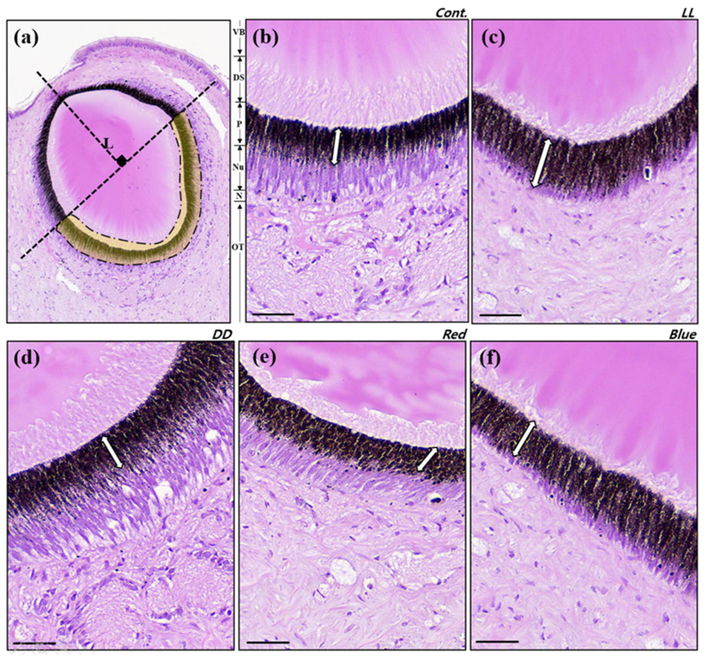
The retinas of conches were observed in each of the five light experimental groups (LD control, LL, DD, R, B). The distal segments and neuropile layer represent the light-absorbing region of the retina (Figure 2). In addition, a significant difference in the thickness of the pigment layer (TPL) was observed in each group. The pigment layer between the photoperiod groups (LD, LL, DD) was significantly (p < 0.05) thickest in the LL experimental group. Additionally, there was no significant difference (p > 0.05) in the pigment layer between light wavelength groups (LD vs. R, B). Overall, when exposed to the DD, R, and B light environments, there was no significant difference from the LD control, and a significant difference was found only in the LL environments. These results showed the same tendency when TPL was divided by each lens diameter (Table 1).

3.3. H2O2 Levels and TAC and MDA Contents

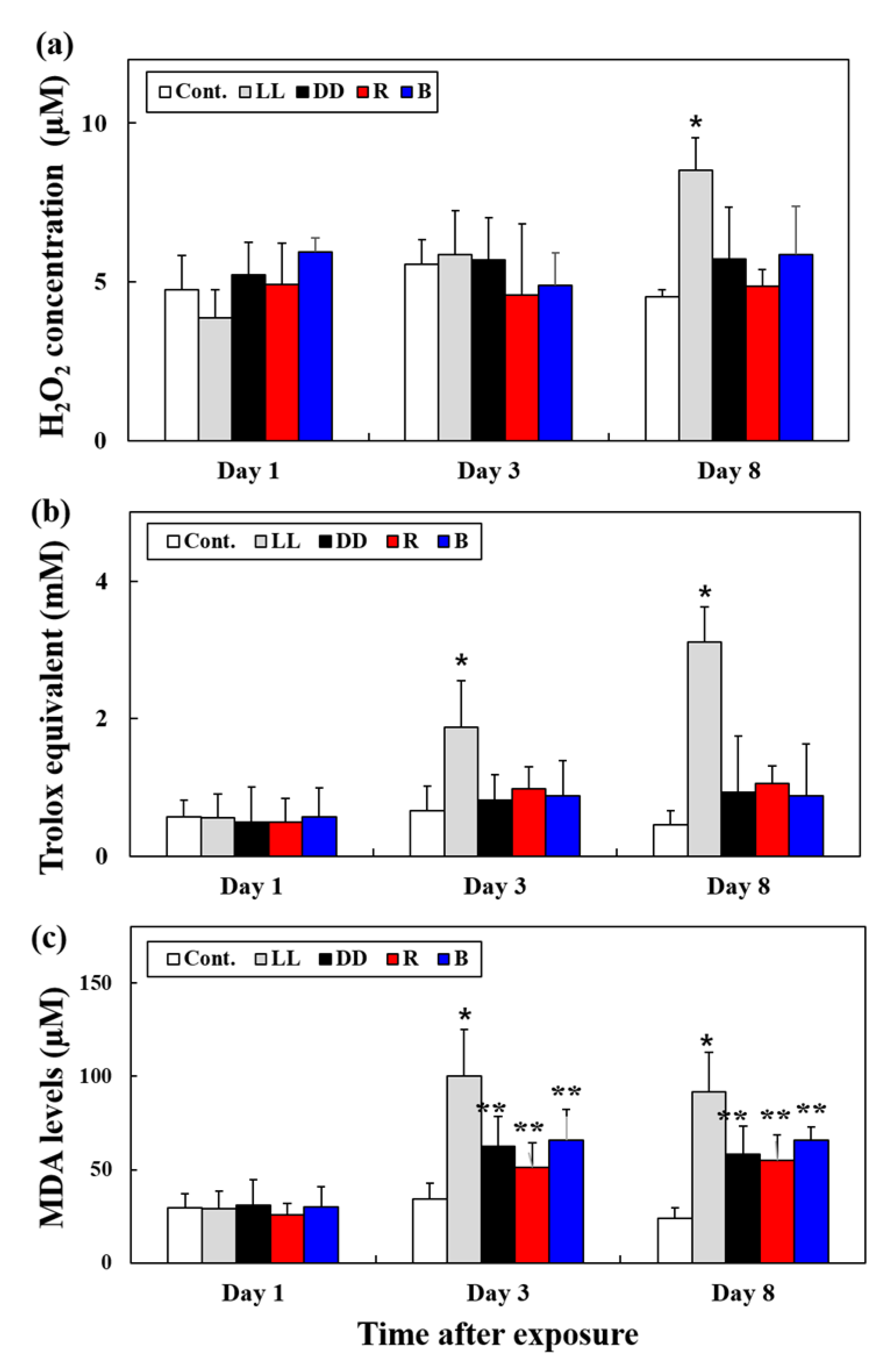
There was no significant difference in the H2O2 levels of the digestive diverticula according to the light wavelength and photoperiod up to the third day after exposure (p > 0.05, Figure 3a). However, when exposed to the LL environment for 8 days, a significantly higher H2O2 concentration was observed than in the other experimental groups (Day 8, LD: 4.54 ± 0.23; LL: 8.51 ± 1.02; p < 0.01].
TAC and MDA showed no significant change under the different conditions up to the first day of exposure (p > 0.05, Figure 3b,c). However, when exposed to the LL environment for more than 3 days, significantly higher TAC and MDA levels were observed compared with other photoperiod and light wavelength experimental groups. When exposed to the various light environments for 8 days, the MDA contents showed LL, DD, R, and B were significantly different from the control, and the most significant relationship was in the LL light environment (p < 0.01).

3.4. Immunohistochemical Staining of Rhodopsin

To observe the photoperiod-induced expression of rhodopsin protein, we performed immunohistochemical (IHC) staining for rhodopsin (Figure 4). The expression of rhodopsin protein levels (brown) in the neuropile (N) of the retinal layer was higher when exposed to light for a relatively short time (DD > LD > LL). Rhodopsin in LL was expressed lower than in the LD control, while rhodopsin in DD was expressed higher than in LD (p < 0.05).

4. Discussion

In the present study, we exposed T. rustica, a primitive nocturnal gastropod, to various light environments to investigate the effects of light wavelength and photoperiod on retinal cytological changes and oxidative stress.
Tegula rustica has a highly developed camera-type eye, as confirmed by this study. The eyes are elliptical (spherical), 397 × 359 μm (average) in size, and characterised by a large vitreous body. Most strombid gastropods studied so far, such as the Florida fighting conch Strombus alatus, the hawk-wing conch Lobatus raninus, and the marine gastropod Conomurex luhuanus, have camera-type eyes with spherical lenses [15,28,29], and this includes T. rustica.
Histological observation of the intraocular retina of the specimens under different light conditions confirmed that the pigment layer was significantly thicker in the LL group, which was irradiated with light for a long period. In contrast, light wavelength had no significant effect on the results. To date, there has been no research into why changes in intraocular pigment layer thickness occur in gastropods. However, it has been reported that when fish (Atlantic salmon Salmo salar and Goldfish Carrasius auratus) are strongly stimulated by light, the melanin granular layers in the retina become thicker [30,31]. It has been reported that when aquatic animals are stressed, melanin pigments in the retina are produced as byproducts, which aggregate to form a melanin granule layer [32].
Considering this, the thick pigment layers observed in the group irradiated with light for 24 h (LL) in this study may have formed to protect their retinas in an environment continuously irradiated with light. However, the thickness of the pigment layer did not exhibit a significant difference in response to light wavelengths. To support this, the concentration of H2O2, a type of ROS generated by stress, was measured. We found that conches exposed to photoperiod and light wavelength changes did not show significant changes in H2O2 concentration until 5 days after the change in the light environment. However, a significantly higher H2O2 concentration was observed in the LL test group on the eighth day compared with the other groups.
Previous studies have reported that stress-induced ROS production occurs in gastropods exposed to environmental changes, such as oxidative stress induced by high water temperatures [12,33]. To date, there have been few studies on the induction of oxidative stress in gastropods by radiance conditions. This may be a factor that induces oxidative stress and is more affected by the photoperiod (the existence of light) than by the light wavelength.
In addition, as a result of TAC and MDA levels, significantly higher levels were confirmed in the LL experimental group from the third day after the change in light environment. These results suggest the possibility that a large amount of various reactive oxygen species or reactive nitrogen species other than H2O2 that can affect the antioxidant response were generated from the third day after exposure. To remove the generated oxidation substances, the level of TAC increased from the third day, and it is believed that the concentration of MDA increased due to the oxidation substances that were not removed through this process.
Rhodopsin is a light-sensitive opsin that enables vision to be used in low-light environments. To confirm that rhodopsin is sensitive to light, even in T. rustica, IHC was used to compare the degree of RH protein expression with photoperiod (LD, LL, DD). We observed that the group exposed to the dark environment for 24 h (DD) had a far higher degree of RH protein expression.
Other conch species are also nocturnal and known to be more active at dusk and at night [28,34]. Given the nocturnal behaviour of conches, it is clear that greater RH expression was observed in the light-free environment to enhance ocular light sensitivity for nocturnal activity. Seyer [28] also suggested the possibility that the output from the rhabdoms in L. raninus may be neutrally pooled at night, which would increase sensitivity at the cost of resolution. Similar to previous research findings, our study also suggests that the absence of light affects opsin expression in T. rustica, and that RH is regulated by light.
These results show that exposure to continuous light induces excessive ROS in conches and causes oxidative stress. The conch protects itself from light stimulation by thickening the pigment layer within the retina, and this stimulation is believed to be more greatly affected by continuous light irradiation than the light wavelength.
To date, various studies have been actively conducted on large conch species and abalones, which are the main cultivated species of primitive gastropods. However, starting with this study, it may be important to obtain data on physiological responses to different light environments (photoperiod, light intensity, light wavelength, etc.) in a variety of gastropods, including small conch species, to improve aquaculture practices and husbandry. So, it is considered necessary to investigate the relationship between food intake and locomotive activities and oxidative stress according to the light environment.

Supplementary Materials

The following supporting information can be downloaded at: https://www.mdpi.com/article/10.3390/fishes9060226/s1, Figure S1: Western blotting of rhodopsin in T. rustica.

Author Contributions

Conceptualisation, J.A.S. and H.-S.P.; methodology, D.M.C. and C.Y.C.; software, J.A.S. and Y.-H.J.; validation, J.A.S., H.-S.P. and D.-W.L.; formal analysis, D.M.C.; investigation, J.A.S. and Y.-H.J.; resources, Y.-H.J.; data curation, J.A.S.; writing—original draft preparation, J.A.S.; writing—review and editing, H.-S.P. and D.-W.L.; visualisation, J.A.S.; supervision, D.-W.L.; project administration, D.-W.L.; funding acquisition, H.-S.P. All authors have read and agreed to the published version of the manuscript.

Funding

This study was funded by the Korea Institute of Ocean Science and Technology (Grant number: PEA0205), Republic of Korea.

Institutional Review Board Statement

Not applicable.

Informed Consent Statement

Not applicable.

Data Availability Statement

No new data were created or analysed in this study. Data sharing is not applicable to this article.

Acknowledgments

The authors would like to thank the anonymous reviewers who critically reviewed the manuscript and colleagues who helped with sample collection.

Conflicts of Interest

The authors declare no conflicts of interest.

References

  1. Miller, L.P.; Denny, M.W. Importance of behavior and morphological traits for controlling body temperature in littorinid snails. Biol. Bull. 2011, 220, 209–223. [Google Scholar] [CrossRef]
  2. Beuchel, F.; Gulliksen, B.; Carroll, M.L. Long-term patterns of rocky bottom macrobenthic community structure in an Arctic fjord (Kongsfjorden, Svalbard) in relation to climate variability (1980–2003). J. Mar. Syst. 2006, 63, 35–48. [Google Scholar] [CrossRef]
  3. Kulikova, V.A.; Omel’yanenko, V.A. Reproduction and larval development of the gastropod mollusk Tegula rustica in Peter the Great Bay, Sea of Japan. Russ. J. Mar. Biol. 2000, 26, 128–130. [Google Scholar] [CrossRef]
  4. Serb, J.M. Toward developing models to study the disease, ecology, and evolution of the eye in Mollusca. Am. Malacol. Bull. 2008, 26, 3–18. [Google Scholar] [CrossRef]
  5. Young, J.Z. The Anatomy of the Nervous System of Octopus Vulgaris; Clarendon Press: Oxford, UK, 1971. [Google Scholar]
  6. Muntz, W.R.A.; Raj, U. On the visual system of Nautilus pompilius. J. Exp. Biol. 1984, 109, 253–263. [Google Scholar] [CrossRef]
  7. Hanlon, R.T.; Shashar, N. Aspects of the sensory ecology of cephalopods. In Sensory Processing in Aquatic Environments; Springer: New York, NY, USA, 2003; pp. 266–282. [Google Scholar]
  8. Zieger, M.V.; Meyer-Rochow, V.B. Understanding the cephalic eyes of pulmonate gastropods: A review. Am. Malacol. Bull. 2008, 26, 47–66. [Google Scholar] [CrossRef]
  9. Salvini-Plawen, L.V.; Mayr, E. On the evolution of photoreceptors and eyes. Evol. Biol. 1977, 10, 207–263. [Google Scholar]
  10. Serb, J.M.; Eernisse, D.J. Charting evolution’s trajectory: Using molluscan eye diversity to understand parallel and convergent evolution. Evol. Educ. Outreach. 2008, 1, 439–447. [Google Scholar] [CrossRef]
  11. Katagiri, N.; Terakita, A.; Shichida, Y.; Katagiri, Y. Demonstration of a rhodopsin-retinochrome system in the stalk eye of a marine gastropod, Onchidium, by immunohistochemistry. J. Comp. Neurol. 2001, 433, 380–389. [Google Scholar] [CrossRef]
  12. Ambekar, A.A.; Sivaperumal, P.; Kamala, K.; Kubal, P.; Prakash, C. Effect of temperature changes on antioxidant enzymes and oxidative stress in gastropod Nerita oryzarum collected along India’s first Tarapur Atomic Power Plant site. Environ. Res. 2023, 216, 114334. [Google Scholar] [CrossRef]
  13. Kingston, A.C.N.; Kuzirian, A.M.; Hanlon, R.T.; Cronin, T.W. Visual phototransduction components in cephalopod chromatophores suggest dermal photoreception. J. Exp. Biol. 2015, 218, 1596–1602. [Google Scholar] [CrossRef] [PubMed]
  14. Kingston, A.C.N.; Wardill, T.J.; Hanlon, R.T.; Cronin, T.W. An Unexpected Diversity of Photoreceptor Classes in the Longfin Squid, Doryteuthis pealeii. PLoS ONE 2015, 10, e0135381. [Google Scholar] [CrossRef] [PubMed]
  15. Clark, J.M. Restoration of Visual Performance and Opsin Expression within the Retina during Eye Regeneration in the Florida Fighting Conch (Strombus alatus). Master’s Thesis, University of South Carolina, Columbia, SC, USA, 2018. [Google Scholar]
  16. Fridovich, I. Superoxide dismutases. Annu. Rev. Biochem. 1975, 44, 147–159. [Google Scholar] [CrossRef]
  17. Jiang, Q.; Zhou, Z.; Wang, L.L.; Shi, X.W.; Wang, J.J.; Yue, F.; Yi, Q.L.; Yang, C.Y.; Song, L.S. The immunomodulation of inducible nitric oxide in scallop Chlamys farreri. Fish. Shellfish Immunol. 2013, 34, 100–108. [Google Scholar] [CrossRef] [PubMed]
  18. Pan, L.Q.; Ren, J.; Liu, J. Responses of antioxidant systems and LPO level to benzo[α]pyrene and benzo(k)uoranthene in the haemolymph of the scallop Chlamys ferrari. Environ. Pollut. 2006, 141, 443–451. [Google Scholar] [CrossRef] [PubMed]
  19. Li, Q.; Wang, M.; Duan, L.; Qiu, Y.; Ma, T.; Chen, L.; Breitholtz, M.; Bergman, Å.; Zhao, J.; Hecker, M.; et al. Multiple biomarker responses in caged benthic gastropods Bellamya aeruginosa after in situ exposure to Taihu Lake in China. Environ. Sci. Eur. 2018, 30, 34. [Google Scholar] [CrossRef] [PubMed]
  20. Campoy-Diaz, A.D.; Malanga, G.; Giraud-Billoud, M.; Vega, I.A. Changes in the oxidative status and damage by non-essential elements in the digestive gland of the gastropod Pomacea canaliculata. Front. Physiol. 2023, 14, 1123977. [Google Scholar] [CrossRef] [PubMed]
  21. Shin, H.S.; Lee, J.; Choi, C.Y. Effects of LED light spectra on the growth of the yellowtail clownfish Amphiprion clarkii. Fish. Sci. 2012, 78, 549–556. [Google Scholar] [CrossRef]
  22. Song, J.A.; Lee, Y.S.; Choi, Y.U.; Choi, C.Y. Effects of light-emitting diodes on thermally-induced oxidative stress in the bay scallop Argopecten irradians. Molluscan Res. 2020, 40, 130–141. [Google Scholar] [CrossRef]
  23. Liu, C.; Yang, X.; Sun, Y.; Yang, Y.; Wang, A.; He, L.; Gu, Z. Effects of the daily light/dark cycle on photosynthetic performance, oxidative stress and illumination-related genes in boring giant clam Tridacna crocea. Mar. Biol. 2021, 168, 71. [Google Scholar] [CrossRef]
  24. Ma, S.; Li, L.; Chen, X.; Chen, S.; Dong, Y.; Gao, Q.; Zhou, Y.; Dong, S. Influence of daily rhythmic light spectra and intensity changes on the growth and physiological status of juvenile steelhead trout (Oncorhynchus mykiss). Front. Mar. Sci. 2023, 10, 36. [Google Scholar] [CrossRef]
  25. Kim, J.A.; Kim, M.J.; Kim, J.H.; Choi, C.Y. Effects of Red LED Spectra and Different Photoperiods on the Circadian Rhythm of Abalones (Haliotis discus hannai). Ocean Polar Res. 2024, 46, 55–64. [Google Scholar]
  26. Gao, X.; Li, X.; Zhang, M.; Chi, L.; Song, C.; Liu, Y. Effects of LED light quality on the growth, survival and metamorphosis of Haliotis discus hannai Ino larvae. Aquac. Res. 2016, 47, 3705–3717. [Google Scholar] [CrossRef]
  27. Takata, T.; Zhao, M.; Uchida, T.; Kudo, Y.; Sato, S.; Nikai, H. Immunohistochemical demonstration of an enamel sheath protein, sheathlin, in odontogenic tumors. Virchows Arch. 2000, 436, 324–329. [Google Scholar] [CrossRef] [PubMed]
  28. Seyer, J.O. Structure and optics of the eye of the hawk-wing conch, Strombus raninus (L.). J. Exp. Zool. 1994, 268, 200–207. [Google Scholar] [CrossRef]
  29. Irwin, A.R.; Williams, S.T.; Speiser, D.I.; Roberts, N.W. The marine gastropod Conomurex luhuanus (Strombidae) has high-resolution spatial vision and eyes with complex retinas. J. Exp. Biol. 2022, 225, jeb243927. [Google Scholar] [CrossRef] [PubMed]
  30. Migaud, H.; Cowan, M.; Taylor, J.; Ferguson, H.W. The effect of spectral composition and light intensity on melatonin, stress and retinal damage in post-smolt Atlantic salmon, Salmo salar. Aquaculture 2007, 270, 390–404. [Google Scholar] [CrossRef]
  31. Song, J.A.; Choi, C.Y. Effects of blue light spectra on retinal stress and damage in goldfish (Carassius auratus). Fish Physiol. Biochem. 2019, 45, 391–400. [Google Scholar] [CrossRef] [PubMed]
  32. Bogerts, B. A brainstem atlas of catecholaminergic neurons in man, using melanin as a natural marker. J. Comp. Neurol. 1981, 197, 63–80. [Google Scholar] [CrossRef]
  33. Dieterich, A.; Troschinski, S.; Schwarz, S.; Di Lellis, M.A.; Henneberg, A.; Fischbach, U.; Ludwig, M.; Gärtner, U.; Triebskorn, R.; Köhler, H.R. Hsp70 and lipid peroxide levels following heat stress in Xeropicta derbentina (Krynicki 1836) (Gastropoda, Pulmonata) with regard to different colour morphs. Cell Stress Chaperones 2015, 20, 159–168. [Google Scholar] [CrossRef]
  34. Sandt, V.J.; Stoner, A.W. Ontogenic shift in habitat by early juvenile queen conch, strombus-gigas-patterns and potential mechanisms. Fish. Bull. 1993, 91, 516–525. [Google Scholar]
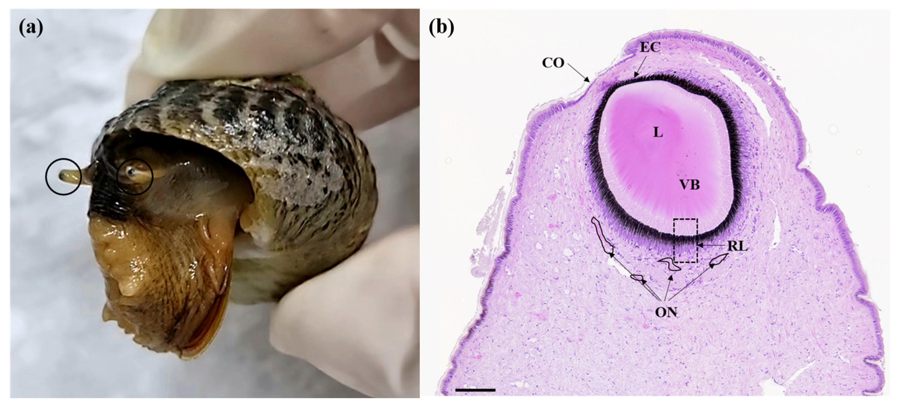
Figure 1. (a) Tegula rustica as an experimental model with extended eyestalks (circle). (b) Hematoxylin and eosin (H&E) staining from the eyestalk of T. rustica in longitudinal section. CO, cornea; L, lens; VB, vitreous body; ON, optic nerve; RL, retinal layer, EC; eye capsule. Scale bar = 100 μm.
Fishes 09 00226 g001
Figure 2. Representative photomicrographs of retina specimens prepared from the retinal layers of Tegula rustica reared under illumination of five different photoenvironments for a period of 8 days: (a) the position of the retinal sections (transparent yellow) used for analyses; (b) 12L: 12D using white LED (LD control); (c) 24L using white LED (LL); (d) 24D (DD); (e) 12L: 12D using red LED (R); (f) 12L: 12D using blue LED (B). The white arrow indicates the thickness of the pigment layer. VB, vitreous body; DS, distal segments; P, pigmented layer; Nu, nuclear layer; N; neuropile; OT, optic tentacle. Prepared using H&E staining. Scale bar = 30 μm.
Fishes 09 00226 g002
Figure 3. Concentration of (a) H2O2, (b) total antioxidant capacity, and (c) MDA levels in the digestive diverticula of Tegula rustica exposed to five different photoenvironments for 8 days. Asterisks (*) indicate significant differences among groups exposed to different photic environments (* p < 0.01; ** p < 0.05). Values indicate means ± SD (n = 7).
Fishes 09 00226 g003
Figure 4. Images of rhodopsin immunohistochemistry for the retinal tissue of Tegula rustica exposed to three different photoenvironments for 8 days: (a) Negative control without antibody; (b) 12 L: 12D using white LED (LD control); (c) 24 L using white LED (LL); (d) 24D (DD); (e) analysis of immunohistochemistry staining intensity using ImageJ. Scale bars = 30 μm. Rhodopsin (brown) is present in the neuropile (N) and between the distal segments (DS) within the back of the retinal layer. P, pigmented layer; Nu, nuclear layer; OT, optic tentacle; ON, optic nerve; RC, photoreceptor cells; PC, pigment cells. The yellow arrow indicates the direction of light. The different lowercase letters above bars indicate significant differences among groups exposed to different photic environments (p < 0.05).
Fishes 09 00226 g004
Table 1. The thickness of retinal pigment layer.
Cont.LLDDRedBlue
Thickness of pigment layer (μm, TPL)21.65
± 4.66
29.38
± 7.03 *
22.02
± 7.40
22.01
± 6.85
25.10
± 4.85
TPL⁄(lens length)0.18 ± 0.040.24 ± 0.06 *0.18 ± 0.060.18 ± 0.050.20 ± 0.04
* Indicate significant differences among groups exposed to different photic environments (p < 0.05).
Disclaimer/Publisher’s Note: The statements, opinions and data contained in all publications are solely those of the individual author(s) and contributor(s) and not of MDPI and/or the editor(s). MDPI and/or the editor(s) disclaim responsibility for any injury to people or property resulting from any ideas, methods, instructions or products referred to in the content.