Article

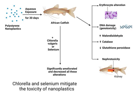
Impact of Chlorella vulgaris Bioremediation and Selenium on Genotoxicity, Nephrotoxicity and Oxidative/Antioxidant Imbalance Induced by Polystyrene Nanoplastics in African Catfish (Clarias gariepinus)

1
Department of Forensic Medicine and Toxicology, Faculty of Veterinary Medicine, Assiut University, Assiut 71526, Egypt
2
Department of Pathology, Faculty of Veterinary Medicine, Assiut University, Assiut 71526, Egypt
3
Division of Pharmacology and Toxicology, Department of Pharmaceutical Sciences, University of Vienna, 1090 Vienna, Austria
4
Zoology Department, Faculty of Science, Assiut University, Assiut 71516, Egypt
*
Authors to whom correspondence should be addressed.
Fishes 2024, 9(2), 76; https://doi.org/10.3390/fishes9020076
Submission received: 10 January 2024 / Revised: 5 February 2024 / Accepted: 11 February 2024 / Published: 15 February 2024
(This article belongs to the Special Issue Sustainable Catfish Aquaculture)

Abstract

:
Contamination of the environment with nano- and microplastic particles exerts a threatening impact on the aquatic ecosystems and sustainable catfish aquaculture. The presence of nanoplastics has been found to have a detrimental impact on both aquatic and terrestrial ecosystems. The present study examines the effect of polystyrene nanoplastics (PS NPs) on the DNA, erythrocytes, oxidative status and renal histology of catfish, in addition to the potential protective effects of Chlorella vulgaris bioremediation and selenium to hinder this effect. Six equal groups of fish were used as follows: Group 1 served as a control group and received water free from PS NPs; Group 2 was exposed to PS NPs at a concentration of 5 mg/L; Group 3 was exposed to PS NPs (5 mg/L) + selenium (1 mg/kg diet); Group 4 was exposed to PS NPs (5 mg/L) + C. vulgaris (25 g/kg diet); Group 5 was supplemented with C. vulgaris (25 g/kg diet); and Group 6 was supplemented with selenium (1 mg/kg diet). The exposure period was 30 days. The results indicated that PS NPs induced oxidative stress by significantly elevating malondialdehyde activities and slightly reducing antioxidant biomarkers, resulting in DNA damage, increased frequency of micronuclei, erythrocyte alterations, and numerous histopathological alterations in kidney tissue. Selenium and C. vulgaris significantly ameliorated the oxidative/antioxidant status, reducing DNA damage, micronucleus frequency, erythrocyte alterations, and improving the morphology of kidney tissue. Nevertheless, further research is needed for a profound understanding of the mechanism behind the toxicity of nano-microplatics in aquatic systems.
Key Contribution: Nanoplastic particles have deleterious adverse effects on the DNA integrity oxidative/antioxidant status and kidneys of African catfish. The addition of selenium and C. vulgaris could ameliorate and decrease these alterations.

Graphical Abstract

1. Introduction

Polystyrene nanoplastics (PS NPs) are sub-100 nm sized plastic particles, while polystyrene microplastics (PS MPs) range from 1 µm to 5 mm [1]. They can penetrate biological barriers and accumulate along the food chain, posing significant health risks to humans and animals [2].
MPs and NPs are present in various ecosystems such as soils, air, marine and freshwater environments, as reported by [3]. The issue of water contamination caused by plastics has been recognized as a significant environmental peril in numerous countries worldwide, as stated by [4].
Egypt faces two primary challenges associated with plastics: excessive consumption and inadequate waste management. The nation is identified as the largest plastic polluter in the Mediterranean, contributing 250,000 tons annually [5,6]. Egypt’s annual plastic consumption amounts to approximately 5.4 million tons [6]. The estimated quantity of mismanaged plastic waste in Egypt is 0.97 million tons per year [7,8].
Plastic wastes in the marine environment are subjected to the process of plastic fragmentation after UV exposure and mechanical abrasions to yield huge amounts of MPs [9].
Recent studies have reported that MPs can reach the human food chain in several ways, for example, through contamination of human food during preparation processes, bottled water or through leaching of MPs from plastic packaging of food and drinks [10,11].
PS NPs have the ability to permeate the gut epithelium, leading to systemic exposure in humans. Several in vivo and in vitro studies on animals found that the probable oral bioavailability level of 50 nm polystyrene nanoparticles is ten to one hundred times greater than the level of MPs [12,13].
There are three routes for exposure to MPs and NPs through oral, inhalation and dermal exposure [14,15]. After absorption, MPs and NPs will be able to interact with numerous target cells and induce toxic effects on the kidneys, gut, liver and other organs [16]. Cells can absorb MPs and NPs through different routes, with the main route involving endocytotic uptake of nanoparticles. This mechanism involves the adhesive interaction of nanoparticles with channel or transport proteins, or the inactive permeation of the cell membrane. Various recognized endocytotic pathways include phagocytosis and macropinocytosis, along with clathrin- and caveolae-mediated endocytosis [17,18].
Extensive studies have shown that MPs and NPs can lead to different biological adverse effects, such as oxidative stress, apoptosis and necrosis [19], cytotoxicity, genotoxicity [20], neurotoxicity [21], reproductive and developmental toxicity and immune dysfunction [22]. Furthermore, they have been observed to have detrimental effects on the survival and reproduction of aquatic organisms [23]. The kidney is a significant organ for investigating the detrimental effects of NPs and MPs in aquatic organisms. The adverse effects of NPs on fish kidneys are not yet fully understood and require further exploration.
Clarias gariepinus inhabits various freshwater habitats, such as ponds, lakes and pools. In addition, they are found in rivers, rapids and near dams. They can survive in water with a pH range of 6.5 to 8.0 and exceedingly low oxygen concentrations [24]. Their bodies produce mucus to prevent dehydration [25]. They can tolerate temperatures between 8 and 35 degrees Celsius, as well as low oxygen levels, and persist in environments with high brackish water salinities and nitrogen compounds. The optimal temperature for their growth is between 28 and 30 degrees Celsius [26]. C. gariepinus is widely utilized in aquaculture because it is readily available throughout the year, with a high food conversion ratio and fast growth rate, and adapts well to laboratory conditions and stress, making it an ideal test organism [27].
Probiotics are described as microbial cells capable of positively influencing the development, growth and health of host animals by enhancing the balance of the host-associated microbiota [28].
Chlorella vulgaris, a green unicellular microalga, is used in aquatic bioremediation [29]. It exhibits potent antioxidant properties due to the presence of carotenoids and phenolic bioactive compounds responsible for its antioxidant and radical-scavenging activities [30]. Selenium (Se) is a micronutrient that is necessary for fishes’ development and metabolic processes [31]. Selenium can be found in many essential selenoproteins and enzymes that maintain antioxidant status, DNA synthesis, fertility and reproduction, and reduce inflammation [32].
There is a dearth of research evaluating the genotoxic effect of PS NPs, the effect on erythrocyte morphology and the pathological changes in the kidney of catfish. Also, the potential ameliorative effects of selenium and bioremediation by C. vulgaris. Therefore, the present study assessed the efficacy of L-Selenomethionine and C. vulgaris in mitigating the harmful effects of PS NPs on DNA, RBCs, and kidney of C. gariepinus.

2. Materials and Methods

2.1. Chemicals and Materials

PS NPs were procured from Bangs Laboratories, Inc. The Seleno-L-methionine powder was procured from Sigma-Aldrich, Saint Louis, MO, USA. The powder was high purity (≥98%) and had a molecular weight of 196.11. Additionally, the powder was identified by its CAS number, 3211-76-5. The dried green powder of C. vulgaris microalgae was procured from the Institute of National Research Center, Cairo, Egypt.
Before the conduction of the experiments, a stock solution of PS NPs at a concentration of 5 g L−1 was prepared using deionized water. Then, the dimensions and morphology of NPs were assessed using a JEOL-JEM-100 CX II transmission electron microscope (TEM) (JEOL GmbH, Freising, Germany) located in the electron microscopy unit at Assiut University, Egypt.

2.2. Clarias Gariepinus

For the study, a sample of 144 male juvenile catfish (C. gariepinus) was obtained from an aquaculture fish farm in Assiut, Egypt. The specimens exhibited an average weight of 160 g and a length of 25 and 30 cm. The age of the specimens was determined to be six months. The conveyance of fish to the Fish Biology Laboratory, situated within the Zoology Department of the Faculty of Science at Assiut University in Egypt, was executed. During a 35-day acclimatization period, the fish were subjected to laboratory conditions in which 100 L tanks, dechlorinated tap water and air pumps were used; this was executed before the initiation of the exposure. The piscine subjects were administered a diet comprising a commercial substrate with a crude protein content of 30%. The nutritional needs of C. gariepinus were satisfied by creating a fundamental diet comprising diverse constituents, with the estimated chemical makeup of each component outlined in Supplementary Table S1.

2.3. Ethics Statement

The research protocol mentioned above has received approval from the veterinary ethical committee of the Faculty of Veterinary Medicine at Assiut University, Egypt, under the reference number 06/2022/0016.

2.4. Experimental Design

The fish were randomly put into six groups of 24 each, with three tanks in each group. The first group (C) was used as a control and was kept in water that did not contain PS NPs. The second group (NPs) was exposed to 5 mg/L PS NPs in water according to [33]. The third group (NPs+Se) was given PS NPs (5 mg/L) and selenium (1 mg/kg diet) according to [34]. The fourth group (NPs+Ch) was exposed to PS NPs (5 mg/L) and C. vulgaris (25 g/kg diet) according to [35,36]. The fifth group (Ch) was exposed to only C. vulgaris (25 g/kg diet). The sixth group (Se) was only exposed to selenium (1 mg/kg diet).
For 30 days, the animals were kept in tanks with 60 L of naturally dechlorinated water and air blowers for regular aeration. The experiment was performed with steady water quality (temperature of 22 °C, dissolved oxygen (mg/L) of 6.85, pH of 7.38 and a 12:12 light–dark cycle). About 50% of the water was changed daily throughout the experiment, and PS NPs were added in the abovementioned amounts. In addition, waste products like feces were removed daily to keep the ammonia levels in the water as low as possible.

2.5. Sample Collection

2.5.1. Blood Samples

Following a period of 30 days of exposure, six distinct groups of fish comprising both control and treatment groups, were captured at random using a hand net and subsequently transferred to ice in order to mitigate any potential stress, based on the study conducted by [37]. The blood collection methodology and the approach were described by [38]. First, EDTA-containing tubes were utilized to obtain blood samples from the caudal vein of C. gariepinus to detect DNA damage through the comet assay. Next, additional blood samples were collected using standard vacutainer tubes and subsequently subjected to centrifugation at 3000 revolutions per minute for 15 min to procure serum samples, which were then stored at a temperature of −20 °C. These samples were taken to assess malondialdehyde, catalase enzyme and glutathione peroxidase enzyme levels. Finally, the blood samples were subjected to the preparation of blood smears, followed by drying at ambient temperature and fixation in absolute methanol to conduct the micronucleus assay and assess other nuclear abnormalities and erythrocyte morphological changes.

2.5.2. Tissue Samples

After dissecting the fish, kidney tissues were rapidly removed, cleaned in normal saline to eliminate blood traces and stored in 10% buffered formalin for histological investigation.

2.6. Oxidative Stress Biomarkers Measurements

2.6.1. Determination of Serum Malondialdehyde (MDA) Activity

MDA was measured by the spectrophotometric method (V-630 UV-Vis Spectrophotometer, JASCO, Easton, MD, USA), using a commercial kit supplied by Bio Diagnostics, Egypt, with a catalog number MD 25 30 and according to the method described by [39].

2.6.2. Determination of Serum Catalase Enzyme Activity

Catalase assay was performed by the spectrophotometric method (V-630 UV-Vis Spectrophotometer, JASCO, Easton, MD, USA). According to [40] catalase was measured using test kits provided by Bio-diagnostic (Dokki, Giza, Egypt).

2.6.3. Determination of Serum Glutathione Peroxidase (GPX) Activity

Glutathione peroxidase activity was measured in serum samples using test kits provided by Bio-diagnostic (Dokki, Giza, Egypt). This was performed by using a spectrophotometric method (V-630 UV-Vis Spectrophotometer, Easton, MD, USA) to detect the activity of the GPX enzyme [41].

2.7. Genotoxicity Assays

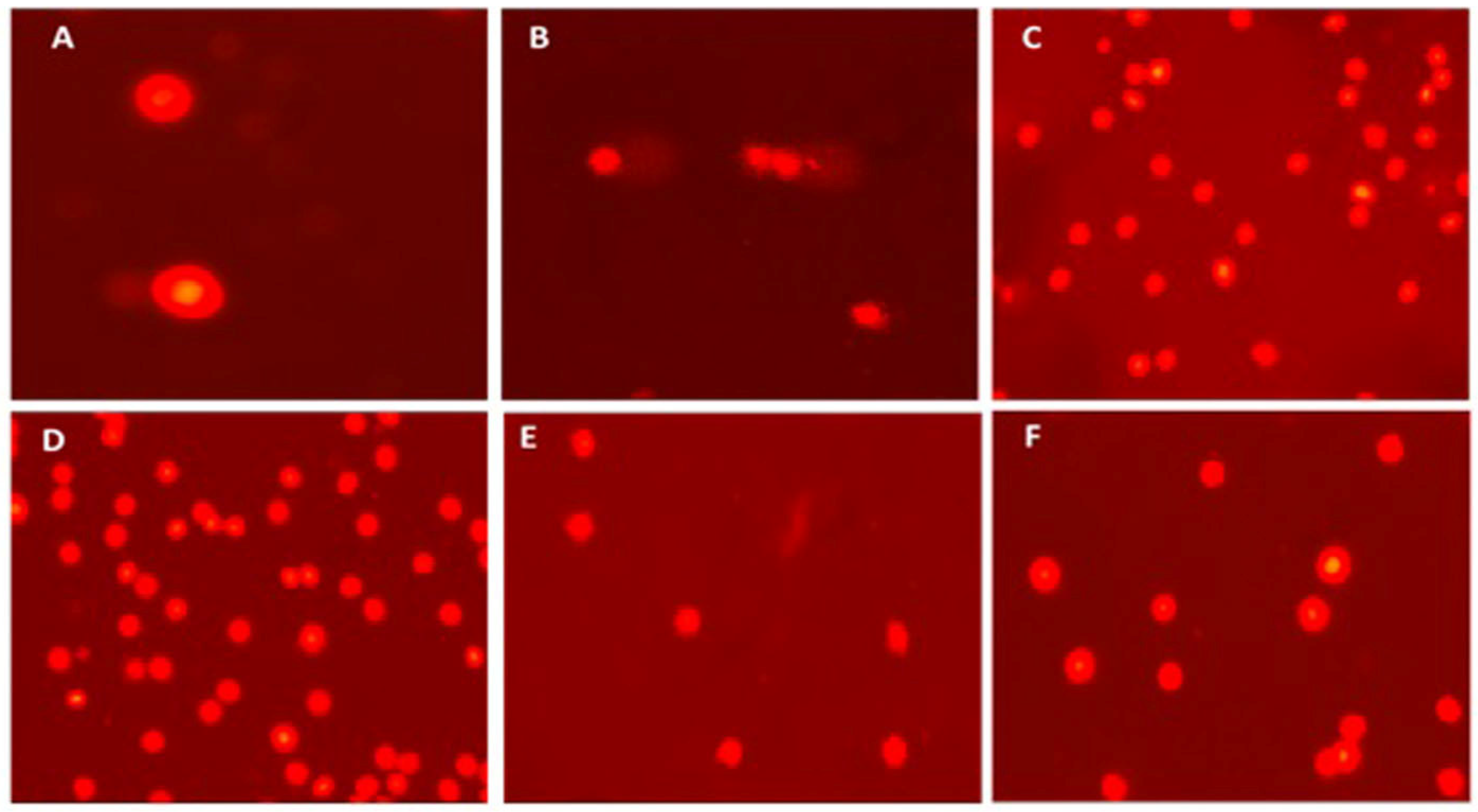
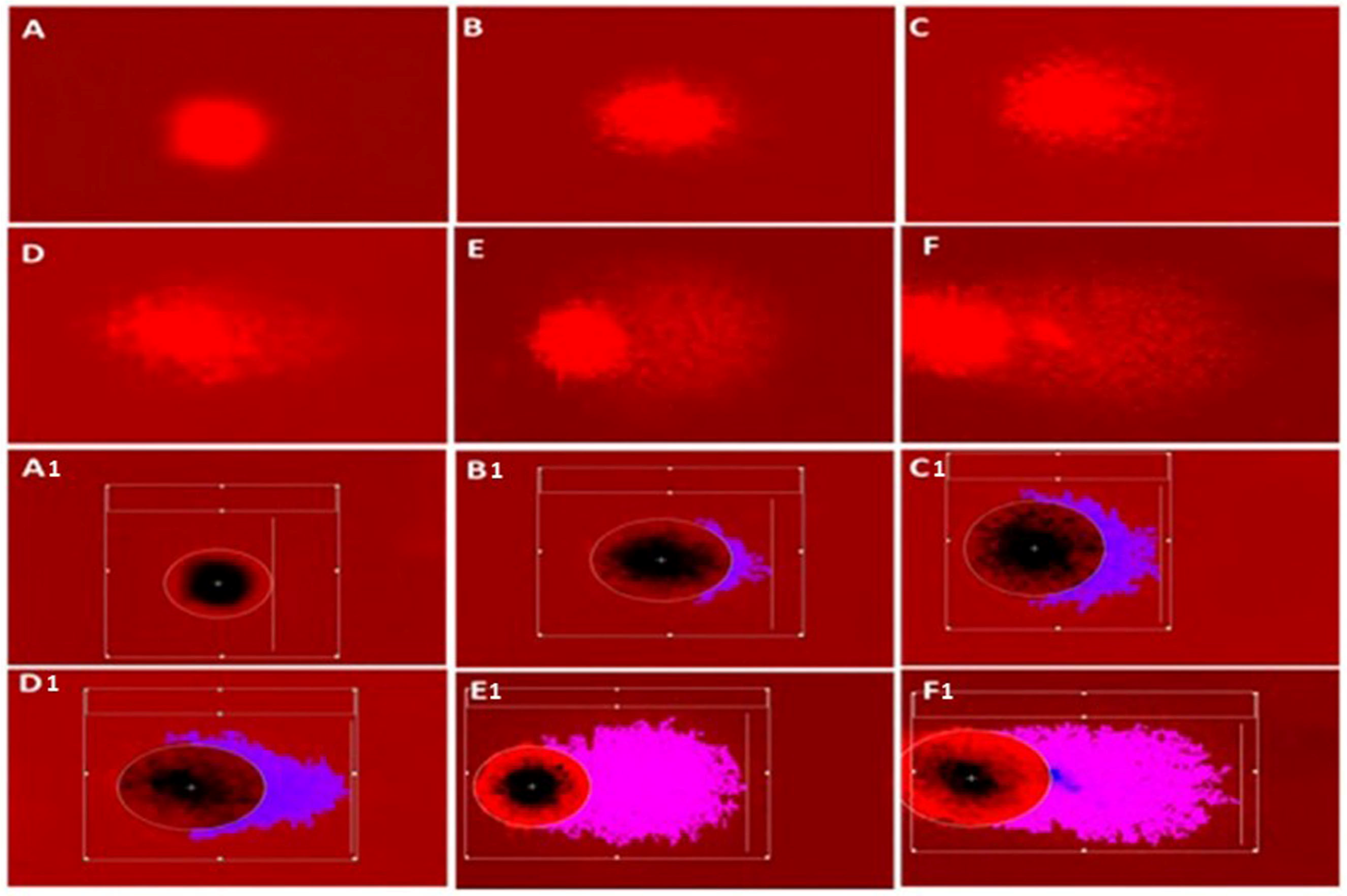
2.7.1. Detection of DNA Damage by Single-Cell Gel Electrophoresis (Comet Assay)

DNA damage in the erythrocyte of C. gariepinus was estimated using a comet assay according to the method of [42], as follows.
(a)
Preparation of slides
Slides were double coated with 1% standard agarose; 0.3 mL of blood was diluted in 1000 µL PBS; next, ten microliters of cell suspension was mixed with 120 µL of 2% low-melting-point agarose at 37 °C. A layer of the mixture was placed on the previously prepared slides, cover-slipped and placed in the refrigerator for 5 min to solidify. Finally, 1% agarose (100 μL) was layered on top and allowed to gel.
(b)
Lysis and electrophoresis
All slides were kept in a cold lysis solution (10 mM Trizma, 100 mM Na4 EDTA, 2.5 M NaCl, 10% dimethyl sulfoxide, 0.1% sodium lauryl sulfate (SDS), and Triton X-100) at 4 °C for 60 min. Then, they were transferred to the electrophoresis tank (Cleaver Scientific Ltd., Rugby, UK) containing chilled alkaline solution (1 mM Na2 EDTA and 300 mM NaOH) at pH 13 for 25 min in the dark for unwinding of the DNA strands. Electrophoresis was run for 25 min at 25 V and approximately 300 mA. Next, slides were neutralized in 400 mM Tris buffer (0.4 M Tris-HCl, pH 7.5) for 7 min. Finally, the neutralized slides were dehydrated in ethanol for a few minutes and then dried at room temperature.
(c)
Slide examination
The slides were examined through a fluorescence microscope (Olympus BX-43, Olympus Life Science, Tokyo, Japan) with a green filter at 200-fold magnification. In addition, 50 μL of ethidium bromide at 20 μg/mL concentration was used to stain all the slides. In order to assess DNA migration, a minimum of 50 nuclei were examined per slide, with three slides per sample. In addition, the Comet Assay Software Project (CASP-version 1.2.3b) was utilized to measure the diameter of the comet’s head and the length of its tail.

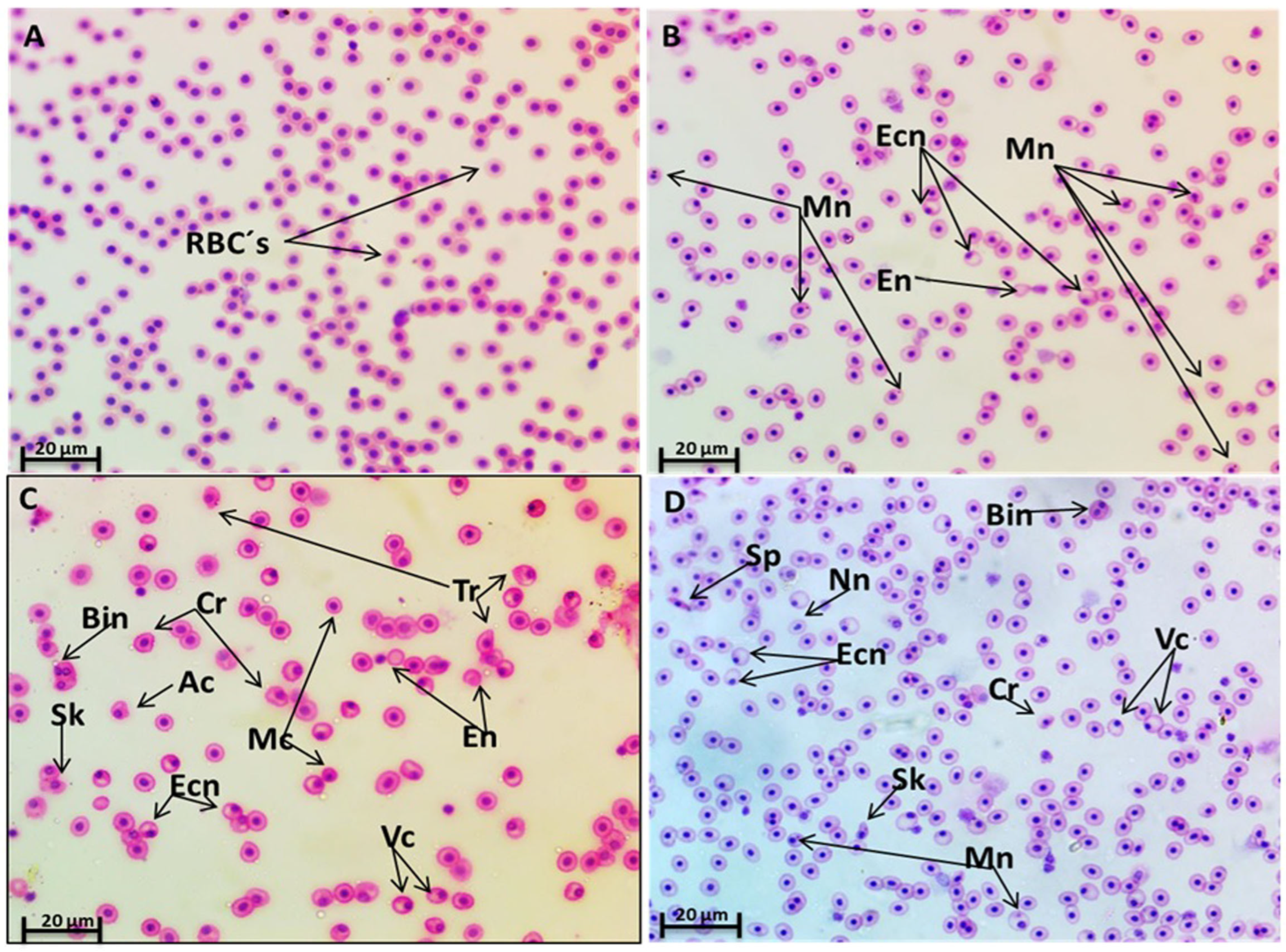
2.7.2. Micronucleus and Erythrocyte Morphological Alterations

Three blood smears were prepared and dried at room temperature. For 10 min, the smears were fixed in methanol (100%) and stained with Hematoxylin and Eosin. The slides were then dehydrated with alcohol with increasing concentrations (30, 50, 70, 90 and 100 percent) before being cleaned in xylene and permanently mounted with DPX [43]. According to [44], each nuclear abnormality observed during slide examination, including micronuclei formation, was noted. Next, 10 photomicrographs of random fields were obtained for each slide, totaling 30 photomicrographs per experimental group, and one hundred cells were examined in each analysis field, totaling 3000 erythrocytes/group.
M N % = N u m b e r   o f   c e l l s   w i t h   m i c r o n u c l e i × 100 T o t a l   n u m b e r   o f   c e l l s   c o u n t e d

2.8. Histopathological Studies

Kidney specimens were fixed in 10% neutral-buffered formalin, then dehydrated in serial alcohol concentrations, cleared in xylene and impregnated in paraffin. Next, 5 μm paraffin sections were prepared and then stained with the following stains: Hematoxylin and Eosin (H and E) according to [45], Periodic Acid Schiff (PAS) and Hematoxylin for polysaccharides (glycogen) [46] and Picrosirius red stain for collagen fibers [47]. A histopathologist, unaware of the groups’ treatments, examined three sections per specimen under Olympus light microscope (New York Microscope Company, NY, USA).

2.9. Statistical Analysis

The statistical analysis used SPSS software version 16.0 (SPSS Inc., Chicago, IL, USA). Initially, the statistical method of one-way analysis of variance (ANOVA) was employed, subsequently followed by a post hoc least significant difference (LSD) multiple range test to make comparisons between the control and exposed groups. Finally, the mean ± SE representing all outcomes for the control and experimental groups was calculated. The statistical significance level was established at a value of p < 0.05.

3. Results

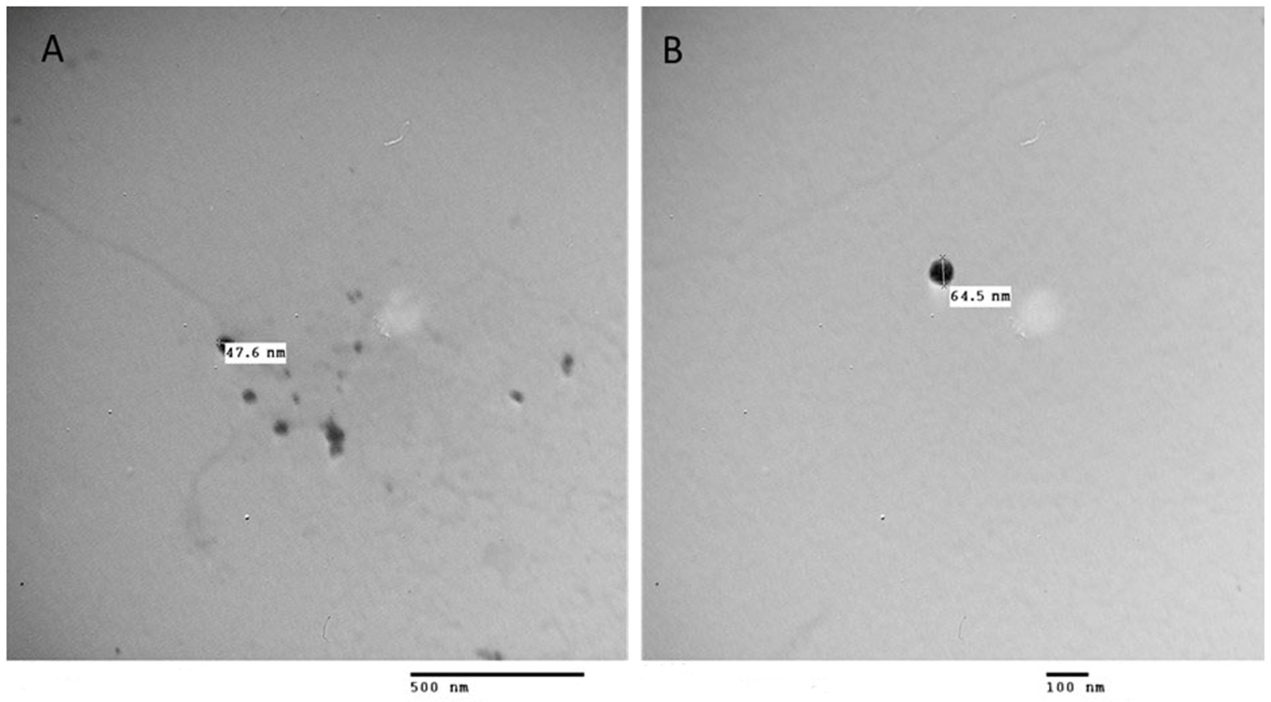
3.1. PS NPs Characterization

PS NPs were analyzed using transmission electron microscopy (TEM), which revealed that they are made up of spherical particles with a diameter of 50.1 ± 13.4 nm (Figure 1).

3.2. Mortality Rate

The results showed no deaths were recorded in the control group, while the NPs-exposed group showed a significant increase in mortality to 36.3% (p < 0.001) compared to the control. The administration of selenium significantly reduced the rate of mortality to 14.8% (p < 0.05) compared to the NPs-exposed group. Similarly, the administration of C. vulgaris significantly reduced the percentage of mortality to 8.3% (p < 0.01) compared to the NPs-exposed group. explanation for A and B

3.3. Oxidative Status

3.3.1. Lipid Peroxide

Serum MDA activity significantly increased (p < 0.01) in C. gariepinus exposed only to NPs compared to the control group. Conversely, treatment of fish with Se and Chlorella showed a significant (p < 0.01) decrease in the MDA activity in comparison with the NPs-exposed group in Table 1.

3.3.2. Catalase and Glutathione Peroxidase Activities

Catalase and glutathione peroxidase activities in the serum of C. gariepinus exposed to NPs showed a mild, non-significant (p > 0.05) decrease compared to the control group, while other groups showed no change in the activity of Catalase and GPX (Table 1).

3.4. Genotoxic Biomarkers for DNA Damage

3.4.1. DNA Damage Parameters (Comet Assay)

The study’s findings indicate that the administration of NPs resulted in a notable increase in DNA damage in the blood of C. gariepinus compared to the control group. In addition, the study observed a rise in damage indicators, namely tail DNA%, tail length, tail moment, and Olive tail moment, as depicted in (Figure 2 and Table 2). The different levels of DNA damage and the technique employed to evaluate DNA damage using CASP software (CASP-version 1.2.3b) are presented in Figure 3.
All parameters of DNA damage were significantly increased in the NPs-exposed group (p < 0.01) compared with the control and other treated groups. Conversely, Chlorella (NPs+Ch) and selenium (NPs+SE)-treated groups demonstrated a significant decrease (p < 0.01) for all these parameters in comparison with the NPs-exposed group (Figure 2 and Table 2).

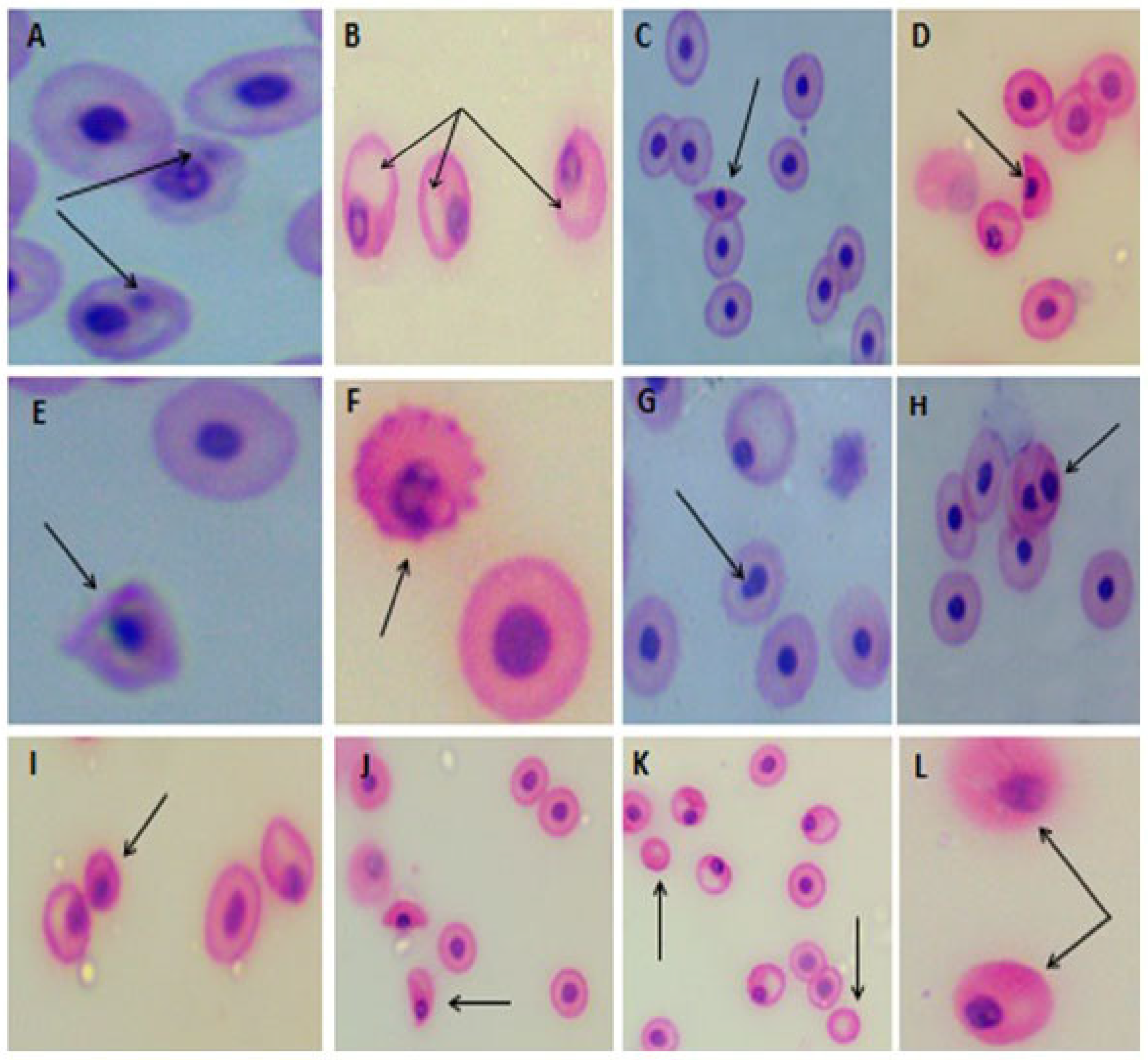
3.4.2. Erythrocyte Micronucleus (EMn) and Morphological Alterations

The normal erythrocytes of C. gariepinus are rounded with a centrally located nucleus (Figure 4A). Micronucleus (Mn) analysis of erythrocytes of C. gariepinus exposed to NPs indicated a significant (p < 0.01) increase in comparison with the control group (Figure 4B, Figure 5A and Table 3). In addition, using Se and C. vulgaris significantly (p < 0.01) reduced the EMn frequency compared with the NPs-exposed group.
Erythrocyte morphological alterations and nuclear abnormalities of RBCs were significantly elevated (p < 0.01) in the NPs group compared with the control group.
Both groups exposed to NPs and treated with Se or Chlorella showed a significant decrease (p < 0.01) in nuclear abnormalities and erythrocyte alterations compared to those only exposed to NPs.
The predominant alterations of erythrocytes in the NPs-exposed group included micronucleated cells (Mn), with one or more Mn per cell present in most observations; vacuolated cells (Vc); acanthocytes (Ac); crenated cells with fewer surface projections, eccentric nuclei (Ecn); spindle-shaped cells (Sp); crenated cells (echinocytes) (Cr), in which the RBCs develop an irregular surface with numerous projections; sickle cells (Sk), which vary in shape between ellipsoidal, boat-shaped, and genuine sickles; tear-drop cells (Tr), whose body looks like a tear with pointed apices; bilobed nuclei (Bin); microcytes (Mc); enucleated cells (En); and notched nuclei (Nn) (Figure 4B–D, Figure 5A–L and Table 3).

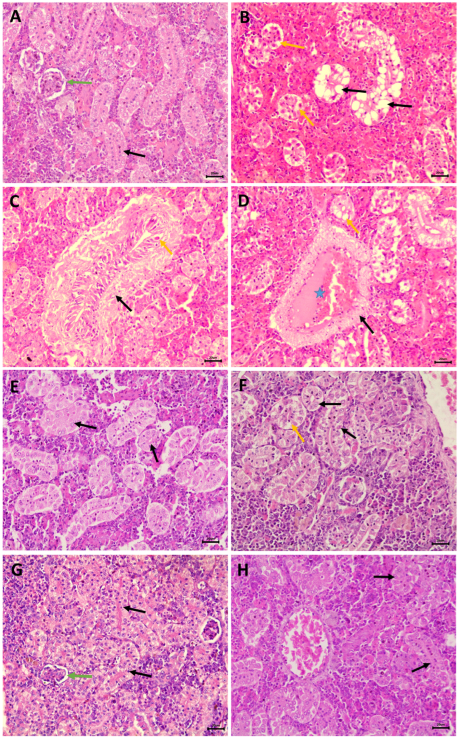
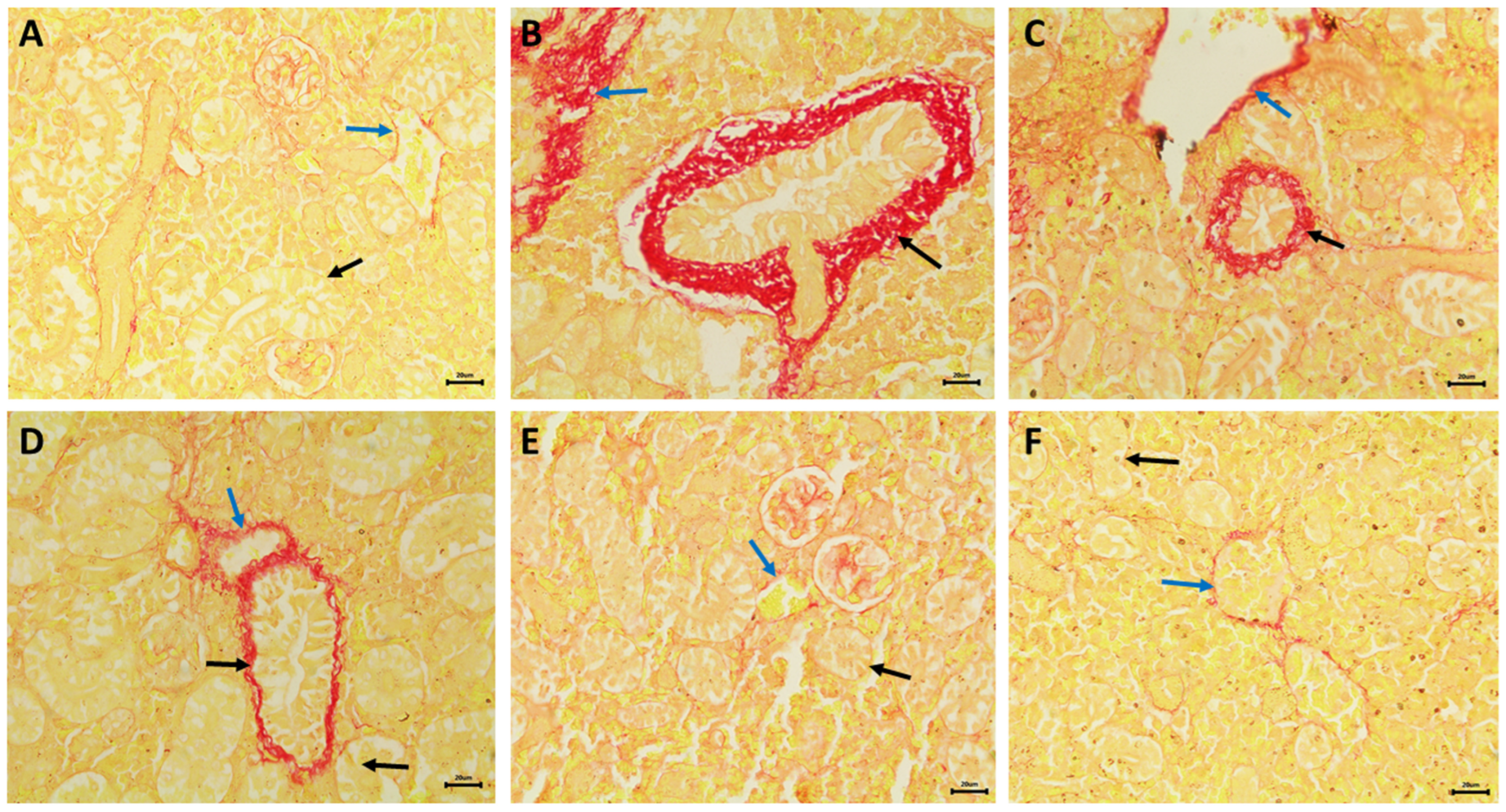
3.5. Nephrotoxic Effects

Histological examination of transverse kidney sections from control, selenium-treated, and C. vulgaris-treated catfish showed typical histological structures of rounded glomeruli, which are lined by the flat epithelium of Bowman’s capsule, and proximal and distal convoluted tubules that are lined by simple cuboidal epithelium. The apical part of the proximal tubule cell is covered by a brush border of microvilli (Figure 6A,G,H). PAS and Hematoxylin stains demonstrated increased carbohydrate substances in the brush borders of the epithelium of proximal tubules and basement membranes (Figure 7A,E,F). Transverse kidney sections of NPs-exposed catfish showed several pathological alterations, mainly in the renal tubules. They consisted of vacuolar degeneration, apoptosis and coagulative necrosis of cells lining the renal tubules that later dissociated, causing detachment of the tubular epithelium (Figure 6B). The nuclei of the necrotic tubular epithelium showed necrotic changes in the form of pyknosis, karyorrhexis or complete loss of nuclei. There was also connective tissue proliferation around the renal tubules (peritubular fibrosis), hemorrhages and tubular dilatation (Figure 6C). Perivascular connective tissue proliferation around the renal blood vessels and thrombosis were also observed (Figure 6D). PAS and Hematoxylin-stained transverse kidney sections showed a decline in the carbohydrate materials in the apical brush borders of proximal tubules, confirming the loss of brush borders (Figure 7B). Picrosirius red-stained transverse sections demonstrated the increment of the connective tissue around the renal tubules and blood vessels (Figure 8B) in comparison with the control, selenium-treated and C. vulgaris-treated groups that showed a scarcity of connective tissue surrounding the renal tubules and blood vessels (Figure 8A,E,F).
Administration of selenium in the NPs+Se treated group ameliorated and inhibited the nephrotoxic effects of NPs. Minimal alterative changes in the tubular epithelium were noticed. The typical histological appearance of the kidney was nearly restored with less vacuolar degeneration and necrotic changes in the tubular epithelium (Figure 6E). Hematoxylin–PAS- and Hematoxylin-stained transverse sections showed increased carbohydrate materials in the apical brush borders of proximal tubules, revealing the restoration of their typical structures (Figure 7C). In addition, transverse kidney sections stained with picrosirius red stain showed a decrease in the amount of connective tissue surrounding renal tubules and blood vessels compared to that found in the NPs-exposed group (Figure 8C).
Administration of Chlorella in the NPs+Ch treated group restored almost all pathological alterations to near their control levels. Most renal tubules appeared nearly normal, and a few exhibited vacuolar degeneration and tubular necrosis (Figure 6F). PAS and Hematoxylin-stained transverse kidney sections showed moderate carbohydrate material localization in the apical brush borders of proximal tubules (Figure 7D). The utilization of picrosirius red stain on transverse kidney sections revealed that the quantity of connective tissue surrounding the renal tubules and blood vessels in the experimental group was lower than that observed in the NPs-exposed group (Figure 8D).

4. Discussion

MPs and NPs are contaminants of growing worldwide concern that could be hazardous to the aquatic ecosystem and human beings [48].
The current results showed a high mortality rate in the NPs-exposed group compared to the control group, which demonstrated no deaths. A previous study on juvenile seabreams demonstrated that exposure to MPs (10–20 μm diameter) at a concentration of 5 μg of MP per gram of fish for 35 days increased the mortality rate among exposed fish [49]. The rise in the mortality rate in NPs exposed group may be attributed to intestinal blockage of the gut or tissue abrasions, which may cause lining epithelium injury, morbidity and mortality [50,51].
The present study revealed that using selenium reduced the fish mortality percentage, which is similar to results obtained by [52], who reported that a diet supplemented with Se (0.21–0.8 mg Se/kg) increased the survival rate of cobia (Rachycentron canadum L.) juveniles. Furthermore, use of Chlorella reduced the percentage of fish mortalities and this result is in harmony with [53], who mentioned that adding C. vulgaris to the diet of rohu Labeo rohita enhanced the survival rate and phagocytic and antioxidant activity.
The obtained data showed that NPs enhanced oxidative stress by elevating the MDA level in catfish serum. Previous studies demonstrated that exposure to MPs triggers ROS overproduction and oxidative stress, and this is consistent with the current findings [54,55]. Moreover, [56] reported that exposing A. franciscana to PS NPs (50 nm) for 48 h significantly increased lipid peroxidation.
The alkaline comet assay was used in the current study for DNA damage detection in C. gariepinus blood. The results revealed that fish exposed to PS NPs showed a significant increase in DNA damage parameters (tail length, tail DNA %, tail moment and Olive tail moment). This result was similar to that of [57], who reported that short-term exposure to different concentrations of 100 nm PS NPs (from 0.05 up to 50 mg/L) induced DNA damage in the hemocytes of the Mediterranean mussel (Mytilus galloprovincialis). Our results are also confirmed by [58], who stated that exposing Ctenopharyngodon idella to low concentrations of PS NPs (0.04 ng/L, 34 ng/L and 34 μg/L) for 20 days, resulted in DNA damage and had mutagenic and cytotoxic impacts on their erythrocytes.
DNA damage induced by exposing fish to PS NPs in this study could be a result of the increased state of oxidative stress due to increased levels of MDA and decreased levels of CAT and GPX that play crucial roles as antioxidant enzymes, working to counteract reactive oxygen species and free radicals. They protect against oxidative modification of lipids, DNA and proteins. This observation and explanation are in agreement with the previous studies conducted on both MPs and NPs [58,59,60,61].
Based on the previous data, it has been observed that both MPs and NPs can induce DNA strand breaks, with the extent of the damage being influenced by their respective size and surface charge. Although the exact mechanism by which MPs/NPs induce DNA damage has yet to be fully understood, several investigations suggest that oxidative stress and physical interaction are pivotal in instigating DNA strand breaks by MPs and NPs [62,63]. Additionally, synthetic polymers may have low-molecular-weight fragments of mono- and oligomers, catalysts, artificial stabilizers and a variety of particular chemical additives such as (phthalates, bisphenol A, stabilizers, pigments, flame retardants and polychlorinated biphenyls (PCBs), each of which has genotoxic properties [64].
The results obtained by other studies in mussels [65], fish [58,66] and human blood cells [59,67] support the current findings, reinforcing the idea that PS-NPs are genotoxic contaminants.
Conversely, certain studies have documented the lack of genotoxic damage in hemocytes following exposure to PS MPs [68,69]. The presence of contradictory data is not unexpected, given that the model experiments were conducted under varied conditions, encompassing differences in the type, shape and concentration of plastic microparticles, as well as variations in temperature and the duration of the experiments.
The present investigation opted for the erythrocyte micronucleus (EMn) test due to the robust consistency observed between the comet and micronucleus assays across various nanomaterials [70]. The outcomes indicated that the exposure of catfish to PS NPs resulted in a noteworthy increase in Mn frequency and several modifications in RBCs. These findings align with a study conducted by [71] on erythrocytes of Sparus aurata exposed to 45 nm PMMA-NPs (0.01–10 mg/L). Additionally, Guimarães et al. [58]. reported erythrocyte morphometric changes in C. idella juveniles following exposure to PS NPs.
Thomas et al. [72] elucidated that the occurrence of micronuclei (Mn) is attributed to disruptions in the structure or function of the mitotic apparatus, leading to a failure in the integration of chromosomes or chromosome fragments into the main nucleus.
The rise in erythrocyte nuclear abnormalities noted following exposure to PS NPs could be attributed to either the oxidative stress induced by the NPs or their interaction with chromosomal material, both being plausible explanations. Oxidative stress has been extensively documented as a repercussion of NP exposure [20,73].
Erythrocyte anomalies have been documented in hypoxic conditions [74] and under factors inducing apoptosis in blood cells, such as radiation [75]. The presence of vacuoles in erythrocytes may stem from the uneven distribution of hemoglobin, aligning with findings by [76]. Oxygen plays a vital role in fish respiration, and deformities in erythrocytes can lead to reduced oxygen levels, impacting the circulatory system and causing respiratory dysfunction. This respiratory stress has the potential to influence both the number and morphology of fish erythrocytes [77]. Similar alterations in erythrocytes were observed in catfish and D. rerio exposed to microplastics, exhibiting a notable increase in the frequency of nuclear abnormalities and morphological changes in RBCs [78,79].
Some studies emphasized the positive impacts of dietary administration of probiotic Lactobacillus rhamnosu in protecting developing zebrafish larvae from the aquatic toxicities of perfluorobutanesulfonate (PFBS) and modulated the lipid, glucose and retinoid metabolic disorders induced by PFBS pollution. Moreover, they highlighted the potential practical applications of probiotic formulations in aquaculture and ecological conservation [80,81,82,83].
The current study emphasized the same concept and revealed that selenium and C. vulgaris supplementation improved all distorted parameters induced by NPs exposure. MDA activity, DNA damage, Mn, and erythrocyte abnormalities were decreased. These outcomes are consistent with those obtained by [84,85], who reported that Se reduces MDA activity and increases the antioxidant levels in fish.
Bera et al. [86] explained that the protection of selenium against DNA damage might be because selenium contains essential selenoproteins as thioredoxin reductases and glutathione peroxidases that play a crucial role in the antioxidant defense mechanism. Selenium, therefore, may protect against DNA damage by elevating DNA glycosylase repair enzyme activity and enhancing pathways of DNA damage repair involving the p53 gene.
The protective role of C. vulgaris could be attributed to its natural antioxidant contents, such as chlorophyll, polyphenol, vitamins and sulfur-containing compounds, which are capable of neutralizing oxidative free radicals [87].
On the other hand, Bengwayan et al. [88] explained that C. vulgaris has an inhibitory effect on lipid peroxidation. Also, Wu et al. [89] stated that C. vulgaris possesses various therapeutic characteristics, such as anti-inflammatory, immunomodulatory and antioxidant activities, which may be extremely important for animal health. These antioxidant components of C. vulgaris increase SOD and CAT activity, guard against DNA oxidation and lipid peroxidation and activate cellular antioxidant enzymes.
The results revealed that C. vulgaris reduced micronucleus frequency and other morphological abnormalities in the erythrocyte. These findings were matched with [78], who demonstrated that treating MPs-intoxicated C. gariepinus with Chlorella reduced genotoxicity and cytotoxicity in the blood.
Pathological changes observed in NPs-exposed C. gariepinus kidney were comparable to those demonstrated by [90], who studied the nephrotoxic effect of NPs and MPs in mice and observed that intoxicated kidneys showed necrosis of the tubular epithelium, associated with a loss of tubular brush border and tubulointerstitial fibrosis. Chen et al. [91] found that PS MPs had an oxidative nephrotoxic effect on HEK293 cells induced by inhibiting the antioxidant heme oxygenase-1. Also, they observed that MPs induced apoptosis and autophagy via the depolarization of mitochondrial membranes and the formation of autophagosomes. These results support our hypothesis that PS NPs could induce nephrotoxic effects through oxidative–antioxidant homeostasis disturbance. The hepatotoxic effects of NPs exposure and the ameliorative effects of selenium and C. vulgaris in C. gariepinus were described in detail by [92].
Administration of selenium in the NPs+Se group ameliorated and reduced the nephrotoxic effects of NPs. Minimal pathological changes in the tubular epithelium were noticed, and the normal histological appearance was moderately restored. This ameliorative role of Se was also confirmed by [93], who mentions that Se has a protective role against oxidative stress, apoptosis, and kidney damage caused by zearalenone (ZEA) in mice. Zhang et al. [93] found that Se restored alterations in the biochemical and antioxidant indicators of ZEA-induced kidney damage. Moreover, Se reduced the expression of proteins and genes associated with endoplasmic reticulum stress and apoptosis caused by ZEA.
Administration of C. vulgaris in the NPs+Ch treated group prevents the nephrotoxic effects of NPs and moderately improves the kidney’s histology. Consistent with the current results, Latif et al., [94] stated that C. vulgaris reduces the adverse effects of paracetamol intoxication on hematological, biochemical, oxidative stress and histopathological levels. In addition, they found that C. vulgaris had antioxidant, nephro-, hepato-, and cardioprotective effects against paracetamol-induced toxic effects in Wistar rats.

5. Conclusions

The occurrence of MPs and NPs particles in the aquatic environment poses a significant threat to sustainable catfish aquaculture. This research emphasized that PS NPs have deleterious adverse effects on the DNA integrity, erythrocytes morphology, oxidative/antioxidant status and kidneys of African catfish. Administration of selenium and C. vulgaris mitigated these deleterious effects and ameliorated these alterations. Therefore, in sustainable catfish aquaculture, selenium and C. vulgaris could be prophylactic agents against the aquatic toxins, and more research should be directed toward the use of probiotics in sustainable catfish aquaculture.

Supplementary Materials

The following supporting information can be downloaded at https://www.mdpi.com/article/10.3390/fishes9020076/s1, Table S1: Ingredients and chemical composition of the basal diet.

Author Contributions

Conceptualization, Z.M.Z., D.Y. and A.E.-D.H.S.; Methodology, S.A.A., D.Y., M.H.K., M.A.A., M.A. and A.E.-D.H.S.; Validation, M.H.K.; Formal analysis, S.A.A., M.A.A. and M.A.; Investigation, S.A.A. and M.H.K.; Resources, A.E.-D.H.S.; Data curation, M.A.A.; Writing—original draft, S.A.A. and M.H.K.; Writing—review and editing, Z.M.Z., D.Y., M.H.K., M.A.A., M.A. and A.E.-D.H.S.; Visualization, M.A.; Supervision, Z.M.Z., D.Y. and A.E.-D.H.S.; Project administration, Z.M.Z. All authors have read and agreed to the published version of the manuscript.

Funding

The authors received no funding for this experiment. Open Access Funding by the University of Vienna.

Institutional Review Board Statement

The research protocol mentioned above has received approval from the veterinary ethical committee of the Faculty of Veterinary Medicine at Assiut University, Egypt, under the reference number 06/2022/0016.

Data Availability Statement

Data are contained within the article and Supplementary Materials.

Conflicts of Interest

The authors declare no conflicts of interest.

References

  1. Nag, A.; Baksi, A.; Ghosh, J.; Kumar, V.; Bag, S.; Mondal, B.; Ahuja, T.; Pradeep, T. Tribochemical degradation of polytetrafluoroethylene in water and generation of nanoplastics. ACS Sustain. Chem. Eng. 2019, 7, 17554–17558. [Google Scholar] [CrossRef]
  2. Mattsson, K.; Johnson, E.V.; Malmendal, A.; Linse, S.; Hansson, L.A.; Cedervall, T. Brain damage and behavioural disorders in fish induced by plastic nanoparticles delivered through the food chain. Sci. Rep. 2017, 7, 11452. [Google Scholar] [CrossRef] [PubMed]
  3. Zhu, F.; Zhu, C.; Wang, C.; Gu, C. Occurrence and ecological impacts of microplastics in soil systems: A review. Bull. Environ. Contam. Toxicol. 2019, 102, 741–749. [Google Scholar] [CrossRef] [PubMed]
  4. Álvarez-Ruiz, R.; Picó, Y. Analysis of emerging and related pollutants in aquaticbiota. Trends Environ. Anal. Chem. 2020, 25, e00082. [Google Scholar] [CrossRef]
  5. WWF Mediterranean Marine Initiative, Stop the Flood of Plastic: How Mediterranean Countries Can Save Their Sea. 2019. Available online: https://wwfeu.awsassets.panda.org/downloads/wwfmmi_stop_the_flood_of_plastic_mediterranean.pdf (accessed on 7 June 2019).
  6. Ospina, L.M.D. Behavioral Analysis of Single-use Plastic Consumption in Cairo. In Proceedings of the International Conference of Contemporary Affairs in Architecture and Urbanism—ICCAUA, Antalya, Turkey, 11–13 May 2022; Volume 5. [Google Scholar]
  7. Babayemi, J.O.; Nnorom, I.C.; Osibanjo, O.; Weber, R. Ensuring sustainability in plastics use in Africa: Consumption, waste generation, and projections. Environ. Sci. Europe 2019, 31, 60. [Google Scholar] [CrossRef]
  8. Jambeck, J.R.; Geyer, R.; Wilcox, C.; Siegler, T.R.; Perryman, M.; Andrady, A.; Narayan, R.; Law, K.L. Plastic waste inputs from land into the ocean. Science 2015, 347, 768–771. [Google Scholar] [CrossRef] [PubMed]
  9. Song, Y.K.; Hong, S.H.; Jang, M.; Han, G.M.; Jung, S.W.; Shim, W.J. Combined effects of UV exposure duration and mechanical abrasion on microplastic fragmentation by polymer type. Environ. Sci. Technol. 2017, 51, 4368–4376. [Google Scholar] [CrossRef] [PubMed]
  10. Santillo, D.; Miller, K.; Johnston, P. Microplastics as contaminants in commercially important seafood species. Integr. Environ. Assess. Manag. 2017, 13, 516–521. [Google Scholar] [CrossRef]
  11. Mason, S.A.; Welch, V.G.; Neratko, J. Synthetic polymer contamination in bottled water. Front. Chem. 2018, 6, 407. [Google Scholar] [CrossRef]
  12. Jani, P.; Halbert, G.W.; Langridge, J.; Florence, A.T. Nanoparticle uptake by the rat gastrointestinal mucosa: Quantitation and particle size dependency. J. Pharm. Pharmacol. 1990, 42, 821–826. [Google Scholar] [CrossRef]
  13. Walczak, A.P.; Kramer, E.; Hendriksen, P.J.; Tromp, P.; Helsper, J.P.; van der Zande, M.; Rietjens, I.M.C.M.; Bouwmeester, H.; Bouwmeester, H. Translocation of differently sized and charged polystyrene nanoparticles in in vitro intestinal cell models of increasing complexity. Nanotoxicology 2015, 9, 453–461. [Google Scholar] [CrossRef]
  14. Prata, J.C.; da Costa, J.P.; Lopes, I.; Duarte, A.C.; Rocha-Santos, T. Environmental exposure to microplastics: An overview on possible human health effects. Sci. Total Environ. 2020, 702, 134455. [Google Scholar] [CrossRef] [PubMed]
  15. Rahman, A.; Sarkar, A.; Yadav, O.P.; Achari, G.; Slobodnik, J. Potential human health risks due to environmental exposure to nano-and microplastics and knowledge gaps: A scoping review. Sci. Total Environ. 2021, 757, 143872. [Google Scholar] [CrossRef] [PubMed]
  16. Yee, M.S.L.; Hii, L.W.; Looi, C.K.; Lim, W.M.; Wong, S.F.; Kok, Y.Y.; Tan, B.K.; Wong, C.Y.; Leong, C.O.; Leong, C.O. Impact of microplastics and nanoplastics on human health. Nanomaterials 2021, 11, 496. [Google Scholar] [CrossRef] [PubMed]
  17. Doherty, G.J.; McMahon, H.T. Mechanisms of Endocytosis. Annu. Rev. Biochem. 2009, 78, 857–902. [Google Scholar] [CrossRef] [PubMed]
  18. Kaksonen, M.; Roux, A. Mechanisms of clathrin-mediated endocytosis. Nat. Rev. Mol. Cell Biol. 2018, 19, 313–326. [Google Scholar] [CrossRef] [PubMed]
  19. Jeong, C.B.; Kang, H.M.; Lee, M.C.; Kim, D.H.; Han, J.; Hwang, D.S.; Souissi, S.; Lee, S.J.; Shin, K.H.; Park, H.G.; et al. Adverse effects of microplastics and oxidative stress-induced MAPK/Nrf2 pathway-mediated defense mechanisms in the marine copepod Paracyclopina nana. Sci. Rep. 2017, 7, 41323. [Google Scholar] [CrossRef]
  20. Brandts, I.; Cánovas, M.; Tvarijonaviciute, A.; Llorca, M.; Vega, A.; Farré, M.; Teles, M. Nanoplastics are bioaccumulated in fish liver and muscle and cause DNA damage after a chronic exposure. Environ. Res. 2022, 212, 113433. [Google Scholar] [CrossRef]
  21. Chen, Q.; Yin, D.; Jia, Y.; Schiwy, S.; Legradi, J.; Yang, S.; Hollert, H. Enhanced uptake of BPA in the presence of nanoplastics can lead to neurotoxic effects in adult zebrafish. Sci. Total Environ. 2017, 609, 1312–1321. [Google Scholar] [CrossRef]
  22. Canesi, L.; Ciacci, C.; Fabbri, R.; Balbi, T.; Salis, A.; Damonte, G.; Cortese, K.; Caratto, V.; Monopoli, M.P.; Dawson, K.; et al. Interactions of cationic polystyrene nanoparticles with marine bivalve hemocytes in a physiological environment: Role of soluble hemolymph proteins. Environ. Res. 2016, 150, 73–81. [Google Scholar] [CrossRef]
  23. Han, Y.; Lian, F.; Xiao, Z.; Gu, S.; Cao, X.; Wang, Z.; Xing, B. Potential toxicity of nanoplastics to fish and aquatic invertebrates: Current understanding, mechanistic interpretation, and meta-analysis. J. Hazard Mater. 2021, 427, 127870. [Google Scholar] [CrossRef]
  24. Pienaar, U.D.V. The freshwater fished of the Kruger National Park. Koedoe 1968, 11, 1–82. [Google Scholar] [CrossRef]
  25. Skelton, P. A Complete Guide to the Freshwater Fishes of Southern Africa; Southern Book Publishers Ltd.: Johannesburg, South Africa, 1993. [Google Scholar]
  26. Teugels, G. A systematic revision of the African species of the genus Clarias (Pisces: Clariidae). Ann. Mus. R. Afr. Cent. 1986, 247, 1–199. [Google Scholar]
  27. Nwani, C.D.; Ifo, C.T.; Nwamba, H.O.; Ejere, V.C.; Onyishi, G.C.; Oluah, S.N.; Ikwuagwu, O.E.; Odo, G.E. Oxidative stress and biochemical responses in the tissues of African catfish Clarias gariepinus juvenile following exposure to primextra herbicide. Drug Chem. Toxicol. 2015, 38, 278–285. [Google Scholar] [CrossRef] [PubMed]
  28. Dimitroglou, A.; Merrifield, D.L.; Carnevali, O.; Picchietti, S.; Avella, M.; Daniels, C.; Davies, S.J. Microbial manipulations to improve fish health and production—A Mediterranean perspective. Fish Shellfish Immunol. 2011, 30, 1–16. [Google Scholar] [CrossRef]
  29. Brar, A.; Kumar, M.; Vivekanand, V.; Pareek, N. Photoautotrophic microorganisms and bioremediation of industrial effluents: Current status and future prospects. 3 Biotech 2017, 7, 18. [Google Scholar] [CrossRef]
  30. Safafar, H.; Wagenen, J.V.; Moller, P.; Jacobsen, C. Carotenoids, phenolic compounds and tocopherols contribute to the antioxidative properties of some microalgae species grown on industrial wastewater. Marine Drugs 2015, 13, 7339–7356. [Google Scholar] [CrossRef]
  31. Hamilton, S.J. Review of selenium toxicity in the aquatic food chain. Sci. Total Environ. 2004, 326, 1–31. [Google Scholar] [CrossRef] [PubMed]
  32. Rayman, M.P. The importance of selenium to human health. Lancet 2000, 356, 233–241. [Google Scholar] [CrossRef]
  33. Skjolding, L.M.; Ašmonaitė, G.; Jølck, R.I.; Andresen, T.L.; Selck, H.; Baun, A.; Sturve, J. An assessment of the importance of exposure routes to the uptake and internal localisation of fluorescent nanoparticles in zebrafish (Danio rerio), using light sheet microscopy. Nanotoxicology 2017, 11, 351–359. [Google Scholar] [CrossRef]
  34. Durigon, E.G.; Kunz, D.F.; Peixoto, N.C.; Uczay, J.; Lazzari, R. Diet selenium improves the antioxidant defense system of juveniles Nile tilapia (Oreochromis niloticus L.). Braz. J. Biol. 2018, 79, 527–532. [Google Scholar] [CrossRef]
  35. Hamed, M.; Soliman, H.A.; Eid, Z.; Al Naggar, Y.; Sayed, A.E.D.H. Dietary feeding lycopene, citric acid, and Chlorella alleviated the neurotoxicity of polyethylene microplastics in African catfish (Clarias gariepinus). Front. Environ. Sci. 2022, 10, 869727. [Google Scholar] [CrossRef]
  36. Bai, S.; Koo, J.; Kim, K.; Kim, S. Effects of Chlorella powder as a feed additive on growth performance in juvenile Korean rockfish, Sebastes schlegeli (Hilgendorf). Aquacult Res. 2001, 32, 92–98. [Google Scholar] [CrossRef]
  37. Wilson, J.M.; Bunte, R.M.; Carty, A.J. Evaluation of rapid cooling and tricaine methanesulfonate (ms222) as methods of euthanasia in zebrafish (Danio rerio). J. Am. Assoc. Lab. Anim. Sci. 2009, 48, 785–789. [Google Scholar] [PubMed]
  38. Bello, O.S.; Olaifa, F.E.; Emikpe, B.O. Haematological and blood biochemical changes in African catfish, Clarias gariepinus fed walnut (Tetracarpidium conophorum Mull Arg) leaf and onion (Allium cepa Linn.) bulb supplemented diets. Am. J. Exp. Agric. 2014, 4, 1593–1603. [Google Scholar] [CrossRef]
  39. Kei, S. Serum lipid peroxide in cerebrovascular disorders determined by a new colorimetric method. Clin. Chim. Acta 1978, 90, 37–43. [Google Scholar] [CrossRef] [PubMed]
  40. Aebi, H. Catalase in vitro. Methods Enzymol. 1984, 105, 121–126. [Google Scholar] [PubMed]
  41. Abd Ellah, M.R.; Niishimori, K.; Goryo, M.; Okada, K.; Yasuda, J. Glutathion peroxidase and glucose-6-phosphate dehydrogenase activities in bovine blood and liver. J. Vet. Med. Sci. 2004, 66, 1219–1221. [Google Scholar] [CrossRef]
  42. Villela, I.V.; Oliveira, I.M.; Silva, J.; Henriques, J.A.P. DNA damage and repair in haemolynph cells of golden mussel (Limnoperna fortunei) exposed to environmental contaminants. Mutat. Res. 2006, 605, 78–86. [Google Scholar] [CrossRef]
  43. Pascoe, S.; Gatehouse, D. The use of a simple haematoxylin and eosin staining procedure to demonstrate micronuclei within rodent bone marrow. Mutat. Res. 1986, 164, 237–243. [Google Scholar] [CrossRef]
  44. Carrasco, K.R.; Tilbury, K.L.; Myers, M.S. Assessment of the piscine micronucleus test as an in situ biological indicator of chemical contaminant effects. Can. Aquat. Sci. 1990, 47, 2123–2136. [Google Scholar] [CrossRef]
  45. Fischer, A.H.; Jacobson, K.A.; Rose, J.; Zeller, R. Hematoxylin and Eosin Staining of Tissue and Cell Sections. Cold Spring Harb. Protoc. 2008, 2008, 4986. [Google Scholar] [CrossRef]
  46. McManus, J.F.A. Histological demonstration of mucin after periodic acid. Nature 1946, 158, 202. [Google Scholar] [CrossRef]
  47. Bhutda, S.; Surve, M.V.; Anil, A.; Kamath, K.; Singh, N.; Modi, D.; Banerjee, A. Histochemical Staining of Collagen and Identification of Its Subtypes by Picrosirius Red Dye in Mouse Reproductive Tissues. Bio-Protocol 2017, 7, 2592. [Google Scholar] [CrossRef]
  48. Singh, S.; Naik, T.S.K.; Anil, A.G.; Dhiman, J.; Kumar, V.; Dhanjal, D.S.; Ramamurthy, P.C. Micro (nano) plastics in wastewater: A critical review on toxicity risk assessment, behaviour, environmental impact and challenges. Chemosphere 2022, 290, 133169. [Google Scholar] [CrossRef]
  49. Jacob, H.; Besson, M.; Oberhaensli, F.; Taylor, A.; Gillet, B.; Hughes, S.; Melvi, S.D.; Bustamante, P.; Swarzenski, P.W.; Lecchini, D.; et al. A multifaceted assessment of the effects of polyethylene microplastics on juvenile gilthead seabreams (Sparus aurata). Aquat. Toxicol. 2021, 241, 106004. [Google Scholar] [CrossRef]
  50. Guerrera, M.C.; Aragona, M.; Porcino, C.; Fazio, F.; Laurà, R.; Levanti, M.; Montalbano, G.; Germanà, G.; Abbate, A.; Germanà, A. Micro and nano plastics distribution in fish as model organisms: Histopathology, blood response and bioaccumulation in different organs. Appl. Sci. 2021, 11, 5768. [Google Scholar] [CrossRef]
  51. Lu, Y.F.; Zhang, Y.; Deng, Y.F.; Jiang, W.; Zhao, Y.P.; Geng, J.J.; Ding, L.L.; Zhao, Y. Uptake and accumulation of polystyrene microplastics in zebrafish (Danio rerio) and toxic effects in liver. Environ. Sci. Technol. 2016, 50, 4054–4060. [Google Scholar] [CrossRef] [PubMed]
  52. Liu, K.; Wang, X.; Ai, Q.; Mai, K.; Zhang, W. Dietary selenium requirement for juvenile cobia, Rachycentron canadum L. Aquac. Res. 2010, 41, 594–601. [Google Scholar] [CrossRef]
  53. Andrews, S.R.; Sahu, N.P.; Pal, A.K.; Kumar, S. Haematological modulation and growth of Labeo rohita fingerlings: Effect of dietary mannan-oligosaccharide, yeast extract, protein hydrolysate and Chlorella sp. Aquacult. Res. 2009, 41, 61–69. [Google Scholar] [CrossRef]
  54. Eom, H.J.; Nam, S.E.; Rhee, J.S. Polystyrene Microplastics Induce Mortality through Acute Cell Stress and Inhibition of Cholinergic Activity in a Brine Shrimp. Mol. Cell. Toxicol. 2020, 16, 233–243. [Google Scholar] [CrossRef]
  55. Lee, D.H.; Lee, S.; Rhee, J.S. Consistent exposure to microplastics induces age-specific physiological and biochemical changes in a marine mysid. Mar. Pollut. Bull. 2021, 162, 111850. [Google Scholar] [CrossRef]
  56. Varó, I.; Perini, A.; Torreblanca, A.; Garcia, Y.; Bergami, E.; Vannuccini, M.L.; Corsi, I. Time-dependent effects of polystyrene nanoparticles in brine shrimp Artemia franciscana at physiological, biochemical and molecular levels. Sci. Total Environ. 2019, 675, 570–580. [Google Scholar] [CrossRef] [PubMed]
  57. Brandts, I.; Teles, M.; Gonçalves, A.P.; Barreto, A.; Franco-Martinez, L.; Tvarijonaviciute, A.; Oliveira, M. Effects of nanoplastics on Mytilus galloprovincialis after individual and combined exposure with carbamazepine. Sci. Total Environ. 2018, 643, 775–784. [Google Scholar] [CrossRef] [PubMed]
  58. Guimarães, A.T.B.; Estrela, F.N.; Pereira, P.S.; de Andrade Vieira, J.E.; de Lima Rodrigues, A.S.; Silva, F.G.; Malafaia, G. Toxicity of polystyrene nanoplastics in Ctenopharyngodon idella juveniles: A genotoxic, mutagenic and cytotoxic perspective. Sci. Total Environ. 2021, 752, 141937. [Google Scholar] [CrossRef]
  59. Gopinath, P.M.; Saranya, V.; Vijayakumar, S.; Mythili Meera, M.; Ruprekha, S.; Kunal, R.; Pranay, A.; Thomas, J.; Mukherjee, A.; Chandrasekaran, N. Assessment on interactive prospectives of nanoplastics with plasma proteins and the toxicological impacts of virgin, coronated and environmentally released-nanoplastics. Sci. Rep. 2019, 9, 8860. [Google Scholar] [CrossRef]
  60. Shah, N.; Khan, A.; Habib Khan, N.; Khisroon, M. Genotoxic consequences in common grass carp (Ctenopharyngodon idella Valenciennes, 1844) exposed to selected toxic metals. Biol. Trace Elem. Res. 2020, 199, 305–314. [Google Scholar] [CrossRef]
  61. Ribeiro, F.; Garcia, A.R.; Pereira, B.P.; Fonseca, M.; Mestre, N.C.; Fonseca, T.G.; Bebianno, M.J. Microplastics effects in Scrobicularia plana. Mar. Pollut. Bull. 2017, 122, 379–391. [Google Scholar] [CrossRef]
  62. Hu, M.; Palić, D. Micro-and nano-plastics activation of oxidative and inflammatory adverse outcome pathways. Redox Biol. 2020, 37, 101620. [Google Scholar] [CrossRef]
  63. Mazur, A.A.; Chelomin, V.P.; Zhuravel, E.V.; Kukla, S.P.; Slobodskova, V.V.; Dovzhenko, N.V. Genotoxicity of polystyrene (PS) microspheres in short-term exposure to gametes of the sand dollar Scaphechinus mirabilis (Agassiz, 1864)(Echinodermata, Echinoidea). J. Mar. Sci. Eng. 2021, 9, 1088. [Google Scholar] [CrossRef]
  64. Hamlin, H.J.; Marciano, K.; Downs, C.A. Migration of nonylphenol from food-grade plastic is toxic to the coralreef fish species Pseudochromis fridmani. Chemosphere 2015, 139, 223–228. [Google Scholar] [CrossRef]
  65. Gonçalves, J.M.; Sousa, V.S.; Teixeira, M.R.; Bebianno, M.J. Chronic toxicity of polystyrene nanoparticles in the marine mussel Mytilus galloprovincialis. Chemosphere 2022, 287, 132356. [Google Scholar] [CrossRef]
  66. Estrela, F.N.; Batista Guimarães, A.T.; Silva, F.G.; Marinho da Luz, T.; Silva, A.M.; Pereira, P.S.; Malafaia, G. Effects of polystyrene nanoplastics on Ctenopharyngodon idella (grass carp) after individual and combined exposure with zinc oxide nanoparticles. J. Hazard Mater. 2021, 403, 123879. [Google Scholar] [CrossRef]
  67. Ballesteros, S.; Domenech, J.; Barguilla, I.; Cortés, C.; Marcos, R.; Hernández, A. Genotoxic and immunomodulatory effects in human white blood cells after ex vivo exposure to polystyrene nanoplastics. Environ. Sci. Nano 2020, 7, 3431–3446. [Google Scholar] [CrossRef]
  68. Magni, S.; Gagné, F.; André, C.; Della Torre, C.; Auclair, J.; Hanana, H.; Parenti, C.C.; Bonasoro, F.; Binelli, A. Evaluation of uptake and chronic toxicity of virgin polystyrene microbeads in freshwater zebra mussel Dreissena polymorpha (Mollusca: Bivalvia). Sci. Total Environ. 2018, 631–632, 778–788. [Google Scholar] [CrossRef] [PubMed]
  69. Xia, B.; Zhang, J.; Zhao, X.; Feng, J.; Teng, Y.; Chen, B.; Sun, X.; Zhu, L.; Sun, X.; Qu, K. Polystyrene microplastics increase uptake, elimination and cytotoxicity of decabromodiphenyl ether (BDE-209) in the marine scallop Chlamys farreri. Environ. Pollut. 2020, 258, 113657. [Google Scholar] [CrossRef] [PubMed]
  70. Karlsson, H.L. The comet assay in nanotoxicology research. Anal. Bioanal. Chem. 2010, 398, 651–666. [Google Scholar] [CrossRef] [PubMed]
  71. Brandts, I.; Barría, C.; Martins, M.A.; Franco-Martínez, L.; Barreto, A.; Tvarijonaviciute, A.; Tort, L.; Oliveira, M.; Teles, M. Waterborne exposure of gilthead seabream (Sparus aurata) to polymethylmethacrylate nanoplastics causes effects at cellular and molecular levels. J. Hazard. Mater. 2021, 644, 123590. [Google Scholar] [CrossRef] [PubMed]
  72. Thomas, P.; Holland, N.; Bolognesi, C.; Kirsch-Volders, M.; Bonassi, S.; Zeiger, E.; Knasmueller, S.; Fenech, M. Buccal micronucleus cytome assay. Nat. Protoc. 2009, 4, 825–837. [Google Scholar] [CrossRef] [PubMed]
  73. Jacob, H.; Besson, M.; Swarzenski, P.W.; Lecchini, D.; Metian, M. Effects of virgin micro- and nanoplastics on fish: Trends, meta-analysis, and perspectives. Environ. Sci. Technol. 2020, 54, 4733–4745. [Google Scholar] [CrossRef] [PubMed]
  74. Sawhney, A.K.; Johal, M.S. Erythrocyte alterations induced by malathion in Channa punctatus (Bloch). Bull. Environ. Contam. Toxicol. 2000, 64, 398–405. [Google Scholar] [CrossRef] [PubMed]
  75. Chukhlovin, A.B. Apoptosis and red blood cell echinocytosis: Common features. Scanning Microsc. 1996, 10, 795–803. [Google Scholar] [PubMed]
  76. Bushra, A.; Abul farah, M.; Niamat, M.A.; Waseem, A. Induction of micronuclei and erythrocyte alterations in the catfish clarias battrachus by 2,4-dichlorophenoxyacetic acid and butachlor. Mutat. Res./Genet. Toxicol. Environ. Mutagen. 2002, 518, 135–144. [Google Scholar]
  77. Brown, L. Aquaculture for Veterinarians: Fish Husbandry and Medicine; Pergamon Press: Oxford, NC, USA, 1993; 440 p. [Google Scholar]
  78. Sayed, A.H.; Hamed, M.; Badrey, A.E.A.; Soliman, H.A.M. Bioremediation of hemotoxic and oxidative stress induced by polyethylene microplastic in Clarias gariepinus using lycopene, citric acid, and Chlorella. Comp. Biochem. Physiol. 2021, 250, 109189. [Google Scholar] [CrossRef]
  79. Da Costa Araujo, A.P.; de Andrade Vieira, J.E.; Malafaia, G. Toxicity and trophic transfer of polyethylene microplastics from Poecilia reticulata to Danio rerio. Sci. Total Environ. 2020, 742, 140217. [Google Scholar] [CrossRef]
  80. Sun, B.; Liu, M.; Tang, L.; Hu, C.; Huang, Z.; Zhou, X.; Chen, L. Probiotic supplementation mitigates the developmental toxicity of perfluorobutanesulfonate in zebrafish larvae. Sci. Total Environ. 2021, 799, 149458. [Google Scholar] [CrossRef]
  81. Hu, C.; Liu, M.; Tang, L.; Liu, H.; Sun, B.; Chen, L. Probiotic intervention mitigates the metabolic disturbances of perfluorobutanesulfonate along the gut-liver axis of zebrafish. Chemosphere 2021, 284, 131374. [Google Scholar] [CrossRef]
  82. Hu, C.; Tang, L.; Liu, M.; Lam, P.K.; Lam, J.C.; Chen, L. Probiotic modulation of perfluorobutanesulfonate toxicity in zebrafish: Disturbances in retinoid metabolism and visual physiology. Chemosphere 2020, 258, 127409. [Google Scholar] [CrossRef]
  83. Liu, M.; Tang, L.; Hu, C.; Huang, Z.; Sun, B.; Lam, J.C.; Lam, P.K.; Chen, L. Antagonistic interaction between perfluorobutanesulfonate and probiotic on lipid and glucose metabolisms in the liver of zebrafish. Aquat. Toxicol. 2021, 237, 105897. [Google Scholar] [CrossRef] [PubMed]
  84. Ashouri, S.; Keyvanshokooh, S.; Salati, A.P.; Johari, S.A.; Pasha-Zanoosi, H. Effects of different levels of dietary selenium nanoparticles on growth performance, muscle composition, blood biochemical profiles and antioxidant status of common carp (Cyprinus carpio). Aquaculture 2015, 446, 25–29. [Google Scholar] [CrossRef]
  85. Saffari, S.; Keyvanshokooh, S.; Zakeri, M.; Johari, S.A.; Pasha-Zanoosi, H.J.A.N. Effects of different dietary selenium sources (sodium selenite, selenomethionine and nanoselenium) on growth performance, muscle composition, blood enzymes and antioxidant status of common carp (Cyprinus carpio). Aquac. Nutr. 2017, 23, 611–617. [Google Scholar] [CrossRef]
  86. Bera, S.; Rosa, V.D.; Rachidi, W.; Diamond, A.M. Does a role for selenium in DNA damage repair explain apparent controversies in its use in chemoprevention? Mutagenesis 2013, 28, 127–134. [Google Scholar] [CrossRef] [PubMed]
  87. Abdelhamid, F.M.; Elshopakey, G.E.; Aziza, A.E. Ameliorative effects of dietary Chlorella vulgaris and β-glucan against diazinon-induced toxicity in Nile tilapia (Oreochromis niloticus). Fish Shellfish Immunol. 2020, 96, 213–222. [Google Scholar] [CrossRef] [PubMed]
  88. Bengwayan, P.T.; Laygo, J.C.; Pacio, A.E.; Poyaoan, J.L.Z.; Rebugio, J.F.; Yuson, A.L.L. A comparative study on the antioxidant property of Chlorella (Chlorella sp.) tablet and glutathione tablet. Int. J. Sci. Res. 2010, 2, 25–35. [Google Scholar]
  89. Wu, Q.; Liu, L.; Miron, A.; Klímová, B.; Wan, D.; Kuča, K. The antioxidant, immunomodulatory, and anti-inflammatory activities of Spirulina: An overview. Arch. Toxicol. 2016, 90, 1817–1840. [Google Scholar] [CrossRef] [PubMed]
  90. Meng, X.; Zhang, J.; Wang, W.; Gonzalez-Gil, G.; Vrouwenvelder, J.S.; Li, Z. Effects of nano-and microplastics on kidney: Physicochemospherical properties, bioaccumulation, oxidative stress and immunoreaction. Chemosphere 2022, 288, 132631. [Google Scholar] [CrossRef]
  91. Chen, Y.C.; Chen, K.F.; Lin, K.Y.A.; Chen, J.K.; Jiang, X.Y.; Lin, C.H. The nephrotoxic potential of polystyrene microplastics at realistic environmental concentrations. J. Hazard. Mat. 2022, 427, 127871. [Google Scholar] [CrossRef] [PubMed]
  92. Abdelbaky, S.A.; Zaky, Z.; Yahia, D.; Ali, M.; Sayed, A.E.D.; Abd-Elkareem, M.; Kotob, M.H. Ameliorative Effects of Selenium and Chlorella vulgaris against Polystyrene Nanoplastics-induced Hepatotoxicity in African Catfish (Clarias gariepinus). J. Adv. Vet. Res. 2022, 12, 308–317. [Google Scholar]
  93. Zhang, Y.; Hu, B.; Wang, M.; Tong, J.; Pan, J.; Wang, N.; Gong, P.; Long, M. Selenium protects against zearalenone-induced oxidative stress and apoptosis in the mouse kidney by inhibiting endoplasmic reticulum stress. Oxidative Med. Cell. Longev. 2020, 2020, 6059058. [Google Scholar] [CrossRef]
  94. Latif, A.A.E.; Assar, D.H.; Elkaw, E.M.; Hamza, H.A.; Alkhalifah, D.H.M.; Hozzein, W.N.; Hamouda, R.A. Protective role of Chlorella vulgaris with Thiamine against Paracetamol induced toxic effects on haematological, biochemical, oxidative stress parameters and histopathological changes in Wistar rats. Sci. Rep. 2021, 11, 3911. [Google Scholar] [CrossRef]
Figure 1. Transmission electron photomicrographs of the polystyrene nanoplastics. (A). Direct magnification 48,000×. (B). Direct magnification 58,000×.
Fishes 09 00076 g001
Figure 2. DNA damage in whole blood of C. gariepinus in all the experimental groups. (A): Control group; (B): NPs group; (C): (NPs+Se) group; (D): (NPs+Ch); (E): Ch; (F): Se.
Fishes 09 00076 g002
Figure 3. Assessment of different degrees of DNA damage induced by polystyrene nanoplastics using CASP software (CASP-version 1.2.3b) shows different degrees of DNA migration. (A,A1): intact nucleus (no DNA damage). (B,B1): mild DNA damage. (C,C1): moderate degree of DNA damage. (D,D1): severe DNA damage. (E,E1,F,F1): extensive DNA damage.
Fishes 09 00076 g003
Figure 4. Blood smears stained by H and E show the morphological alterations and nuclear abnormalities in erythrocytes of C. gariepiuns exposed to polystyrene nanoplastics. (A) Control fish showing normal red blood cells (RBCs). (BD) Fish exposed to PS NPs showing Mn: micronucleated cell; Vc: vacuolated cell; Bin: bilobed nucleus cell; Ac: acanthocyte; Ecn: eccentric nucleus; Sp: spindle-shaped cell; Cr: crenated cell; Sk: sickle cell; Tr: tear-drop cell; En: enucluated cell; Mc: microcyte; Nn: notched nucleated cell. Scale bar = 20 μm.
Fishes 09 00076 g004
Figure 5. Different erythrocyte alterations were recorded in PS NPs-exposed group. (A) micronucleated cell; (B) vacuolated cell, (C) spindle cell; (D) sickle cell; (E) acanthocyte; (F) crenated cell; (G) notched nucleated cell; (H) bilobed nucleus; (I) microcell; (J) tear-drop like cell; (K) enucleated cell; (L) eccentric nucleus.
Fishes 09 00076 g005
Figure 6. Photomicrographs of kidney transverse sections from control, NPs-exposed and -treated C. gariepinus: Kidney sections of (A) control, (G) selenium- and (H) C. vulgaris-treated catfish show normal histological structures of the kidney that consisted of normally rounded glomeruli (green arrow) and renal tubules that covered by simple columnar epithelium with apical brush border (black arrow). (BD). Kidney transverse sections of NPs-exposed catfish in (B) shows the presence of vacuolar degeneration in the cytoplasm of tubular epithelium with peripherally located pyknotic nuclei (black arrow) and epithelial necrotic changes with nuclear alterations, dissociation of renal cells and epithelium desquamation (yellow arrow) in (C) shows connective tissue proliferation around the renal tubules (peritubular fibrosis) (black arrow) and necrosis and apoptosis of tubular epithelium (yellow arrow) in (D) shows perivascular connective tissue proliferation (black arrow), thrombosis (star) and vacuolar degeneration (yellow arrow). (E) Kidney transverse section of NPs+Se-treated catfish shows minimal vacuolar degenerative changes and necrotic changes in tubular epithelium; their nuclei appeared round and vesicular and were centrally located (arrow). (F). Kidney transverse section of NPs+Ch-treated catfish shows vacuolar degenerative changes and necrotic changes in tubular epithelium and desquamation (yellow arrow). Some renal tubules appeared normal with minimal cytoplasmic and nuclear alterations (black arrow). H and E staining was performed. Scale bar = 20 μm.
Fishes 09 00076 g006
Figure 7. Photomicrographs of kidney transverse sections from control, NPs-exposed and treated C. gariepinus: Kidney transverse sections of (A) control, (E) selenium- and (F) C. vulgaris-treated catfish show positive PAS reactivity in the brush border (BB) and basement membrane (BM) of the renal tubules. (B). Kidney transverse section of NPs-exposed catfish exhibits a decrease in carbohydrate materials in the brush borders and the basement membranes of the renal tubules. (C). Kidney transverse section of NPs+Se treated catfish shows an increase in the amount of carbohydrate observed in the basement membranes and brush borders. (D). Kidney transverse section of NPs+Ch treated catfish shows moderate carbohydrate materials localization in the brush border and basement membrane of renal tissue. PAS and Hematoxylin staining was performed. Scale bar = 20 μm.
Fishes 09 00076 g007
Figure 8. Photomicrographs of kidney transverse sections from control, NPs-exposed and treated C. gariepinus: Kidney transverse sections of (A) control, (E) selenium- and (F) C. vulgaris-treated catfish show a scanty amount of connective tissue fibers around the renal tubules and blood vessels. (B) Kidney transverse sections of NPs-exposed catfish demonstrate an increased amount of connective tissue fibers around the renal tubules and blood vessels with a thickened wall of blood vessels. Kidney transverse section of NPs+Se (C) and NPs+Ch (D)-treated catfish show a decreased amount of connective tissue fibers around the renal tubules and blood vessels. Renal tubules (black arrow) and blood vessels (blue arrow). Picrosirius red stain. Scale bar = 20 μm.
Fishes 09 00076 g008
Table 1. Malondialdehyde (nmol/mL), catalase (U/L) and glutathione (U/L) activities in serum of C. gariepinus in control, NPs, (NPs+Se), (NPs+Ch), Ch and Se groups.
GroupsControlNPsNPs+SeNPs+ChChSe
Parameters
MDA3.71 ± 0.7 b10.22 ± 2.4 a5.56 ± 0.5 b4.98 ± 0.7 b3.72 ± 0.8 b4.02 ± 0.9 b
CAT225.9 ± 13.6 b201.43 ± 5.2 b235 ± 62.3 b202.43 ± 40.5 b232.93 ± 11.2 b352.9 ± 33.4 a
GPX27.5 ± 5.6 a19.1 ± 6.4 a33.7 ± 10.4 a25.57 ± 4.2 a24.33 ± 2.99 a32.43 ± 8.0 a
Data are presented as means ± SE. Different letters a and b indicate significant changes among groups. p < 0.05. n = 6.
Table 2. DNA damage in the blood of C. gariepinus in control, NPs, (NPs+Se), (NPs+Ch), Ch and Se groups.
ParametersControlNPsNPs+SeNPs+ChChSe
Tail DNA%0.57 ± 0.12 bc3.43 ± 0.53 a1.26 ± 0.19 b0.98 ± 0.17 b0.56 ± 0.16 bc0.08 ± 0.03 c
Tail Length
(µm)
8.24 ± 0.62 c32.14 ± 2.60 a15.42 ± 1.21 b9.77 ± 0.60 c7.50 ± 0.45 cd4.22 ± 0.28 d
Tail moment0.19 ± 0.05 b3.60 ± 0.88 a0.44 ± 0.07 b0.20 ± 0.06 b0.15 ± 0.03 b0.09 ± 0.01 b
Olive tail moment0.46 ± 0.09 b4.58 ± 0.66 a1.15 ± 0.15 b0.59 ± 0.11 b0.30 ± 0.05 b0.11 ± 0.02 b
Data are presented as mean ± SE. Values followed by different letters indicate significant changes among groups. p < 0.05. n = 6.
Table 3. Erythrocytes morphological alterations and nuclear abnormalities in C. gariepinus in control, NPs, NPs+Se, NPs+Ch, Ch and Se groups.
GroupsControlNPsNPs+SeNPs+ChChSe
Parameters
Mn2.3 ± 0.88 b22.3 ± 2.00 a4.3 ± 1.20 b3.3 ± 1.2 b2.3 ± 0.33 b3.0 ± 0.57 b
Vc7.0 ± 0.57 b47.3 ± 5.78 a9.3 ± 1.45 b8.3 ± 2.6 b6.0 ± 1.52 b5.3 ± 0.88 b
Tr5.0 ± 1.50 b14.3 ± 1.45 a7.6 ± 0.88 b6 ± 0.57 b5.0 ± 1.15 b4.3 ± 1.20 b
Ac4.3 ± 1.20 b11.3 ± 1.45 a5.0 ± 1.15 b4.3 ± 0.88 b3.0 ± 1.00 b3.3 ± 0.88 b
Cr3.6 ± 0.88 b12.6 ± 1.76 a5.6 ± 1.20 b5.3 ± 0.88 b4.3 ± 1.20 b4.0 ± 1.5 b
Sk2.0 ± 0.57 b13.3 ± 0.88 a4.6 ± 0.80 b3.6 ± 1.20 b2.0 ± 0.57 b2.3 ± 0.88 b
Sp1.6 ± 0.33 b7.3 ± 1.45 a2.3 ± 0.80 b2.6 ± 1.20 b1.6 ± 0.66 b2.0 ± 0.57 b
Mc4.6 ± 1.20 b19.0 ± 1.15 a5.3 ± 1.20 b5.6 ± 1.70 b2.6 ± 1.20 b3.6 ± 0.88 b
Ecn7.6 ± 1.76 bc50.0 ± 3.20 a12.0 ± 2.50 b7.0 ± 2.00 bc6.3 ± 1.2 bc4.6 ± 0.88 d
Bin3.0 ± 0.57 b10 ± 1.50 a4.3 ± 1.40 b4.0 ± 0.57 b1.3 ± 0.33 b1.3 ± 0.33 b
Nn1.3 ± 0.33 bc8.3 ± 1.20 a3.6 ± 0.80 b2.3 ± 0.88 bc1.0 ± 0.33 d1.6 ± 0.33 bc
En2.6 ± 0.88 b9.0 ± 1.15 a3.0 ± 0.57 b2.0 ± 0.57 b1.3 ± 0.33 b2.0 ± 0.57 b
Data are presented as mean ± SE. Values followed by different letters indicate significant changes among groups. p < 0.05. Mn: micronucleated cell; Vc: vacuolated cell; Tr: tear-drop cell; Ac: acanthocyte; Cr: crenated cell; Sk: sickle cell; Sp: spindle-shaped cell; Mc: microcyte; Ecn: eccentric nucleus; Bin: bilobed nucleus; Nn: notched nucleated cell; En: enucleated cell. n = 6.
Disclaimer/Publisher’s Note: The statements, opinions and data contained in all publications are solely those of the individual author(s) and contributor(s) and not of MDPI and/or the editor(s). MDPI and/or the editor(s) disclaim responsibility for any injury to people or property resulting from any ideas, methods, instructions or products referred to in the content.

