Article

First Report of Vibrio vulnificus Outbreak in Farm-Raised Sorubim (Pseudoplatystoma sp.) from Brazil

by
Peter C. Janampa-Sarmiento
1,
Francisco Y. T. Reis
1,
Renata C. Egger
1,
Santiago B. de Pádua
2,
Sóstenes A. C. Marcelino
3,
João L. R. Cunha
4,
Felipe Pierezan
3,
Henrique C. P. Figueiredo
1 and
Guilherme C. Tavares
1,*
1
Department of Preventive Veterinary Medicine, School of Veterinary Medicine, Federal University of Minas Gerais, Belo Horizonte 31270-901, MG, Brazil
2
Aquivet Saúde Aquática, Santa Fé do Sul 15085-310, SP, Brazil
3
Department of Veterinary Clinics and Surgery, School of Veterinary Medicine, Federal University of Minas Gerais, Belo Horizonte 31270-901, MG, Brazil
4
Department of Biology, Biomedical Research Institute, University of York, York YO10 5DD, UK
*
Author to whom correspondence should be addressed.
Fishes 2024, 9(2), 54; https://doi.org/10.3390/fishes9020054
Submission received: 20 December 2023 / Revised: 25 January 2024 / Accepted: 27 January 2024 / Published: 29 January 2024
(This article belongs to the Special Issue Fish Pathogens: Infection and Biological Control)

Abstract

:
Vibrio vulnificus is an opportunistic pathogen in humans and exhibits pathogenic behavior in several aquaculture fish species. To date, in Brazil, there are no reports of V. vulnificus outbreaks in farmed fish. However, in 2019, sorubim (Pseudoplatystoma sp.) in the grow-out phase with clinical signs and cumulative mortality of 40% was registered. We aimed to identify and characterize the etiological agents of this outbreak. Seven moribund fish were sampled for bacteriological studies, and isolates were obtained from the brain and kidneys. Bacterial identification using matrix-assisted laser desorption ionization mass spectrometry (MALDI-TOF/MS) revealed V. vulnificus. One isolate, RP4, was used for identification via dnaJ and 16S rRNA gene sequencing and antimicrobial susceptibility evaluation. Sorubim juveniles were experimentally challenged with RP4 isolate via intracelomic injection (IC, 107 colony-forming units [CFU] fish−1) and immersion bath (IB, 106 CFU mL−1 for 30 min). Identities to V. vulnificus of dnaJ and 16S rRNA genes by BLAST analysis were higher than 92% and 98%, respectively. Susceptibility to oxytetracycline, florfenicol, and other antimicrobial molecules was also observed. In the IC-challenged group, the mortality rate was 100% with V. vulnificus recovered from fish organs (brain, liver, spleen, and kidney). Additionally, splenic endothelium alterations were observed in the IC group. On the contrary, the control and IB groups did not develop any clinical signs, mortality, or bacterial recovery after 7 days of challenge. To the best of our knowledge, this is the first report of pathogenic V. vulnificus in farmed Pseudoplatystoma sp.
Key Contribution: This is the first report of vibriosis in farm-raised sorubim associated with high cumulative mortality. Vibrio vulnificus RP4 isolate has pathogenic activity in sorubim following intracoelomic injection and is susceptible to florfenicol and oxytetracycline.

1. Introduction

Vibrio vulnificus is a flagellated halophilic Gram-negative bacterium that has been associated with opportunistic human illnesses transmitted through contaminated seafood ingestion or via direct wound contact with contaminated water or seafood [1]. V. vulnificus has been isolated from freshwater, brackish water, and diseased seawater fish species of aquaculture importance, including Oreochromis niloticus [2,3], Anguilla sp. [4], Ctenopharyngodon idellus [5], and Trachinotus ovatus [6] from Asia and Europe since the 1970s [7]. In Brazil, there have been reports of isolation and molecular identification of V. vulnificus from aquatic environments [8,9], human blood (patient who died of septicemia after ingestion of seafood) [10], wild and farmed healthy shellfish [11], and wild captured fish such as Paralichthys orbignyanus and Micropogonias furnieri [12]. In addition, there are reports of presumptive identification of V. vulnificus in sea mammals [13] and sea birds [14] in the country; however, these isolates have not been confirmed at the species level. Accurate V. vulnificus identification can be achieved through dnaJ gene sequencing [15] or the detection of specific biomarkers, such as the cytotoxin-hemolysin vvh gene [16].
Sorubim (Pseudoplatystoma sp.) is a neotropical siluriform fish native to South America [17] that has important commercial value in Brazilian aquaculture. Sorubim production reached 11,058 tons in 2022 [18], making it the third most-produced native fish in Brazil. However, with increasingly intensive commercial systems for this fish, propitious conditions for disease occurrence, such as higher stocking density and husbandry stress, are emerging.
To date, there have been reports of Gram-negative bacteria such as Flavobacterium columnare [19], Edwardsiella ictaluri [20], and Aeromonas hydrophila [21], as well as Gram-positive bacteria such as Streptococcus agalactiae, S. iniae, S. dysgalactiae [22], and Lactococcus garvieae [23,24] being involved in natural outbreaks in Brazilian sorubim aquaculture. In this study, we describe vibriosis in a Brazilian fish farm of freshwater Pseudoplatystoma sp. V. vulnificus identification was achieved using matrix-assisted laser desorption ionization mass spectrometry (MALDI-TOF/MS) and gene sequencing. Additionally, we assessed the pathogenicity to Pseudoplatystoma sp. and evaluated the antimicrobial susceptibility of the RP4 V. vulnificus isolate.

2. Materials and Methods

2.1. Outbreak Characterization

The outbreak occurred on a fish farm located at the São Simon reservoir (18°38′31″ S, 50°03′57″ W), Goiás state, in August 2019. Sorubim in the grow-out phase (weighing 400–700 g) showed clinical signs of infection (erratic swimming, melanosis, and hyporexia) and abrupt mortality. Fish mortality of 40% (5400 out of a population of 13,500 individuals) occurred within 1 week of the onset of the disease. The water temperature of the affected net cage was approximately 24 °C.

2.2. Vibrio Vulnificus Identification and Phylogenetic Analysis

2.2.1. Isolation and MALDI-TOF/MS Identification

Seven moribund fish were sampled and subjected to a bacteriological examination. Brain and kidney samples were streaked on Columbia agar (Kasvi, Parana, Brazil) and incubated at 28 °C for 24 h, resulting in the isolation of seven cultures. Bacterial identification was performed using mass spectrometry (MALDI-TOF/MS) with a FlexControl MicroFlex LT mass spectrometer (Bruker Daltonics, Billerica, MA, USA), as previously described by Assis et al. [25]. Subsequently, the isolates were stored at −70 °C, but only RP4 (V. vulnificus) was able to reactivate and was used for further analysis.

2.2.2. dnaJ and 16S rRNA Sequencing and Phylogenetic Analysis

The frozen RP4 isolate was thawed and reactivated on marine agar (HiMedia, Mumbai, India) at 28 °C for 24 h. Pure colonies obtained were subjected to DNA extraction using the Maxwell® 16 Tissue DNA Purification Kit in the Maxwell 16 Research Instrument (Promega, Madison, WI, USA). The DNA’s quality and quantity were verified using a NanoDrop spectrophotometer (Thermo Fisher Scientific, Waltham, MA, USA) before being submitted for amplification and sequencing of the dnaJ and 16S rRNA genes. Briefly, dnaJ was amplified by PCR using primers DN1-1F (5′-GATYTRCGHTAYAACATGGA-3′) and DN1-2R (5′-TTCACRCCRTYDAAGAARC-3′) as reported by Nhung et al. [15]. Concurrently, 16S rRNA amplification was conducted using universal primers B37 (5′-TACGGYTACCTTGTTACGA-3′) and C70 (5′-AGAGTTTGATYMTGGC-3′) as reported by Fox et al. [26]. Forward and reverse PCR amplicons were sequenced using an ABI 3500 Genetic Analyzer (Life Technologies, Carlsbad, CA, USA) and subjected to quality control; sequence trimming and contig generation were performed using BioEdit, version 7.2 [27]. Both sequences were evaluated using NCBI BLAST (https://blast.ncbi.nlm.nih.gov/Blast.cgi, accessed on 10 November 2023) in the nt/nr database. Sequences from Vibrio sp. strains with identity ≥ 83% and query coverage ≥ 96% were considered for alignment assessment (Table S1) and polishing using the AliView software, version 1.28 [28]. The phylogeny of these sequences was estimated with MrBAYES software, version 3.2 [29] using the GTR nucleotide substitution model (nst = 6) with different substitution rates (rates = invgamma) and two million Markov chain Monte Carlo iterations, as previously described [30]. The resulting phylogram was edited using the graphical viewer FigTree version 3.4.4 [31].

2.3. Antimicrobial Susceptibility Testing

Antimicrobial susceptibility testing was performed using the disc diffusion method according to the CLSI VET03 guidelines [32]. Briefly, RP4 colonies cultured on marine agar were suspended in a sterile saline solution (NaCl 0.9%) until an absorbance of 0.1 was obtained at 625 nm. This solution was streaked on Muller Hinton agar plates (HiMedia, Mumbai, India) supplemented with NaCl at 1%. Following this, nine antibiotic discs (Oxoid, Basingstoke, UK) were positioned on the plates as follows: lincomycin (10 µg), neomycin (10 µg), erythromycin (15 µg), amoxicillin (10 µg), sulfamethoxazole/trimethoprim (25 µg), nitrofurantoin (300 µg), norfloxacin (10 µg), oxytetracycline (30 µg), and florfenicol (30 µg). The plates were then incubated at 28 °C and the zone of inhibition was measured after 24 h of incubation.

2.4. Experimental Infection Challenge

The pathogenicity of the RP4 isolate was evaluated through experimental infection (Ethics Committee Protocol No. 315/2019). Amazon catfish (Leiarius marmoratus × Pseudoplatystoma corruscans) juveniles (88 ± 41.23 g) were acquired from a local commercial farm with no disease antecedents. Upon arrival at the laboratory facility, 10% of the total batch of fish were subjected to parasitological (gill and skin biopsy) and microbiological (brain, kidney, and spleen streaking on marine agar plates) analyses. Fish were acclimated in glass aquaria supplied with flow-through freshwater at 28 °C and aerated by a stone diffusor. Fish were fed commercial extruded food for carnivorous species (42% crude protein) two times daily until apparent satiation.
The RP4 isolate stored at −70 °C was reactivated on a marine agar plate at 28 °C. After 24 h incubation, one colony was selected and used for inoculum preparation by bacterial suspension in brain–heart infusion broth (Kasvi, Parana, Brazil). Bacterial growth occurred at 28 °C under 100 rpm agitation until a 0.3 OD600 was achieved. Bacterial challenge doses were administered to sorubim juveniles via 0.1 mL of intracoelomic injection (IC, 107 colony-forming units [CFU] fish−1, n = 6) and 30-min immersion baths (IB, 106 CFU mL−1, n = 6). A control group (C, 0.1 mL IC of brain–heart infusion broth, n = 4) was included in this study. All fish in the experimental challenge were kept in 60 L glass aquaria (one aquarium per group) during a 7-d monitoring period under conditions similar to those in the acclimation phase. Fish that died during the experimental challenge and fish that survived at the end of the experiment were subjected to microbiological analysis (brain, kidney, liver, and spleen streaking on sterile marine agar plates).

2.5. Histopathological Evaluation

Organs (brain, spleen, heart, midgut, stomach, swim bladder, kidney, and liver) from all fish (dead and surviving the experimental challenge) were collected and fixed in 10% buffered formalin. Then, paraffin wax sections (4 µm) were obtained after organ immersion in ethanol solutions of increasing concentrations (70–100%), cleared with xylene, embedded in paraffin wax, and sectioned using a semi-automated rotary microtome Leica RM2245 (Leica Biosystems, Wetzlar, Germany). Hematoxylin and eosin staining was routinely performed for all organ sections, whereas GoodPasture staining was performed on selected tissues for Gram-negative bacterial observation as described by Luna [33].

3. Results and Discussion

3.1. Vibrio Vulnificus Identification and Phylogenetic Analysis

3.1.1. Isolation and MALDI-TOF/MS Identification

During this disease outbreak, bacterial colonies were isolated from the brains (n = 3) and kidneys (n = 4) of the diseased aquaculture species. Bacteriological studies resulted in the isolation of three colonies with different morphologies. The MALDI Biotyper results identified V. vulnificus (scores between 1.755 and 2.233) isolates from five fish, and Edwardsiella tarda (score 2.348) and Enterococcus saccharolyticus (score 2.008) from one fish.
V. vulnificus was identified based on the MALDI-TOF/MS results. The suitability of MALDI-TOF/MS as a tool for V. vulnificus identification has been previously reported [34,35]. Vibrio species belong to a monophyletic group with high similarity. Thus, phylogenetic analyses provide additional information to support this identification.

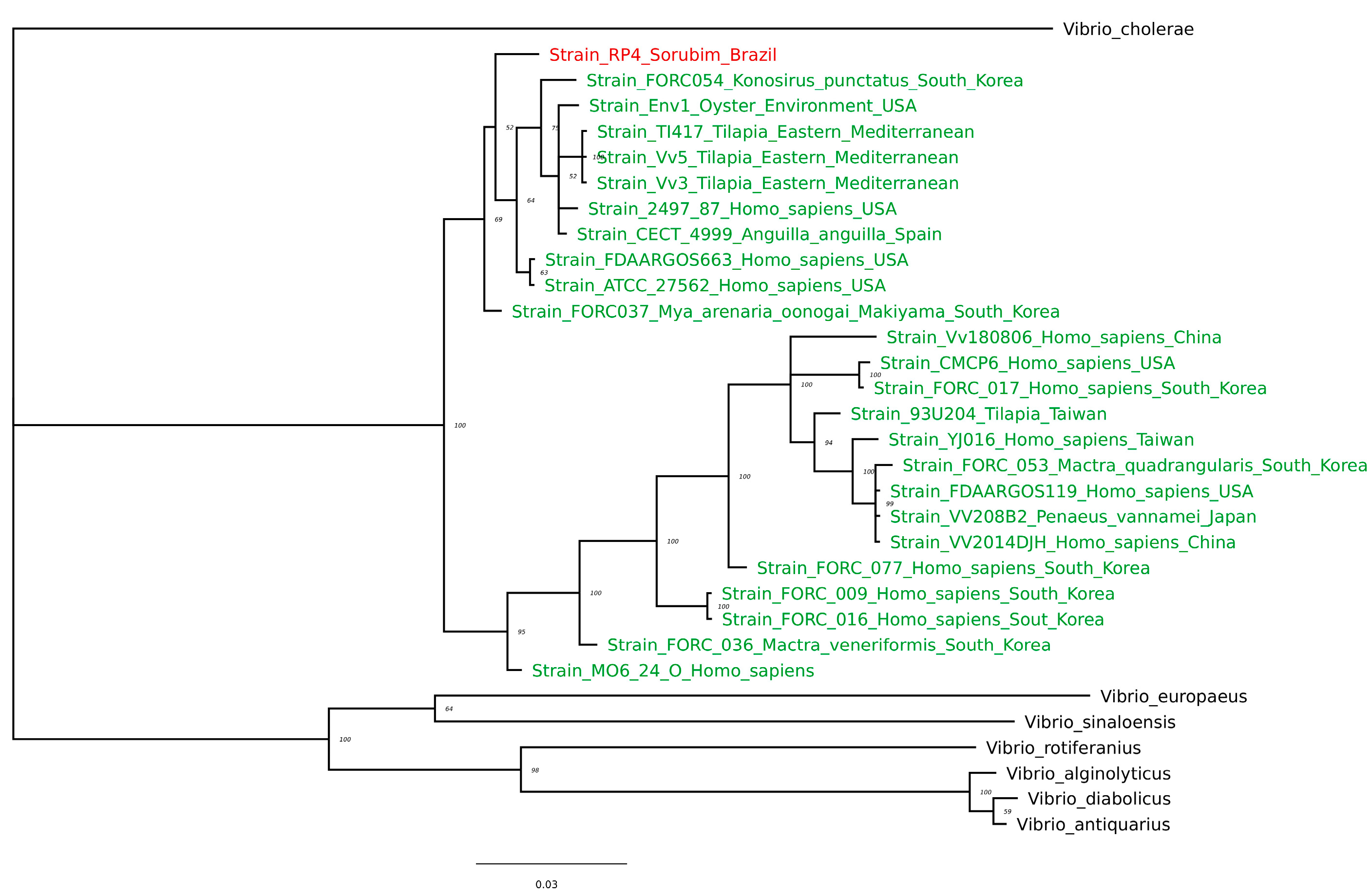
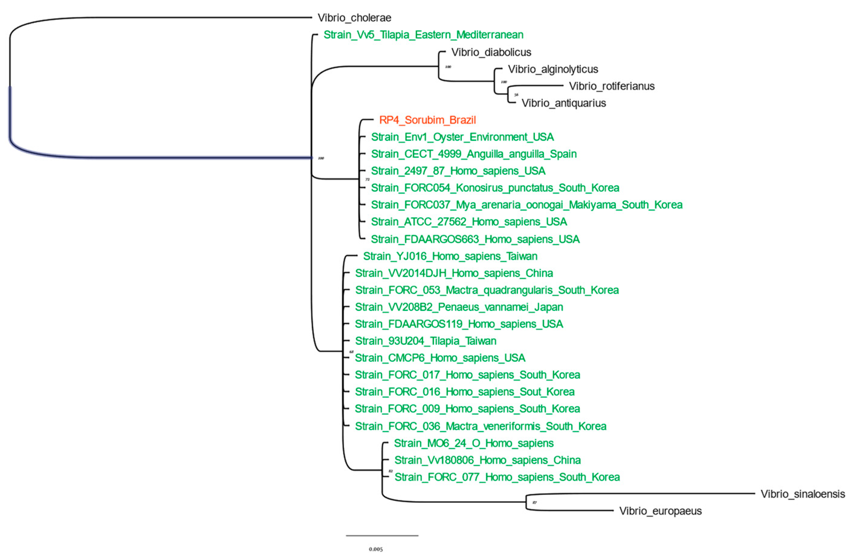
3.1.2. dnaJ and 16S rRNA Sequencing and Phylogenetic Analysis

Partial dnaJ (727 bp) and 16S rRNA (1397 bp) sequences obtained from the RP4 isolate were deposited in NCBI (GenBank accession numbers: OP690609 and OP692644) and submitted to BLAST against the NCBI nt/nr database. The RP4 isolate obtained identities between 98.56% and 92.98% with 24 dnaJ sequences of strains identified as V. vulnificus (Table S1). Comparably, identities between 99.71% and 98.57% with 22 16S rRNA sequences identified as V. vulnificus were observed (Table S1).
Phylogenetic trees based on dnaJ and 16S rRNA were constructed using these strains. In addition, seven Vibrio spp. strains were included in the analysis. Markov chain Monte Carlo simulations with <0.01 average standard deviation of split frequencies allowed the generation of two phylograms (Figure 1 and Figure 2). Mean branches with posterior probability (prob) of 100% for an exclusively V. vulnificus cluster were only obtained by dnaJ phylogenetic analysis, where the RP4 isolate was located (Figure 1).
The identity results for 16S rRNA showed poor species differentiation for V. vulnificus, as identities higher than 97% were reached with other Vibrio species (e.g., Vibrio europaeus, V. diabolicus, V. antiquarius, V. alginolyticus, and V. rotiferanius). Even with a clustering threshold of 99% identity for complete 16S rRNA as proposed by Edgard [36], lower identities were obtained for 16S rRNA with several V. vulnificus strains from NCBI. This explains the failure to cluster all V. vulnificus strains into an exclusive species branch, as shown in previous studies [15].
In contrast, the phylogenetic tree based on dnaJ identified the RP4 isolate in the exclusive V. vulnificus branch. Although dnaJ of the RP4 isolate displayed variable identities with other V. vulnificus strains, it was sufficient for species discrimination. This can be explained by the lower identity (<86%) of dnaJ of the RP4 isolate compared with the other Vibrio species evaluated in this study. In addition, a trend of concentrating fish-pathogenic V. vulnificus strains (including the RP4 isolate) in a distinct branch was observed. Further evaluations to confirm whether the RP4 isolate belongs to pathovar piscis can be conducted using protocols previously reported [37].
Currently, the Vibrionaceae family comprises 190 species distributed in 51 clades where V. vulnificus is grouped in the “Vulnificus clade” with two other closely related species: V. navarrensis and V. cidicii [38]. In the present study, we characterized the RP4 isolate as V. vulnificus using the dnaJ sequence. In addition, the RP4 isolate showed identity values lower than 85% with closely related species such as V. navarrensis and V. cidicii (Table S2).

3.2. Antimicrobial Susceptibility Testing

The antimicrobial susceptibility profile of the RP4 isolate (Table 1) was obtained and verified according to the breakpoints described by CLSI and EUCAST [39,40,41].
The isolate was susceptible to amoxicillin, sulfamethoxazole/trimethoprim, nitrofurantoin, norfloxacin, oxytetracycline, florfenicol, neomycin, and erythromycin. This was contrary to the typical antimicrobial resistance traits observed in other Brazilian isolates of V. vulnificus [12,42,43]. On the other hand, despite lincomycin having the lowest inhibition zone (8 mm), it was not possible to find an actual reference for breakpoints for appropriate comparison. However, V. vulnificus (strain H 1828/94) isolated from a human clinical case showed the same inhibition zone behavior [44].
Overall, the RP4 isolate was susceptible to various antimicrobial molecules, including two antimicrobials licensed for aquaculture purposes in Brazil: florfenicol and oxytetracycline. This information may be useful for the management of fish vibriosis in Brazil.

3.3. Experimental Infection Challenge and Histopathological Evaluation

Mortality rates of 100% and 0% were obtained in the IC (<24 h after inoculation) and IB (7-day observation period) groups, respectively. In addition, recovery of V. vulnificus from IC-treated fish organs (brain, liver, spleen, and kidney) was possible after cultivation in Marine agar for 24 h at 28 °C. One fish in the IC group exhibited erratic swimming. Externally, in fish from the IC group, the ventral skin was reddish and the anus was swollen. Internal lesions, including organ congestion and reddish ascitic fluid, were also observed in these fish (Figure 3A,B). Conversely, the Control and IB groups did not develop any clinical signs, internal lesions, or bacterial growth at the end of the assay.
Histopathological examination showed that major lesions were restricted to the spleen of the fish in the IC group, with early necrotic foci and proliferation of Gram-negative bacteria around the necrotized endothelium of the splenic vessels (Figure 3C,D). The other groups (Control and IB) did not show any cellular or tissue alterations.
Complete mortality was observed at a dose of 107 CFU fish−1 via IC. These results demonstrate the pathogenic behavior of V. vulnificus isolate RP4 towards sorubim. Other authors have observed similar mortality and clinical signs 24 h after inoculation with V. vulnificus at a lower dose (103 CFU fish−1) in tilapia [3] or a higher dose (1010 CFU fish−1) in eels [45]. The authors attributed this rapid mortality to peracute septic shock. In the present study, septicemia occurred because it was possible to recover V. vulnificus colonies from several organs, including the brain. In addition, necrosis of the endothelial vessels associated with Gram-negative bacillus proliferation observed in the spleen of the IC group supports this peracute septic shock caused by V. vulnificus in sorubim. Similarly, histopathological findings associated with vascular disturbances were reported in the renal capillaries of eels [4] and the liver of tilapia [3] challenged with V. vulnificus.
Conversely, infection via immersion did not result in mortality or clinical signs after the 7-day observation period. One explanation for the lack of infectivity via immersion could be the relatively short exposure time (30 min) of the fish to the bacterial suspension bath. Previous reports of experimental V. vulnificus infection via immersion in fish have shown varying immersion times, ranging from 60 min [46] to over 24 h [3]. However, it is worth noting that successful infections in fish have been reported with shorter experimental immersion times (less than 60 min) involving other pathogenic bacteria [47], and longer exposure times did not necessarily lead to pathogenic activity in suspected bacteria-infected fish [48].
On the other hand, failure of the immersion infection could be associated with the inability of V. vulnificus to adhere and invade mucosal surfaces. A report [49] suggested that the natural portal of entry for V. vulnificus into fish could be the mucosal surfaces of the gills. Additionally, it was observed that the skin mucosa could serve as a reservoir, as V. vulnificus can colonize and develop a biofilm structure on fish skin [49]. However, the adhesion and invasion capabilities to fish mucosal surfaces by V. vulnificus are not fully understood. There is a report of a lack of pathogenicity in fish after immersion in freshwater (0% salinity) with V. vulnificus strains originally isolated from diseased fish inhabiting water with salinities ≥ 0.3% [46]. In that report, it was suggested that the inconsistency between water salinity in the infection bath and the strain’s salinity isolation may contribute to the failure of immersion infection. Recent studies suggest that exposure to freshwater has a detrimental effect on V. vulnificus virulence, specifically with respect to a compound contributing to water salinity. It has been shown that calcium in water (an important salinity contributor in seawater) can trigger biofilm formation while suppressing the motility of V. vulnificus [50,51]. In our immersion infection assay, the challenge was conducted in a freshwater bath with V. vulnificus RP4 isolate originating from ill freshwater fish. We speculated that the RP4 isolate was adapted to freshwater conditions and would express virulence in an immersion infection under similar conditions. Unfortunately, this assumption was not validated, and further studies are necessary to verify the effect of salinity on the virulence of RP4 isolate and to elucidate the primary mode of infection of sorubim vibriosis.

4. Conclusions

Taken together, the first disease outbreak caused by pathogenic V. vulnificus in farmed Pseudoplatystoma sp. in Brazil is described using MALDI-TOF identification, dnaJ and 16S rRNA sequencing, and experimental challenge. The pathogenic behavior was demonstrated to cause mortality and histological damage following intracelomic injection. Under in vitro conditions, this bacterium is susceptible to different antimicrobials, including florfenicol and oxytetracycline, which are licensed for aquaculture purposes by the Brazilian Sanitary Livestock Authority.

Supplementary Materials

The following supporting information can be downloaded at: https://www.mdpi.com/article/10.3390/fishes9020054/s1, Table S1: Vibrio spp. strains used for dnaJ and 16S rRNA genes phylogenetic evaluation.

Author Contributions

Conceptualization, P.C.J.-S. and G.C.T.; Methodology, P.C.J.-S. and G.C.T.; Validation, H.C.P.F. and J.L.R.C.; Formal Analysis, P.C.J.-S., G.C.T. and J.L.R.C.; Investigation, P.C.J.-S., F.Y.T.R., S.B.d.P., S.A.C.M., F.P. and R.C.E.; Resources, G.C.T., F.P. and H.C.P.F.; Data Curation, P.C.J.-S.; Writing—Original Draft Preparation, P.C.J.-S.; Writing—Review and Editing, G.C.T., H.C.P.F., F.Y.T.R. and R.C.E.; Visualization, P.C.J.-S., S.A.C.M. and F.P.; Supervision G.C.T. and H.C.P.F.; Project Administration, G.C.T.; Funding Acquisition, G.C.T. and H.C.P.F. All authors have read and agreed to the published version of the manuscript.

Funding

This study was financed in part by the Coordenação de Aperfeiçoamento de Pessoal de Nível Superior—Brasil (CAPES), Fundação de Amparo à Pesquisa do Estado de Minas Gerais (FAPEMIG, grant numbers APQ-01227-22 and APQ-04309-22), and Conselho Nacional de Desenvolvimento Científico e Tecnológico—Brasil (CNPq, grant number 141889/2019-5).

Institutional Review Board Statement

The study was conducted according to the guidelines of the CONCEA (Conselho Nacional de Controle da Experimentação Animal) and approved by the Ethics Committee of Federal University of Minas Gerais (Protocol No. 315/2019).

Data Availability Statement

GenBank accession numbers: OP690609 and OP692644.

Acknowledgments

We thank Joaquim Campos Júnior (Agrobell Alevinos) for providing sorubim juveniles for this study.

Conflicts of Interest

Author Santiago Benites De Pádua was employed by the company Aquivet Saúde Aquática. The remaining authors declare that the research was conducted in the absence of any commercial or financial relationships that could be construed as a potential conflict of interest.

References

  1. Haftel, A.; Sharman, T. Vibrio vulnificus Infection. In StatPearls; StatPearls Publishing: Treasure Island, FL, USA, 2023. [Google Scholar]
  2. Carmona-Salido, H.; Fouz, B.; Sanjuán, E.; Carda-Diéguez, M.; Delannoy, C.M.J.; García-González, N.; González-Candelas, F.; Amaro, C. Draft Genome Sequences of Vibrio vulnificus Strains Recovered from Moribund Tilapia. Microbiol. Resour. Announc. 2021, 10, e00094-21. [Google Scholar] [CrossRef] [PubMed]
  3. Sumithra, T.G.; Reshma, K.J.; Anusree, V.N.; Sayooj, P.; Sharma, S.R.K.; Suja, G.; Amala, P.V.; Joseph, S.; Sanil, N.K. Pathological Investigations of Vibrio vulnificus Infection in Genetically Improved Farmed Tilapia (Oreochromis niloticus L.) Cultured at a Floating Cage Farm of India. Aquaculture 2019, 511, 734217. [Google Scholar] [CrossRef]
  4. Amaro, C.; Sanjuán, E.; Fouz, B.; Pajuelo, D.; Lee, C.-T.; Hor, L.-I.; Barrera, R. The Fish Pathogen Vibrio vulnificus Biotype 2: Epidemiology, Phylogeny, and Virulence Factors Involved in Warm-Water Vibriosis. Microbiol. Spectr. 2015, 3. [Google Scholar] [CrossRef] [PubMed]
  5. Liu, R.; Lian, Z.; Hu, X.; Lü, A.; Sun, J.; Chen, C.; Liu, X.; Song, Y.; Yiksung, Y. First Report of Vibrio vulnificus Infection in Grass Carp Ctenopharyngodon idellus in China. Aquaculture 2019, 499, 283–289. [Google Scholar] [CrossRef]
  6. Li, G.; Zhao, D.; Huang, L.; Sun, J.; Gao, D.; Wang, H.; Tan, Y.; Liang, L. Identification and Phylogenetic Analysis of Vibrio vulnificus Isolated from Diseased Trachinotus ovatus in Cage Mariculture. Aquaculture 2006, 261, 17–25. [Google Scholar] [CrossRef]
  7. Hernández-Cabanyero, C.; Amaro, C. Phylogeny and Life Cycle of the Zoonotic Pathogen Vibrio vulnificus. Environ. Microbiol. 2020, 22, 4133–4148. [Google Scholar] [CrossRef]
  8. Castelo-Branco, D.D.S.C.M.; Sales, J.A.; Brilhante, R.S.N.; De Melo Guedes, G.M.; De Ponte, Y.B.; De Souza Sampaio, C.M.; Bandeira, T.D.J.P.G.; Moreira, J.L.B.; De Alencar, L.P.; Paiva, M.D.A.N.; et al. Enterobacteria and Vibrio from Macrobrachium amazonicum Prawn Farming in Fortaleza, Ceará, Brazil. Asian Pac. J. Trop. Med. 2016, 9, 27–31. [Google Scholar] [CrossRef]
  9. Toubiana, M.; Fernandez, D.; Filho, J.-E.M.; Garnier, J.; De Castro, R.R.; Bonnet, M.-P.; Colwell, R.R.; Monfort, P. Occurence of Vibrio parahaemolyticus, Vibrio vulnificus and Vibrio cholerae in the Urbanized Guaraja Bay, Amazonia, Brasil. In Proceedings of the 8th Biennial International Conference on the Biology of Vibrios, McGill University, Montréal, Canada, 18–20 November 2019. [Google Scholar]
  10. Tiba-Casas, M.R.; Lemes-Marques, E.G.; Almeida, E.A.; Soares, F.B.; Camargo, C.H. Draft Genome Sequence of a Pathogenic Vibrio vulnificus Strain Isolated in Brazil. Genome Announc. 2018, 6, e01274-17. [Google Scholar] [CrossRef]
  11. Raszl, S.M.; Froelich, B.A.; Vieira, C.R.W.; Blackwood, A.D.; Noble, R.T. Vibrio parahaemolyticus and Vibrio vulnificus in South America: Water, Seafood and Human Infections. J. Appl. Microbiol. 2016, 121, 1201–1222. [Google Scholar] [CrossRef]
  12. Silveira, D.R.; Da Rosa, J.V.; Kaefer, K.; Bach, L.G.; Barbosa, A.D.O.; Timm, C.D. Ability of Vibrio vulnificus Isolated from Fish of the Lagoa Dos Patos Estuary in South Brazil to Form Biofilms after Sublethal Stress and Bacterial Resistance to Antibiotics and Sanitizers. Int. J. Food Microbiol. 2019, 303, 19–25. [Google Scholar] [CrossRef]
  13. Pereira, C.S.; Amorim, S.D.; Santos, A.F.M.; Siciliano, S.; Moreno, I.M.B.; Ott, P.H.; Rodrigues, D.P. Vibrio spp. isolados de mamíferos marinhos capturados na região litorânea do Sudeste ao Sul do Brasil. Pesq. Vet. Bras. 2007, 27, 81–83. [Google Scholar] [CrossRef]
  14. Cardoso, M.D.; Lemos, L.S.; Roges, E.M.; De Moura, J.F.; Tavares, D.C.; Matias, C.a.R.; Rodrigues, D.P.; Siciliano, S. A Comprehensive Survey of Aeromonas sp. and Vibrio sp. in Seabirds from Southeastern Brazil: Outcomes for Public Health. J. Appl. Microbiol. 2018, 124, 1283–1293. [Google Scholar] [CrossRef] [PubMed]
  15. Nhung, P.H.; Shah, M.M.; Ohkusu, K.; Noda, M.; Hata, H.; Sun, X.S.; Iihara, H.; Goto, K.; Masaki, T.; Miyasaka, J.; et al. The dnaJ Gene as a Novel Phylogenetic Marker for Identification of Vibrio Species. Syst. Appl. Microbiol. 2007, 30, 309–315. [Google Scholar] [CrossRef] [PubMed]
  16. King, W.L.; Siboni, N.; Kahlke, T.; Green, T.J.; Labbate, M.; Seymour, J.R. A New High Throughput Sequencing Assay for Characterizing the Diversity of Natural Vibrio Communities and Its Application to a Pacific Oyster Mortality Event. Front. Microbiol. 2019, 10, 2907. [Google Scholar] [CrossRef] [PubMed]
  17. Brambilla, L.; Toledo, M.J.; Ibarra, D.A. First Fossil Record of Pseudoplatystoma corruscans (Siluriformes, Pimelodidae) from the Late Pleistocene, Santa Fe, Argentina. J. South. Am. Earth Sci. 2021, 105, 102987. [Google Scholar] [CrossRef]
  18. IBGE. Pesquisa da Pecuária Municipal. BRASIL. Available online: https://cidades.ibge.gov.br/brasil/pesquisa/18/16459 (accessed on 10 November 2023).
  19. Barony, G.; Tavares, G.; Assis, G.; Luz, R.; Figueiredo, H.; Leal, C. New Hosts and Genetic Diversity of Flavobacterium columnare Isolated from Brazilian Native Species and Nile Tilapia. Dis. Aquat. Org. 2015, 117, 1–11. [Google Scholar] [CrossRef]
  20. Da Costa, A.R.; De Abreu, D.C.; Torres Chideroli, R.; Santo, K.; Dib Gonçalves, D.; Di Santis, G.W.; De Pádua Pereira, U. Interspecies Transmission of Edwardsiella ictaluri in Brazilian Catfish (Pseudoplatystoma corruscans) from Exotic Invasive Fish Species. Dis. Aquat. Organ. 2021, 145, 197–208. [Google Scholar] [CrossRef]
  21. Da Silva, B.C.; Mouriño, J.L.P.; Vieira, F.N.; Jatobá, A.; Seiffert, W.Q.; Martins, M.L. Haemorrhagic Septicaemia in the Hybrid Surubim (Pseudoplatystoma corruscans×Pseudoplatystoma fasciatum) Caused by Aeromonas hydrophila: Haemorrhagic Septicaemia in the Hybrid Surubim. Aquac. Res. 2012, 43, 908–916. [Google Scholar] [CrossRef]
  22. Tavares, G.C.; De Queiroz, G.A.; Assis, G.B.N.; Leibowitz, M.P.; Teixeira, J.P.; Figueiredo, H.C.P.; Leal, C.A.G. Disease Outbreaks in Farmed Amazon Catfish (Leiarius marmoratus × Pseudoplatystoma corruscans) Caused by Streptococcus agalactiae, S. iniae, and S. Dysgalactiae. Aquaculture 2018, 495, 384–392. [Google Scholar] [CrossRef]
  23. Evans, J.J.; Klesius, P.H.; Shoemaker, C.A. First Isolation and Characterization of Lactococcus garvieae from Brazilian Nile Tilapia, Oreochromis niloticus (L.), and Pintado, Pseudoplathystoma corruscans (Spix & Agassiz). J. Fish. Dis. 2009, 32, 943–951. [Google Scholar] [CrossRef]
  24. Fukushima, H.C.S.; Leal, C.a.G.; Cavalcante, R.B.; Figueiredo, H.C.P.; Arijo, S.; Moriñigo, M.A.; Ishikawa, M.; Borra, R.C.; Ranzani-Paiva, M.J.T. Lactococcus garvieae Outbreaks in Brazilian Farms Lactococcosis in Pseudoplatystoma Sp.—Development of an Autogenous Vaccine as a Control Strategy. J. Fish. Dis. 2017, 40, 263–272. [Google Scholar] [CrossRef]
  25. Assis, G.B.N.; Pereira, F.L.; Zegarra, A.U.; Tavares, G.C.; Leal, C.A.; Figueiredo, H.C.P. Use of MALDI-TOF Mass Spectrometry for the Fast Identification of Gram-Positive Fish Pathogens. Front. Microbiol. 2017, 8, 1492. [Google Scholar] [CrossRef]
  26. Fox, J.G.; Yan, L.L.; Dewhirst, F.E.; Paster, B.J.; Shames, B.; Murphy, J.C.; Hayward, A.; Belcher, J.C.; Mendes, E.N. Helicobacter bilis sp. Nov., a Novel Helicobacter Species Isolated from Bile, Livers, and Intestines of Aged, Inbred Mice. J. Clin. Microbiol. 1995, 33, 445–454. [Google Scholar] [CrossRef]
  27. Hall, T.A. BioEdit A User-Friendly Biological Sequence Alignment Editor and Analysis Program for Windows 95/98/NT. Nucleic Acids Symp. Ser. 1999, 41, 95–98. Available online: https://www.scirp.org/(S(351jmbntvnsjt1aadkposzje))/reference/ReferencesPapers.aspx?ReferenceID=1383440 (accessed on 10 November 2023).
  28. Larsson, A. AliView: A Fast and Lightweight Alignment Viewer and Editor for Large Datasets. Bioinformatics 2014, 30, 3276–3278. [Google Scholar] [CrossRef] [PubMed]
  29. Ronquist, F.; Teslenko, M.; Van Der Mark, P.; Ayres, D.L.; Darling, A.; Höhna, S.; Larget, B.; Liu, L.; Suchard, M.A.; Huelsenbeck, J.P. MrBayes 3.2: Efficient Bayesian Phylogenetic Inference and Model Choice Across a Large Model Space. Syst. Biol. 2012, 61, 539–542. [Google Scholar] [CrossRef] [PubMed]
  30. Nascimento, F.F.; Reis, M.D.; Yang, Z. A Biologist’s Guide to Bayesian Phylogenetic Analysis. Nat. Ecol. Evol. 2017, 1, 1446–1454. [Google Scholar] [CrossRef] [PubMed]
  31. Rambaut, A. FigTree v1.3.1. Institute of Evolutionary Biology, University of Edinburgh, Edinburgh. 2010. Available online: http://tree.bio.ed.ac.uk/software/figtree/ (accessed on 10 November 2023).
  32. CLSI. VET03|Methods for Antimicrobial Broth Dilution and Disk Diffusion Susceptibility Testing of Bacteria Isolated from Aquatic Animals, 2nd Edition. Clinical & Laboratory Standards Institute. Available online: https://clsi.org/standards/products/veterinary-medicine/documents/vet03/ (accessed on 18 December 2023).
  33. Luna, L.G. Manual of Histologic Staining Methods of the Armed Forces Institute of Pathology, 3rd ed.; McGraw-Hill: New York, NY, USA, 1968; Available online: https://www.scirp.org/(S(351jmbntvnsjt1aadkposzje))/reference/ReferencesPapers.aspx?ReferenceID=854813 (accessed on 10 November 2023).
  34. Cheng, W.-C.; Jan, I.-S.; Chen, J.-M.; Teng, S.-H.; Teng, L.-J.; Sheng, W.-H.; Ko, W.-C.; Hsueh, P.-R. Evaluation of the Bruker Biotyper Matrix-Assisted Laser Desorption Ionization-Time of Flight Mass Spectrometry System for Identification of Blood Isolates of Vibrio Species. J. Clin. Microbiol. 2015, 53, 1741–1744. [Google Scholar] [CrossRef] [PubMed]
  35. Liu, T.; Kang, L.; Xu, J.; Wang, J.; Gao, S.; Li, Y.; Li, J.; Yuan, Y.; Yuan, B.; Wang, J.; et al. PVBase: A MALDI-TOF MS Database for Fast Identification and Characterization of Potentially Pathogenic Vibrio Species from Multiple Regions of China. Front. Microbiol. 2022, 13, 872825. [Google Scholar] [CrossRef] [PubMed]
  36. Edgar, R.C. Updating the 97% Identity Threshold for 16S Ribosomal RNA OTUs. Bioinformatics 2018, 34, 2371–2375. [Google Scholar] [CrossRef] [PubMed]
  37. Carmona-Salido, H.; Fouz, B.; Sanjuán, E.; Carda, M.; Delannoy, C.M.J.; García-González, N.; González-Candelas, F.; Amaro, C. The Widespread Presence of a Family of Fish Virulence Plasmids in Vibrio vulnificus Stresses Its Relevance as a Zoonotic Pathogen Linked to Fish Farms. Emerg. Microbes Infect. 2021, 10, 2128–2140. [Google Scholar] [CrossRef]
  38. Jiang, C.; Tanaka, M.; Nishikawa, S.; Mino, S.; Romalde, J.L.; Thompson, F.L.; Gomez-Gil, B.; Sawabe, T. Vibrio Clade 3.0: New Vibrionaceae Evolutionary Units Using Genome-Based Approach. Curr. Microbiol. 2022, 79, 10. [Google Scholar] [CrossRef]
  39. EUCAST: Clinical Breakpoints and Dosing of Antibiotics. Version 12.0. Available online: https://www.eucast.org/clinical_breakpoints (accessed on 10 November 2023).
  40. CLSI M100; Performance Standards for Antimicrobial Susceptibility Testing. 27th ed. Clinical & Laboratory Standards Institute: Wayne, NJ, USA, 2017.
  41. CLSI M45; Methods for Antimicrobial Dilution and Disk Susceptibility Testing of Infrequently Isolated or Fastidious Bacteria. 3rd ed. Clinical & Laboratory Standards Institute: Wayne, NJ, USA, 2015.
  42. Nascimento, S.M.M.D.; Vieira, R.H.S.D.F.; Theophilo, G.N.D.; Rodrigues, D.D.P.; Vieira, G.H.F. Vibrio vulnificus as a Health Hazard for Shrimp Consumers. Rev. Inst. Med. Trop. São Paulo 2001, 43, 263–266. [Google Scholar] [CrossRef] [PubMed]
  43. Vilela, F.P.; Falcão, J.P. Analysis of the Antimicrobial Resistance Gene Frequency in Whole-Genome Sequenced Vibrio from Latin American Countries. J. Med. Microbiol. 2021, 70, 001428. [Google Scholar] [CrossRef] [PubMed]
  44. Reimer, L.C.; Sarda Carbasse, J.; Koblitz, J.; Podstawka, A.; Overmann, J. Vibrio vulnificus (Reichelt et al. 1979) Farmer 1980 Emend. West et al. 1986. 2023. Available online: https://bacdive.dsmz.de/pdf-view/17277?site=pdf_view&id=17277&doi=doi%3A10.13145%2Fbacdive17277.20230509.8 (accessed on 10 November 2023).
  45. Valiente, E.; Padrós, F.; Lamas, J.; Llorens, A.; Amaro, C. Microbial and Histopathological Study of the Vibriosis Caused by Vibrio vulnificus Serovar E in Eels: The Metalloprotease Vvp Is Not an Essential Lesional Factor. Microb. Pathog. 2008, 45, 386–393. [Google Scholar] [CrossRef] [PubMed]
  46. Amaro, C.; Biosca, E.G.; Fouz, B.; Alcaide, E.; Esteve, C. Evidence that Water Transmits Vibrio vulnificus Biotype 2 Infections to Eels. Appl. Environ. Microbiol. 1995, 61, 1133–1137. [Google Scholar] [CrossRef]
  47. Oh, M.-J.; Kim, W.-S.; Kitamura, S.-I.; Lee, H.; Son, B.W.; Jung, T.-S.; Jung, S.-J. Change of Pathogenicity in Olive Flounder Paralichthys olivaceus by Co-Infection of Vibrio harveyi, Edwardsiella tarda and Marine Birnavirus. Aquaculture 2006, 257, 156–160. [Google Scholar] [CrossRef]
  48. López, J.; De La Roca, E.; Núñez, S.; De La Herran, R.; Navas, J.; Manchado, M.; Herrera, M.; Toranzo, A. Identification of Vibrio harveyi Isolated from Diseased Cultured Wedge Sole Dicologoglossa cuneata. Dis. Aquat. Org. 2009, 84, 209–217. [Google Scholar] [CrossRef] [PubMed]
  49. Marco-Noales, E.; Milán, M.; Fouz, B.; Sanjuán, E.; Amaro, C. Transmission to Eels, Portals of Entry, and Putative Reservoirs of Vibrio vulnificus Serovar E (Biotype 2). Appl. Environ. Microbiol. 2001, 67, 4717–4725. [Google Scholar] [CrossRef]
  50. Chodur, D.M.; Coulter, P.; Isaacs, J.; Pu, M.; Fernandez, N.; Waters, C.M.; Rowe-Magnus, D.A. Environmental Calcium Initiates a Feed-Forward Signaling Circuit That Regulates Biofilm Formation and Rugosity in Vibrio vulnificus. mBio 2018, 9, e01377-18. [Google Scholar] [CrossRef]
  51. Chen, T.; Pu, M.; Subramanian, S.; Kearns, D.; Rowe-Magnus, D. PlzD Modifies Vibrio vulnificus Foraging Behavior and Virulence in Response to Elevated c-di-GMP. mBio 2023, 14, 1–22. [Google Scholar] [CrossRef] [PubMed]
Figure 1. Phylogeny of V. vulnificus RP4 isolate from Pseudoplatystoma sp. (taxa in red) by dnaJ sequence. Phylogenetic tree obtained by Bayesian analysis of 24 V. vulnificus (taxa in green), 7 Vibrio sp. (taxa in black), and with Vibrio cholerae as the root. All dnaJ gene sequences were obtained from NCBI. Prob node values are shown as a percent (%).
Fishes 09 00054 g001
Figure 2. Phylogeny of V. vulnificus RP4 isolate from Pseudoplatystoma sp. (taxa in red) by 16S rRNA. Phylogenetic tree obtained by Bayesian analysis of 22 V. vulnificus (taxa in green), 7 Vibrio sp. (taxa in black), and with Vibrio cholerae as the root. All 16S rRNA gene sequences were obtained from NCBI. Prob node values are shown as a percent (%).
Fishes 09 00054 g002
Figure 3. Pathological signs in fish subjected to experimental challenge via intracelomic injection of V. vulnificus RP4 isolate (107 CFU fish−1): (A), External lesions as reddish skin (red arrows) and swollen anus (yellow arrows); (B), Internal lesions as reddish ascitic fluid (red arrow) and congested swim bladder (yellow arrow); (C), foci necrosis areas on spleen (red doted ellipses); (D), splenic vessel (blue arrow) with endothelial necrosis and presence of eosinophilic bacilli into the vessel lumen.
Fishes 09 00054 g003
Table 1. Antimicrobial susceptibility results of V. vulnificus RP4 isolate.
AntibioticInhibition Diameter of RP4 Isolate (mm)CLSI * (mm)EUCAST ** (mm)
S aI bR cSR
Neomycin (10 µg)14 (S)>12 1<12 1
Erythromycin (15 µg)22 (S)>12 2<12 2
Amoxicillin (10 µg)23 (S)>17 214–16 2<13 2>14 3<14 3
Sulfamethoxazole/trimethoprim (25 µg)26 (S)>16 211–15 2<10 2>18 2<18 2
Nitrofurantoin (300 µg)29 (S)>17 415–16 4<14 4xx
Norfloxacin (10 µg)32 (S)>17 413–16 4<12 4>22 3<22 3
Oxytetracycline (30 µg)35 (S)>15 512–14 5<11 5>20 5<20 5
Florfenicol (30 µg)34 (S)>18 613–17 6<12 6>17 7<17 7
Lincomycin (10 µg)8
(*): CLSI, Clinical and Laboratory Standards Institute breakpoints (M100-27th Edition and M45-3rd Edition); (**): EUCAST, European Committee for Antimicrobial Susceptibility Testing breakpoints. Version 12.0, 2022; (a): Susceptibility; (b): Intermediate; (c): Resistance; (–): Whit not description on CLSI and/or EUCAST guidelines.; (x): Whit description but with different disc concentrations used in this essay. (1): Antimicrobial breakpoint for Enterobacterales submitted against agents of topical use. (2): Exclusive antimicrobial breakpoint for Vibrio spp. (3): Antimicrobial breakpoint for Enterobacterales. (4): Antimicrobial breakpoint for Enterobacteriaceae. (5): Antimicrobial breakpoint for a closely related chemical molecule, that is, tetracycline, against Vibrio spp. (6): Antimicrobial breakpoint for a closely related chemical molecule, that is, chloramphenicol, against Vibrio spp. (7): Antimicrobial breakpoint for a closely related chemical molecule, that is, chloramphenicol, against Enterobacterales.
Disclaimer/Publisher’s Note: The statements, opinions and data contained in all publications are solely those of the individual author(s) and contributor(s) and not of MDPI and/or the editor(s). MDPI and/or the editor(s) disclaim responsibility for any injury to people or property resulting from any ideas, methods, instructions or products referred to in the content.

