Article

Tissue-Specific Toxicity in Common Carp (Cyprinus carpio) Caused by Combined Exposure to Triphenyltin and Norfloxacin

1
Marine College, Shandong University, Weihai 264209, China
2
Yangtze River Fisheries Research Institute, Chinese Academy of Fishery Sciences, Wuhan 430223, China
*
Authors to whom correspondence should be addressed.
These authors contributed equally to this work.
Fishes 2024, 9(10), 415; https://doi.org/10.3390/fishes9100415
Submission received: 4 September 2024 / Revised: 6 October 2024 / Accepted: 14 October 2024 / Published: 17 October 2024

Abstract

:
Triphenyltin (TPT) is a commonly encountered organotin compound known for its endocrine-disrupting properties; it frequently interacts with antibiotics in aquatic environments. In this study, common carp (Cyprinus carpio) (17.43 ± 4.34 g, 11.84 ± 0.88 cm) were chosen as the experimental organisms. According to the environmental concentration in the heavily polluted area, the control group and the experimental groups were exposed for 21 days to the following treatments: 1 μg/L TPT, 1 mg/L NOR, and a combination of 1 μg/L TPT plus 1 mg/L NOR. The investigation examined the individual and combined toxicities of TPT and norfloxacin (NOR) on the gill, liver, and gut tissues of common carp in highly polluted areas. The findings revealed tissue-specific variations in 1L-1β enzyme activity; specifically, 1L-1β enzyme activity exhibited a significant reduction in liver tissue under both NOR exposure and combined exposure, indicating that high concentrations of NOR had the most pronounced impact on the immune system of liver tissue. Furthermore, the gene expression levels of IL-1β, Lysozyme-C, NKA, and CPT1 in the liver, intestinal, and gill tissues showed differences after exposure. In addition, TPT exerted the most significant effect on intestinal tissue, followed by the liver and gill tissues. Interestingly, when TPT and NOR were exposed together, the toxic effects on all tissues were reduced, suggesting the existence of antagonistic effects.
Key Contribution: Existing studies on the combined toxicity of the two pollutants mainly measured different indicators for different tissues. This study explored the combined toxicity of TPT and NOR through a horizontal comparison on intestine, liver, and gill tissues. On the basis of the obtained experimental data, the study strengthens the demonstration of a correlation between genes and other aspects beyond the limitations of previous studies.

Graphical Abstract

1. Introduction

Triphenyltin (TPT) is a typical organotin compound commonly found in forms of chloride, hydroxide, and acetate compounds. It can be used as a fungicide and stabilizer for plastic processing [1,2], and as an antifouling agent in the aquaculture industry [1]. In recent years, TPT has been commonly detected in the environment [3,4,5]; it has seriously polluted the marine environment and has toxic effects on aquatic organisms. TPT can have an impact on the endocrine systems of fish. For example, TPT can affect reproduction by inhibiting the frequency of spawning and reducing the number of eggs laid by females (Oryzias latipes) [6]. In addition, TPT can also cause embryo deformities in amphibians such as the African clawed frog (Xenopus tropicalis) [7].
Studies have indicated that the concentration of TPT in the freshwater of the Yangtze River and Jialing River is 37.2 ng Sn/L [8]; in China’s coastal waters, it measures 17.2 ng Sn/L [9].
Additionally, different concentrations of TPT have been observed not only in the environment, but also in sediments and in several organs of aquatic organisms [9,10,11,12]. Studies have revealed that in red mullet (Mullus barbatus), the liver tends to accumulate higher concentrations of TPT than other organs; such differences have also been observed in white croaker (Pennehia argentatus) [13,14]. In addition, previous research has demonstrated that chronic exposure to TPT significantly increases the risk of physiological function impairment and has toxic effects on aquatic organisms [10], affecting their endocrine system, development, reproduction, and immune function. For example, TPT can impact reproductive function by changing the frequency of spawning and lowering the quantity of eggs laid by female medaka (Oryzias latipes) [6], or by preventing male groupers (Sebastiscus marmoratus) from developing their testicles [15]. Norfloxacin (NOR) is a common quinol antibiotic. In a previous study, it was shown that TPT combined with NOR amplifies the immunotoxicity and metabolic toxicity to carp [16]. There is growing concern about the joint toxicity mechanism of TPT and other pollutants on aquatic organisms, as well as the existing ecological risks, which warrant further exploration.
Quinol antibiotics represent an emerging pollutant that have gained widespread usage in human medical treatment and livestock breeding due to their broad-spectrum antibacterial properties and minimal side effects [17]. Studies have shown that the excessive use of antibiotics can lead to an increase in bacterial resistance, consequently posing significant environmental impacts [17]. Recent studies demonstrate that quinol antibiotics are present in water [18,19], sediments [20], and soils [21]. For example, the concentration of ciprofloxacin in the sewage of a hospital in India was recorded as 0.2366 mg/L [22]. The detected concentrations of quinolone antibiotics in some Swedish medical wastewater ranged from 0.0036 mg to 0.1010 mg [23]. The mass concentration of furazolidone in upstream water samples of the Cengang Reservoir (China) in the Zhoushan water area was recorded as 53.73 ng/L. Additionally, there is growing concern regarding antibiotic pollution in the Yangtze River (China). Antibiotics were found in 74.7% of urine samples collected in the Shanghai area of China [24].Veterinary antibiotics have been detected in the urine of nearly 80 percent of children in the Yangtze River Delta [25].
Additionally, quinolones have a negative impact on aquatic life, including the inhibition of animal neurotransmitters, reproductive dysfunction, and gastrointestinal diseases. It has been discovered that green algae’s antioxidant system is upset by exposure to ciprofloxacin and norfloxacin (NOR), which can cause cellular damage [26]. Previous research has shown that NOR significantly affected the expression of CYP1A, CYP3A, and GST genes in swordtails [27]. Additionally, it has been noted that NOR damages male goldfish (Carassius auratus) DNA in concentration- and time-dependent ways at concentrations higher than 0.4 mg/L [28]. In addition, concentrations of ciprofloxacin exceeding 12.5 mg/L have been shown to decrease the chlorophyll concentration of algae and severely impact their growth [29].
Furthermore, limited data exist regarding the chronic toxicity of quinol antibiotics at environmental concentrations. It has been documented that extended exposure to norfloxacin can harm the common carp’s intestinal barrier and structure, cause in vivo stress reactions, alter how the intestinal digestive system is regulated in common carp, activate the pathway of NF-B signaling, and increase the expression of inflammatory factors within this pathway [28].
To investigate the tissue-specific toxicity of TPT and NOR to the liver, intestine, and gills of common carp, four representative biochemical parameters, including acid phosphatase (ACP), Na+-K+-ATPase (NKA), interleukin-1 beta (IL-1β), Lysozyme-C, and carnitine palmitoyl transferase 1 (CPT1), were selected to comprehensively evaluate the physiological status of fish in this study.
ACP is a widely used immune parameter for assessing the health status of fish [30]. It plays a role in various metabolic processes and contributes to the synthesis of energy macromolecules essential for diverse functions [31]. Additionally, ACP is closely associated with tissue and biochemical damage affecting cell function [32]; it can be employed to detect lysosomal marker enzymes in cells [33]. Na+-K+-ATPase actively transports Na+ out of cells and K+ into cells, thereby playing a crucial role in maintaining cell homeostasis [34,35,36]. The principal initiator of other ion conveyor systems involved in penetrative control is NKA [22,37]. Research has established that the presence of pollutants can influence ATPase activity [21]. IL-1β, as a pro-inflammatory cytokine that induces inflammation and immune responses [38], plays a crucial role in controlling the immune system and inflammatory processes in the body. CPT1 is involved in regulating signal pathways related to liver lipid metabolism and is closely associated with body fat deposition [39], as well as the occurrence of various metabolic diseases.
The objective of this study was to evaluate the differences in toxicity of two representative contaminants, TPT and norfloxacin, to the liver, intestine, and gill tissues of common carp under single and combined exposure, and to evaluate the toxicity of both contaminants to common carp, alone or in combination. The assessment included the detection of Na+-K+-ATPase, Ca+-Mg+-ATPase, interleukin-1β, lysozyme, and acid phosphatase levels in the liver, intestinal and gill tissues of common carp. Despite the widespread presence of organotin and antibiotics in natural or contaminated water bodies, and their known toxic effects on aquatic organisms, few studies have investigated combined toxicity. Therefore, in this study, we aimed to, as follows: (1) explain the tissue-specific differential toxicity of TPT and NOR to the liver, intestine, and gill tissues of common carp; (2) assess the chronic toxicity of TPT and NOR to common carp and whether there is a combined effect; and (3) identify potential associations between basic biochemical markers of liver, intestine, and gill tissues under single or combined exposure to TPT and NOR.
Building on these objectives, the present study will further delve into the combined ecological risks associated with organotin and antibiotic exposure, aiming to enhance our understanding of the tissue specificity of these contaminants in the liver, intestine, and gills of fish. By analyzing the interactions and potential synergistic or antagonistic effects of TPT and norfloxacin, we can better predict how these contaminants may impact fish health in aquatic environments where they coexist. The results of this study will not only contribute to a more comprehensive understanding of the tissue-specific responses to these contaminants but also provide a theoretical foundation for integrated ecological risk assessments. This is crucial for developing effective strategies to mitigate the impacts of organotin and antibiotic pollution on aquatic ecosystems, ensuring the long-term health and sustainability of these vital natural resources.

2. Materials and Methods

2.1. Test Chemicals

The reagents and kits used in this study are listed in Table S1 of the Supplementary Materials.

2.2. Experimental Animals and Experimental Design

After selecting the 6-month-old carp (17.43 ± 4.34 g, 11.84 ± 0.88 cm) [40] from Tianjin Xinda Aquaculture Co., Ltd., Tianjin, China. they were temporarily housed in the laboratory for one week prior to the experiment. The adaptation conditions for the carp align with those used in our previous studies. The primary water parameters, including a pH of 7.6 ± 0.3, a temperature of 23 ± 1 °C, and a 14:10 h light–dark cycle were maintained. Commercial bait was administered twice daily at 9 am and 4 pm, with the feeding amount set at 6% of the fish’s body weight. The water was changed every 2 days, and waste and food were cleaned up.
For this investigation, the control group and treatment groups exposed to 1 μg/L TPT, 1 mg/L NOR, and a combined 1 μg/L TPT + 1 mg/L NOR were set up according to the environmental concentrations in the severely polluted area [22,23,34,35,36,41] and included in the study involving common carp over a 21-day period. Each experimental group comprised three duplicate fish tanks, with each tank holding 30 L of water housing 12 healthy carp at a density of 6.89 g of fish per liter of water. The temporary aquarium is a temperature-controlled circulation system with several inflatable stones to ensure adequate oxygen in the water. The water-quality parameters and feeding regimen were consistent with the temporary storage period. To maintain stable pollutant concentrations, 2/3 of the water were replenished every two days and a reserve solution of pollutants was introduced. All solution samples underwent analysis every two days. The measured concentrations of TPT (0.91 ± 0.10 μg/L, equivalent to 1.0 μg/L) and NOR (0.82 ± 0.08 mg/L, equivalent to 1.0 mg/L) were within 20% of the nominal concentrations, complying with OECD guidelines (OECD Guideline for the Testing of Chemicals No. 204, “Fish, Long-term Toxicity Tests”).

2.3. Sample Collection

The idea behind the sampling strategy was to use as few live carp samples as possible. After 21 days, the carp were dissected and the gill, liver, and intestinal tissues were removed. The exact sampling procedure was as follows. Feeding of carp was prohibited 24 h before sampling. When sampling, 6 samples were randomly drawn from each tank, with three tanks per group, (i.e., 18 samples per group, n = 18). Carp were anesthetized in MS-222 (110~130 mg/L), and 0.1 g of liver, intestinal, and gill tissues were quickly taken from each fish during dissection, and then frozen with liquid nitrogen quickly. After sampling, the tissue specimens were stored in a refrigerator at −80 °C.

2.4. Biochemical Analysis

A total of 0.1 g of intestinal, liver and gill tissue samples from each experimental group were, respectively, weighed and cut into pieces with scissors and placed in test tubes. Nine times the volume of normal saline was added to the test tube and the tissue was homogenized by ultrasonic crushing. The homogenate was centrifuged at 737.5× g for 10 min; the supernatant was collected and diluted into different concentrations for subsequent experiments. The kit’s instructions were followed when evaluating the intestine, liver, and gill tissues for acid phosphatase (ACP), interleukin, Na+-K+-ATPase, and Ca2+-Mg2+-ATPase activity. The quantitative protein concentration was determined by Bradford [37]. The analysis was highly repeatable and fast, with little or no interference from cations such as sodium under proper control.

2.5. Quantitative Real-Time PCR (qPCR) Assay

Using AG21101, ACCURATE BIOTECHNOLOGY in Hunan Province, RNA was obtained from the liver, gill, and gut tissues. A micro-spectrophotometer (Nanodrop-300, Hangzhou Allsheng, Hangzhou, China) was used to check RNA concentration and purity. The qRT-PCR was conducted with a Light Cycler 96 system (Roche, Little Falls, NJ, USA). The reaction system included 5 μL SYBR® Green Premix Pro Taq HS, 0.2 μL PCR forward primer (10 μmol/L), 0.2 μL PCR reverse primer (10 μmol/L), 2 μL cDNA template, and 2.6 μL DEPC-treated water. The reaction conditions consisted of initial denaturation at 95 °C for 10 min, followed by 44 cycles at 95 °C for 15 s, 56 °C for 15 s, and 68 °C for 30 s. Melting curve data were collected at 70–95 °C (0.5 °C/s). All reactions were performed with three replicates. For each run, a negative control without a template was included. Efficiencies of amplifications were determined by running a standard curve with serial dilutions of cDNA. The 2−ΔΔCT method was used to evaluate the data. Table 1 lists the specific primer sequences for RT-qPCR in each tissue [42,43,44,45]. All reagents utilized in this study were procured from Precision Biotechnology (Hunan) Co., Ltd. (Changsha, China).

2.6. Statistical Analysis

The mean value ± standard error was used to report the experimental results and SPSS (version 23.0, Armonk, NY, USA) was used for the statistical analysis. Before the data analysis, we ensured that the data conformed to the normal distribution and homogeneity of variance. Dunnett’s unimodal analysis of variance (ANOVA) was utilized to ascertain noteworthy distinctions between the treatment and control cohorts. A significant difference was deemed to be indicated by a p-value of less than 0.05.

3. Results

3.1. The Change of Biochemical Index

3.1.1. Effect on ACP Activity

The ACP activity in the liver, intestine, and gill tissues of carp after 21 days of single and combined exposure to TPT and NOR is illustrated in Figure 1. The biomarker results indicate that there was a significant increase in ACP activity in the intestine, liver, and gill tissues after TPT exposure.

3.1.2. Effect of Interleukin

Figure 2 shows the levels of IL-1β in the liver, gut, and gill tissues of carp after 21 days of single and combination exposure to both TPT and NOR. The IL-1β levels in the intestine were much higher in both the single- and combined-exposure groups than in the control group. The TPT single-exposure group showed a significant rise in IL-1β content in the liver tissue, whereas the NOR single-exposure group and the combined-exposure group showed significant decreases in IL-1β content.

3.1.3. Effects on ATPase Activity

Figure 3 shows the changes in ATPase activity in the liver, gut, and gill tissues of carp after 21 days of single and combined exposure to TPT and NOR. There was a substantial drop in NKA activity in the intestinal tissues as compared to the other groups. Notably, when compared to the other two groups, the TPT exposure group and the combined-exposure group had significantly lower levels of both NKA and CMA in the liver tissue. Conversely, in the gill tissue, NKA activity in the NOR exposure group displayed a significant increase compared to the other groups. CMA activity in the combined-exposure group was also remarkably increased compared to that of the other groups.

3.2. Analysis of Changes Expression Levels of Related Genes

3.2.1. Differences in Expression Levels of Related Genes

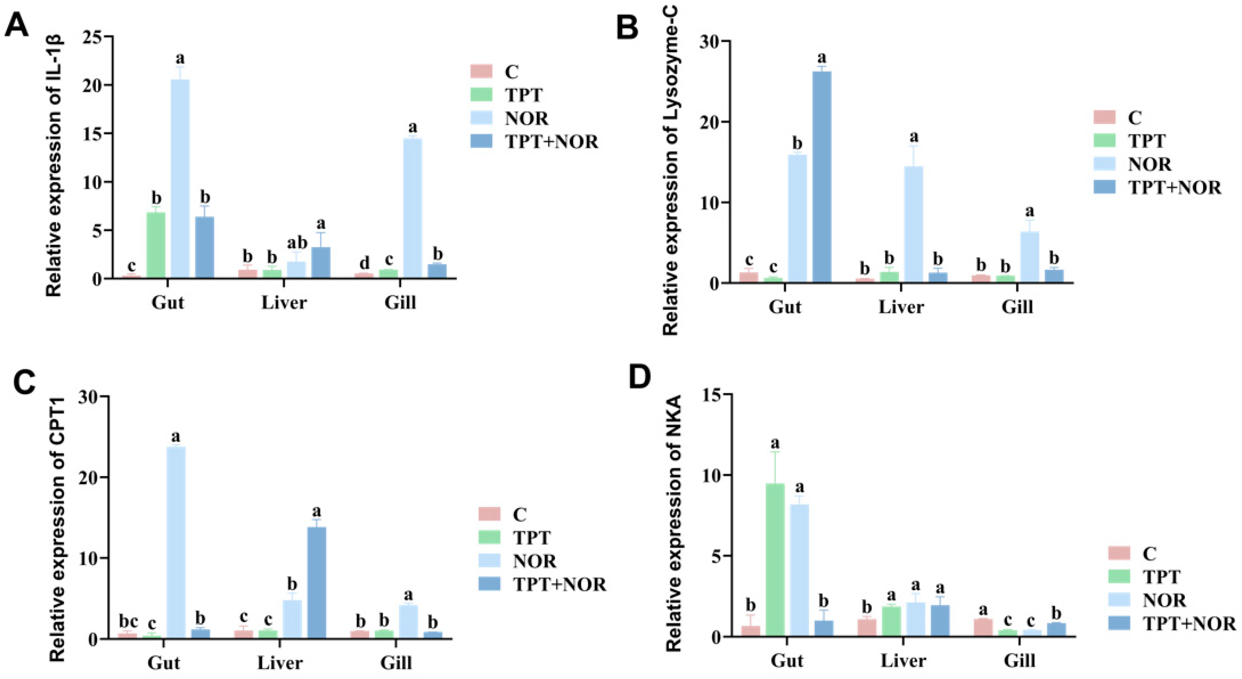
Figure 4 shows that gene expressions of IL-1β, Lysozyme-C, and CPT1 in the liver, intestine, and gill tissues in the NOR group were markedly greater compared with those in other groups. NKA gene expression was highest in the intestine, followed by the liver, and lastly, the gills. This trend corresponds with findings in previous studies [46] and supports the results of this experiment. The activity of NKA and CMA in the intestinal tissue was significantly decreased in the TPT group.

3.2.2. Analysis of Related Gene Expression Levels

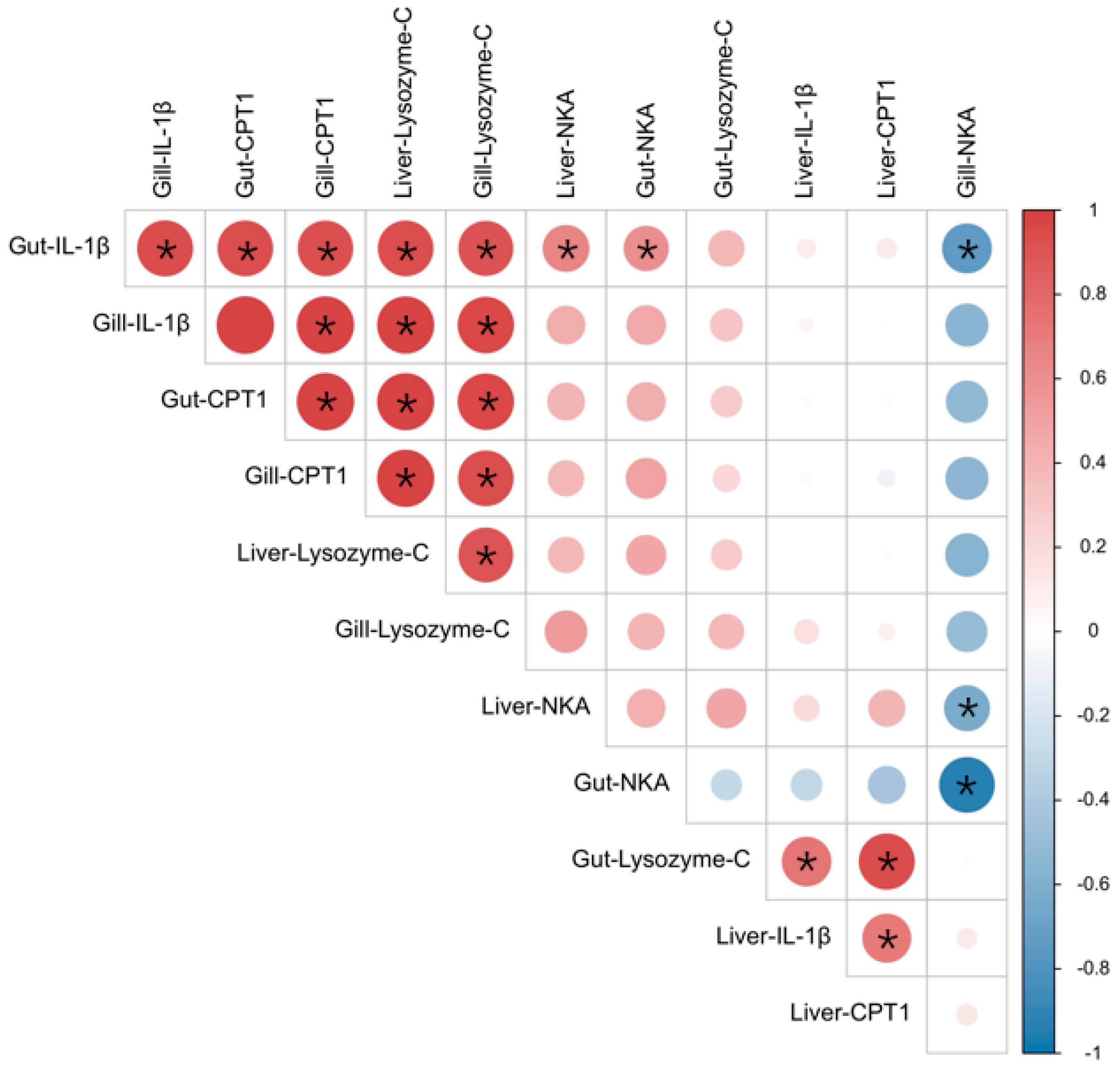
The correlation heat map of gene expressions in each tissue is displayed in Figure 5 to uncover the relationships between different parameters and evaluate their correlation coefficients. Based on the data presented in Figure 5, the expression levels of IL-1β, Lysozyme-C, and CPT1 genes in the intestinal and gill tissues exhibited strong positive correlations. Additionally, the expression levels of Lysozyme-C in the intestinal tissues showed significant positive correlations with the expression levels of CPT1 genes in the liver tissues. Notably, NKA expression in the intestinal tissue demonstrated a negative correlation with NKA expression in the gill tissue, while IL-1β expression in the intestinal tissue displayed a substantial negative correlation with NKA expression in the gill tissue.
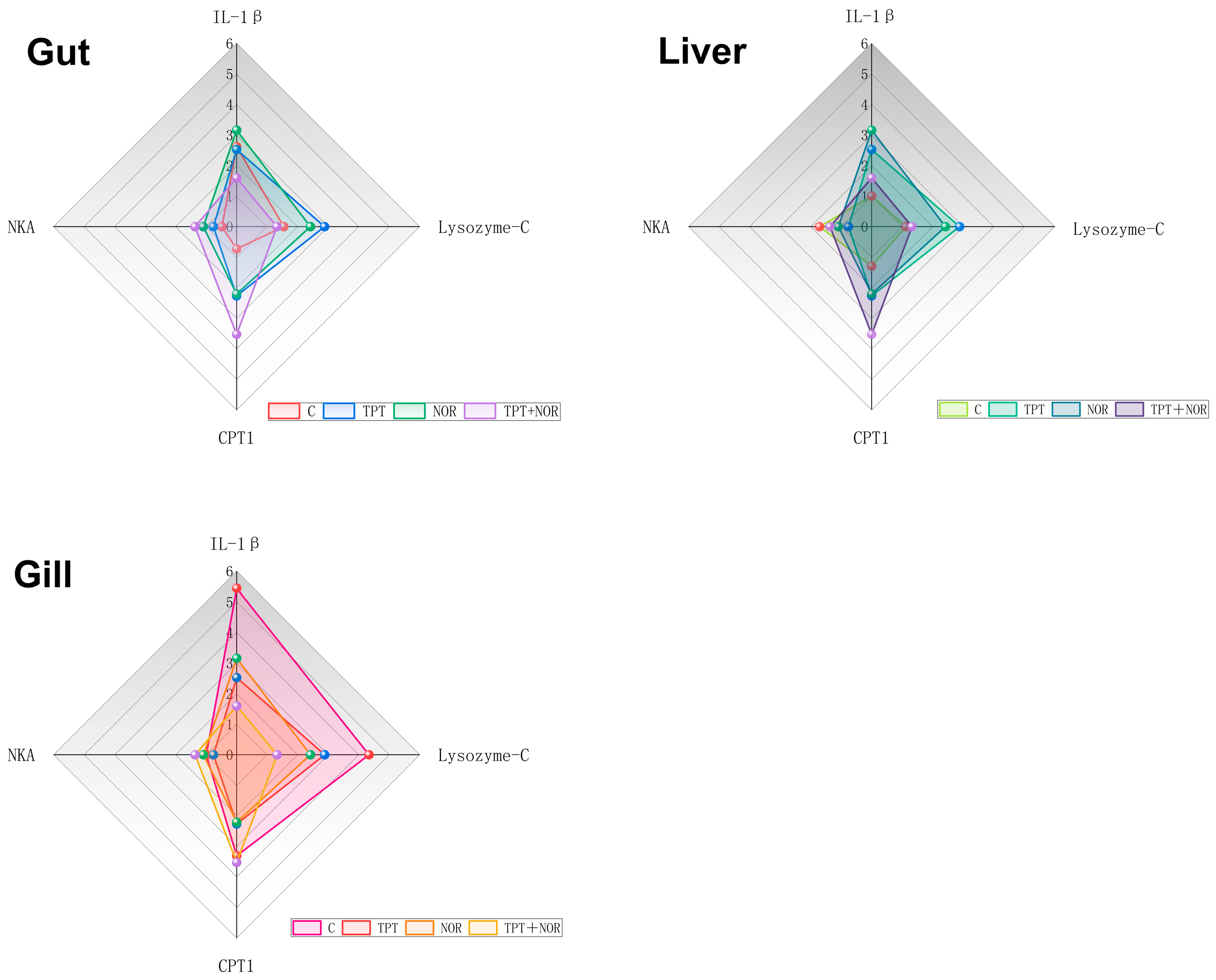
The radar map displays the standardized gene expression levels in different tissues of various carp groups (Figure 6), revealing differential responses to the IBR index [47] values as biomarkers. According to this index, it is evident that the intestinal and liver tissues experienced the least stress in the control group, while intriguingly, the gill tissues appeared to be subjected to higher stress. In contrast, the gene expression levels in the intestinal and liver tissues across all treatment groups were notably similar. Under single exposure to TPT and a high concentration of NOR, all gene expression levels were significantly elevated, while under combined exposure, the expression levels of CPT1 exhibited a notable increase. Notably, CPT1 gene expression is closely associated with lipid metabolism.

4. Discussion

Consistent with a plethora of studies that have demonstrated the alteration of lipid metabolism in organisms under adverse conditions [48], our results provide compelling evidence that common carp upregulate the expression of CPT1 in response to TPT exposure. This upregulation was observed across all three tissues, suggesting a systemic response to the environmental stressor. The significant increase in CPT1 gene expression in the TPT-exposed group likely reflects an attempt by the carp to enhance energy production through lipid metabolism, thereby supporting the physiological changes necessary to withstand the toxic effects of TPT. Interestingly, during combined exposure to TPT and NOR, the upregulation of CPT1 was confined to the liver. This tissue-specific response may be attributed to the liver’s critical function in metabolic homeostasis [49]. The liver’s role in detoxification and energy regulation positions it as a primary site for metabolic adjustments in response to environmental stress. As such, the liver’s heightened sensitivity to combined stressors may underlie the observed upregulation of CPT1, highlighting its adaptive role in maintaining metabolic balance.
The immunosuppressive effects of TPT on abalone (Abalone) have been observed [50]. Once fish are exposed to environmental contaminants, the cytokines that cause inflammation mRNA expression are increased [51,52]. A separate study indicated increased expression of the IL-1β gene in mice after TPT exposure [53]. Another experiment showed that low concentrations of pollutants inhibited the expression of inflammatory cytokine genes [54], with a similar occurrence observed in abalone (Abalone) [55]. Prior research has indicated increased renal IL-1β expression in carp (Cyprinus carpio) exposed to a concentration of 0.75 ppm indoxacarb [56]. In this experiment, the interleukin content and IL-1β gene expression levels in liver, intestine, and gill tissues of the TPT single-exposure group were significantly increased compared with the control group. The pollutants increase IL-1β expression by activating inflammasomes. For example, studies have found that certain pesticides can activate caspase-1 through the NLRP3 inflammasome, which promotes the maturation and release of IL-1β [57] Elevated levels of interleukin and expression of the IL-1β gene contribute to the survival and recovery of fish exposed to pollutants [58].
In mussels (Lamellidens corrianus) exposed to heavy metals, gill ACP activity remained elevated throughout the exposure period [59]. This suggests that in the face of toxic stress from heavy metals, the gill tissue of mussels may activate its defense mechanisms to maintain physiological function. Moreover, exposure to organophosphorus insecticides resulted in increased ACP activity in the euryhaline fish species (Oreochromis mossambicus) [60]. However, the situation observed in this experiment is quite different. ACP activity in the liver, intestine and gill tissues of common carp was significantly decreased in both the single- and combined-exposure groups. This reduction may indicate tissue damage or dysfunction, affecting the carp’s ability to adapt to environmental stress. Exposure to environmental antibiotic residues reduces the activity of ACP and AKP in the intestines of zebrafish (Danio rerio) [61]. For this experiment, both the significant increase of ACP activity in TPT environment and the decrease in NOR environment can be regarded as negative effects on the liver, intestine, and gills of common carp, representing two different injury pathways and coping mechanisms. The elevation of ACP activity in the TPT group can be seen as a direct response to metal toxicity, which means that the defense mechanism is disrupted, or the recovery is delayed. The decreased activity of the NOR group can be seen as the presence of antibiotics indirectly interfering with an early event in the inflammatory process, namely, the increase in pro-inflammatory cytokines, resulting in a reduced adaptability of ACP activity, enabling organisms to respond to a wide variety of inflammations [62].
Na+-K+-ATPase (NKA), also known as the Na+, K+ pump, indirectly drives numerous transport processes and is the primary source of intracellular ATP consumption [63,64]. Studies have shown that in chronic diseases, such as epilepsy and diabetic cardiomyopathy, the change in NKA activity is particularly complex, and the decrease in NKA activity may be related to a variety of pathophysiological mechanisms [65]. In this study, intestinal tissue from the combined-exposure group showed a significant reduction in NKA activity. This decreased activity may be related to stress from external pollutants; in order to maintain the balance of the environment in the body, intestinal tissues may respond to this stress by reducing the activity of NKA to reduce ion secretion [66]. Pollutants can cause oxidative stress, which may also lead to a decrease in NKA activity. For example, in one study, nitrite exposure led to a significant decrease in Na+/K+-ATPase activity and mRNA levels [67].
In this study, the expression level of IL-1β gene in the liver, intestine and gill tissues of carp exposed to TPT was significantly increased. The decrease in IL-1β gene expression in the NOR single- and combined-exposure groups indicated the adjuvant effect of antibiotics on the immune system [68].
The IBR index also showed that the gill tissue of carp was most affected by stress. The reason for this phenomenon may be the direct contact between gills and the water environment as the main site for material exchange between the fish and the water environment [69]. In order to inhibit the effect of pollutants on organisms, gills can achieve resistance to pollutants by preventing pollutants from entering the body or by compensating [69,70,71]. As such, gills are extremely vulnerable to contaminants and frequently display the early signs of unfavorable environmental circumstances [72,73]. Within 21 days of single or combined exposure, the gill tissue demonstrated the earliest and most significant damage, potentially indicating impaired regulatory capacity, leading to the negative gene expression patterns observed in this experiment. It has also been shown that TBT and TPT may cause adipocyte inflammation or other TNF-α-associated adipocyte dysfunction in addition to causing lipid accumulation in rainbow trout (Oncorhynchus mykiss) cells [74]. Furthermore, Zhang et al. found that NOR may cause lipid buildup in carp and that TPT might interfere with lipid metabolism in brain tissue [40]. According to the IBR index, when TPT and NOR were jointly exposed to the intestinal and liver tissues of carp, several indicators were very similar to those of the control group. In this study, CPT1 expression levels were elevated in liver, intestine, and gill tissues in the TPT-exposed group, which may be due to the fact that environmental chemical pollutants can mimic, antagonize, or inappropriately regulate specific metabolic pathways, alter adipocyte programming, and increase energy storage in adipose tissue [75]. Studies have also shown that overexpression of CPT1A in adipocytes has been shown to reduce obesity and improve glucose intolerance in mice while also enhancing the expression of mitochondrial respiratory chain complexes in adipose tissue [76]. Conversely, the knockdown of CPT1A in goat intramuscular precursor adipocytes increased lipid deposition and inhibited cell proliferation, indicating a potential role of CPT1A in regulating intramuscular fat (IMF) formation [77]. Therefore, this shows that high concentrations of TPT exposure affect the liver and intestines of common carp and the regulation of lipids in gill tissue.
However, this study did not include a histological section analysis, which could provide valuable insights into the structural changes and potential damage in the tissues of common carp exposed to TPT and NOR. In future studies, incorporating histological assessments would be beneficial to complement the molecular and biochemical data, offering a more comprehensive understanding of the toxic effects and the physiological responses of common carp to environmental stressors.

5. Conclusions

The current findings demonstrate substantial alterations in various carp tissues following single or combined exposure to TPT and NOR. Specifically, the presence of NOR significantly decreased the interleukin composition of common carp liver tissue. Moreover, the expression levels of IL-1β, Lysozyme-C, CPT1, and NKA were markedly altered across different tissues. The correlation analysis indicated closely related expressions of IL-1β, Lysozyme-C, CPT1, and NKA in intestinal and gill tissues. The IBR index showed that, compared with the TPT exposure group, the indexes of intestinal tissue and liver tissue in the NOR and TPT combined-exposure group were more similar to those in the control group, indicating that NOR could reduce the toxicity of TPT in the intestinal tissue and liver tissue of common carp to a certain extent, and may have antagonistic effects, and that high concentrations of TPT exposure affect the liver and intestine of common carp and the regulation of lipids in gill tissue. Overall, the intestinal tissue was most affected by the presence of TPT, followed by the liver and gills. This study investigated the single or combined toxicity of TPT and NOR in different tissues of common carp, providing a foundation for a more comprehensive assessment of potential pollutant toxicity to diverse aquatic organisms.

Supplementary Materials

The following supporting information can be downloaded at: https://www.mdpi.com/article/10.3390/fishes9100415/s1, Figure S1. Standard curves of the genes in PCR; Table S1. There agents and their sources; Table S2. Specific Information of PCR.

Author Contributions

Conceptualization, Z.L. and L.L. (Ling Liu); methodology, L.L. (Luoxin Li); software, S.Z. and M.Y.; validation, T.L. and B.Z.; formal analysis, P.L.; writing—original draft preparation, Y.L.; writing—review and editing, Z.L.; supervision, Z.L. and L.L. (Ling Liu); project administration, Z.L.; funding acquisition, Z.L. All authors have read and agreed to the published version of the manuscript.

Funding

This work was financially supported by the National Natural Science Foundation of China (42277269).

Institutional Review Board Statement

Because carp is a common economic fish and not a restricted protected species, experiments related to this fish in China do not require special ethical authorization. Therefore, we currently do not have a special ethical authorization for carp. The research was conducted according to the usual ethical standards, which has been addressed as “All procedures in this study complied with the guidelines of the Chinese Association for Laboratory Animal Sciences and the ethical principles for animal experimentation at Shandong University”.

Informed Consent Statement

Not applicable.

Data Availability Statement

Dataset available on request from the authors.

Conflicts of Interest

The authors declare no conflict of interest.

References

  1. Anastasiou, T.I.; Chatzinikolaou, E.; Mandalakis, M.; Arvanitidis, C. Imposex and organotin compounds in ports of the Mediterranean and the Atlantic: Is the story over? Sci. Total Environ. 2016, 569–570, 1315–1329. [Google Scholar] [CrossRef]
  2. Snoeij, N.J.; Penninks, A.H.; Seinen, W. Biological activity of organotin compounds—An overview. Environ. Res. 1987, 44, 335–353. [Google Scholar] [CrossRef]
  3. Chen, C.; Chen, L.; Huang, Q.; Chen, Z.; Zhang, W. Organotin contamination in commercial and wild oysters from China: Increasing occurrence of triphenyltin. Sci. Total Environ. 2019, 650, 2527–2534. [Google Scholar] [CrossRef]
  4. Guérin, T.; Sirot, V.; Volatier, J.L.; Leblanc, J.C. Organotin levels in seafood and its implications for health risk in high-seafood consumers. Sci. Total Environ. 2007, 388, 66–77. [Google Scholar] [CrossRef]
  5. Liu, L.; Du, R.Y.; Jia, R.L.; Wang, J.X.; Chen, C.Z.; Li, P.; Li, Z.H.; Kong, L.M. Micro(nano)plastics in marine medaka: Entry pathways and cardiotoxicity with triphenyltin. Environ. Pollut. 2024, 342, 123079. [Google Scholar] [CrossRef]
  6. Zhang, Z.B.; Hu, J.Y.; Zhen, H.J.; Wu, X.Q.; Huang, C. Reproductive Inhibition and Transgenerational Toxicity of Triphenyltin on Medaka (Oryzias latipes) at Environmentally Relevant tip Levels. Environ. Sci. Technol. 2008, 42, 8133–8139. [Google Scholar] [CrossRef]
  7. Yuan, J.; Zhang, X.; Yu, L.; Sun, Z.; Zhu, P.; Wang, X.; Shi, H. Stage-specific malformations and phenotypic changes induced in embryos of amphibian (Xenopus tropicalis) by triphenyltin. Ecotoxicol. Environ. Saf. 2011, 74, 1960–1966. [Google Scholar] [CrossRef]
  8. Gao, J.M.; Zhang, Y.; Guo, J.S.; Jin, F.; Zhang, K. Occurrence of organotins in the Yangtze River and the Jialing River in the urban section of Chongqing, China. Environ. Monit. Assess. 2013, 185, 3831–3837. [Google Scholar] [CrossRef]
  9. Liu, L.-L.; Wang, J.-T.; Chung, K.-N.; Leu, M.-Y.; Meng, P.-J. Distribution and accumulation of organotin species in seawater, sediments and organisms collected from a Taiwan mariculture area. Mar. Pollut. Bull. 2011, 63, 535–540. [Google Scholar] [CrossRef]
  10. Li, Y.; Mu, D.; Wu, H.-Q.; Liu, H.-J.; Wang, Y.-H.; Ma, G.-C.; Duan, X.-M.; Zhou, J.-J.; Zhang, C.-M.; Lu, X.-H.; et al. Derivation of copper water quality criteria in Bohai Bay for the protection of local aquatic life and the ecological risk assessment. Mar. Pollut. Bull. 2023, 190, 114863. [Google Scholar] [CrossRef]
  11. Li, P.; Li, Z.H.; Zhong, L. Effects of low concentrations of triphenyltin on neurobehavior and the thyroid endocrine system in zebrafish. Ecotoxicol. Environ. Saf. 2019, 186, 109776. [Google Scholar] [CrossRef]
  12. Zhang, S.Q.; Li, P.; He, S.W.; Xing, S.Y.; Cao, Z.H.; Zhao, X.L.; Sun, C.C.; Li, Z.H. Combined effect of microplastic and triphenyltin: Insights from the gut-brain axis. Environ. Sci. Ecotechnol. 2023, 16, 100266. [Google Scholar] [CrossRef]
  13. Morcillo, Y.; Porte, C. Interaction of tributyl- and triphenyltin with the microsomal monooxygenase system of molluscs and fish from the Western Mediterranean. Aquat. Toxicol. 1997, 38, 35–46. [Google Scholar] [CrossRef]
  14. Harino, H.; Fukushima, M.; Kawai, S. Accumulation of butyltin and phenyltin compounds in various fish species. Arch. Environ. Contam. Toxicol. 2000, 39, 13–19. [Google Scholar] [CrossRef]
  15. Sun, L.; Zhang, J.; Zuo, Z.; Chen, Y.; Wang, X.; Huang, X.; Wang, C. Influence of triphenyltin exposure on the hypothalamus–pituitary–gonad axis in male Sebastiscus marmoratus. Aquat. Toxicol. 2011, 104, 263–269. [Google Scholar] [CrossRef]
  16. Zhang, S.-Q.; Li, P.; Zhao, X.-L.; He, S.-W.; Xing, S.-Y.; Cao, Z.-H.; Zhang, H.-Q.; Li, Z.-H. Hepatotoxicity in carp (Cyprinus carpio) exposed to environmental levels of norfloxacin (NOR): Some latest evidences from transcriptomics analysis, biochemical parameters and histopathological changes. Chemosphere 2021, 283, 131210. [Google Scholar] [CrossRef]
  17. Yang, C.; Wu, T. A comprehensive review on quinolone contamination in environments: Current research progress. Environ. Sci. Pollut. Res. Int. 2023, 30, 48778–48792. [Google Scholar] [CrossRef]
  18. Chen, H.; Liu, S.; Xu, X.-R.; Zhou, G.-J.; Liu, S.-S.; Yue, W.-Z.; Sun, K.-F.; Ying, G.-G. Antibiotics in the coastal environment of the Hailing Bay region, South China Sea: Spatial distribution, source analysis and ecological risks. Mar. Pollut. Bull. 2015, 95, 365–373. [Google Scholar] [CrossRef]
  19. Zhao, X.L.; Li, P.; Zhang, S.Q.; He, S.W.; Xing, S.Y.; Cao, Z.H.; Lu, R.; Li, Z.H. Effects of environmental norfloxacin concentrations on the intestinal health and function of juvenile common carp and potential risk to humans. Environ. Pollut. 2021, 287, 117612. [Google Scholar] [CrossRef]
  20. Shi, H.; Yang, Y.; Liu, M.; Yan, C.; Yue, H.; Zhou, J. Occurrence and distribution of antibiotics in the surface sediments of the Yangtze Estuary and nearby coastal areas. Mar. Pollut. Bull. 2014, 83, 317–323. [Google Scholar] [CrossRef]
  21. Golet, E.M.; Xifra, I.; Siegrist, H.; Alder, A.C.; Giger, W. Environmental exposure assessment of fluoroquinolone antibacterial agents from sewage to soil. Environ. Sci. Technol. 2003, 37, 3243–3249. [Google Scholar] [CrossRef]
  22. Diwan, V.; Tamhankar, A.J.; Aggarwal, M.; Sen, S.; Khandal, R.K.; Lundborg, C.S. Detection of antibiotics in hospital effluents in India. Curr. Sci. 2009, 97, 1752–1755. [Google Scholar]
  23. Lindberg, R.; Jarnheimer, P.-Å.; Olsen, B.; Johansson, M.; Tysklind, M. Determination of antibiotic substances in hospital sewage water using solid phase extraction and liquid chromatography/mass spectrometry and group analogue internal standards. Chemosphere 2004, 57, 1479–1488. [Google Scholar] [CrossRef]
  24. Liu, Y.J.; Wang, S.Q.; Pan, J.L.; Zhu, F.; Wu, M.H.; Xu, G. Antibiotics in urine of the general population: Exposure, health risk assessment, and food factors. J. Environ. Sci. Health Part B-Pestic. Food Contam. Agric. Wastes 2022, 57, 1–12. [Google Scholar] [CrossRef]
  25. Ho, K.K.Y.; Zhou, G.J.; Xu, E.G.B.; Wang, X.; Leung, K.M.Y. Long-term spatio-temporal trends of organotin contaminations in the marine environment of Hong Kong. PLoS ONE 2016, 11, e0155632. [Google Scholar] [CrossRef]
  26. Ebert, I.; Bachmann, J.; Kühnen, U.; Küster, A.; Kussatz, C.; Maletzki, D.; Schlüter, C. Toxicity of the fluoroquinolone antibiotics enrofloxacin and ciprofloxacin to photoautotrophic aquatic organisms. Environ. Toxicol. Chem. 2011, 30, 2786–2792. [Google Scholar] [CrossRef]
  27. Liang, X.M.; Wang, L.; Ou, R.K.; Nie, X.P.; Yang, Y.F.; Wang, F.; Li, K.B. Effects of norfioxacin on hepatic genes expression of P450 isoforms (CYP1A and CYP3A), GST and P-glycoprotein (P-gp) in Swordtail fish (Xiphophorus Helleri). Ecotoxicology 2015, 24, 1566–1573. [Google Scholar] [CrossRef]
  28. Liu, J.; Lu, G.; Wu, D.; Yan, Z. A multi-biomarker assessment of single and combined effects of norfloxacin and sulfamethoxazole on male goldfish (Carassius auratus). Ecotoxicol. Environ. Saf. 2014, 102, 12–17. [Google Scholar] [CrossRef]
  29. Janecko, N.; Pokludova, L.; Blahova, J.; Svobodova, Z.; Literak, I. Implications of fluoroquinolone contamination for the aquatic environment—A review. Environ. Toxicol. Chem. 2016, 35, 2647–2656. [Google Scholar] [CrossRef]
  30. Zhang, C.-N.; Zhang, J.-L.; Ren, H.-T.; Zhou, B.-H.; Wu, Q.-J.; Sun, P. Effect of tributyltin on antioxidant ability and immune responses of zebrafish (Danio rerio). Ecotoxicol. Environ. Saf. 2017, 138, 1–8. [Google Scholar] [CrossRef]
  31. Rahman, M.F.; Siddiqui, M.K.J. Biochemical effects of vepacide (from Azadirachta indica) on Wistar rats during subchronic exposure. Ecotoxicol. Environ. Saf. 2004, 59, 332–339. [Google Scholar] [CrossRef]
  32. Enan, E.E.; Enan, O.H.; El-Sebae, A.E. Biochemical targets affected by sublethal doses of organophosphorus insecticides. J. Int. Pest Control 1982, 24, 120. [Google Scholar]
  33. Cajaraville, M.P.; Bebianno, M.J.; Blasco, J.; Porte, C.; Sarasquete, C.; Viarengo, A. The use of biomarkers to assess the impact of pollution in coastal environments of the Iberian Peninsula: A practical approach. Sci. Total Environ. 2000, 247, 295–311. [Google Scholar] [CrossRef]
  34. Ho, K.K.Y.; Leung, K.M.Y. Imposex status associated with organotin contamination in Reishia clavigera after reciprocal transplantation between clean and polluted sites in Hong Kong. Reg. Stud. Mar. Sci. 2016, 8, 480–486. [Google Scholar] [CrossRef]
  35. Okoro, H.K.; Fatoki, O.S.; Adekola, F.A.; Ximba, B.J.; Snyman, R.G. Spatio-temporal variation of organotin compounds in seawater and sediments from Cape Town harbour, South Africa using gas chromatography with flame photometric detector (GC-FPD). Arab. J. Chem. 2016, 9, 95–104. [Google Scholar] [CrossRef]
  36. Le Page, G.; Gunnarsson, L.; Snape, J.; Tyler, C.R. Integrating human and environmental health in antibiotic risk assessment: A critical analysis of protection goals, species sensitivity and antimicrobial resistance. Environ. Int. 2017, 109, 155–169. [Google Scholar] [CrossRef]
  37. Bradford, M.M. A rapid and sensitive method for the quantitation of microgram quantities of protein utilizing the principle of protein-dye binding. Anal. Biochem. 1976, 72, 248–254. [Google Scholar] [CrossRef]
  38. Corripio-Miyar, Y.; Bird, S.; Tsamopoulos, K.; Secombes, C.J. Cloning and expression analysis of two pro-inflammatory cytokines, IL-1β and IL-8, in haddock (Melanogrammus aeglefinus). Mol. Immunol. 2007, 44, 1361–1373. [Google Scholar] [CrossRef]
  39. Kim, J.Y.; Hickner, R.C.; Cortright, R.L.; Dohm, G.L.; Houmard, J.A. Lipid oxidation is reduced in obese human skeletal muscle. Am. J. Physiol.-Endocrinol. Metab. 2000, 279, E1039–E1044. [Google Scholar] [CrossRef]
  40. Zhang, S.-Q.; Li, P.; He, S.-W.; Xing, S.-Y.; Cao, Z.-H.; Zhao, X.-L.; Sun, C.; Li, Z.-H. Assessing the ecotoxicity of combined exposure to triphenyltin and norfloxacin at environmental levels: A case study of immunotoxicity and metabolic regulation in carp (Cyprinus carpio). Chemosphere 2023, 313, 137381. [Google Scholar] [CrossRef]
  41. Wen, J.; Cui, X.; Gibson, M.; Li, Z. Water quality criteria derivation and ecological risk assessment for triphenyltin in China. Ecotoxicol. Environ. Saf. 2018, 161, 397–401. [Google Scholar] [CrossRef]
  42. Hoseini, S.M.; Yousefi, M.; Hoseinifar, S.H.; Van Doan, H. Effects of dietary arginine supplementation on growth, biochemical, and immunological responses of common carp (Cyprinus carpio L.), stressed by stocking density. Aquaculture 2019, 503, 452–459. [Google Scholar] [CrossRef]
  43. Yuan, C.; Pan, X.; Gong, Y.; Xia, A.; Wu, G.; Tang, J.; Han, X. Effects of Astragalus polysaccharides (APS) on the expression of immune response genes in head kidney, gill and spleen of the common carp, Cyprinus carpio L. Int. Immunopharmacol. 2008, 8, 51–58. [Google Scholar] [CrossRef]
  44. Zhang, Z.; Liu, Q.; Cai, J.; Yang, J.; Shen, Q.; Xu, S. Chlorpyrifos exposure in common carp (Cyprinus carpio L.) leads to oxidative stress and immune responses. Fish Shellfish Immunol. 2017, 67, 604–611. [Google Scholar] [CrossRef]
  45. Yuan, X.; Wang, C.; Huang, Y.; Dai, Y.; Desouky, H.E. A comparative study on intestinal morphology and function of normal and injured intestines of Jian carp (Cyprinus carpio var. Jian). Aquaculture 2020, 528, 735496. [Google Scholar] [CrossRef]
  46. Lim, Y.; Lee, V.; Blanco, A.; Kelly, S.P.; Unniappan, S. Ion-poor water and dietary salt deprivation upregulate the ghrelinergic system in the goldfish (Carassius auratus). J. Fish Biol. 2021, 99, 1100–1109. [Google Scholar] [CrossRef]
  47. Chen, C.-Z.; Li, P.; Wang, W.-B.; Li, Z.-H. Response of growth performance, serum biochemical parameters, antioxidant capacity, and digestive enzyme activity to different feeding strategies in common carp (Cyprinus carpio) under high-temperature stress. Aquaculture 2022, 548, 737636. [Google Scholar] [CrossRef]
  48. Yu, J.; Wang, X.; Qian, S.; Liu, P.; Li, X.; Li, J. Exposure to nitrate induces alterations in blood parameter responses, liver immunity, and lipid metabolism in juvenile turbot (Scophthalmus maximus). Aquat. Toxicol. 2022, 251, 106280. [Google Scholar] [CrossRef]
  49. Vandenberghe, G. The role of the liver in metabolic homeostasis—Implications for inborn-errors of metabolism. J. Inherit. Metab. Dis. 1991, 14, 407–420. [Google Scholar] [CrossRef]
  50. Gopalakrishnan, S.; Huang, W.-B.; Wang, Q.-W.; Wu, M.-L.; Liu, J.; Wang, K.-J. Effects of tributyltin and benzo[a]pyrene on the immune-associated activities of hemocytes and recovery responses in the gastropod abalone, Haliotis diversicolor. Comp. Biochem. Physiol. Part C Toxicol. Pharmacol. 2011, 154, 120–128. [Google Scholar] [CrossRef]
  51. Liu, Z.; Fu, Z.; Jin, Y. Immunotoxic effects of atrazine and its main metabolites at environmental relevant concentrations on larval zebrafish (Danio rerio). Chemosphere 2017, 166, 212–220. [Google Scholar] [CrossRef]
  52. Li, Z.H.; Xu, H.Y.; Zheng, W.L.; Lam, S.H.; Gong, Z.Y. RNA-Sequencing Analysis of TCDD-Induced Responses in Zebrafish Liver Reveals High Relatedness to Mammalian Models and Conserved Biological Pathways. PLoS ONE 2013, 8, e77292. [Google Scholar] [CrossRef]
  53. Chen, X.; Zhu, D.; Ge, R.; Bao, Z. Fecal transplantation of young mouse donors effectively improves enterotoxicity in elderly recipients exposed to triphenyltin. Ecotoxicol. Environ. Saf. 2024, 273, 116140. [Google Scholar] [CrossRef]
  54. Ma, J.; Li, Y.; Li, W.; Li, X. Hepatotoxicity of paraquat on common carp (Cyprinus carpio L.). Sci. Total Environ. 2018, 616–617, 889–898. [Google Scholar] [CrossRef]
  55. Horiguchi, T.; Kojima, M.; Kaya, M.; Matsuo, T.; Shiraishi, H.; Morita, M.; Adachi, Y. Tributyltin and triphenyltin induce spermatogenesis in ovary of female abalone, Haliotis gigantea. Mar. Environ. Res. 2002, 54, 679–684. [Google Scholar] [CrossRef]
  56. Ghelichpour, M.; Taheri Mirghaed, A.; Hoseinifar, S.H.; Khalili, M.; Yousefi, M.; Van Doan, H.; Perez-Jimenez, A. Expression of immune, antioxidant and stress related genes in different organs of common carp exposed to indoxacarb. Aquat. Toxicol. 2019, 208, 208–216. [Google Scholar] [CrossRef]
  57. Yang, C.; Lim, W.; Song, G. Immunotoxicological effects of insecticides in exposed fishes. Comp. Biochem. Physiol. Part C Toxicol. Pharmacol. 2021, 247, 109064. [Google Scholar] [CrossRef]
  58. Joshi, P.K.; Bose, M.; Harish, D. Changes in certain haematological parameters in a siluroid cat fish Clarias batrachus (Linn) exposed to cadmium chloride. Pollut. Res. 2002, 21, 129–131. [Google Scholar]
  59. Rajalakshmi, S.; Mohandas, A. Copper-induced changes in tissue enzyme activity in a freshwater mussel. Ecotoxicol. Environ. Saf. 2005, 62, 140–143. [Google Scholar] [CrossRef]
  60. McClellan-Green, P.; Robbins, J. Effects of TBT and 3-MC co-exposure on cytochrome P450 expression and activity in marine organisms. Mar. Environ. Res. 2000, 50, 243. [Google Scholar] [CrossRef]
  61. Zhou, L.; Limbu, S.M.; Shen, M.; Zhai, W.; Qiao, F.; He, A.; Du, Z.-Y.; Zhang, M. Environmental concentrations of antibiotics impair zebrafish gut health. Environ. Pollut. 2018, 235, 245–254. [Google Scholar] [CrossRef]
  62. Chen, G.Y.; Nuñez, G. Sterile inflammation: Sensing and reacting to damage. Nat. Rev. Immunol. 2010, 10, 826–837. [Google Scholar] [CrossRef]
  63. Evans, D.H.; Piermarini, P.M.; Choe, K.P. The multifunctional fish gill: Dominant site of gas exchange, osmoregulation, acid-base regulation, and excretion of nitrogenous waste. Physiol. Rev. 2005, 85, 97–177. [Google Scholar] [CrossRef]
  64. Hwang, P.-P.; Lee, T.-H. New insights into fish ion regulation and mitochondrion-rich cells. Comp. Biochem. Physiol. Part A Mol. Integr. Physiol. 2007, 148, 479–497. [Google Scholar] [CrossRef]
  65. Zhu, M.Y.; Sun, H.J.; Cao, L.; Wu, Z.Y.; Leng, B.; Bian, J.S. Role of Na+/K+-ATPase in ischemic stroke: In-depth perspectives from physiology to pharmacology. J. Mol. Med.-JMM 2022, 100, 395–410. [Google Scholar] [CrossRef]
  66. Lu, W.; Long, L.; Zhao, P.; Zhang, X.; Yan, C.; Dong, S.; Huang, Q. Perfluorinated compounds disrupted osmoregulation in Oryzias melastigma during acclimation to hypoosmotic environment. Ecotoxicol. Environ. Saf. 2021, 223, 112613. [Google Scholar] [CrossRef]
  67. Wang, J.; Tang, H.; Zhang, X.; Xue, X.; Zhu, X.; Chen, Y.; Yang, Z. Mitigation of nitrite toxicity by increased salinity is associated with multiple physiological responses: A case study using an economically important model species, the juvenile obscure puffer (Takifugu obscurus). Environ. Pollut. 2018, 232, 137–145. [Google Scholar] [CrossRef]
  68. Ngugi, C.C.; Oyoo-Okoth, E.; Mugo-Bundi, J.; Orina, P.S.; Chemoiwa, E.J.; Aloo, P.A. Effects of dietary administration of stinging nettle (Urtica dioica) on the growth performance, biochemical, hematological and immunological parameters in juvenile and adult Victoria Labeo (Labeo victorianus) challenged with Aeromonas hydrophila. Fish Shellfish Immunol. 2015, 44, 533–541. [Google Scholar] [CrossRef]
  69. Marcon, L.; Lopes, D.S.; Mounteer, A.H.; Goulart, A.M.A.; Leandro, M.V.; dos Anjos Benjamin, L. Pathological and histometric analysis of the gills of female Hyphessobrycon eques (Teleostei:Characidae) exposed to different concentrations of the insecticide Dimilin®. Ecotoxicol. Environ. Saf. 2016, 131, 135–142. [Google Scholar] [CrossRef]
  70. Crestani, M.; Menezes, C.; Glusczak, L.; Santos Miron, D.d.; Spanevello, R.; Silveira, A.; Gonçalves, F.F.; Zanella, R.; Loro, V.L. Effect of clomazone herbicide on biochemical and histological aspects of silver catfish (Rhamdia quelen) and recovery pattern. Chemosphere 2007, 67, 2305–2311. [Google Scholar] [CrossRef]
  71. Paulino, M.G.; Souza, N.E.S.; Fernandes, M.N. Subchronic exposure to atrazine induces biochemical and histopathological changes in the gills of a Neotropical freshwater fish, Prochilodus lineatus. Ecotoxicol. Environ. Saf. 2012, 80, 6–13. [Google Scholar] [CrossRef]
  72. Boran, H.; Capkin, E.; Altinok, I.; Terzi, E. Assessment of acute toxicity and histopathology of the fungicide captan in rainbow trout. Exp. Toxicol. Pathol. 2012, 64, 175–179. [Google Scholar] [CrossRef]
  73. Sonne, C.; Bach, L.; Søndergaard, J.; Rigét, F.F.; Dietz, R.; Mosbech, A.; Leifsson, P.S.; Gustavson, K. Evaluation of the use of common sculpin (Myoxocephalus scorpius) organ histology as bioindicator for element exposure in the fjord of the mining area Maarmorilik, West Greenland. Environ. Res. 2014, 133, 304–311. [Google Scholar] [CrossRef]
  74. Lutfi, E.; Riera-Heredia, N.; Córdoba, M.; Porte, C.; Gutiérrez, J.; Capilla, E.; Navarro, I. Tributyltin and triphenyltin exposure promotes in vitro adipogenic differentiation but alters the adipocyte phenotype in rainbow trout. Aquat. Toxicol. 2017, 188, 148–158. [Google Scholar] [CrossRef]
  75. Lubrano, C.; Genovesi, G.; Specchia, P.; Costantini, D.; Mariani, S.; Petrangeli, E.; Lenzi, A.; Gnessi, L. Obesity and metabolic comorbidities: Environmental diseases? Oxidative Med. Cell. Longev. 2013, 2013, 640673. [Google Scholar] [CrossRef]
  76. Soler-Vázquez, M.C.; Romero, M.d.M.; Todorcevic, M.; Delgado, K.; Calatayud, C.; Benitez-Amaro, A.; La Chica Lhoëst, M.T.; Mera, P.; Zagmutt, S.; Bastías-Pérez, M.; et al. Implantation of CPT1AM-expressing adipocytes reduces obesity and glucose intolerance in mice. Metab. Eng. 2023, 77, 256–272. [Google Scholar] [CrossRef]
  77. Tang, Y.M.; Zhang, W.Y.; Wang, Y.G.; Li, H.Y.; Zhang, C.H.; Wang, Y.; Lin, Y.Q.; Shi, H.B.; Xiang, H.; Huang, L.; et al. Expression Variation of CPT1A Induces Lipid Reconstruction in Goat Intramuscular Precursor Adipocytes. Int. J. Mol. Sci. 2023, 24, 13415. [Google Scholar] [CrossRef]
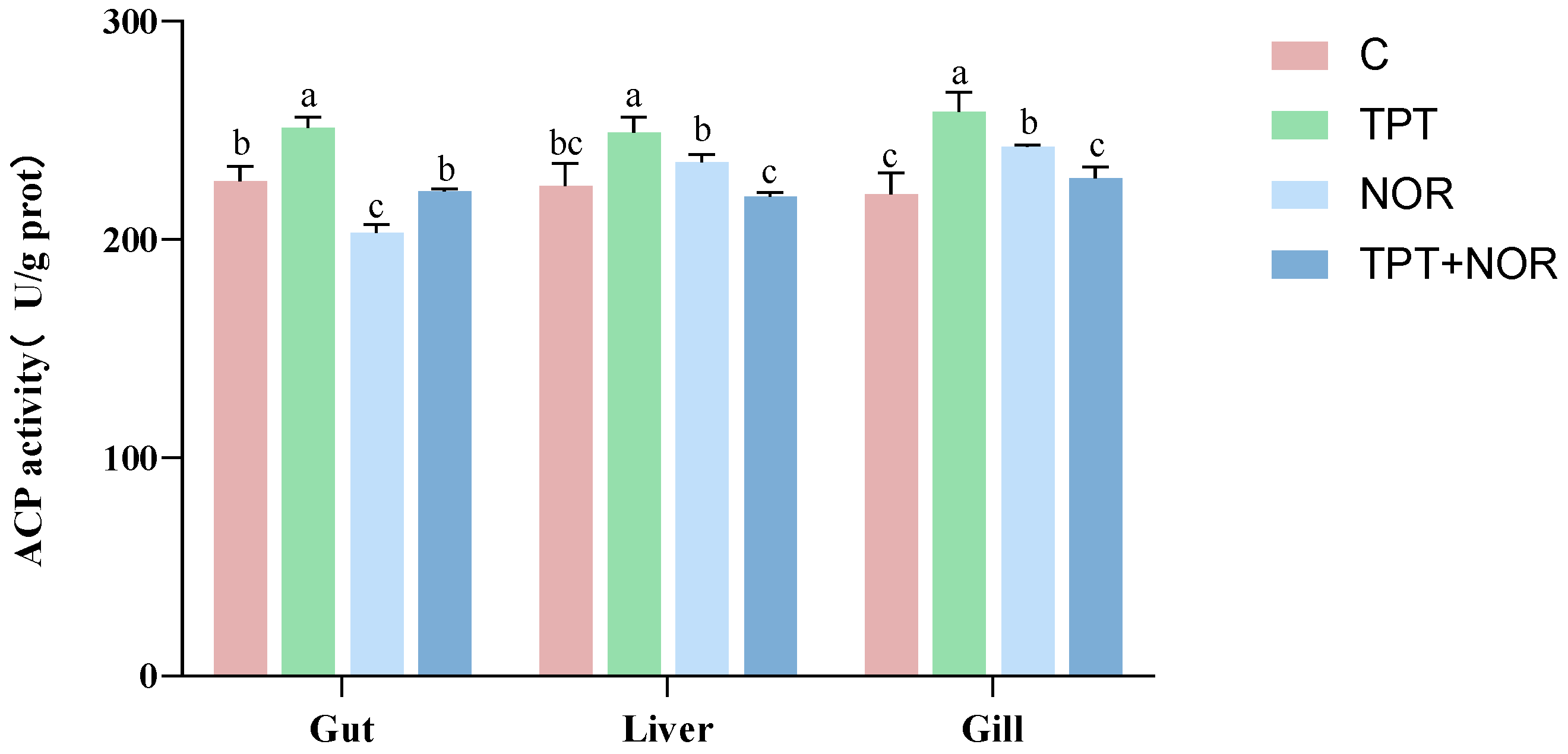
Figure 1. The activity of ACP enzyme in the lower intestine, liver and gill tissues was demonstrated after single or combined exposure to TPT and NOR. Vertical bars represent the mean ± SE (n = 18). Letters indicate significant differences (p < 0.05).
Fishes 09 00415 g001
Figure 2. The 1L-1β enzyme activity is shown in three different tissues of the lower intestine, liver and gills exposed to TPT and NOR alone, or in combination. Vertical bars represent the mean ± SE (n = 18). Letters indicate significant differences (p < 0.05).
Fishes 09 00415 g002
Figure 3. The activity of NKA and CMA enzymes in three different tissues of the lower intestine, liver and gills exposed to TPT and NOR alone, or in combination: (A) NKA enzyme activity; and (B) CMA enzyme activity. Vertical bars represent the mean ± SE (n = 18). Letters indicate significant differences (p < 0.05).
Fishes 09 00415 g003
Figure 4. The expression levels of IL-1β, Lysozyme-C, CPT1 and NKA in three different tissues of the lower intestine, liver and gill after single or combined exposure of TPT and NOR: (A) IL-1β level; (B) Lysozyme-C level; (C) CPT1 level; and (D) NKA level. Vertical bars represent the mean ± SE (n = 18). Letters indicate significant differences (p < 0.05).
Fishes 09 00415 g004
Figure 5. Heat map of correlations between the various parameters studied. Vibrant red denotes a positive correlation and brilliant blue denotes a negative correlation on the color scale, which shows the correlation value between −1 and 1. “*” represents a significant difference (p < 0.05).
Fishes 09 00415 g005
Figure 6. Radar plots of biomarker data in different groups and integrated biomarker response (IBR) index values for each group.
Fishes 09 00415 g006
Table 1. The primer sequences for the tested genes.
GeneSequences of Primers (5′−3′)Accession No.qPCR Efficiency
IL-1βF: ACCAGCTGGATTTGTCAGAAG
R: ACATACTGAATTGAACTTTG
AB010701.198.7190%
Lysozyme-CF: GTGTCTGATGTGGCTGTGCT
R: TTCCCCAGGTATCCCATGAT
AB02730598.0435%
CPT1F: CAGATGGAAAGTGTTGCTAATGAC
R: TGTGTAGAAGTTGCTGTTGACCA
JF72883998.9315%
NKAF: TGCCAGAACTTCTCCACA
R: AGCGATACCCATAGCCAC
JN03275995.7576%
β-actinF: GGCTGTGCTGTCCCTGTA
R: GGCGTAACCCTCGTAG
M2501397.5814%
Disclaimer/Publisher’s Note: The statements, opinions and data contained in all publications are solely those of the individual author(s) and contributor(s) and not of MDPI and/or the editor(s). MDPI and/or the editor(s) disclaim responsibility for any injury to people or property resulting from any ideas, methods, instructions or products referred to in the content.