Article

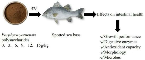
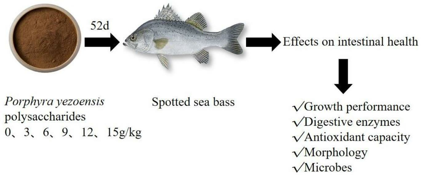
The Effects of Porphyra yezoensis Polysaccharides on Intestinal Health of Spotted Sea Bass, Lateolabrax maculatus

1
Fisheries College, Jimei University, Xiamen 361021, China
2
Fujian Provincial Key Laboratory of Marine Fishery Resources and Eco-Environment, Fisheries College, Jimei University, Xiamen 361021, China
*
Author to whom correspondence should be addressed.
Fishes 2023, 8(8), 419; https://doi.org/10.3390/fishes8080419
Submission received: 19 July 2023 / Revised: 10 August 2023 / Accepted: 12 August 2023 / Published: 15 August 2023
(This article belongs to the Section Sustainable Aquaculture)

Abstract

:
Porphyra yezoensis polysaccharides (PPs) have biological activities such as promoting digestion, functioning as antioxidants, and improving intestinal health. The aim of this study was to investigate the effects of PPs on the intestinal health of spotted sea bass (Lateolabrax maculatus). A total of 360 spotted sea bass (10.53 ± 0.02 g) were randomly divided into six groups. Fish in each group were fed with varying PP concentrations (0, 3, 6, 9, 12, 15 g/kg) for 52 days. The results showed that 12 g/kg PPs significantly increased the body weight gain, specific growth rate, and condition factor, while there was no significant change in the feed conversion ratio. A supplementation dosage of 9 g/kg PPs significantly increased intestinal amylase activity, and 12 g/kg PPs supplementation significantly increased intestinal trypsin activity. In addition, compared with the control group, there was no difference in lipase in the experimental groups. When fed 9 g/kg, the intestinal glutathione content was significantly increased, and the malondialdehyde content was significantly decreased; the effect of PPs on the catalase activity was not significant. PPs improved intestinal morphology, specifically by improving the intestinal villus morphology and increasing the intestinal lining surface area. Compared with the control group, PPs increased the abundance of Firmicutes and Bacteroides. At the genus level, Cyanobacteria, Muribaculaceae, and Lachnospiraceae were the main flora in the intestinal tract of spotted sea bass. In addition, PPs increased ACE and Chao1 indices of the intestinal microorganisms, while the Simpson index and Shannon index did not change significantly, indicating that intestinal microbial composition and abundance had increased to a certain extent. The results indicated that different levels of PPs in feed can improve the intestinal physiological and biochemical indexes, intestinal morphology, and microbial composition, and thus improve the intestinal health of spotted sea bass.
Key Contribution: Porphyra yezoensis polysaccharides as a dietary supplementation improved the growth performance, intestinal digestive enzyme activities, antioxidant capacity, intestinal morphology, and microbial composition of spotted sea bass.

Graphical Abstract

1. Introduction

The intestinal tract plays a crucial role in the digestion and absorption of nutrients, as well as serving as a site for metabolism and material exchange. In addition, the gut can provide a tight barrier against pathogenic bacteria and coexist with a variety of commensal microorganisms [1,2]. Therefore, the intestinal barrier is of great significance for the growth and development of fish. However, different factors can lead to disruption of the intestinal barrier. For example, under stress conditions, the balance of the intestinal microbiota can be disturbed, which may lead to intestinal infection with harmful bacteria, such as Aeromonas hydrophila and Edwardella tarda [3,4]. The invasion of pathogenic bacteria can change the composition of intestinal microbiota [5]. Intestinal parasites can damage the intestinal mucosal epithelium [6]. Some environmental pollutants, such as antibiotics, heavy metals, and pesticides, may alter the gut microbiome and adversely affect gut health [7]. Dysregulation of the intestinal mucosal immune response leads to intestinal inflammation [8,9]. Therefore, the development of non-toxic and harmless dietary supplements is necessary.
The long-term use of antibiotics to combat diseases has led to the development of antibiotic resistance in intestinal microbes, which affects the health of the host [10]. Furthermore, in aquaculture, the discharge of most antibiotics and their secondary metabolites into the aquatic environment has a significant impact on human and animal health [11]. Therefore, it is necessary to replace antibiotics with positive and pollution-free supplements. In recent years, natural compounds such as Chinese herbs, polyphenols, flavonoids, polysaccharides, and amino acids have been utilized to improve intestinal health [12,13,14,15,16,17]. Polysaccharides, in particular, are frequently employed in drug development due to their high biological activity, minimal side effects, and low toxicity [18]. Dictyophora indusiata polysaccharides have the functions of regulating intestinal flora and promoting the production of short-chain fatty acids [19]. Lycium barbarum polysaccharides can improve the activity of intestinal digestive enzymes and the structure of intestinal flora [20]. Thus, polysaccharides have positive effects in improving intestinal health.
Seaweed is a common marine living resource. Studies have shown that seaweed polysaccharides have biological activities such as resisting ammonia stress, anti-viral activity, improving immunity, improving intestinal flora, promoting growth, promoting digestion, and anti-oxidation activity in fish [21,22,23,24,25,26,27] so that it has a broad space waiting for development. Laver (Porphyra), an economic seaweed, is rich in resources and is widely distributed in cold, temperate, subtropical seas, and tropical seas [28]. Dried Porphyra contains 25–40% carbohydrates, 25–50% proteins, and 0.5–1% fats [29]. Porphyra yezoensis polysaccharides (PPs) are one of the active components in Porphyra, which is mainly composed of galactose, 3, 6-endolether galactose, glucose, mannose, fucose, xylose, and sulfate groups, etc., belonging to galactan sulfate [30]. PPs have good biological activities, and they are easy to obtain. Spotted sea bass (Lateolabrax maculatus) are one of the most popular aquaculture species in East Asia and are widely farmed for their rapid growth, delicious meat, high nutrition, and economic benefits. Accordingly, PPs can be studied in spotted sea bass as a substance to improve intestinal health. In this study, PPs were added to the feedstock to explore the effects of PPs on the intestinal physical and chemical indexes, histomorphology, and microbiota of spotted sea bass. The results may provide a theoretical basis for the use of PPs as a dietary supplement to improve the intestinal health of spotted sea bass.

2. Materials and Methods

2.1. Experiment Materials

The feed ingredients in this experiment are shown in Table 1. All feed materials were mixed and added with 0%, 0.3%, 0.6%, 0.9%, 1.2%, and 1.5% PPs with 50% purity (the other 50% contains 46.7% maltodextrin and 1.8% water), and the PPs were balanced with wheat flour in the feed. Pure water was added to make a dough, which was passed through a 40-mesh screen, stirred well until even, placed into a spiral extruder and extruded into strips, cut into particles with a diameter of 2.5 mm with a cutting machine, dried in a drying oven at 55 °C, and stored at −20 °C for use. All feed materials were purchased from Xiamen Jiakang Feed Co., Ltd. (Xiamen, China), and PPs were purchased from Shaanxi Haosen Biotechnology Co., Ltd. (Xi’an, China).

2.2. Feeding and Management

Spotted sea bass were cultured for two weeks to allow the fish to adapt to the new water environment and diet, during which time the fish were fed basic diets. Before the experiment began, the spotted sea bass were fasted for 24 h and anesthetized with clove oil (1:10,000), and weighed. A total of 360 spotted sea bass with an average body weight of 10.53 ± 0.02 g were randomly divided into 6 groups (0 g/kg PP: K; 3 g/kg PP: PP1; 6 g/kg PP: PP2; 9 g/kg PP: PP3; 12 g/kg PP: PP4; 15 g/kg PP: PP5) with 3 tanks in each group and placed into 18 individual 200L tanks with 20 fish in each tank. The experiment lasted for 52 days, and the fish were fed twice a day (9:00–9:30 and 17:30–18:00); feces were cleaned up 30 min after feeding, and then 40% to 50% of the water was replaced. The aquaculture water conditions were as follows: temperature, 24–26 °C; salinity, 1–1.8; dissolved oxygen, 7.1 mg/L; pH, 7.5–8.0; nitrite 0.01 mg/L; ammonia 3 mg/L; photoperiod, 12L and 12D. The experiment was conducted at the Aquatic Products Test Ground of Jimei University (Xiamen, China). The spotted sea bass used in the experiment were purchased from Huifeng Aquatic Products Development Company (Zhangzhou, China), and the clove oil was purchased from Guangzhou Heshanfu Technology Co., Ltd. (Guangzhou, China).

2.3. Sample Collection

After the experiment, the spotted sea bass were fasted for 24 h and anesthetized with clove oil (1:10,000). The body length and weight of the fish in each tank were measured and recorded to analyze growth performance. A total of 11 fish were randomly selected from each culture tank for dissection, and intestinal samples were collected, among which 6 intestinal samples were used to detect physiological and biochemical indexes, 3 intestinal samples were used for histological analysis, and 2 intestinal samples were used for intestinal microbes analysis. The intestinal samples for histological analysis were stored in paraformaldehyde fixative, and the remaining samples were stored in liquid nitrogen.

2.4. Growth Performance Analysis

The growth performance includes weight gain (WG), specific growth rate (SGR), condition factor (CF), and feed conversion ratio (FCR) were calculated using the following formulas:
WG(%) = (WT − Wo)/Wo × 100
SGR(%) = [(lnWT − lnWo)/d] × 100
CF(%) = Wt/L3 × 100
FCR = F/(Wt − Wo)
In the formulas: WO and WT are the initial weight (g) and the final weight (g) of the fish, respectively; F is the feed intake (g) during the culture process; d is the number of culture days; L is the length of the fish (cm).

2.5. Intestinal Digestive Enzyme Activity and Antioxidant Capacity Analysis

Intestinal digestive enzyme activities included amylase (AMS) activity, lipase (LPS) activity, and trypsin (TRS) activity, and antioxidant capacity included catalase (CAT) activity, glutathione (GSH), and malondialdehyde (MDA) content. Commercial kits (Nanjing Jiancheng Bioengineering Institute, Nanjing, China) were used to detect intestinal digestive enzyme activity (kit number AMS: C016-1, LPS: A054-1-1, TRS: A080-2) and antioxidant capacity (kit number CAT: A007-1-1, GSH: A006-2-1, MDA: A003-1). The intestinal samples were rinsed with 0.9% normal saline, then the intestinal tissues and 0.9% normal saline were put into the tissue homogenizer at a ratio of 1:9 for 30 s, and then into a high-speed low-temperature centrifuge at 2500 g for 10 min, and the supernatant was taken for the determination of digestive enzyme activities and antioxidant capacity.

2.6. Preparation and Observation of Intestinal Tissue Section

Intestinal tissue samples were fixed with paraformaldehyde fixative for 48 h before section preparation. The intestinal tissues were pruned and flattened with a scalpel, rinsed with 75% ethanol, and soaked and dehydrated in 75%, 95%, and 100% ethanol, respectively. After the tissues were dehydrated, they were placed in xylene to make them transparent. After the tissues were dehydrated and transparent, they were placed in paraffin and put into a paraffin embedding machine (JT-12J, Wuhan Junjie Electronics Co., Ltd., Wuhan, China) to make wax blocks. A total of 2 sections were cut from each sample using a microtome (RM2016, Leica Instruments Co., Ltd., Shanghai, China) and stained with hematoxylin and eosin (H&E). Slices of intestinal tissue were observed on an optical microscope (Eclipse E100, Nikon Corporation, Tokyo, Japan) and photographed using WT-1000GM X86 3.7.7246 software.

2.7. Intestinal Microbes Analysis

After the total DNA of the bacterial genome was extracted from intestinal tissue, primers were designed with the sequence of 16S rDNA “V3 + V4” highly variable region, that is, universal primers; the total DNA of intestinal flora was amplified using PCR. The primers used were 338F (5′-ACTCCTACGGGAGGCAGCA-3′) and 806R (5′-GGACTACHVGGGTWTCTAAT-3′). PCR amplicons were quantitatively detected using 1.8% agarose gel and Image J 1.8.0 software, and the quality of PCR amplicons was detected using the Qsep-400 method. After passing a quality check, sequencing was performed using an Illumina Novaseq6000 instrument (San Diego, CA, USA). The DATA2 method in QIIME2 2016.2 was used to de-noise these experimental data, and a Venn diagram was used to display the obtained OTUs. The alpha diversity of intestinal microorganisms was analyzed using QIIME2 2020.6, and a Student’s t-test was used to verify the significance of the difference, then draw a box plot. Characteristic sequences were classified and annotated using naive Bayesian classifiers to calculate the composition and abundance of the intestinal microbes in each sample at the classification level of phylum and genus.

2.8. Statistical Analysis

All experimental data were statistically analyzed and recorded in Microsoft Excel 2016. Then, the one-way analysis of variance was performed using SPSS 25.0. If there were significant differences, multiple comparisons were performed using Duncan’s method, expressed as mean + standard deviation (M ± SD), with a significance level of p < 0.05. These experimental data were also tested for a normal distribution, which was tested using histogram, and Q-Q plot, and the results were compound normal distribution.

3. Results

3.1. The Effect of PPs on the Growth Performance of Spotted Sea Bass

The growth performance data are shown in Table 2. Compared with the control group, PPs significantly increased WG, SGR, and CF of spotted sea bass (p < 0.05). WG, SGR, and CF reached the highest when the PP supplementation level was 12 g/kg (p < 0.05). In addition, PPs had no significant effect on the FCR.

3.2. The Effects of PPs on Intestinal Physiological and Biochemical Indexes

3.2.1. The Effects of PPs on the Activity of Intestinal Digestive Enzymes

The activity data of intestinal digestive enzymes are shown in Table 3. Fish fed 9 g/kg presented higher amylase activity than fish fed the control diet (p < 0.05). Compared with the control group, there was no significant difference in intestinal LPS activity in the experimental groups (p > 0.05). Compared with the control group, when the PP supplementation level was 12 g/kg, the activity of intestinal TRS was highest (p < 0.05).

3.2.2. The Effects of PPs on Intestinal Antioxidant Capacity

The effects of PPs on intestinal antioxidant capacity are shown in Table 4. As the table shows, there was no significant difference in intestinal CAT activity of the spotted sea bass in the experimental groups (p > 0.05). Compared with the control group, when the PP supplementation level was 9 g/kg, the GSH content was the highest, and the MDA content was the lowest (p < 0.05).

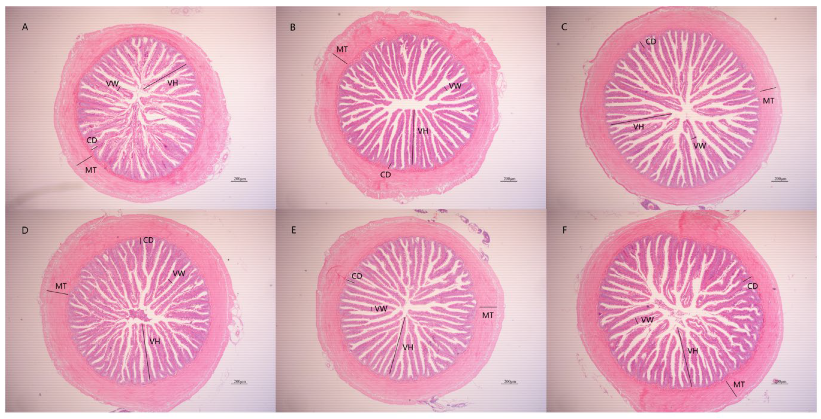
3.3. The Effects of PPs on Intestinal Tissue Morphology

The effects of PPs on intestinal tissue morphology are shown in Figure 1 and Table 5. Compared with the control group, PPs significantly increased villus height and significantly decreased villus width when the PP supplementation level was 12 g/kg, the villus height was the highest, and the villus width was the lowest (p < 0.05). Compared with the control group, PPs had no significant effects on the depth of the intestinal crypt and the thickness of the intestinal muscular (p > 0.05).

3.4. The Effects of PPs on Intestinal Microbes

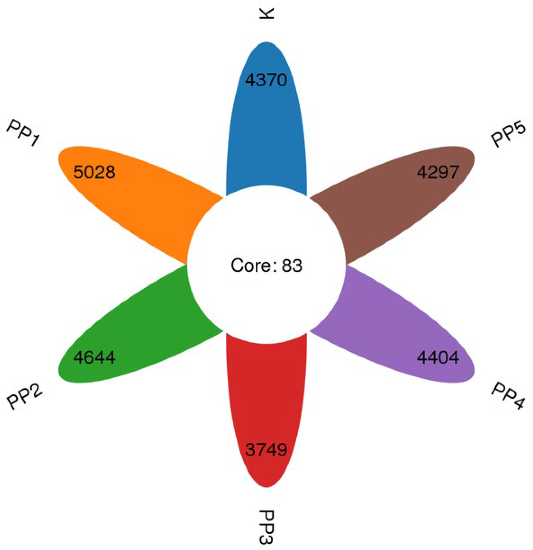
3.4.1. The Effects of PPs on Intestinal Microbial OTUs

A total of 1,440,449 sequences were obtained from the spotted sea bass intestinal flora. After being quality filtered and DADA2 denoised, 1,273,402 high-quality sequences were obtained. A total of 28,111 OTU homologous alignments were identified upon screening unique representative sequences. The Venn diagram shows the number of specific OTUs. As shown in Figure 2, 4453, 5111, 4727, 3832, 4487, and 4380 OTUs were detected in K, PP1, PP2, PP3, PP4, and PP5, respectively. The specific OTUs of each group were 4370, 5028, 4644, 3749, 4404, and 4297. The total of mutual OTUs was 83.

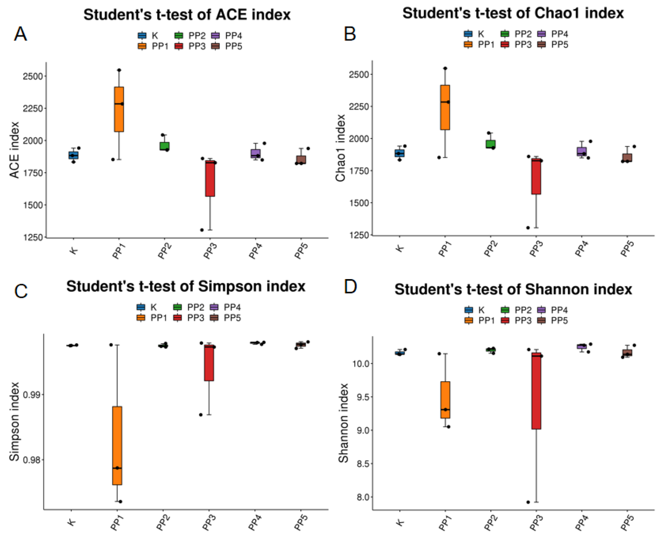
3.4.2. The Effects of PPs on Intestinal Microbial Alpha Diversity

The effects of PPs on the alpha diversity of intestinal microbes in spotted sea bass are shown in Figure 3 and Table 6. Compared with the control group, PPs increased the ACE and Chao1 indices of intestinal microbes of spotted sea bass. When the PP supplementation level was 3 g/kg, ACE and Chao1 indices were significantly higher than the control group (p < 0.05). In addition, when the PP supplementation level was 3 g/kg, the Simpson index was significantly lower than the control group (p < 0.05). Compared with the control group, there was no significant change in the Shannon index in the experimental groups (p > 0.05).

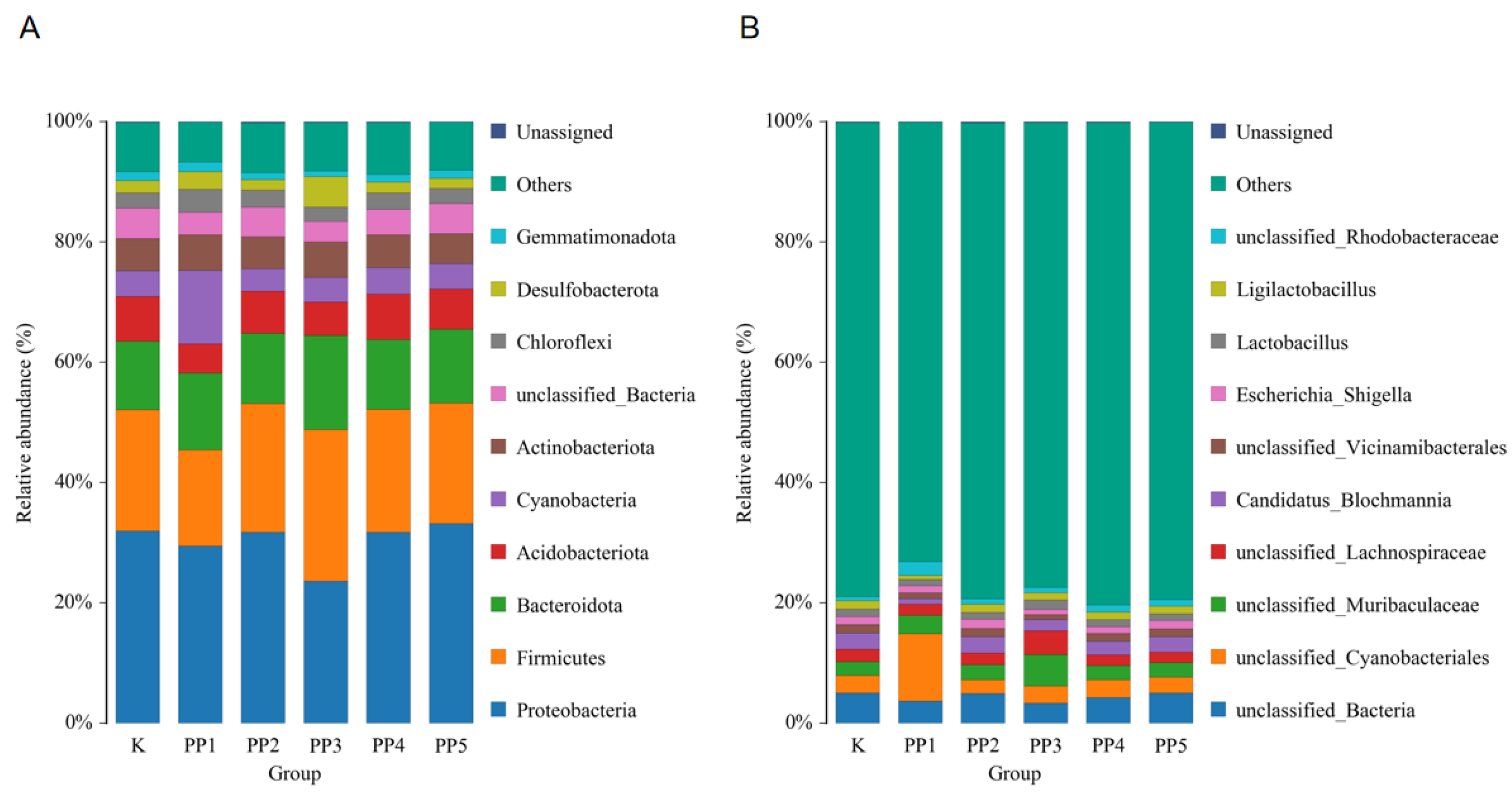
3.4.3. The Effects of PPs on Species Composition and Abundance of Intestinal Microbes

There were 51 phyla, 129 classes, 390 orders, 865 families, and 2032 genera in the intestinal microbes of spotted sea bass. The top 10 phyla based on relative abundance in each group were classified, and their composition was analyzed. At the phylum level (Figure 4A), Proteobacteria, Firmicutes, Bacteroides, and Acidobacteria were the main flora in the intestinal tract of spotted sea bass. Compared with the control group, PPs increased the abundance of Firmicutes and Bacteroides. At the genus level (Figure 4B), Cyanobacteria, Muribaculaceae, and Lachnospiraceae were the main flora in the intestinal tract of spotted sea bass. Compared with the control group, PPs increased the abundance of Cyanobacteria, Muribaculaceae, and Lachnospiraceae.

4. Discussion

WG, SGR, and CF can intuitively reflect the growth performance of the body. In this study, PPs increased WG, SGR, and CF of spotted sea bass, and they promoted the growth of spotted sea bass. In addition, PPs did not significantly affect the FCR. Previous studies found that 3 g/kg and 5 g/kg PPs could promote the WG and SGR of grass carp (Ctenopharyngodon idella), which promoted the growth of these fish [31]. However, in this study, the growth performance of fish was not proportional to the concentration of PPs added. Supplemental levels of PPs did not further improve the growth of spotted sea bass compared to lower levels. Polysaccharides can cause immune fatigue in the body [32]. In this study, the reason that 15 g/kg PPs could not improve the growth indices well may be that the high dose of polysaccharide caused immune fatigue and had adverse effects on growth indices. Therefore, the higher supplementation levels of PPs did not better promote the growth performance of spotted sea bass. In conclusion, PPs have the function of promoting the growth of spotted sea bass.
The intestinal tract is an important organ for digestion and absorption of nutrients, which affects the body’s growth and immunity [33]. Spotted sea bass are carnivorous fish, and protein is their main energy source [34]; starch can only be used as a small part of their energy intake. AMS can be involved in the hydrolysis of starch and glycogen, and it can provide a part of the energy for spotted sea bass. LPS can hydrolyze triglycerides and release free fatty acids and monoacylglycerol for tissue absorption or storage in adipose tissue [35]. TRS is a serine proteolytic enzyme that breaks down proteins in food into amino acids for cell absorption [36]. Therefore, the activity of digestive enzymes in the intestine is closely related to the healthy growth of the organism. Studies have shown that 3 g/kg PPs significantly increased the activity of intestinal AMS and TRS in grass carp [31]. In this study, 9 g/kg PPs significantly increased the AMS activity, and 12 g/kg PPs significantly increased the TRS activity. These results indicate that the effects of PPs on the digestive enzyme activities of fish with different feeding habits are different, and it also indicates that PPs have a promoting effect on the digestive enzyme activities of spotted sea bass. Intestinal digestive enzymes break down carbohydrates, fats, and proteins in food into small molecules that can be absorbed by the intestine. Spotted sea bass are carnivorous fish, and protein is their main energy intake. Therefore, PPs can promote intestinal digestive function by promoting intestinal TRS activity in spotted sea bass. In conclusion, PPs can increase the activity of AMS and TRS in the intestinal tract of spotted sea bass. The reason for this may be related to the growth-promoting effect of PPs and the dilution of amylase and protease by PPs. Thus, high expression of intestinal digestive enzymes was induced and promoted the intestinal digestive function, which further enhanced the growth of these fish.
The body’s defense system is closely related to its antioxidant capacity. Reactive oxygen species (ROS) exist widely in the body and can participate in the immune regulation of the body [37]. When the body is subjected to harmful stimuli, the increase in free radicals in the body or the weakening of antioxidant capacity leads to an imbalance in the oxidation–antioxidant system [38], thus accumulating excessive reactive oxygen species, leading to oxidative damage [39]. CAT is an antioxidant enzyme widely existing in organisms, which can protect the stable internal environment of the body and the normal function of cells [40]. In addition, there are also non-enzymatic antioxidants, such as GSH and MDA. GSH is a very powerful molecule with various functions, such as an antioxidant, regulator of DNA synthesis and repair, and protector of mercaptan groups in proteins [41]. MDA is a product of lipid oxidation [42], and the content of MDA in the body can reflect the antioxidant capacity of organisms to a certain extent [43]. The results showed that PPs significantly increased the content of GSH and decreased the content of MDA, while CAT activity did not change significantly. Studies have shown that PPs have reducing power and free radical scavenging ability, and high sulfate content in PPs is beneficial to their antioxidant activity [44]. Different extraction methods can lead to different purities of PPs. PPs show DPPH and OH free radical scavenging ability, which are significantly improved with an increase in the concentration and purity of PPs [45]. In this study, the level of GSH in the intestinal tract of spotted sea bass reached the highest level at a PPs concentration of 9 g/kg but decreased with an increase in PPs concentration. The reason for this may be that higher concentrations of PPs did not have a favorable effect on the intestinal antioxidant capacity of spotted sea bass. PPs can remove excessive free radicals, peroxides, and other reactive oxygen species in the intestine by increasing the GSH content in the intestine of spotted sea bass and preventing the damage caused by reactive oxygen species to intestinal cells, thus, maintaining the normal function of intestinal cells and improving the antioxidant capacity of the intestinal tract. The decrease in MDA content in the intestinal tract also indicates that PPs reduce lipid oxidation in the intestinal tract to a certain extent. This is consistent with the results of the effects of PPs on the antioxidant capacity of hyperlipidemia mice [46]. The results indicate that PPs can improve the antioxidant capacity of the intestinal tract of spotted sea bass by regulating the contents of GSH and MDA, thus improving intestinal health.
The intestinal tract is the largest surface in the body facing the external environment. It is in direct contact with symbiotic microorganisms and dietary antigens and plays an important role in separating intestinal materials and preventing the invasion of pathogenic substances [47]. Normal intestinal morphology is the basis for ensuring intestinal function and maintaining intestinal homeostasis [48,49]. It has been reported that the increased villus height of the intestine will increase the area of intestinal contact with nutrients, thus enhancing the absorption of nutrients by the intestine, so the morphology of the intestinal villus is related to the growth and development of the body [50]. The crypt depth reflects the proliferation rate and maturity of crypt cells. The results of this study showed that PPs significantly increased intestinal villus length and crypt depth and decreased villus width, while there was no significant effect on muscular thickness. PPs promote intestinal digestive enzyme activity and antioxidant capacity, which has a favorable effect on the development of intestinal morphology. Therefore, PPs improved intestinal morphology to some extent. In addition, as can be seen from Figure 3, the number of intestinal villi in the control group was small, and their morphology was short and thick, while the number of intestinal villi in the experimental groups was large, and their morphology was thin and long. In conclusion, PPs can improve the intestinal tissue structure and intestinal health of spotted sea bass to a certain extent.
The intestinal improvement effects of polysaccharides were closely related to their effects on intestinal flora [51,52]. The structure of fish intestinal microbes is affected by the environment and feed [53]. The influence of the environment is mainly determined by the colonizing bacteria at the larval stage [54], while the influence of feed is determined by its composition and nutrient composition [55]. In this study, Venn analysis found that the common OTUs of each group was 83, which indicated that the core flora in the intestine of spotted sea bass was not much. In terms of alpha diversity, ACE and Chao1 determined that OTUs abundance, the Shannon and Simpson indexes were correlated with population diversity in the samples [56,57]. In this study, the ACE and Chao1 indexes reached their maximum when the added amount of PPs was 3 g/kg and showed a decreasing trend with an increase in PPs, which indicated that the richness of intestinal microorganisms decreased with an increase in PP supplementation level. Intestinal microbiota richness is very important for fish, but its impact on fish should be judged according to the number of beneficial or harmful bacteria. The diversity indexes (Simpson and Shannon) are inversely proportional to the diversity of microbial species. When the PP supplementation level was 3 g/kg, the Simpson index was significantly lower than that of the control group, and there was no significant change upon an increase in PP supplementation level. In addition, the Shannon index did not change significantly. It has been reported that when PPs are used as a carbon source for intestinal microbiota, PPs with a high total sugar content have good effects in promoting intestinal microbial diversity [58]. The purity of PPs used in this experiment was 50%, and the sugar content was not high, which may have contributed to the lack of significant changes in the intestinal microbial diversity index of the experimental groups. In conclusion, PPs can improve the intestinal microbial abundance of spotted sea bass, but the effects of PPs on the intestinal microbial diversity of spotted sea bass are not significant.
The species, richness, and proportion of intestinal microbes can affect intestinal health and nutrition utilization, and dominant intestinal flora plays a leading role in improving intestinal functions [59]. Some bacterial species may interact with host metabolism through metabolite-mediated stimulation of intestinal hormones and other systems outside the gastrointestinal tract (such as the endocannabinoid system), leading to a decline in the richness of dominant species of intestinal flora, which is ultimately reflected in the deterioration of metabolic health of the host [60]. As prebiotics, polysaccharides can regulate the balance between probiotics and opportunistic pathogens, thus balancing intestinal flora [61]. On the one hand, polysaccharides are substances that cannot be digested by the host, but they can be converted into short-chain fatty acids by beneficial bacteria, which play a role in optimizing the microecology. On the other hand, the proliferation of beneficial bacteria preferentially occupies the bacterial colony niche in the intestinal tract, thus inhibiting the colonization and growth of opportunistic bacteria [62]. In this study, the relative abundance of some opportunistic pathogens varied. When the PP supplementation level was 9 g/kg, the relative abundance of proteobacteria was lower than in the control group, whereas no significant changes were observed in the other experimental groups. The relative abundance of Cyanobacteria at the genus level was higher than that in the blank group when the PP supplementation level was 3 g/kg, while the relative abundance of Cyanobacteria in other experimental groups did not change significantly. Cyanobacteria are a kind of harmful bacteria, and their metabolites, cyanobacterial toxins, can seriously affect the health of the body [63]. The digestive enzyme activities and antioxidant capacity of fish did not change significantly when the PP supplementation level was 3 g/kg. The reason may be that cyanobacteria inhibit the antioxidant capacity of the intestine and further inhibit the intestinal digestive function. In addition, the abundance of probiotics was up-regulated. For example, Firmicutes and Bacteroides are at the level of phylum, and Muribaculaceae and Lachnospiraceae are at the level of genus. The results of this study showed that PPs reduced the abundance of some opportunistic pathogens and increased the appreciation of some probiotics. The reason may be that PPs are used by beneficial bacteria, which leads to an increase in beneficial bacteria and the first step to occupy the niche, and then leads to a reduction in harmful bacteria. Studies have shown that seaweed extract improved the intestinal flora composition of Atlantic salmon (Salmo salar), increased the abundance of beneficial bacteria, and decreased the abundance of harmful bacteria [64], and two seaweed polysaccharides improved the intestinal flora structure of mice [65], which is consistent with the results of this study. In conclusion, PPs can improve the intestinal microbial structure of spotted sea bass.

5. Conclusions

PPs are seaweed polysaccharides with various biological activities. Different concentrations of PPs were added to the feed to explore their effects on the intestinal health of spotted sea bass. The results showed that PPs improved growth performance. PPs improved intestinal digestive enzyme activities, intestinal antioxidant capacity, intestinal flora diversity and richness, and intestinal tissue structure in spotted sea bass. PPs promoted amylase and trypsin activities in the intestinal tract and improved the digestive capacity of spotted sea bass. PPs increased the GSH content and decreased the MDA content in the intestine, indicating that PPs increased the antioxidant capacity of the intestine to a certain extent. PPs improved the morphology of intestinal tissue and increased the area in the intestinal inner wall, which was conducive to the digestion and absorption of nutrients in the intestine. In addition, PPs improved the diversity of gut microbes and the richness of dominant bacteria. In conclusion, PPs improved the intestinal health of spotted sea bass, which enriched the research focus area of PPs in the culture of these fish and provided a theoretical basis for using seaweed polysaccharides as a feed additive. Therefore, PPs have broad prospects as feed additives for aquatic animals.

Supplementary Materials

The following supporting information can be downloaded at: https://www.mdpi.com/article/10.3390/fishes8080419/s1, Figure S1: CF histogram, Figure S2: CF Q-Q plot, Figure S3: FCR histogram, Figure S4: FCR Q-Q plot, Figure S5: SGR histogram, Figure S6: SGR Q-Q plot, Figure S7: WG histogram, Figure S8: WG Q-Q plot, Figure S9: AMS histogram, Figure S10: AMS Q-Q plot, Figure S11: LPS histogram, Figure S12: LPS Q-Q plot, Figure S13: TRS histogram, Figure S14: TRS Q-Q plot, Figure S15: CAT histogram, Figure S16: CAT Q-Q plot, Figure S17: GSH histogram, Figure S18: GSH Q-Q plot, Figure S19: MDA histogram, Figure S20: MDA Q-Q plot.

Author Contributions

Conceptualization, H.L.; methodology, H.L.; software, H.L.; validation, H.L.; formal analysis, H.L.; investigation, H.L., Z.H., S.Z., J.M., L.K., Y.L., Z.L. (Zhongying Long), H.Q., L.L. and Y.Z.; resources, Z.L. (Zhongbao Li); data curation, H.L.; writing—original draft preparation, H.L.; writing—review and editing, H.L.; visualization, H.L.; supervision, Z.L. (Zhongbao Li); project administration, Z.L. (Zhongbao Li); funding acquisition, Z.L. (Zhongbao Li). All authors have read and agreed to the published version of the manuscript.

Funding

This research was funded by Science and Technology Planning Project in Fujian, China (Grant No. 2015N0010) and Science and Technology Planning Project in Xiamen, China (Grant No. 3502Z20143017).

Institutional Review Board Statement

All procedures of this study are subject to the Regulations on the Management of Experimental Animals (National Animal Control Regulations of China No. 11, Law and Regulation of The State Council No. 676 amended on 1 March 2017), which has been approved by the Ethics Committee of Animal Experiments of Jimei University (Xiamen, China), approval code: JMU202103009.

Informed Consent Statement

Not applicable.

Data Availability Statement

The data presented in this study are available in the article. The data presented in this study are available in the Supplementary Materials.

Acknowledgments

The authors are thankful to Jimei University (Xiamen, China) for providing the test site and the Beijing Biomarker Biotechnologies Co., Ltd. (Beijing, China) for providing technical services.

Conflicts of Interest

The authors have no conflict of interest.

References

  1. Julia, K.; Jerry, W.; Cani, P.D.; García-Ródenas, C.L.; Tom, M.; Annick, M.; Jacqueline, W.; Freddy, T.; Robert-Jan, B. Human intestinal barrier function in health and disease. Clin. Transl. Gastroenterol. 2016, 7, 196. [Google Scholar]
  2. Abreu, A.M.; Masayuki, F.; Moshe, A. TLR signaling in the gut in health and disease. J. Immunol. 2005, 174, 4453–4460. [Google Scholar] [CrossRef] [PubMed]
  3. Liu, X.; Chang, X.; Wu, H.; Xiao, J.; Gao, Y.; Zhang, Y. Role of intestinal inflammation in predisposition of Edwardsiella tarda infection in zebrafish (Danio rerio). Fish Shellfish Immunol. 2014, 41, 271–278. [Google Scholar] [CrossRef] [PubMed]
  4. Liu, Z.; Liu, W.; Ran, C.; Hu, J.; Zhou, Z. Abrupt suspension of probiotics administration may increase host pathogen susceptibility by inducing gut dysbiosis. Sci. Rep. 2016, 6, 23214. [Google Scholar] [CrossRef] [PubMed] [Green Version]
  5. Yang, H.; Zou, S.; Zhai, L.; Wang, Y.; Zhang, F.; An, L.; Yang, G. Pathogen invasion changes the intestinal microbiota composition and induces innate immune responses in the zebrafish intestine. Fish Shellfish Immunol. 2017, 71, 35–42. [Google Scholar] [CrossRef]
  6. Sayyaf, D.B.; Castaldelli, G.; Giari, L. Histopathological and ultrastructural assessment of two mugilid species infected with myxozoans and helminths. J. Fish Dis. 2018, 41, 299–307. [Google Scholar] [CrossRef]
  7. Jin, Y.; Wu, S.; Zeng, Z.; Fu, Z. Effects of environmental pollutants on gut microbiota. Environ. Pollut. 2017, 222, 1–9. [Google Scholar] [CrossRef]
  8. Reinoso, W.C.; Koboziev, I.; Furr, K.L.; Grisham, M.B. Protective and pro-inflammatory roles of intestinal bacteria. Pathophysiology 2016, 23, 67–80. [Google Scholar] [CrossRef]
  9. Rombout, J.H.; Abelli, L.; Picchietti, S.; Scapigliati, G.; Kiron, V. Teleost intestinal immunology. Fish Shellfish Immunol. 2011, 31, 616–626. [Google Scholar] [CrossRef] [Green Version]
  10. Gough, E.K. The impact of mass drug administration of antibiotics on the gut microbiota of target populations. Infect. Dis. Poverty 2022, 11, 76. [Google Scholar] [CrossRef]
  11. Sun, S.; Korheina, D.K.A.; Fu, H.; Ge, X. Chronic exposure to dietary antibiotics affects intestinal health and antibiotic resistance gene abundance in oriental river prawn (Macrobrachium nipponense), and provokes human health risk. Sci. Total Environ. 2020, 720, 137478. [Google Scholar] [CrossRef] [PubMed]
  12. Yin, S.; Sun, C.; Ji, Y.; Abdolmaleky, H.; Zhou, J.; Wang, S.; Bao, C. Herbal medicine improves gastrointestinal health in mice via modulation of intestinal tight junctions and gut microbiota and inhibition of inflammation. Biomed. Pharmacother. 2021, 138, 111426. [Google Scholar] [CrossRef] [PubMed]
  13. Bao, N.; Chen, F.; Dai, D. The regulation of host intestinal microbiota by polyphenols in the development and prevention of chronic kidney disease. Front. Immunol. 2019, 10, 2981. [Google Scholar] [CrossRef] [PubMed] [Green Version]
  14. Zhuang, Y.; Huang, H.; Liu, S.; Liu, F.; Tu, Q.; Yin, Y.; He, S. Resveratrol improves growth performance, intestinal morphology, and microbiota composition and metabolism in mice. Front. Microbiol. 2021, 12, 726878. [Google Scholar] [CrossRef]
  15. Layla, A.N.; Aaron, K. Soy isoflavones and gastrointestinal health. J. Curr. Nutr. Rep. 2020, 9, 193–201. [Google Scholar]
  16. Feng, Y.Q.; Song, Y.T.; Zhou, J.; Duan, Y.Q.; Kong, T.Y.; Ma, H.L.; Zhang, H.H. Recent progress of Lycium barbarum polysaccharides on intestinal microbiota, microbial metabolites and health: A review. Crit. Rev. Food Sci. Nutr. 2022, 28, 21–24. [Google Scholar]
  17. Song, B.; Zheng, C.; Zha, C.; Hu, S.; Yang, X.; Wang, L.; Xiao, H. Dietary leucine supplementation improves intestinal health of mice through intestinal SIgA secretion. J. Appl. Microbiol. 2020, 128, 574–583. [Google Scholar] [CrossRef]
  18. Song, Q.; Wang, Y.; Huang, L.; Shen, M.; Yu, Y.; Yu, Q.; Chen, Y.; Xie, J.H. Review of the relationships among polysaccharides, gut microbiota, and human health. Food Res. Int. 2020, 140, 109858. [Google Scholar] [CrossRef]
  19. Lai, Y.; Fang, Q.; Guo, X.; Lei, H.; Zhou, Q.; Wu, N.; Song, C. Effect of polysaccharides from Dictyophora indusiata on regulating gut microbiota and short-chain fatty acids in mice. J. Food Meas. Charact. 2022, 17, 1–11. [Google Scholar] [CrossRef]
  20. Sun, Y.; Meng, X.; Hu, X.; Liu, R.; Zhao, Z.; Wang, S.; Zhang, R.; Guo, K.; Luo, L. Dietary supplementation with Lycium barbarum polysaccharides conducive to maintaining the health of Luciobarbus capito via the enhancement of enzyme activities and the modulation of gut microbiota. Int. J. Biol. Macromol. 2023, 232, 123500. [Google Scholar] [CrossRef]
  21. Zuo, Z.; Wang, S.; Wang, Q.; Wang, D.; Wu, Q.; Xie, S.; Zou, J. Effects of partial replacement of dietary flour meal with seaweed polysaccharides on the resistance to ammonia stress in the intestine of hybrid snakehead (Channa maculatus female × Channa argus male). Fish Shellfish Immunol. 2022, 127, 271–279. [Google Scholar] [CrossRef]
  22. Jyotsna; Vijayakumar, P.; Dhas, S.T.; Mani, R.; Raguraman, V. Antiviral activity of sulfated polysaccharides from Sargassum ilicifolium against fish Betanodavirus infection. Aquac. Int. 2021, 29, 1049–1067. [Google Scholar] [CrossRef]
  23. Rajendran, P.; Subramani, A.P.; Michael, D. Polysaccharides from marine macroalga, Padina gymnospora improve the nonspecific and specific immune responses of Cyprinus carpio and protect it from different pathogens. Fish Shellfish. Immunol. 2016, 58, 220–228. [Google Scholar] [CrossRef] [PubMed]
  24. Chen, X.; Sun, Y.; Hu, L.; Liu, S.; Yu, H.; Xing, R.; Li, R.; Wang, X.; Li, P. In vitro prebiotic effects of seaweed polysaccharides. J. Oceanol. Limnol. 2018, 36, 926–932. [Google Scholar] [CrossRef]
  25. Hasan, A.M.B.; Tuan, N.T.; Zhang, Y.; Hu, H.; Lin, H.; Zhang, M.; Liang, H.; Zhang, Y.; Li, S. Effects of dietary supplementation of Gracilaria lemaneiformis-derived sulfated polysaccharides on the growth, antioxidant capacity, and innate immunity of rabbitfish (Siganus canaliculatus). Fish Shellfish. Immunol. 2023, 139, 108933. [Google Scholar]
  26. Peixoto, J.M.; Salas-Leitón, E.; Pereira, F.L.; Queiroz, A.; Magalhães, F.; Pereira, R.; Abreu, H.; Reis, P.A.; Gonçalves, J.F.M.; Ozório, R.O.A. Role of dietary seaweed supplementation on growth performance, digestive capacity and immune and stress responsiveness in European seabass (Dicentrarchus labrax). Aquac. Rep. 2016, 3, 189–197. [Google Scholar] [CrossRef] [Green Version]
  27. Abdelrhman, A.M.; Mohamed, A.; Al-Zahaby, M.A.; Sharawy, Z.Z.; Nazmi, H.; Zaki, M.A.A.; Ahmed, N.H.; Ahmed, S.R.; El-Haroun, E.; Van, D.H.; et al. Effect of polysaccharides derived from brown macroalgae Sargassum dentifolium on growth performance, serum biochemical, digestive histology and enzyme activity of hybrid red tilapia. Aquac. Rep. 2022, 25, 101212. [Google Scholar] [CrossRef]
  28. Yoshida, T.; Notoya, M.; Kikuchi, N.; Miyata, M. Catalogue of species of Porphyra in the world, with special reference to the type locality and bibliography. J. Nat. Hist. Res. 1997, 3, 5–18. [Google Scholar]
  29. Lahaye, M.; Jegou, D. Chemical and physical-chemical characteristics of dietary fibres from Ulva lactuca (L.) Thuret and Enteromorpha compressa (L.) Grev. J. Appl. Phycol. 1993, 5, 195–200. [Google Scholar] [CrossRef]
  30. Geng, Y. Structure and bioactivities of Porphyrans and Oligoporphyrans. Curr. Pharm. Des. 2019, 25, 1163–1171. [Google Scholar] [CrossRef]
  31. Chen, L.; Zhang, Y. The growth performance and nonspecific immunity of juvenile grass carp (Ctenopharyngodon idella) affected by dietary Porphyra yezoensis polysaccharide supplementation. Fish Shellfish Immunol. 2019, 87, 615–619. [Google Scholar] [CrossRef]
  32. Yu, W.; Yang, Y.; Zhou, Q.; Huang, X.; Huang, Z.; Li, T.; Wu, Q.; Zhou, C.; Ma, Z.; Lin, H. Effects of dietary Astragalus polysaccharides on growth, health and resistance to Vibrio harveyi of Lates calcarifer. Int. J. Biol. Macromol. 2022, 207, 850–858. [Google Scholar] [CrossRef]
  33. Martin, S.A.M.; Dehler, C.E.; Król, E. Transcriptomic responses in the fish intestine. Dev. Comp. Immunol. 2016, 64, 103–117. [Google Scholar] [CrossRef] [PubMed]
  34. Ai, Q.; Mai, K.; Li, H.; Zhang, C.; Zhang, L.; Duan, Q.; Tan, B.; Xu, W.; Ma, H.; Zhang, W.; et al. Effects of dietary protein to energy ratios on growth and body composition of juvenile Japanese seabass, Lateolabrax japonicus. Aquaculture 2003, 230, 507–516. [Google Scholar] [CrossRef]
  35. Young, S.G.; Fong, L.G.; Beigneux, A.P.; Allan, C.M.; He, C.; Jiang, H.; Nakajima, K.; Meiyappan, M.; Birrane, G.; Ploug, M. GPIHBP1 and lipoprotein lipase, partners in plasma triglyceride metabolism. Cell Metab. 2019, 30, 51–65. [Google Scholar] [CrossRef] [PubMed]
  36. Zvereva, E.A.; Zaichik, B.T.; Eremin, S.A.; Zherdev, A.V.; Dzantiev, B.B. Enzyme immunoassay for detection of Sudan I dye and its application to the control of foodstuffs. J. Anal. Chem. 2016, 71, 944–948. [Google Scholar] [CrossRef]
  37. Nauseef, W.M. Nox enzymes in immune cells. Semin. Immunopathol. 2008, 30, 195–208. [Google Scholar] [CrossRef]
  38. Pisoschi, A.M.; Pop, A. The role of antioxidants in the chemistry of oxidative stress: A review. Eur. J. Med. Chem. 2015, 97, 55–74. [Google Scholar] [CrossRef] [PubMed]
  39. Emanuele, S.; Calvaruso, G.; Lauricella, M.; Giuliano, M.; Bellavia, G.; D’Anneo, A.; Vento, R.; Tesoriere, G. Apoptosis induced in hepatoblastoma HepG2 cells by the proteasome inhibitor MG132 is associated with hydrogen peroxide production, expression of Bcl-XS and activation of caspase-3. Int. J. Oncol. 2002, 21, 857–865. [Google Scholar] [CrossRef]
  40. Christophe, G.; Buc, C.P. Catalase, a remarkable enzyme: Targeting the oldest antioxidant enzyme to find a new cancer treatment approach. Biol. Chem. 2017, 398, 1095–1108. [Google Scholar]
  41. Iskusnykh, I.Y.; Zakharova, A.A.; Dhruba, P. Glutathione in brain disorders and aging. Molecules 2022, 27, 324. [Google Scholar] [CrossRef]
  42. Wang, Z.; He, Z.; Emara, A.M.; Gan, X.; Li, H. Effects of malondialdehyde as a byproduct of lipid oxidation on protein oxidation in rabbit meat. Food Chem. 2019, 288, 405–412. [Google Scholar] [CrossRef] [PubMed]
  43. Tsikas, D. Assessment of lipid peroxidation by measuring malondialdehyde (MDA) and relatives in biological samples: Analytical and biological challenges. Anal. Biochem. 2016, 524, 13–30. [Google Scholar] [CrossRef]
  44. Wu, Y.; Huo, Y.; Xu, L.; Xu, Y.; Wang, X.; Zhou, T. Purification, characterization and antioxidant activity of polysaccharides from Porphyra haitanensis. Int. J. Biol. Macromol. 2020, 165, 2116–2125. [Google Scholar] [CrossRef]
  45. Zheng, M.; Ma, M.; Yang, Y.; Liu, Z.; Liu, S.; Hong, T.; Ni, H.; Jiang, Z. Structural characterization and antioxidant activity of polysaccharides extracted from Porphyra haitanensis by different methods. Int. J. Biol. Macromol. 2023, 242, 125003. [Google Scholar] [CrossRef]
  46. Wang, X.; Li, W.; Xiao, L.; Liu, C.; Qi, H.; Zhang, Z. In vivo antihyperlipidemic and antioxidant activity of porphyran in hyperlipidemic mice. Carbohydr. Polym. 2017, 174, 417–420. [Google Scholar] [CrossRef]
  47. Schug, H.; Yue, Y.; Krese, R.; Fischer, S.; Kortner, T.M.; Schirmer, K. Time- and concentration-dependent expression of immune and barrier genes in the RTgutGC fish intestinal model following immune stimulation. Fish Shellfish Immunol. 2019, 88, 308–317. [Google Scholar] [CrossRef] [PubMed] [Green Version]
  48. Baker, R.; Buckland, A.; Sheaves, M. Fish gut content analysis: Robust measures of diet composition. Fish Fish. 2014, 15, 170–177. [Google Scholar] [CrossRef]
  49. Wu, N.; Song, Y.; Wang, B.; Zhang, X.; Zhang, X.; Wang, Y.; Cheng, Y.; Chen, D.; Xia, X.; Lu, Y.; et al. Fish gut-liver immunity during homeostasis or inflammation revealed by integrative transcriptome and proteome studies. Sci. Rep. 2016, 6, 36048. [Google Scholar] [CrossRef] [PubMed] [Green Version]
  50. Caspary, W.F. Physiology and pathophysiology ofintes-tinal absorption. Am. J. Clin. Nutr. 1992, 55, 299–308. [Google Scholar] [CrossRef]
  51. Liu, A.; Kim, E.; Cui, J.; Li, J.; Lee, Y.; Zhang, G. Laminaria Japonica polysaccharide improved the productivities and systemic health of ducks by mediating the gut microbiota and metabolome. J. Agric. Food Chem. 2023, 71, 7382–7395. [Google Scholar] [CrossRef] [PubMed]
  52. Wu, Q.; Wu, S.; Cheng, Y.; Zhang, Z.; Mao, G.; Li, S.; Yang, Y.; Zhang, X.; Wu, M.; Tong, H. Sargassum fusiforme fucoidan modifies gut microbiota and intestinal metabolites during alleviation of hyperglycemia in type 2 diabetic mice. Food Funct. 2021, 12, 3572–3585. [Google Scholar] [CrossRef] [PubMed]
  53. Arthur, E.; Christophe, A.J.; Amandine, A.; Raphaël, S.; Antoine, G.; Lucie, B.; Sébastien, V. Ecological specialization within a carnivorous fish family is supported by a herbivorous microbiome shaped by a combination of gut traits and specific diet. Front. Mar. Sci. 2021, 8, 91. [Google Scholar]
  54. Tarnecki, A.M.; Burgos, F.A.; Ray, C.L.; Arias, C.R. Fish intestinal microbiome: Diversity and symbiosis unravelled by metagenomics. J. Appl. Microbiol. 2017, 123, 2–17. [Google Scholar] [CrossRef] [Green Version]
  55. Pérez, T.; Balcázar, J.L.; Ruiz-Zarzuela, I.; Halaihel, N.; Vendrell, D.; DeBlas, I.; Múzquiz, J.L. Host-microbiota interactions within the fish intestinal ecosystem. Mucosal Immunol. 2010, 3, 355–360. [Google Scholar] [CrossRef] [PubMed] [Green Version]
  56. Tsai, S.M.; Vazoller, R.F.; Pazinato, J.M.; Mendes, L.W.; Paulo, E.N. Molecular characterization of the archaeal community in an amazonian wetland soil and culture-dependent isolation of Methanogenic Archaea. Diversity 2010, 2, 1026–1047. [Google Scholar]
  57. Pélissier, R.; Couteron, P. An operational, additive framework for species diversity partitioning and Beta-diversity analysis. J. Ecol. 2007, 95, 294–300. [Google Scholar] [CrossRef]
  58. Chen, P.; Tong, M.; Zeng, H.; Zheng, B.; Hu, X. Structural characterization and in vitro fermentation by rat intestinal microbiota of a polysaccharide from Porphyra haitanensis. Food Res. Int. 2021, 147, 110546. [Google Scholar] [CrossRef]
  59. Li, L.; Liu, X.; Wang, Y.; Huang, Y.; Wang, C. Effects of alternate feeding between fish meal and novel protein diets on the intestinal health of juvenile largemouth bass (Micropterus salmoides). Aquac. Rep. 2022, 23, 101023. [Google Scholar] [CrossRef]
  60. Elaine, p.; Ryan, P.M.; Cryan, J.F.; Dinan, T.G.; Paul, R.R.; Fitzgerald, G.F.; Catherine, S. Gut microbiota, obesity and diabetes. Postgrad. Med. J. 2016, 92, 286–300. [Google Scholar]
  61. Cheng, W.; Lu, J.; Li, B.; Lin, W.; Zhang, Z.; Wei, X.; Sun, C.; Chi, M.; Bi, W.; Yang, B.; et al. Effect of functional oligosaccharides and ordinary dietary fiber on intestinal microbiota diversity. Front. Microbiol. 2017, 8, 1750. [Google Scholar] [CrossRef] [PubMed] [Green Version]
  62. Yukgehnaish, K.; Kumar, P.; Sivachandran, P.; Marimuthu, K.; Arshad, A.; Paray, B.A.; Arockiaraj, J. Gut microbiota metagenomics in aquaculture: Factors influencing gut microbiome and its physiological role in fish. Rev. Aquac. 2020, 12, 1903–1927. [Google Scholar] [CrossRef]
  63. Carmichael, W.W.; Boyer, L.G. Health impacts from cyanobacteria harmful algae blooms: Implications for the North American Great Lakes. Harmful Algae 2016, 54, 194–212. [Google Scholar] [CrossRef] [PubMed]
  64. Thépot, V.; Campbell, A.H.; Rimmer, M.A.; Jelocnik, M.; Johnston, C.; Evans, B.; Paul, N.A. Dietary inclusion of the red seaweed Asparagopsis taxiformis boosts production, stimulates immune response and modulates gut microbiota in Atlantic salmon, Salmo salar. Aquaculture 2022, 546, 737286. [Google Scholar] [CrossRef]
  65. Zhang, Z.; Wang, X.; Han, S.; Liu, C.; Liu, F. Effect of two seaweed polysaccharides on intestinal microbiota in mice evaluated by illumina PE250 sequencing. Int. J. Biol. Macromol. 2018, 112, 796–802. [Google Scholar] [CrossRef]
Figure 1. The effects of PPs on intestinal tissue morphology. (AF) are HE slices of spotted sea bass intestines from groups K, PP1, PP2, PP3, PP4, and PP5 under a 40× microscope, respectively. VH, VW, CD, and MT in the figure represent villus height, villus width, crypt depth, and muscular thickness, respectively.
Fishes 08 00419 g001
Figure 2. The effects of PPs on intestinal microbial OTUs. The center circle represents the common number of OTUs, and each petal of a different color represents the unique number of OTUs per group. Abbreviations: K, 0 g/kg PP; PP1, 3 g/kg PP; PP2, 6 g/kg PP; PP3, 9 g/kg PP; PP4, 12 g/kg PP; PP5, 15 g/kg PP.
Fishes 08 00419 g002
Figure 3. The effects of PPs on intestinal microbial alpha diversity. (AD) in the figure represent ACE index, Chao1 index, Simpson index and Shannon index, respectively.The abscissa is the group name, and the ordinate is the alpha diversity index. The meaning of each symbol in the figure is as follows: the upper and lower end lines of the box: interquartile range (IQR); median line: median; upper and lower margins: maximum and minimum internal circumference (1.5 times IQR); points outside the upper and lower edges: outliers. p > 0.05.
Fishes 08 00419 g003
Figure 4. The effects of PPs on species composition and abundance at the phylum (A) and genus (B) levels of the intestinal microbes of spotted sea bass. The abscissa is the group name; the ordinate is the relative abundance percentage; different colors indicate different species. Abbreviations: K, 0 g/kg PP; PP1, 3 g/kg PP; PP2, 6 g/kg PP; PP3, 9 g/kg PP; PP4, 12 g/kg PP; PP5, 15 g/kg PP.
Fishes 08 00419 g004
Table 1. Feed composition and nutrient level.
Ingredient (%)Content
Fish meal a49.0
Soybean meal a23.5
Wheat flour15.0
Yeast powder a3.0
Fish oil3.0
Soybean oil2.0
Lecithin1.0
Vitamin premix b0.6
Mineral premix c0.8
Choline0.6
Ca(H2PO4)21.2
Antioxidant0.3
Proximate composition (%)
Crude protein48.01
Crude fat8.60
Crude ash12.22
Carbohydrate10.80
Gross energy (kj/g)16.68
a Fish meal includes crude protein 67% and crude fat 8.4%, Yeast powder includes crude protein 42% and crude fat 3.8%, and soybean meal includes crude protein 44.1% and crude fat 1.8%. b Mineral premix contains: MgSO4·H2O 4000 mg/kg, CoCl2 (1%) 100 mg/kg, MnSO4·4H2O 50 mg/kg, CuSO4·5H2O 20 mg/kg, ZnSO4·H2O 150 mg/kg, FeSO4·H2O 260 mg/kg, Na2SeO3 (1%) 50 mg/kg, KI 100 mg/kg. c Vitamin premix contains Vitamin B12 0.1 mg/kg, Vitamin K3 10 mg/kg, vitamin A acetate 32 mg/kg, Vitamin D3 5 mg/kg, nicotinic acid 200 mg/kg, pyridoxine hydrochloride 20 mg/kg, pantothenic acid 60 mg/kg, biotin 1.2 mg/kg, riboflavin 45 mg/kg, folic acid 20 mg/kg, thiamine 25 mg/kg, ethoxyquin 150 mg/kg, α-tocopherol 120 mg/kg, inositol 800 mg/kg.
Table 2. The effects of PPs on growth performance.
GroupsGrowth Performance
WG (%)SGR (%)CF (%)FCR
K5.72 ± 0.06 a3.66 ± 0.02 a1.46 ± 0.09 a1.04 ± 0.02 a
PP16.63 ± 0.56 bc3.91 ± 0.14 bc1.79 ± 0.22 b0.99 ± 0.03 a
PP26.23 ± 0.05 ab3.80 ± 0.01 ab1.51 ± 0.07 a0.99 ± 0.05 a
PP37.07 ± 0.30 c4.01 ± 0.07 c1.57 ± 0.09 a1.00 ± 0.04 a
PP47.13 ± 0.32 c4.03 ± 0.08 c2.04 ± 0.05 c1.00 ± 0.05 a
PP56.74 ± 0.41 bc3.93 ± 0.10 bc1.78 ± 0.06 b1.01 ± 0.09 a
The results are expressed as mean ± standard deviation from triplicate groups (n = 3), and data values with different superscript letters in the same column were significantly different (p < 0.05). Abbreviations: WG, weight gain; SGR, special growth rate; CF, condition factor; FCR, feed conversion ratio; K, 0 g/kg PP; PP1, 3 g/kg PP; PP2, 6 g/kg PP; PP3, 9 g/kg PP; PP4,12 g/kg PP; PP5, 15 g/kg PP.
Table 3. The effects of PPs on the activity of intestinal digestive enzymes.
GroupsDigestive Enzymes
AMS (U/mgprot)LPS (U/gprot)TRS (U/mgprot)
K0.45 ± 0.18 a1.23 ± 0.42 a758.31 ± 78.34 a
PP10.55 ± 0.09 a1.05 ± 0.39 a1136.13 ± 228.34 ab
PP20.66 ± 0.08 ab1.15 ± 0.36 a833.00 ± 110.18 ab
PP30.91 ± 0.23 b1.58 ± 0.71 a1038.81 ± 569.55 ab
PP40.71 ± 0.12 ab1.47 ± 0.59 a1354.13 ± 310.48 b
PP50.69 ± 0.07 ab1.27 ± 0.30 a719.37 ± 123.00 a
The results are expressed as mean ± standard deviation from triplicate groups (n = 3), and data values with different superscript letters in the same column were significantly different (p < 0.05). Abbreviations: AMS, amylase; LPS, lipase; TRS, trypsin; K, 0 g/kg PP; PP1, 3 g/kg PP; PP2, 6 g/kg PP; PP3, 9 g/kg PP; PP4,12 g/kg PP; PP5, 15 g/kg PP.
Table 4. The effects of PPs on intestinal antioxidant capacity.
GroupsAntioxidant Capacity
CAT (μmol/gprot)GSH (U/mgprot)MDA (nmol/mgprot)
K1.11 ± 0.03 a31.84 ± 1.21 a0.79 ± 0.32 b
PP10.88 ± 0.46 a33.73 ± 5.31 a0.38 ± 0.07 a
PP20.93 ± 0.24 a36.07 ± 5.01 ab0.49 ± 0.23 ab
PP30.91 ± 0.15 a44.20 ± 4.99 b0.37 ± 0.18 a
PP40.92 ± 0.13 a36.74 ± 6.38 ab0.47 ± 0.16 ab
PP50.82 ± 0.35 a32.37 ± 5.70 a0.55 ± 0.45 ab
The results are expressed as mean ± standard deviation from triplicate groups (n = 3), and data values with different superscript letters in the same column were significantly different (p < 0.05). Abbreviations: CAT, catalase; GSH, glutathione; MDA, malondialdehyde; K, 0 g/kg PP; PP1, 3 g/kg PP; PP2, 6 g/kg PP; PP3, 9 g/kg PP; PP4,12 g/kg PP; PP5, 15 g/kg PP.
Table 5. The effects of PPs on intestinal tissue morphology.
GroupsIntestinal Tissue Morphology
Villus Height
(μm)
Villus Width
(μm)
Crypt Depth
(μm)
Muscular Thickness (μm)
K549.28 ± 36.82 a69.51 ± 10.33 b86.85 ± 6.46 a229.48 ± 19.74 a
PP1597.72 ± 17.42 b66.60 ± 5.89 ab91.22 ± 7.94 a219.41 ± 10.75 a
PP2629.62 ± 22.14 bc66.38 ± 5.43 ab88.77 ± 5.54 a218.53 ± 20.03 a
PP3586.91 ± 59.97 ab60.81 ± 4.24 a90.87 ± 7.14 a220.76 ± 53.86 a
PP4647.89 ± 28.19 c59.55 ± 1.54 a91.25 ± 3.37 a208.37 ± 26.79 a
PP5606.72 ± 28.54 bc63.04 ± 2.34 ab86.98 ± 3.76 a212.56 ± 15.69 a
The results are expressed as mean ± standard deviation from triplicate groups (n = 3), and data values with different superscript letters in the same column were significantly different (p < 0.05). Abbreviations: K, 0 g/kg PP; PP1, 3 g/kg PP; PP2, 6 g/kg PP; PP3, 9 g/kg PP; PP4,12 g/kg PP; PP5, 15 g/kg PP.
Table 6. The effects of PPs on intestinal microbial alpha diversity.
GroupsAlpha Diversity
ACEChao1SimpsonShannon
K1885.67 ± 54.05 ab1885.67 ± 54.05 ab1.00 ± 0 b10.16 ± 0.04 a
PP12227.48 ± 350.49 b2227.34 ± 350.45 b0.98 ± 0.01 a9.50 ± 0.57 a
PP21965.33 ± 67.28 ab1965.33 ± 67.28 ab1.00 ± 0 b10.20 ± 0.04 a
PP31664.07 ± 311.40 a1664.00 ± 311.34 a0.99 ± 0.01 b9.41 ± 1.29 a
PP41903.00 ± 67.01 ab1903.00 ± 67.01 ab1.00 ± 0 b10.25 ± 0.06 a
PP51860.73 ± 67.08 ab1860.67 ± 66.97 ab1.00 ± 0 b10.17 ± 0.09 a
The results are expressed as mean ± standard deviation from triplicate groups (n = 3), and data values with different superscript letters in the same row were significantly different (p < 0.05). Abbreviations: K, 0 g/kg PP; PP1, 3 g/kg PP; PP2, 6 g/kg PP; PP3, 9 g/kg PP; PP4, 12 g/kg PP; PP5, 15 g/kg PP.
Disclaimer/Publisher’s Note: The statements, opinions and data contained in all publications are solely those of the individual author(s) and contributor(s) and not of MDPI and/or the editor(s). MDPI and/or the editor(s) disclaim responsibility for any injury to people or property resulting from any ideas, methods, instructions or products referred to in the content.

