Article

Copper-Induced Ionoregulatory Disturbance, Histopathology, and Transcriptome Responses in Freshwater Mussel (Anodonta woodiana) Gills

1
Laboratory of Fishery Microchemistry, Freshwater Fisheries Research Center, Chinese Academy of Fishery Sciences, Wuxi 214081, China
2
Key Laboratory of Integrated Rice-Fish Farming Ecology, Ministry of Agriculture and Rural Affairs, Freshwater Fisheries Research Center, Chinese Academy of Fishery Sciences, Wuxi 214081, China
3
Toxicology Centre, University of Saskatchewan, 44 Campus Drive, Saskatoon, SK S7N 5B3, Canada
*
Author to whom correspondence should be addressed.
Fishes 2023, 8(7), 368; https://doi.org/10.3390/fishes8070368
Submission received: 23 April 2023 / Revised: 13 July 2023 / Accepted: 13 July 2023 / Published: 14 July 2023
(This article belongs to the Section Environment and Climate Change)

Abstract

:
Copper (Cu) contamination has become a severe problem in freshwater environments worldwide. The freshwater mussel Anodonta woodiana is used as a unique bioindicator to monitor Cu contamination in freshwater environments. However, Cu toxicity and response mechanisms in A. woodiana are still largely unknown. A sublethal acute exposure experiment (2.0 mg/L Cu exposure for 72 h) was conducted to investigate the effects of Cu bioaccumulation on ionoregulatory homeostasis, histological features, and transcriptome responses using A. woodiana gills as indicator tissue. The gill bioaccumulation capacity was up to 474. Cu bioaccumulation decreased Na+ and Mg2+ concentrations (p < 0.05) by 82% and 17%, respectively, and induced cilia loss, epithelial desquamation, and filament atrophy of the gills. Transcriptome analysis identified 3160 differentially expressed genes (DEGs), including 1870 upregulated and 1290 downregulated genes. GO enrichment analysis showed that cellular processes, metabolic processes, biological regulation, and responses to stimuli contained the most DEGs in the biological processes. KEGG pathway analysis showed that apoptosis, arginine and proline metabolism, the toll-like receptor signaling pathway, apoptosis-multiple species, histidine metabolism, beta-alanine metabolism, cytokine–cytokine receptor interaction, and the p53 signaling pathway were significantly enriched. These findings provide comprehensive evidence for exploring Cu toxicity and response mechanisms in freshwater mussels.
Key Contribution: This study reveals the toxicity mechanism of Cu overaccumulation in freshwater mussel A. woodiana in terms of ionoregulatory disturbance, histopathology, and transcriptome responses.

1. Introduction

Copper (Cu) contamination has become a severe problem in freshwater environments worldwide owing to industrial emissions, mining discharge, agricultural activities, and fishery use (especially the use of copper sulfate (CuSO4) as an algicide and fungicide) [1,2]. For example, Cu concentrations in surface water could elevate to 1.2–23.8 mg/L in many countries, such as in the USA [3], China [4], Spain [5], and Nigeria [6], severely threatening ecological safety and human health. Cu is an essential metal that is important for numerous physiological processes in aquatic organisms, being a cofactor for oxidation/reduction reaction enzymes [7,8,9]. Cu is also involved in oxygen transport in the hemolymph as a component of hemocyanin in mussels [7,8]. However, elevated concentrations in water can be toxic by disrupting normal metabolic processes [7,8,9]. Notably, Cu is more toxic than other metals (Hg, Cd, Zn, Pb, and Cr) to freshwater organisms [10]. Invertebrates are generally more sensitive to Cu than vertebrates [10]. Freshwater mussels have been experiencing a serious population decline globally [11,12]. Cu stress remains one of the key factors contributing to their decline in many regions [11,12]. However, the toxic mechanisms of Cu in freshwater mussels are still not well understood. The lack of gene sequences of most freshwater mussels [13] is one of the important reasons. In recent years, high-throughput RNA sequencing (RNA-seq) without a reference genome sequence has enabled the transcriptome analysis of freshwater mussels exposed to Cu and will hopefully uncover its molecular mechanisms of toxicity [14].
The freshwater mussel Anodonta woodiana, commonly known as the Chinese pond mussel, is native to the Yangtze and Heilongjiang Rivers of China [15]; however, it has spread across Asia (Indonesia, the Philippines, Turkey, Malaysia, Vietnam, Singapore, Uzbekistan, South Korea, Laos, and Kazakhstan), Europe (Croatia, Montenegro, Russia, Hungary, France, Romania, Italy, Germany, Austria, Slovakia, Czech Republic, Poland, Serbia, Moldova, Ukraine, Sweden, the Netherlands, Belgium, Slovenia, and Bulgaria), and the Americas (Costa Rica, Dominica, America, and Haiti) over the past few decades [16]. This mussel plays an important role as an aquatic ecosystem engineer [16], a food source [17,18], and in the pearl culture industry [19]. Moreover, it has been successfully used as a unique bioindicator in the “Freshwater Mussel Watch” project [20,21,22]. To date, many countries (e.g., China, Poland, Serbia, Bulgaria) have utilized A. woodiana to biomonitor Cu contamination in freshwater environments [21,22,23,24,25]. However, Cu toxicity to A. woodiana has not been explored in addition to reporting the changes in their protein content [26] and antioxidant enzymes [27].
Among all the organs of mussels, gills are one of the focal points in toxicological studies. Mussel gills are vital organs involved in respiration and filtration [28,29]. They are able to accumulate higher concentrations of Cu than other organs [29,30,31] because of their high surface ratio and direct water contact with a high throughput of volume. Therefore, mussel gills have been proposed as an indicator tissue for studying metal toxicity [29,32].
Our previous study showed that the 96 h LC50 of Cu was 3.4 mg/L (95% confidence interval: 2.1–6.5 mg/L) for A. woodiana juveniles [27]. In this study, A. woodiana juveniles were exposed to 2.0 mg/L Cu (approximately 1/2 of the 96 h LC50) for 72 h to reveal the mechanism of Cu toxicity to the mussels in terms of ionoregulatory homeostasis (especially Na+, Mg2+, K+, and Ca2+), histological features, and transcriptome responses using the gills as indicator tissue. This sublethal acute exposure could reveal Cu toxic effects on mussels and avoid mussels’ adaptation to Cu exposure. We hypothesized that excessive Cu accumulation could cause ion loss, histological hazards, and disrupt gene expression. These results provide new insights into Cu toxicity mechanisms in freshwater mussels.

2. Materials and Methods

2.1. Mussels and Treatment

Juveniles of A. woodiana of similar sizes (shell lengths of 5.5 ± 0.3 cm and body weights (including shells and soft tissues) of 21.1 ± 2.0 g) were obtained from the Freshwater Fisheries Research Center, Chinese Academy of Fishery Sciences (Wuxi, China). After removing the epibionts, the mussels were acclimatized in American Society of Testing Materials (ASTM) reconstituted soft water [33] (pH 7.3–7.5, hardness 40–48 mg/L, alkalinity 30–35 mg/L as CaCO3) at 20 ± 1 °C with a 16:8 h light and dark photoperiod for 2 weeks. Cu exposure was carried out according to the ASTM standard guide for conducting laboratory toxicity tests with freshwater mussels [33]. Thirty mussels were randomly divided into control and Cu-treated groups using three replicate tanks (2.5 L solution per tank) with five mussels each. Based on the filtration rate (260 L/h/kg dry weight (d.w.) of soft tissue) of A. woodiana [34], it was estimated that it took approximately 7 h for the solution to be filtered by the mussels. The solution was continuously aerated during the experiment, and dissolved oxygen was higher than 7 mg/L. Cu was not detected (the detection limit is 0.3 μg/L) in the water of the control group. Mussels in the Cu-treated group were exposed to nominal 2.0 mg/L Cu in the form of CuSO4·5H2O (Sinopharm Chemical Reagent Co., Ltd., Shanghai, China) for 72 h. The mean measured concentration of Cu was 2.05 ± 0.1 mg/L. The Cu concentration used in this study was based on the sublethal concentration limit (1/2 of the 96 h LC50 of aqueous Cu) to A. woodiana juveniles [27]. Although this is a concentration characteristic of heavily contaminated natural waters and one that is not that often encountered in the environment, it may be suitable for investigating the mechanisms of acute toxicity. The short 72 h exposure could prevent mussels from adapting to Cu toxicity [30]. The test solutions were 100% renewed every 24 h to maintain Cu concentration stability and to reduce nitrogenous waste.

2.2. Copper Bioaccumulation and Ion Concentration Analyses

After 72 h of Cu exposure, three mussels from each group were randomly chosen, and their gills were dissected using stainless steel scalpels. The concentrations of total Cu and key ions (Na+, Mg2+, K+, and Ca2+) in the gills were determined according to established protocols with slight modifications [22,35]. Briefly, each sample was dried to a constant weight for 24 h at 80 °C, digested in HNO3 (Merck, 65%) with an ETHOS A T260 microwave digestion system (Milestone Inc., Milan, Italy), and analyzed using an Agilent 7500ce ICP-MS (Agilent Technologies, Tokyo, Japan). Concurrent measurements of certified reference material (DOLT-5 dogfish liver; National Research Council of Canada, Ottawa, ON, Canada) indicated that the recoveries of Cu and major ions ranged from 97% to 107%. Concentrations of Cu and ions in the gills were determined on a d.w. basis.

2.3. Histopathologic Analysis

Three mussels from each group were randomly selected for histopathological examination. Two transverse tissue sections and two longitudinal sections were prepared from each individual [36]. Gills were collected, and the samples were fixed using Bouin’s solution. The samples were subjected to alcohol gradation (70%, 80%, 90%, 95%, and 100%) before being embedded in paraffin blocks. Transverse sections of the gills and their longitudinal sections (5 μm thick) were cut and stained using hematoxylin and eosin. An Olympus BX51 microscope (Olympus, Tokyo, Japan) was used for observation and tissue photomicrography.

2.4. RNA Isolation, Library Construction, and Sequencing

Three mussels from each group were used for transcriptome analysis. Total RNA was extracted from gills using the RNAiso Plus Reagent (TaKaRa, Shiga, Japan) according to the manufacturer’s protocol. The RNA integrity and quantity of each sample were determined using an Agilent 2100 Bioanalyzer (Agilent, Shanghai, China) and a NanoDrop 1000 Spectrophotometer (Thermo Scientific, Waltham, MA, USA). Poly(A)+ mRNA was isolated using oligo (dT) beads. The mRNA was randomly interrupted by adding a fragmentation buffer. Single-stranded cDNA was synthesized using random hexamers, and double-stranded cDNA was synthesized by adding buffer, dNTPs, and DNA polymerase I, followed by the purification of double-stranded cDNA using AMPure XP beads. Purified double-stranded cDNA was end-repaired, A-tailed, and ligated to the sequencing linker. AMPure XP beads were used to control fragment size, and polymerase chain reaction (PCR) enrichment was performed to obtain a cDNA library.
After library construction, the quantification and insert size of the library were detected using a Qubit 2.0 Fluorometer (Life Technologies, Carlsbad, CA, USA) and an Agilent 2100 Bioanalyzer instrument (Agilent Technologies, Santa Clara, CA, USA), respectively. The effective concentration of the library was accurately quantified by real-time quantitative RT-PCR (qRT-PCR) to ensure library quality. The cDNA sample was sequenced using the Illumina NovaSeq 6000 strategy with 150 bp paired-end reads. The RNA-seq datasets were deposited in the National Center for Biotechnology Information (NCBI) database under the accession number SRR15677615-SRR15677620 in BioProject PRJNA759042.

2.5. Transcriptome Data Analysis and Identification of Differentially Expressed Genes

Raw data in Fastq format were processed using the Illumina sequencing platform. Adaptor, poly-N, and low-quality sequences were removed from the raw data to obtain clean reads. The Q20, Q30, and GC content and sequence duplication levels of clean reads were calculated. De novo transcriptome (without a reference genome) assembly was performed using Trinity software (v2.4.0) [37].
Bowtie and RNA-seq by expectation maximization (RSEM) were used to estimate the expression levels of all transcripts and to determine mRNA expression levels by calculating fragments per kilobase per million (FPKM). Differentially expressed genes (DEGs) were identified with log2 | fold-change | > 1 and FDR < 0.05 using the R package DESeq2 [38]. The GO database (http://geneontology.org) was used to reveal the biological functions of the DEGs. The KEGG database (http://www.genome.jp/kegg, accessed on 7 October 2021) was used to analyze the signaling pathways mainly involved in the DEGs based on the hypergeometric model. GO enrichment analysis of the DEGs was conducted using GOseq software. KOBAS software was used to identify the statistical enrichment of DEGs in the KEGG pathway analysis.

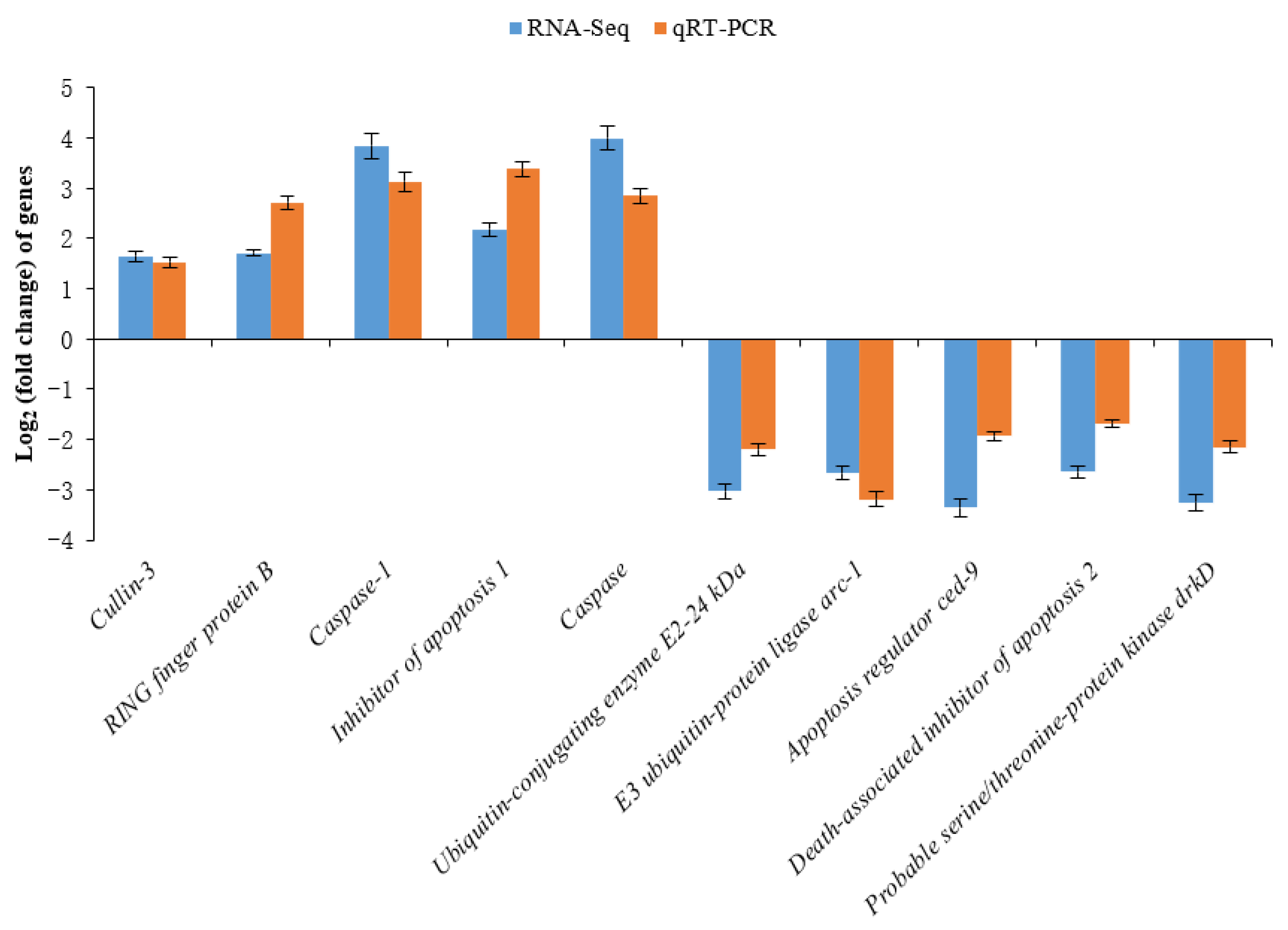
2.6. Real-Time Quantitative RT-PCR Validation

Ten DEGs with immune defense against Cu toxicity were chosen to validate the RNA-seq results. Their primers were designed on the sequences assembled by the transcriptome. These genes were analyzed using qRT-PCR, and the same RNA samples were used for transcriptome analysis. PCR was conducted using the SYBR Green Real-time PCR Kit according to the manufacturer’s instructions. Previous studies showed that the β-actin gene is a suitable housekeeping gene for A. woodiana [39,40,41]. We also verified that the β-actin gene expression is stable in A. woodiana. We judged that using the β-actin as an internal control or using the mean of β-actin and elongation factor 1-alpha (another housekeeping gene commonly used in freshwater mussels [42]) as an internal control should not significantly affect the trend of the DEGs expression. Hence, the β-actin gene was used as an internal control to normalize the expression levels of the target genes. The PCR was carried out in a Roche LightCycler® 480 type II system. The thermal profile for qPCR was 95 °C for 5 min, followed by 40 cycles of 95 °C for 10 s and 60 °C for 1 min. Relative gene expression was calculated using the 2−ΔΔCT method [37,38]. Each qRT-PCR primer sequence is shown in Table 1.

2.7. Statistical Analysis

Concentrations of Cu and other major ions were expressed as mean ± SD. Differences in Cu and major ion concentrations between the control and Cu-treated groups were analyzed using the independent samples t-test in SPSS 22.0 (IBM Corp., New York, NY, USA). Pearson’s correlation was used to investigate the relationship between gene expression identified by qRT-PCR and RNA-seq. Statistical significance was set at p < 0.05. Additionally, the bioaccumulation efficiency of Cu in A. woodiana gills was evaluated using two parameters. First, the bioaccumulation capacity (BAC) was the ratio of the Cu concentration in the gills of the Cu-treated group to the concentration detected in the gills of the control group [43]. Second, the bioconcentration factor (BCF) was the ratio of the Cu concentration in the gills of the Cu-treated group to the concentration in water [44].

3. Results

3.1. Cu Bioaccumulation

The ‘background’ concentrations of Cu in A. woodiana gills were 1.3 ± 0.5 μg/g d.w. Copper concentrations in A. woodiana gills of the control and Cu-treated groups were 0.7 ± 1.2 μg/g d.w. and 331.9 ± 19.8 μg/g d.w., respectively. Cu concentrations in the Cu-treated group were significantly higher than those in the control group (t = −28.961, p < 0.05). Based on BAC, the mussels in the Cu-treated group were found to have bioaccumulated Cu in their gills to a level that, on average, was 474 times higher than that detected in the control group. The BCF values reached 161.9 ± 9.6.

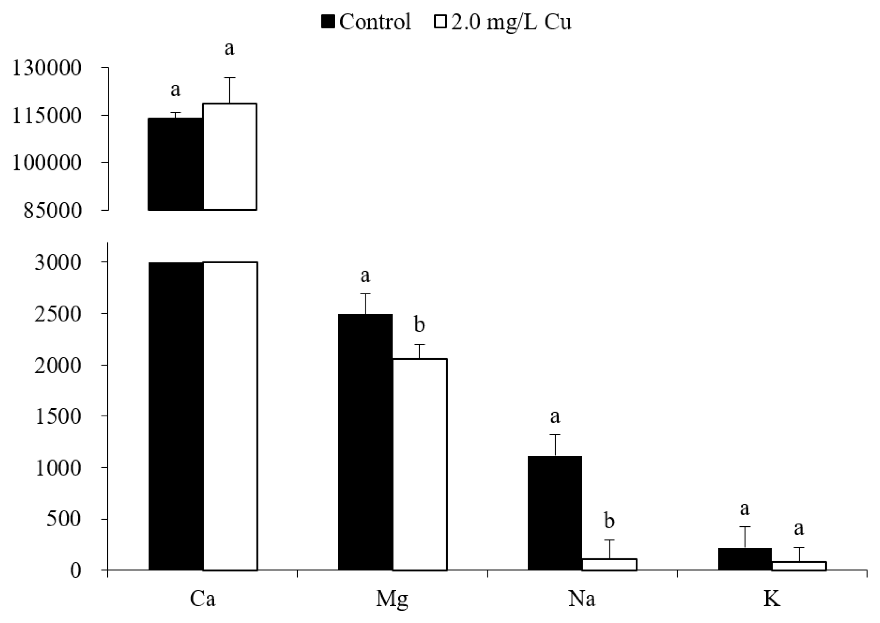
3.2. Ion Concentrations
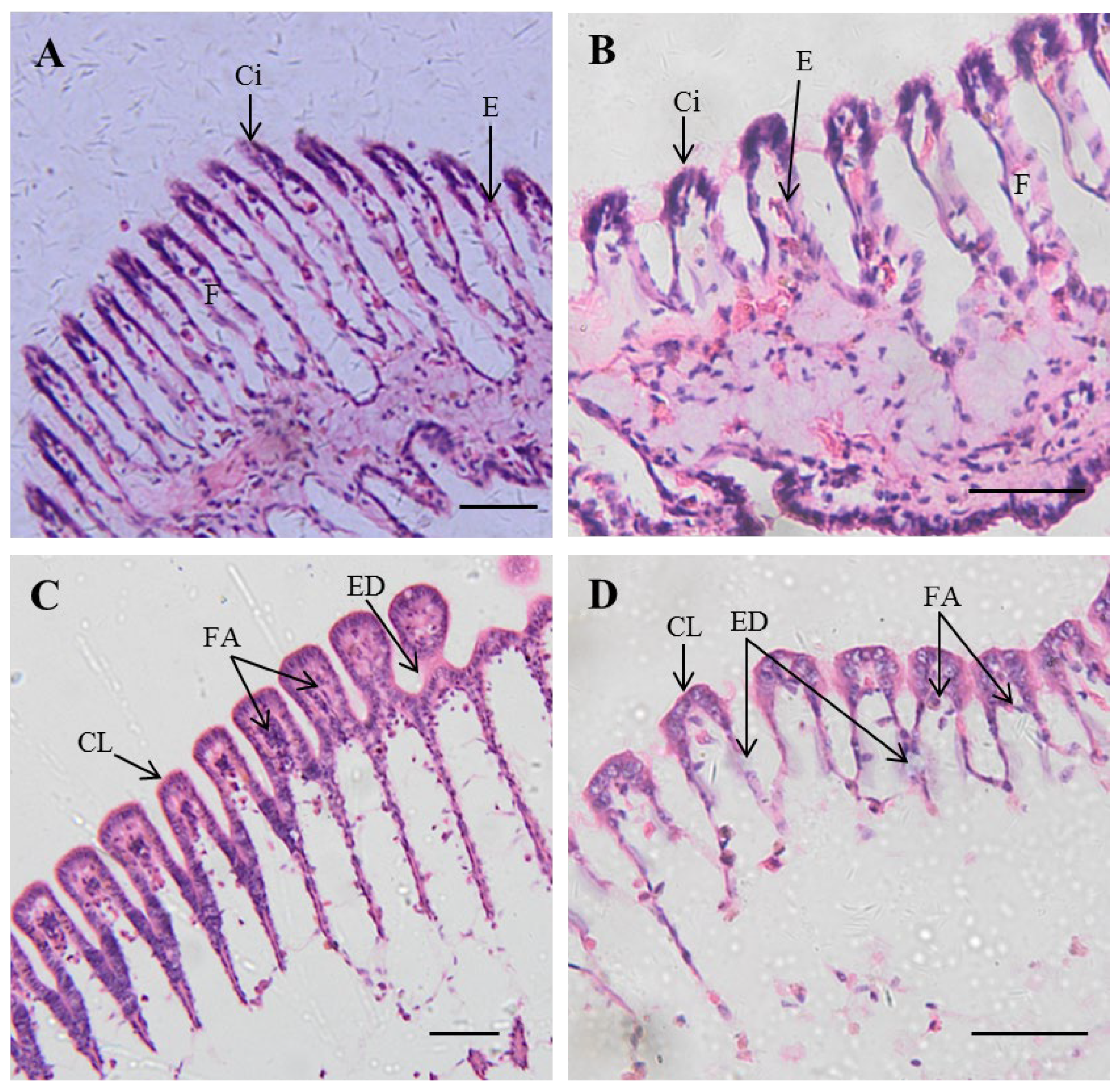
Ion concentrations in A. woodiana gills of both the control and Cu-treated groups generally ranked in decreasing order of Ca2+ > Mg2+ > Na+ > K+ (Figure 1). However, the Na+ (t = 6.346, p < 0.05) and Mg2+ (t = 3.135, p < 0.05) concentrations decreased significantly after Cu exposure, with mean losses of 82% and 17%, respectively (Figure 1). There were no significant differences in K+ and Ca2+ concentrations between the two groups (Figure 1).

3.3. Histopathology

Gill sections of A. woodiana stained with hematoxylin and eosin from the control and Cu-treated groups are shown in Figure 2. The transverse and longitudinal sections show that the gills in the control group exhibited well-preserved structures, and gill filaments were covered with ciliated epithelium on their external surfaces (Figure 2A,B). In contrast, gills from the Cu-treated group exhibited clear histological alterations. The gill filaments lost most of their cilia, desquamation started in the epithelium layer, and atrophy of the gill filaments was observed (Figure 2C,D).

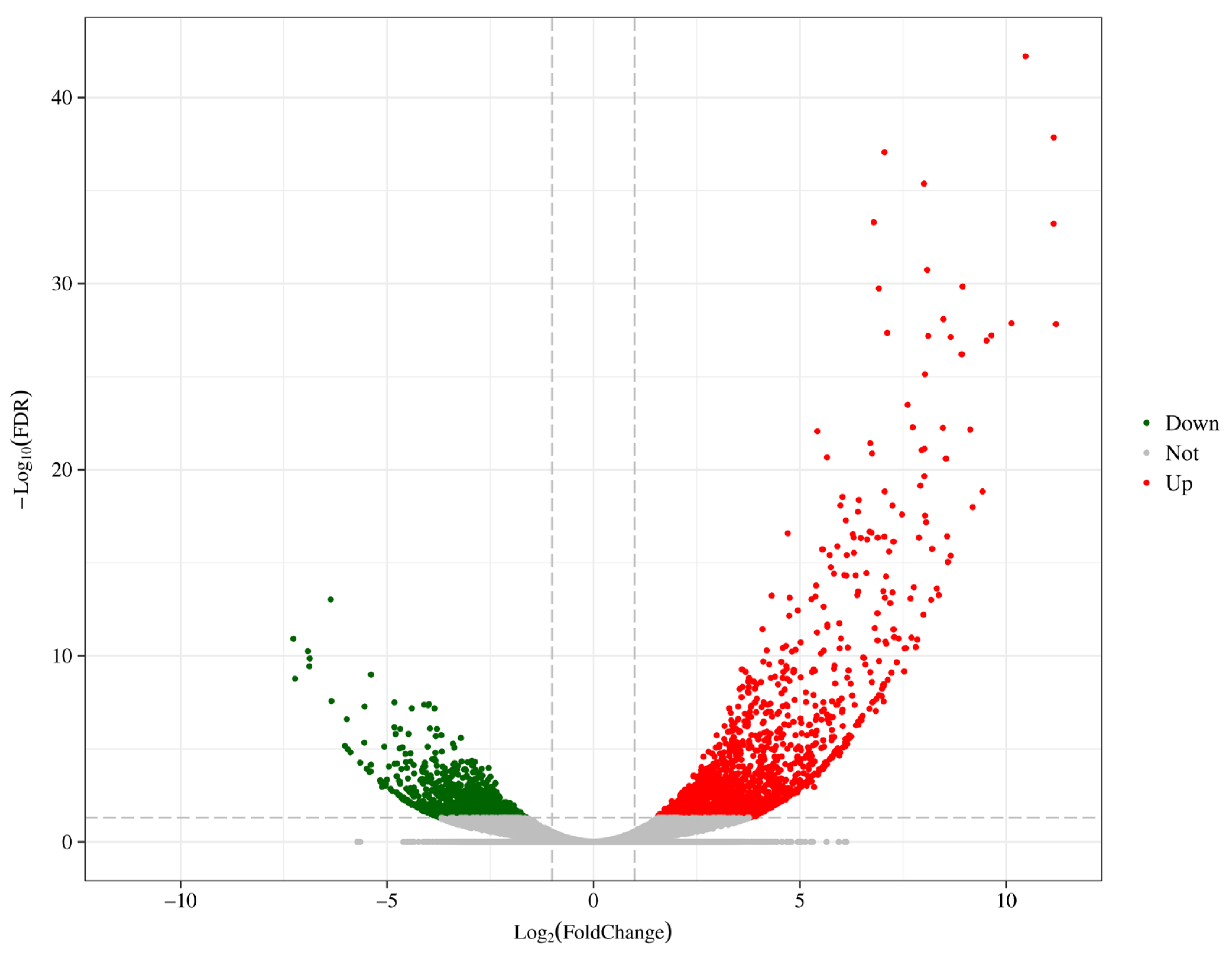
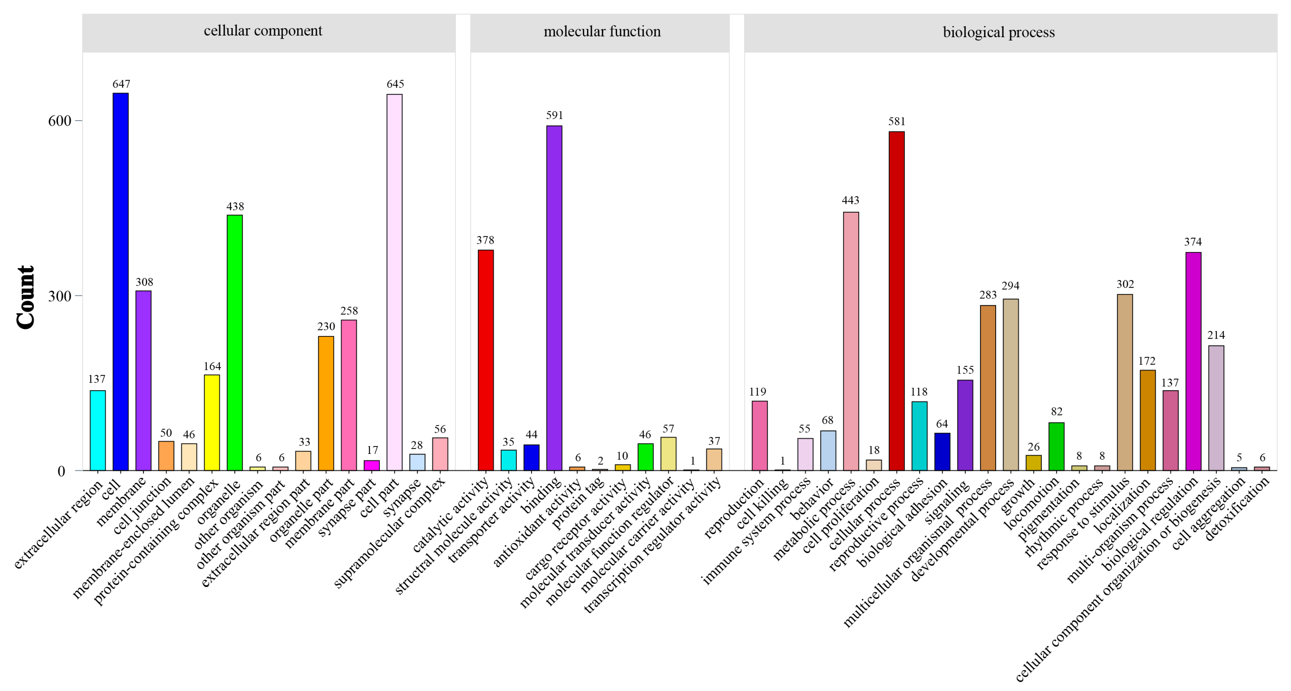
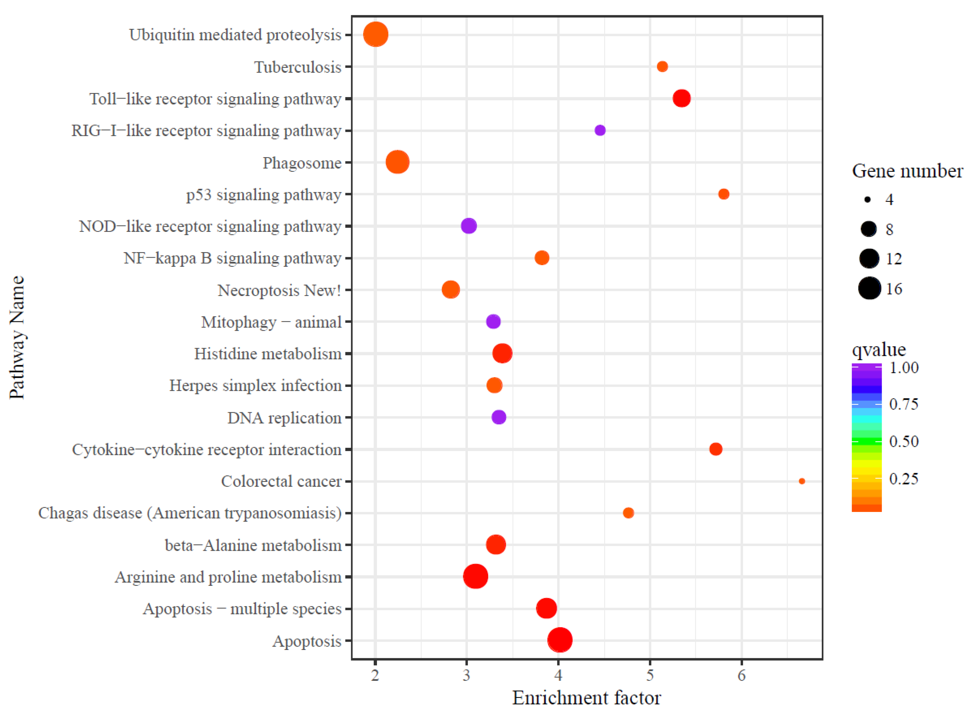
3.4. Differential Expression Analysis

A total of 3160 genes were differentially expressed in the gills of the Cu-treated group compared to in those of the control group, including 1870 upregulated and 1290 downregulated genes (Figure 3). To confirm the veracity and reliability of the DEGs identified by RNA-seq, 10 genes were selected for qRT-PCR analysis. As shown in Figure 4, the qRT-PCR expression patterns of these genes were in agreement with the results of the RNA-seq analysis (R2 = 0.958, p < 0.05).
To obtain insight into the biological functions that can be differentially regulated by Cu exposure, GO enrichment analysis was performed, comparing the Cu and control groups. The GO enrichment analysis included three major functional categories: biological processes, cellular components, and molecular functions. Cellular processes, metabolic processes, biological regulation, and responses to stimuli contained the most DEGs in biological processes (Figure 5). The cell, cell part, organelle, and membrane contained the most DEGs among the cellular components (Figure 5). Binding and catalytic activity contained the most DEGs in terms of molecular functions (Figure 5).
The KEGG database was used to compare the differentially expressed UniGene sequences. In comparison, the Cu-treated group induced eight significantly enriched pathways, including apoptosis, arginine and proline metabolism, the toll-like receptor signaling pathway, apoptosis-multiple species, histidine metabolism, beta-alanine metabolism, cytokine–cytokine receptor interaction, and the p53 signaling pathway (Figure 6).

4. Discussion

4.1. Cu Bioaccumulation

Gills play an important role in respiration and in feeding in filter-feeding species. In A. woodiana, the filtration rate was about 260 L/h/kg d.w. of soft tissue [34]. Aqueous Cu may enter the mussel gills during filtration via facilitated diffusion, active transport, or endocytosis [45]. Therefore, mussel gills are used as indicator tissues to study the bioaccumulation of aqueous Cu. For example, in duck mussel Anodonta anatina exposed to 0.3 μmol/L aqueous Cu for 24 d, approximately 70% of the Cu accumulated in gills with a peak concentration of 75 μmol Cu/kg (4.7 μg/g d.w.); the BAC was 6.5 [46]. In the present study, Cu concentrations in the gills of A. woodiana of the Cu-treated group reached 331.9 ± 19.8 μg/g d.w., and the BAC reached 474. Both Cu concentrations and the BAC of A. woodiana gills in the present study were higher than those of A. anatina [46]. Moreover, the BCF of A. woodiana gills exposed to Cu at 72 h was up to 161.9. These results suggest that A. woodiana gills might have a high bioaccumulation capacity for aqueous Cu.

4.2. Ionoregulatory Disturbance

Na+, Mg2+, K+, and Ca2+ are essential for freshwater mussels, with varied functions, for example, as electrolytes, enzyme constituents, and building materials [47]. A deficiency in these ions leads to serious disorders, such as inhibition of Na+/K+-ATPase activity, thereby reducing osmolarity regulation. In the present study, we found that aqueous Cu exposure/bioaccumulation significantly decreased Na+ and Mg2+ concentrations in the gills of A. woodiana. As documented in freshwater mussel L. cardium larvae, Na+ and Mg2+ concentrations reduced by 30% and 40% after 0.11 μmol/L or 0.27 μmol/L Cu acute exposure for 48 h, paralleled by 20% and 50% mortality, respectively [48]. Freshwater mussel L. siliquoidea juveniles were chronically exposed (28 d) to 2 μg/L or 12 μg/L Cu and mainly exhibited Na+ concentration losses of up to 50% and 70%, respectively; this resulted in 21% and 70% mortality, respectively [49]. Aqueous Cu competes with the Na+/H+ exchanger isoform (NHE-2) for Na+ binding sites [50] and inhibits the activity of Na+/K+-ATPase [51], leading to a significant decrease in Na+ concentrations in freshwater organisms [50,51]. In addition, aqueous Cu exposure inhibits Mg2+-ATPase activity in freshwater mussels [52], which might explain the decreased Mg2+ concentration in the gills of A. woodiana. Considered together, we conclude that increased bioaccumulation of Cu can induce ionoregulatory disturbances in freshwater mussels and further threaten their survival.

4.3. Histological Hazards

Histopathological observation is an effective method for studying Cu toxicology in mussels [43,53]. For example, the freshwater mussel Physa acuta bioaccumulates elevated levels of Cu in the gills but lower amounts in the digestive tract, muscles, and digestive glands [43]. Accordingly, the most serious histopathological changes are observed in the gills, where mucus cells increase, cilia are lost, and columnar cells degenerate in the epithelium [43]. In this study, high bioaccumulation of Cu seemed to induce more serious histological damage than that observed in P. acuta, involving cilia loss, epithelial desquamation, and filament atrophy [43]. From the histology, we propose that triggering cell death by disrupting cell structure and function may be a Cu-specific toxic pathway. Previous studies have also shown that Cu toxicity can cause cell death by damaging the cytoskeleton [54] and disrupting mitochondrial function [55]. Histological damage can affect vital physiological functions such as respiration, filtration, nutrient uptake, and ionic regulation of the gills, which can subsequently affect the growth and survival of freshwater mussels [56]. In freshwater ecosystems, serious Cu contamination events occur frequently [4,5,6], so it is important to pay more attention to the impact of Cu contamination on freshwater mussel resources.

4.4. Transcriptome Responses

Molecular changes can sensitively reflect Cu toxicity [57]. For instance, 3549 DEGs were detected in the gills of Mizuhopecten yessoensis exposed to 0.1 mg/L Cu for 72 h, with 1312 upregulated and 2237 downregulated genes [30]. In contrast, most DEGs were upregulated in A. woodiana gills after aqueous Cu exposure. This implies that gene expressions of A. woodiana were largely activated by aqueous Cu exposure and that the molecular mechanisms of mussels responding to Cu toxicity might be species specific. In addition, these DEGs have the potential to be used for the development of biomarkers of Cu contamination in freshwater ecosystems using A. woodiana as a bioindicator.
GO enrichment analysis showed that DEGs in A. woodiana gills exposed to Cu were mainly involved in cellular and metabolic processes, biological regulation, and responses to stimuli. These generally differed from those observed in other mussels, such as M. yessoensis [30]. This indicates that, although mussels can metabolically fight Cu stress, such as by producing more antioxidants to scavenge intracellular reactive oxygen species (ROS) [27,30], excessive Cu bioaccumulation may cause damage to cell structure and biological function, even extending to histological damage. The molecular toxicity of Cu was confirmed by the gill histological abnormalities, including cilia loss, epithelial desquamation, and filament atrophy, triggered by Cu exposure in this study.
KEGG pathway enrichment analysis showed that DEGs were significantly enriched in pathways related to oxidative stress (the toll-like receptor signaling pathway and cytokine–cytokine receptor interaction), apoptosis (apoptosis, apoptosis-multiple species, and the p53 signaling pathway), and metabolism (arginine and proline metabolism, histidine metabolism, and beta-alanine metabolism). The toll-like receptor signaling pathway is an evolutionarily conserved innate immune pathway that plays a key role in detecting non-self-antigens and immune system activation [30]. Cytokine–cytokine receptor interactions are related to immune defenses [58]. Both the toll-like receptor signaling pathway and cytokine–cytokine receptor interaction may be induced by excessive ROS [59]. These results indicate that Cu bioaccumulation may stimulate ROS generation and activate the mussel’s immune defense. Apoptosis is an important immune response in mussels [60]. It enables the adequate clearance of damaged, senescent, and infected cells without inflammation [60]. Environmental stimulation can induce apoptosis in the cells of mussels [60,61]. For instance, a sudden drop in salinity from 30‰ to 22‰ significantly increased apoptosis and apoptosis-multiple species signaling pathways in the ark shell Anadara kagoshimensis [61]. The p53 signaling pathway is significantly enriched in aquatic organisms, indicating that DNA damage might occur [62]. If DNA damage is extensive, p53 triggers apoptosis [63]. Here, we suggest that Cu toxicity can induce DNA damage and further trigger apoptosis in mussel gills. Amino acids such as histidine and beta-alanine play a substantial role in energy metabolism [64]. Additionally, changes in arginine and proline metabolism are important signatures of apoptosis [65]. The metabolism of eight amino acids (phenylalanine, methionine, histidine, glutamic acid, tryptophan, cysteine, glycine, and alanine) in the hemolymph of the green-shell mussel Perna canaliculus was previously shown to be significantly affected by Cu stress [65]. This indicates that aqueous Cu bioaccumulation may also affect the amino acid metabolism of A. woodiana and may further affect the survival of freshwater mussels.

5. Conclusions

To the best of our knowledge, this study was the first comprehensive screening of the effects of aqueous Cu bioaccumulation on ionoregulatory homeostasis, histological features, and transcriptome responses using A. woodiana gills as the indicator tissue. High bioaccumulation of Cu induced ionoregulatory disturbances (Na+ and Mg2+ losses) and remarkable histological alterations (cilia loss, epithelium desquamation, and filament atrophy). Additionally, the molecular mechanisms of mussel responses to Cu toxicity might be species specific. In A. woodiana, most DEGs were activated by Cu bioaccumulation. These DEGs were mainly involved in cellular processes, metabolic processes, biological regulation, and response to stimuli and were significantly enriched in oxidative-stress-, apoptosis-, and metabolism-related pathways. These results provide valuable insights into Cu toxicity mechanisms in freshwater mussels and benefit the conservation of freshwater mussel resources. In future studies, the DEGs and their enrichment pathways from transcriptome analysis could be used as candidate biomarkers for monitoring Cu contamination in freshwater ecosystems.

Author Contributions

Conceptualization, X.C. and J.Y.; methodology, X.C.; validation, X.C. and J.Y.; formal analysis, X.C.; investigation, X.C., H.L. and T.J.; data curation, X.C.; writing—original draft preparation, X.C.; writing—review and editing, X.C., K.L. and J.Y.; visualization, X.C.; supervision, J.Y. and K.L.; project administration, J.Y.; funding acquisition, X.C. and J.Y. All authors have read and agreed to the published version of the manuscript.

Funding

This research was funded by the Central Public-Interest Scientific Institution Basal Research Fund, CAFS (grant numbers: 2019GH10 and 2022XT0504) and the Central Public-Interest Scientific Institution Basal Research Fund, Freshwater Fisheries Research Center, CAFS (grant number: 2023JBFM01).

Institutional Review Board Statement

The animal study protocol was approved by the Ethics Committee of the Freshwater Fisheries Research Center, Chinese Academy of Fisheries Sciences (protocol code 2011AA1004020012).

Data Availability Statement

Relevant information is included in the article.

Acknowledgments

The authors would like to thank Hui Qiao (Freshwater Fisheries Research Center, Chinese Academy of Fishery Sciences) for help in analyzing transcriptomic data.

Conflicts of Interest

The authors declare no conflict of interest.

References

  1. Johnson, A.C.; Jin, X.; Nakada, N.; Sumpter, J.P. Learning from the past and considering the future of chemicals in the environment. Science 2020, 367, 384–387. [Google Scholar] [CrossRef] [PubMed] [Green Version]
  2. Tavares-Dias, M. Toxic, physiological, histomorphological, growth performance and antiparasitic effects of copper sulphate in fish aquaculture. Aquaculture 2021, 535, 736350. [Google Scholar] [CrossRef]
  3. Dorsey, A.; Ingerman, L.; Swarts, S. Toxicological Profile for Copper; Agency for Toxic Substances and Disease Registry: Atlanta, GA, USA, 2004; pp. 163–167. [Google Scholar]
  4. Xiao, H.Y.; Zhou, W.B.; Zeng, F.P.; Wu, D.S. Water chemistry and heavy metal distribution in an AMD highly contaminated river. Environ. Earth Sci. 2010, 59, 1023–1031. [Google Scholar] [CrossRef]
  5. Grande, J.A.; Aroba, J.; Andújar, J.M.; Gómez, T.; de la Torre, M.L.; Borrego, J.; Romero, S.; Barranco, C.; Santisteban, M. Tinto Versus Odiel: Two A.M.D. polluted rivers and an unresolved issue. An artificial intelligence approach. Water Resour. Manag. 2011, 25, 3575–3594. [Google Scholar] [CrossRef]
  6. Adewumi, A.J.; Laniyan, T.A. Ecological and human health risks associated with metals in water from Anka artisanal gold mining area, Nigeria. Hum. Ecol. Risk Assess. 2021, 27, 307–326. [Google Scholar] [CrossRef]
  7. Pytharopoulou, S.; Kournoutou, G.G.; Leotsinidis, M.; Georgiou, C.D.; Kalpaxis, D.L. Cadmium versus copper toxicity: Insights from an integrated dissection of protein synthesis pathway in the digestive glands of mussel Mytilus galloprovincialis. J. Hazard. Mater. 2013, 260, 263–271. [Google Scholar] [CrossRef]
  8. Liu, H.B.; Chen, X.B.; Su, Y.P.; Kang, I.J.; Qiu, X.C.; Shimasaki, Y.; Oshima, Y.; Yang, J. Effects of calcium and magnesium ions on acute copper toxicity to glochidia and early juveniles of the Chinese pond mussel Anodonta woodiana. Bull. Environ. Contam. Tox. 2016, 97, 504–509. [Google Scholar] [CrossRef]
  9. Shekh, K.; Alcaraz, A.J.; Niyogi, S.; Hecker, M. Comparative analyses of oxidative stress response and metallothionein induction in white sturgeon and rainbow trout during acute waterborne copper exposure. Comp. Biochem. Phys. C 2020, 231, 108723. [Google Scholar] [CrossRef]
  10. Zheng, X.; Zang, W.; Yan, Z.; Hong, Y.; Liu, Z.; Yi, X.; Wang, X.; Liu, T.; Zhou, L. Species sensitivity analysis of heavy metals to freshwater organisms. Ecotoxicology 2015, 24, 1621–1631. [Google Scholar]
  11. Cope, W.G.; Bringolf, R.B.; Buchwalter, D.B.; Newton, T.J.; Ingersoll, C.G.; Wang, N.; Augspurger, T.; Dwyer, F.J.; Barnhart, M.C.; Neves, R.J.; et al. Differential exposure, duration, and sensitivity of unionoidean bivalve life stages to environmental contaminants. J. N. Am. Benthol. Soc. 2008, 27, 451–462. [Google Scholar] [CrossRef]
  12. Bian, B.; Zhou, Y.; Fang, B. Distribution of heavy metals and benthic macroinvertebrates: Impacts from typical inflow river sediments in the Taihu Basin, China. Ecol. Indic. 2016, 69, 348–359. [Google Scholar] [CrossRef]
  13. Renaut, S.; Guerra, D.; Hoeh, W.R.; Stewart, D.T.; Bogan, A.E.; Ghiselli, F.; Milani, L.; Passamonti, M.; Breton, S. Genome survey of the freshwater mussel Venustaconcha ellipsiformis (bivalvia: Unionida) using a hybrid de novo assembly approach. Genome Biol. Evol. 2018, 10, 1637–1646. [Google Scholar] [CrossRef]
  14. Bertucci, A.; Pierron, F.; Thébault, J.; Klopp, C.; Bellec, J.; Gonzalez, P.; Baudrimont, M. Transcriptomic responses of the endangered freshwater mussel Margaritifera margaritifera to trace metal contamination in the Dronne River, France. Environ. Sci. Pollut. Res. 2017, 24, 27145–27159. [Google Scholar] [CrossRef] [PubMed]
  15. Donrovich, S.W.; Douda, K.; Plechingerová, V.; Rylková, K.; Horký, P.; Slavík, O.; Liu, H.Z.; Reichard, M.; Lopes-Lima, M.; Sousa, R. Invasive Chinese pond mussel Sinanodonta woodiana threatens native mussel reproduction by inducing cross-resistance of host fish. Aquat. Conserv. 2017, 27, 1325–1333. [Google Scholar] [CrossRef]
  16. Chen, X.B.; Yang, J.; Liu, H.B.; Jiang, T. Freshwater Mussel Watch: An innovative approach for interpretations of aquatic pollution and toxicology. J. Lake Sci. 2021, 33, 11–27, (In Chinese with English Abstract). [Google Scholar]
  17. Liu, J.; Gu, B.; Bian, J.; Hu, S.; Cheng, X.; Ke, Q.; Yan, H. Antitumor activities of liposome–incorporated aqueous extracts of Anodonta woodiana (Lea, 1834). Eur. Food Res. Technol. 2008, 227, 919–924. [Google Scholar] [CrossRef]
  18. Stangierski, J.; Andrzejewski, W.; Tomaszewska-Gras, J.; Grześ, B.; Konieczny, P.; Urbańska, M. Effect of washing on the quality of surimi-like preparation obtained from soft tissue of freshwater mussel Sinanodonta woodiana (Lea, 1834). J. Aquat. Food Prod. Technol. 2018, 27, 961–974. [Google Scholar] [CrossRef]
  19. Rahayu, S.Y.S.; Solihin, D.D.; Manalu, W.; Affandi, R. Nucleus pearl coating process of freshwater mussel Anodonta woodiana (Unionidae). HAYATI J. Biosci. 2013, 20, 24–30. [Google Scholar] [CrossRef] [Green Version]
  20. Yang, J.; Harino, H.; Liu, H.B.; Miyazaki, N. Monitoring the organotin contamination in the Taihu Lake of China by bivalve mussel Anodonta woodiana. Bull. Environ. Contam. Tox. 2008, 81, 164–168. [Google Scholar] [CrossRef]
  21. Liu, H.B.; Yang, J.; Gan, J.L. Trace element accumulation in bivalves Anodonta woodiana from the Taihu Lake, China. Arch. Environ. Contam. Tox. 2010, 59, 593–601. [Google Scholar] [CrossRef]
  22. Chen, X.B.; Su, Y.P.; Liu, H.B.; Yang, J. Active biomonitoring of metals with cultured Anodonta woodiana: A case study in the Taihu Lake, China. Bull. Environ. Contam. Tox. 2019, 102, 198–203. [Google Scholar] [CrossRef]
  23. Królak, E.; Zdanowski, B. The bioaccumulation of heavy metals by the mussels Anodonta woodiana (Lea, 1834) and Dreissena polymorpha (Pall.) in the Heated Konin Lakes. Arch. Pol. Fish. 2001, 9, 229–237. [Google Scholar]
  24. Kolarević, S.; Knežević-Vukčević, J.; Paunović, M.; Kračun, M.; Vasiljević, B.; Tomović, J.; Vuković-Gačić, B.; Gači, Z. Monitoring of DNA damage in haemocytes of freshwater mussel Sinanodonta woodiana sampled from the Velika Morava River in Serbia with the comet assay. Chemosphere 2013, 93, 243–251. [Google Scholar] [CrossRef] [PubMed]
  25. Gecheva, G.; Yancheva, V.; Velcheva, I.; Georgieva, E.; Stoyanova, S.; Arnaudova, D.; Stefanova, V.; Georgieva, D.; Genina, V.; Todorova, B.; et al. Integrated monitoring with moss-bag and mussel transplants in reservoirs. Water 2020, 12, 1800. [Google Scholar] [CrossRef]
  26. Kurnia, A.I.; Purwanto, E.; Mahajoeno, E. Exposure copper heavy metal (Cu) on freshwater mussel (Anodonta woodiana) and its relation to Cu and protein content in the body shell. Nusant. Biosci. 2010, 2, 48–53. [Google Scholar] [CrossRef]
  27. Liu, H.B.; Chen, X.B.; Oshima, Y.; Shimasaki, Y.; Jiang, T.; Yang, J. Biochemical changes in Chinese pond mussel Anodonta woodiana (Lea, 1834) following exposure to copper. J. Fac. Agric. Kyushu Univ. 2018, 63, 311–318. [Google Scholar]
  28. Chen, X.B.; Liu, H.B.; Huang, H.H.; Liber, K.; Jiang, T.; Yang, J. Cadmium bioaccumulation and distribution in the freshwater bivalve Anodonta woodiana exposed to environmentally relevant Cd levels. Sci. Total Environ. 2021, 791, 148289. [Google Scholar] [CrossRef]
  29. Li, Y.Q.; Chen, C.M.; Liu, N.; Wang, L. Cadmium-induced ultrastructural changes and apoptosis in the gill of freshwater mussel Anodonta woodiana. Environ. Sci. Pollut. Res. 2022, 29, 23338–23351. [Google Scholar] [CrossRef]
  30. Meng, X.; Tian, X.; Liu, M.; Nie, G.; Jiang, K.; Wang, B.; Wang, L. The transcriptomic response to copper exposure by the gill tissue of Japanese scallops (Mizuhopecten yessoensis) using deep-sequencing technology. Fish Shellfish Immun. 2014, 38, 287–293. [Google Scholar] [CrossRef]
  31. Sohail, M.; Khan, M.N.; Qureshi, N.A.; Chaudhry, A.S. Monitoring DNA damage in gills of freshwater mussels (Anodonta anatina) exposed to heavy metals. Pak. J. Zool. 2017, 49, 305–311. [Google Scholar] [CrossRef]
  32. Dragun, Z.; Erk, M.; Ivanković, D.; Žaja, R.; Marijić, V.F.; Raspor, B. Assessment of low-level metal contamination using the Mediterranean mussel gills as the indicator tissue. Environ. Sci. Pollut. Res. 2010, 17, 977–986. [Google Scholar] [CrossRef] [PubMed] [Green Version]
  33. ASTM-E2455-06; Standard Guide for Conducting Laboratory Toxicity Tests with Freshwater Mussels. ASTM International: West Conshohocken, PA, USA, 2013.
  34. Zhou, C.; Huang, J.C.; Liu, F.; He, S.; Zhou, W. Removal of selenium containing algae by the bivalve Sinanodonta woodiana and the potential risk to human health. Environ. Pollut. 2018, 242, 73–81. [Google Scholar] [CrossRef]
  35. Goh, K.S.; Sheu, H.S.; Hua, T.E.; Kang, M.H.; Li, C.W. Uric acid spherulites in the reflector layer of firefly light organ. PLoS ONE 2013, 8, e56406. [Google Scholar] [CrossRef] [PubMed] [Green Version]
  36. Wang, G.; Zhang, C.; Huang, B. Transcriptome analysis and histopathological observations of Geloina erosa gills upon Cr (VI) exposure. Comp. Biochem. Phys. C 2020, 231, 108706. [Google Scholar] [CrossRef] [PubMed]
  37. Zhang, Y.; Li, Z.; Kholodkevich, S.; Sharov, A.; Feng, Y.; Ren, N.; Sun, K. Cadmium-induced oxidative stress, histopathology, and transcriptome changes in the hepatopancreas of freshwater crayfish (Procambarus clarkii). Sci. Total Environ. 2019, 666, 944–955. [Google Scholar] [CrossRef] [PubMed]
  38. Xu, F.; Che, P.; Li, H.; Qiao, S.; Wang, J.; Wang, Y.; Wang, X.; Wu, B.; Liu, H.; Wang, C.; et al. Comparative transcriptome analysis reveals the differential response to cadmium stress of two Pleurotus fungi: Pleurotus cornucopiae and Pleurotus ostreatus. J. Hazard. Mater. 2021, 416, 125814. [Google Scholar] [CrossRef]
  39. Xia, X.; Xue, S.; Wang, X.; Zhang, Q.; Huang, C.; Guo, L.; Yao, L. Response a chronic effects of PBDE-47: Up-regulations of HSP60 and HSP70 expression in freshwater bivalve Anodonta woodiana. Fish Shellfish Immun. 2017, 65, 213–225. [Google Scholar] [CrossRef]
  40. Qu, F.; Li, J.; She, Q.; Zeng, X.; Li, Z.; Lin, Q.; Tang, J.; Yan, Y.; Lu, J.; Li, Y.; et al. Identification and characterization of MKK6 and AP-1 in Anodonta woodiana reveal their potential roles in the host defense response against bacterial challenge. Fish Shellfish Immun. 2022, 124, 261–272. [Google Scholar] [CrossRef]
  41. Qu, F.; She, Q.; Li, J.; Zeng, X.; Li, Y.; Liu, X.; Ren, L.; Liu, Z.; Gao, C.; Lu, X.; et al. Molecular characterization of MyD88 in Anodonta woodiana and its involvement in the innate immune response to bacterial infection. Front. Immunol. 2022, 13, 2893. [Google Scholar] [CrossRef]
  42. Bai, Z.; Lin, J.; Ma, K.; Wang, G.; Niu, D.; Li, J. Identification of housekeeping genes suitable for gene expression analysis in the pearl mussel, Hyriopsis cumingii, during biomineralization. Mol. Genet. Genom. 2014, 289, 717–725. [Google Scholar] [CrossRef]
  43. Dummee, V.; Tanhan, P.; Kruatrachue, M.; Damrongphol, P.; Pokethitiyook, P. Histopathological changes in snail, Pomacea canaliculata, exposed to sub-lethal copper sulfate concentrations. Ecotox. Environ. Safe. 2015, 122, 290–295. [Google Scholar] [CrossRef] [PubMed]
  44. Arnot, J.A.; Gobas, F.A. A review of bioconcentration factor (BCF) and bioaccumulation factor (BAF) assessments for organic chemicals in aquatic organisms. Environ. Rev. 2006, 14, 257–297. [Google Scholar] [CrossRef]
  45. Marigómez, I.; Soto, M.; Cajaraville, M.P.; Angulo, E.; Giamberini, L. Cellular and subcellular distribution of metals in molluscs. Microsc. Res. Tech. 2002, 56, 358–392. [Google Scholar] [CrossRef] [PubMed]
  46. Nugroho, A.P.; Frank, H. Uptake, distribution, and bioaccumulation of copper in the freshwater mussel Anodonta anatina. Toxicol. Environ. Chem. 2011, 93, 1838–1850. [Google Scholar] [CrossRef]
  47. Chen, X.B.; Yang, J.; Liu, H.B.; Su, Y.P.; Sun, L.; Oshima, Y. Element concentrations in a unionid mussel (Anodonta woodiana) at different life stages. J. Fac. Agric. Kyushu Univ. 2012, 57, 139–144. [Google Scholar] [CrossRef]
  48. Jorge, M.B.; Bianchini, A.; Wood, C.M.; Gillis, P.L. Copper uptake, patterns of bioaccumulation, and effects in glochidia (larvae) of the freshwater mussel (Lampsilis cardium). Environ. Toxicol. Chem. 2018, 37, 1092–1103. [Google Scholar] [CrossRef]
  49. Jorge, M.B.; Loro, V.L.; Bianchini, A.; Wood, C.M.; Gillis, P.L. Mortality, bioaccumulation and physiological responses in juvenile freshwater mussels (Lampsilis siliquoidea) chronically exposed to copper. Aquat. Toxicol. 2013, 126, 137–147. [Google Scholar] [CrossRef]
  50. Komjarova, I.; Bury, N.R. Evidence of common cadmium and copper uptake routes in zebrafish Danio rerio. Environ. Sci. Technol. 2014, 48, 12946–12951. [Google Scholar] [CrossRef]
  51. Le, T.T.Y.; Nachev, M.; Grabner, D.; Garcia, M.R.; Balsa-Canto, E.; Hendriks, A.J.; Peijnenburg, W.J.G.M.; Sures, B. Modelling chronic toxicokinetics and toxicodynamics of copper in mussels considering ionoregulatory homeostasis and oxidative stress. Environ. Pollut. 2021, 287, 117645. [Google Scholar] [CrossRef]
  52. Canli, E.G. Alterations on the activities of ion ATPases in the gill and muscle of freshwater mussel (Unio tigridis) exposed to copper. Commagene J. Biol. 2021, 5, 150–155. [Google Scholar] [CrossRef]
  53. Khan, M.I.; Khisroon, M.; Khan, A.; Gulfam, N.; Siraj, M.; Zaidi, F.; Ahmadullah; Abidullah; Fatima, S.H.; Noreen, S.; et al. Bioaccumulation of heavy metals in water, sediments, and tissues and their histopathological effects on Anodonta cygnea (Linea, 1876) in Kabul River, Khyber Pakhtunkhwa, Pakistan. BioMed Res. Int. 2018, 2018, 1910274. [Google Scholar] [CrossRef] [Green Version]
  54. Katsumiti, A.; Thorley, A.J.; Arostegui, I.; Reip, P.; Valsami-Jones, E.; Tetley, T.D.; Cajaraville, M.P. Cytotoxicity and cellular mechanisms of toxicity of CuO NPs in mussel cells in vitro and comparative sensitivity with human cells. Toxicol. In Vitro 2018, 48, 146–158. [Google Scholar] [CrossRef] [PubMed]
  55. Tsvetkov, P.; Coy, S.; Petrova, B.; Dreishpoon, M.; Verma, A.; Abdusamad, M.; Abdusamad, M.; Rossen, J.; Joesch-Cohen, L.; Humeidi, R.; et al. Copper induces cell death by targeting lipoylated TCA cycle proteins. Science 2022, 375, 1254–1261. [Google Scholar] [CrossRef] [PubMed]
  56. Stalin, A.; Musthafa, M.S.; Amanullah, B. Histological alterations in the gills of freshwater mussel Lamellidens marginalis exposed to sub-lethal concentration of an organophosphorus insecticide, chlorpyrifos. Asian J. Microbiol. Biotechnol. Environ. Sci. 2011, 13, 139–142. [Google Scholar]
  57. Wang, W.X.; Meng, J.; Weng, N. Trace metals in oysters: Molecular and cellular mechanisms and ecotoxicological impacts. Environ. Sci. Proc. Imp. 2018, 20, 892–912. [Google Scholar] [CrossRef]
  58. Chen, J.; Xu, Y.; Han, Q.; Yao, Y.; Xing, H.; Teng, X. Immunosuppression, oxidative stress, and glycometabolism disorder caused by cadmium in common carp (Cyprinus carpio L.): Application of transcriptome analysis in risk assessment of environmental contaminant cadmium. J. Hazard. Mater. 2019, 366, 386–394. [Google Scholar] [CrossRef]
  59. Abo-Al-Ela, H.G.; El-Kassas, S.; El-Naggar, K.; Abdo, S.E.; Jahejo, A.R.; Wakeel, R.A.A. Stress and immunity in poultry: Light management and nanotechnology as effective immune enhancers to fight stress. Cell Stress Chaperon. 2021, 26, 457–472. [Google Scholar] [CrossRef]
  60. Terahara, K.; Takahashi, K.G. Mechanisms and immunological roles of apoptosis in molluscs. Curr. Pharm. Des. 2008, 14, 131–137. [Google Scholar] [CrossRef]
  61. Zhang, M.; Li, L.; Liu, Y.; Gao, X. Effects of a sudden drop in salinity on immune response mechanisms of Anadara kagoshimensis. Int. J. Mol. Sci. 2019, 20, 4365. [Google Scholar] [CrossRef] [Green Version]
  62. Wei, F.; Su, T.; Wang, D.; Li, H.; You, J. Transcriptomic analysis reveals common pathways and biomarkers associated with oxidative damage caused by mitochondrial toxicants in Chironomus dilutus. Chemosphere 2020, 254, 126746. [Google Scholar] [CrossRef]
  63. Châtel, A.; Mouneyrac, C. Signaling pathways involved in metal-based nanomaterial toxicity towards aquatic organisms. Comp. Biochem. Phys. C 2017, 196, 61–70. [Google Scholar] [CrossRef] [PubMed]
  64. Huang, W.; Wang, X.; Chen, D.; Xu, E.G.; Luo, X.; Zeng, J.; Huan, T.; Li, L.; Wang, Y. Toxicity mechanisms of polystyrene microplastics in marine mussels revealed by high-coverage quantitative metabolomics using chemical isotope labeling liquid chromatography mass spectrometry. J. Hazard. Mater. 2021, 417, 126003. [Google Scholar] [CrossRef] [PubMed]
  65. Nguyen, T.V.; Alfaro, A.C.; Merien, F.; Lulijwa, R.; Young, T. Copper-induced immunomodulation in mussel (Perna canaliculus) haemocytes. Metallomics 2018, 10, 965. [Google Scholar] [CrossRef] [PubMed]
Figure 1. Ion concentrations in the gills of Anodonta woodiana from the control and Cu-treated groups. Data are presented as mean ± SD of three parallel measurements. Values with different letters indicate a significant difference (t-test, p < 0.05) between the control and Cu-treated groups.
Fishes 08 00368 g001
Figure 2. Light micrographs of transverse and longitudinal gill sections of Anodonta woodiana revealed the histological structure of the control (A,B) and Cu-treated (C,D) groups. Ci: cilia, E: epithelium, F: filament, CL: cilia loss, ED: epithelium desquamation, FA: filament atrophy. Scale bar: 50 μm.
Fishes 08 00368 g002
Figure 3. A volcano plot of differentially expressed genes in the Cu-treated group compared to the control group. Red and green dots represent upregulated and downregulated genes, respectively.
Fishes 08 00368 g003
Figure 4. Comparison of gene expression patterns obtained using RNA-seq and qRT-PCR. The Trinity-assigned genes were annotated using the NR database to obtain the gene names. The X-axis displays 10 selected genes, and the Y-axis represents the relative fold change.
Fishes 08 00368 g004
Figure 5. Gene ontology (GO) classifications of differently expressed genes.
Fishes 08 00368 g005
Figure 6. Kyoto Encyclopedia of Genes and Genomes (KEGG) enrichment of differentially expressed genes (DEGs) after 2.0 mg/L Cu exposure for 72 h. The vertical axis represents different pathways, and the horizontal axis represents the richness factor. Color shades correspond to different q-values, and dot size represents the number of DEGs.
Fishes 08 00368 g006
Table 1. Details of the primer sequence used for qRT-PCR.
Gene NamePrimer SequenceAmplification Efficiency (%)
Cullin-3F-AGGGCGACACACTATTTGGA98
R-CCTGCTCCCTCAAGTAACCA
RING finger protein BF-TGGAAAGCACACAAGTTGCA97
R-GCTTGCCAGTTTCTTCTCGT
Caspase-1F-GCATGCCAGGGACAAAAGTT94
R-AGAGCTTGAACGAACCAGGA
Inhibitor of apoptosis 1F-TCCACATGAGGTTCACACGA100
R-CGCACAAAGGTTCGAATTGC
CaspaseF-CACAATACCTCTGAAGCCGC102
R-AGGACTGGTTGTGATGAGCA
Ubiquitin-conjugating enzyme E2-24 kDaF-CTTTGGTCCAGCACTGCAAT97
R-CGTGGGAAGAAAAGAGCTCG
E3 ubiquitin-protein ligase arc-1F-CTGACATGCCCAGTTTGCTT102
R-GATGTGCGCCTCATTCTCTG
Apoptosis regulator CED-9F-TCAGTGTATGGTATCGGGCC105
R-CACTTTCACGCCCTCTGAAC
Death-associated inhibitor of apoptosis 2F-TCGGGATGCCTGTATTGTGT100
R-AGCTCTTTACTCCCTACCGC
Probable serine/threonine protein kinase drkDF-TCACTGCTGCCCGAATCATA103
R-AGTGACACGGAAGACAGGAG
β-actinF-GTGGCTACTCCTTCACAACC101
R-GAAGCTAGGCTGGAACAAGG
Disclaimer/Publisher’s Note: The statements, opinions and data contained in all publications are solely those of the individual author(s) and contributor(s) and not of MDPI and/or the editor(s). MDPI and/or the editor(s) disclaim responsibility for any injury to people or property resulting from any ideas, methods, instructions or products referred to in the content.

