Article

LC-MS Based Metabolomic Profiling of Largehead Hairtail (Trichiurus japonicus) Ovary Reveals Metabolic Signatures of Ovarian Developmental Process (II–IV)

1
College of Marine Sciences, Shanghai Ocean University, Shanghai 201306, China
2
East China Sea Fisheries Research Institute, Chinese Academy of Fishery Sciences, Shanghai 200090, China
3
Key Laboratory of East China Sea Fishery Resources Exploitation, Ministry of Agriculture and Rural Affairs, Shanghai 200090, China
*
Author to whom correspondence should be addressed.
Fishes 2023, 8(5), 262; https://doi.org/10.3390/fishes8050262
Submission received: 3 April 2023 / Revised: 9 May 2023 / Accepted: 11 May 2023 / Published: 14 May 2023
(This article belongs to the Special Issue Current Trends in Growth and Metabolism of Fishes)

Abstract

:
Trichiurus japonicus is an economically important fish that ranks 11th in global marine fish capture production. However, the reproductive characteristics of this fish have undergone notable changes in recent decades, potentially affecting the quality of offspring and sustainable utilization. To improve our understanding of the physiological regulation of maturation in T. japonicus, untargeted liquid chromatography mass spectrometry was utilized to identify the small molecules that characterize the comprehensive metabolic profiles of ovaries during ovary development from stage II to stage IV. According to the results of OPLS-DA, the ovarian metabolite profiles of the three developmental stages were separated. The concentrations of 124 and 100 metabolites were significantly altered between stage II vs. III and III vs. IV, respectively. Lipids and lipid-like molecules accounted for the largest proportion of the altered metabolites, followed by amino acids, peptides, and analogues. The significantly altered metabolites-enriched pathways differed slightly between stages II and III and stages III and IV. Steroid-related pathways were heavily affected during stages II to III, while significantly altered metabolites from stages III to IV were involved in oocyte-maturation-related pathways. Through metabonomics analysis, potentially important metabolic pathways and metabolites between different ovarian stages were detected, providing basic information for further investigation of maturation mechanisms in wild fish.
Key Contribution: The potentially important metabolic pathways and metabolites during ovary development from stage II to stage IV in largehead hairtail (Trichiurus japonicus) were identified and will pave the way for further investigation of maturation mechanisms in wild fish.

1. Introduction

The largehead hairtail (Trichiurus japonicus) is a member of the Trichiuridae family, within the order Scombriformes. It is a warm-temperature demersal species and is distributed in the tropical and temperate continental shelves and slopes worldwide [1]. According to the FAO report [2], the hairtail production worldwide in 2020 was 1.144 million tons, ranking it 11th in global marine fish capture production. It is also one of China’s most important economic fish, with its yield ranking first in China’s marine fish capture production for many years [3]. T. japonicus is the dominant species among the hairtail yield in China [4]. However, due to fishing pressure and various environmental factors, hairtail resources have declined in recent decades. In 2019, hairtail capture was only 70% of what it was in 1999 [3]. Additionally, various reproductive characteristics have changed, including mature anal length and egg diameter. The East China Sea (ECS) is the primary spawning ground for T. japonicus in China; T. japonicus spawns almost year-round with multiple batches and two dominant spawning periods in the ECS [5,6,7]. It has been reported that T. japonicus follows a group-synchronous oocyte development pattern [8]. In the 1960s, the first sexually mature age of T. japonicus in the ECS was one year, and the minimum anal length of the mature individual was 200–210 mm. However, in the 1990s, the minimum anal length of mature individuals decreased to 140–150 mm. The 50% mature anal length (L50) of the female population decreased to 164.65 mm, and the L50 of the male population decreased to 171.65 mm. When the anal length reached about 210 mm, it was almost 100% mature. Not only did the anal length of maturation decrease, but the diameter of mature eggs also significantly reduced. It decreased from 1.525–1.825 mm in 1963–1964 to 0.9–1.5 mm in 1993–1994 [9,10]. Apart from T. japonicus, early maturation is also common in other major economic fish species in the ECS. This phenomenon is accompanied by a series of problems such as the miniaturization of parent fish, the reduction of egg diameter, and the decline of germplasm of larvae and juvenile, which seriously affects the sustainable utilization of fishery resources and food security [11,12]. Therefore, it is urgent to understand why gonadal development is accelerated in wild fish.
Numerous studies have investigated the potentially possible factors and their fluctuations that affect the development of gonads in wild fish, including temperature [13,14,15], salinity [16,17], photoperiod [18,19], baits [20,21], and fishing [22]. In addition to external environmental factors, the timing and process of gonadal development are also regulated by the internal hypothalamic–pituitary–gonad axis, which involves the balance of various central neurotransmitters and hormones [23]. Gonadotropin-releasing hormone (GnRH) secreted by the hypothalamus is a key factor in initiating gonadal development [24,25]. GnRH promotes the pituitary to release gonadotropin (GTH), which stimulates the synthesis and secretion of steroid hormones in the gonad, such as estradiol and progesterone, ultimately promoting the development of gonads [26]. Additionally, numerous signaling pathways and metabolites related to the metabolism of hormones, amino acids, lipids, and energy play important roles in this process [27,28,29].
To better understand gonadal development, metabolomics has been introduced to the reproduction research of aquatic animals [27,28,29]. Metabolomics is a powerful tool that can comprehensively analyze endogenous metabolites in biological systems, providing a better understanding of their metabolic functional states [30]. This approach is characterized by high sensitivity, high throughput, and rapidity [31]. Untargeted liquid chromatography (LC)–mass spectrometry (MS)-based metabolomics has successfully investigated the metabolic differences between sexes and maturation states of aquatic animals, including blunt snout bream (Megalobrama amblycephala) [27], Chinese sturgeon (Acipenser sinensis) [28], and sea lamprey (Petromyzon marinus) [29].
To improve our understanding of the physiological regulation of fish maturation, untargeted LC-MS was used to identify the small molecules that characterize the comprehensive metabolic profiles of ovaries in T. japonicus during ovary development from stage II to stage V in the present study. The results may provide valuable basic information regarding the reasons for the early maturity of this economically important fish.

2. Materials and Methods

2.1. Sample Collection and Preparation

As the spawning peak period of T. japonicus in the southern offshore area of Zhejiang, China, is from May to July [32], the T. japonicus young-of-the year during May 2021 in the Wentai fishing ground (27°00′~28°00′ N and the west of 125°00′ E) was sampled. The T. japonicus was frozen and transported back to the laboratory. Based on the growth curve of the T. japonicus fitted by the Walford growth transformation method, which showed that the anal length of the one-year-old fish was less than 190 mm [33], the T. japonicus with an anal length of less than 190 mm was chosen for the later analysis. The basic biological information including anal length (AL), body weight, sex, gonad developmental stage, and gonad–somatic index (GSI [34]) was measured and calculated for each T. japonicus. The ovaries were immediately frozen and stored after weighing. The ovarian developmental stages of the T. japonicus were determined based mainly on macroscopic examination with six development classes, which were defined as follows: I = immature, II = developing, III = maturing, IV = mature, V = ripe, and VI = spent [5,35].
Six fish ovaries were randomly selected in each developmental stage from II, III, and IV for LC-MS analysis. An amount of 50 mg ovary was accurately weighed for each sample for subsequent processing [36], which is described in the Supplementary Information. To ensure the stability of the analysis and the metabolomic data quality, pooled quality control (QC) samples were prepared by mixing portions of all the samples. The five QC samples were tested similarly to the analytic samples.

2.2. LC-MS Analysis and Data Processing

Untargeted LC-MS was conducted with the UHPLC-Q Exactive system of Thermo Fisher Scientific. Detailed formation on metabolome analysis conditions is presented in the Supplementary Materials. After the mass spectrometry detection was completed, the raw data of LC/MS were preprocessed by Progenesis QI (Waters Corporation, Milford, CT, USA) software to generate a data matrix that consisted of the retention time (RT), mass-to-charge ratio (m/z) values, and peak intensity. Metabolic features detected at least 80% in any set of samples were retained. At the same time, variables with relative standard deviation (RSD) > 30% of QC samples were removed, and log10 logarithmization was performed to obtain the final data matrix for subsequent analysis [37]. The normalized data were matched against available databases to identify metabolites, especially the HMDB (http://www.hmdb.ca/ (accessed on 1 January 2021)) and KEGG (http://www.kegg.com (accessed on 2 January 2021)) public databases [31,38]. The chemical classification of metabolites in the HMDB can provide better insights into the biological significance of metabolites.

2.3. Statistics

The significance of AL, body weight, and GSI among three developmental stages were detected by one-way ANOVA with Duncan’s multiple range tests [39] in SPSS (Version 21.0, SPSS Inc., Chicago, IL, USA). The data were presented as mean ± SEM, and p < 0.05 was considered as significant [40].
The obtained positive and negative data in the LC-MS analysis were analyzed using the free online platform Majorbio Cloud Platform, https://cloud.majorbio.com (accessed on 1 January 2021) (Shanghai Majorbio Bio-pharm Technology Co., Ltd.). The principal component analysis (PCA) and orthogonal least partial squares discriminant analysis (OPLS-DA) were used for statistical analysis to determine global metabolic changes between II vs. III and III vs. IV [40]. The selection of significantly different metabolites was determined based on the variable importance in the projection (VIP) obtained by the OPLS-DA model and the p-value of the Student’s t-test, and the metabolites with VIP > 1, p < 0.05 were selected as significantly different metabolites [37]. To compare the variation of metabolites among different groups, a Venn plot and heat maps plot were generated. Differential metabolites among the two groups were summarized and mapped into their biochemical pathways through metabolic enrichment and pathway analysis based on KEGG. Statistically significantly enriched pathways were identified using Fisher’s exact test with a p-value of less than 0.05, using the Python package scipy.stats (https://docs.scipy.org/doc/scipy/ (accessed on 1 January 2020)) [36,37].

3. Results

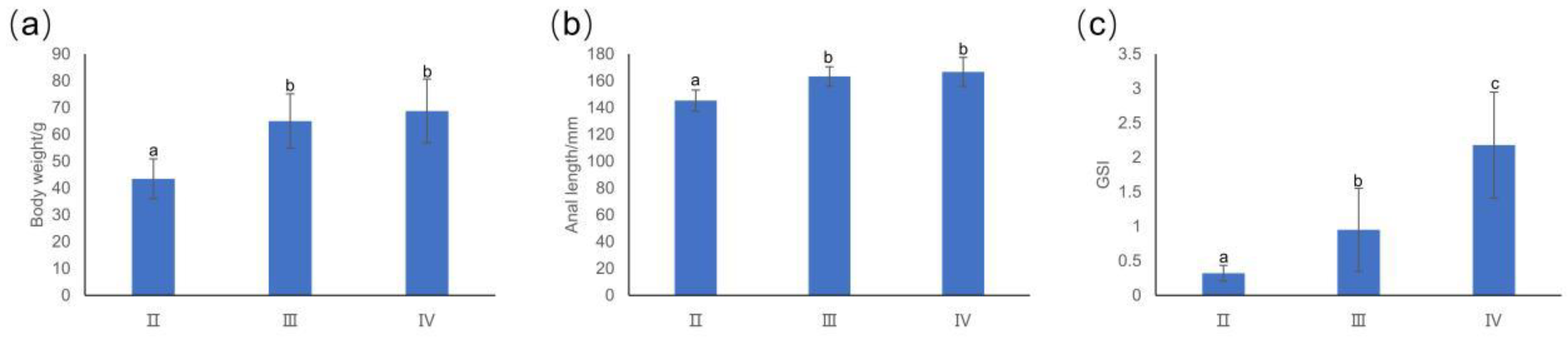
3.1. Basic Information about T. japonicus

As shown in Figure 1, the body weight and anal length of T. japonicus in stage II were significantly lower than those of T. japonicus in stages III and IV, but there were no significant differences in body weight and anal length between stages III and IV. The GSI of different stages had obvious differences, which was consistent with ovarian development.

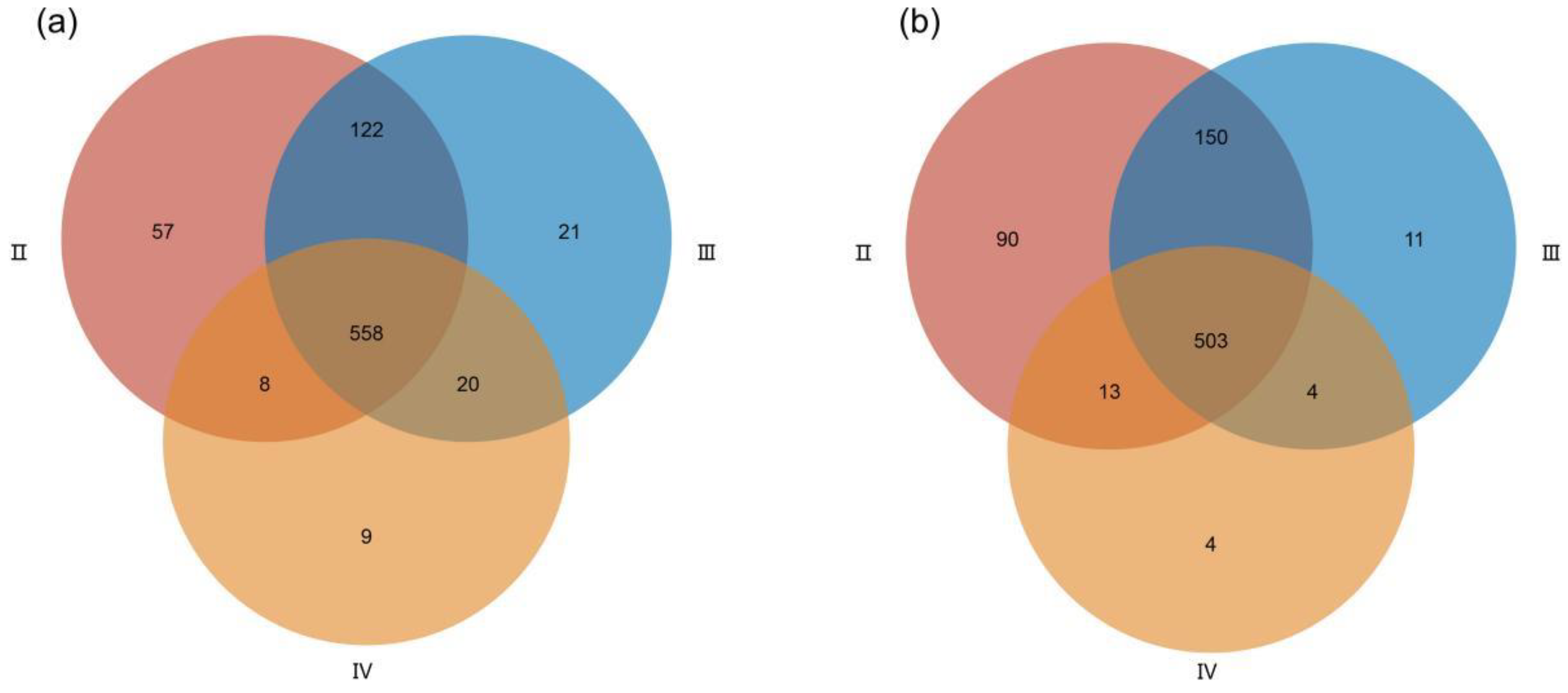
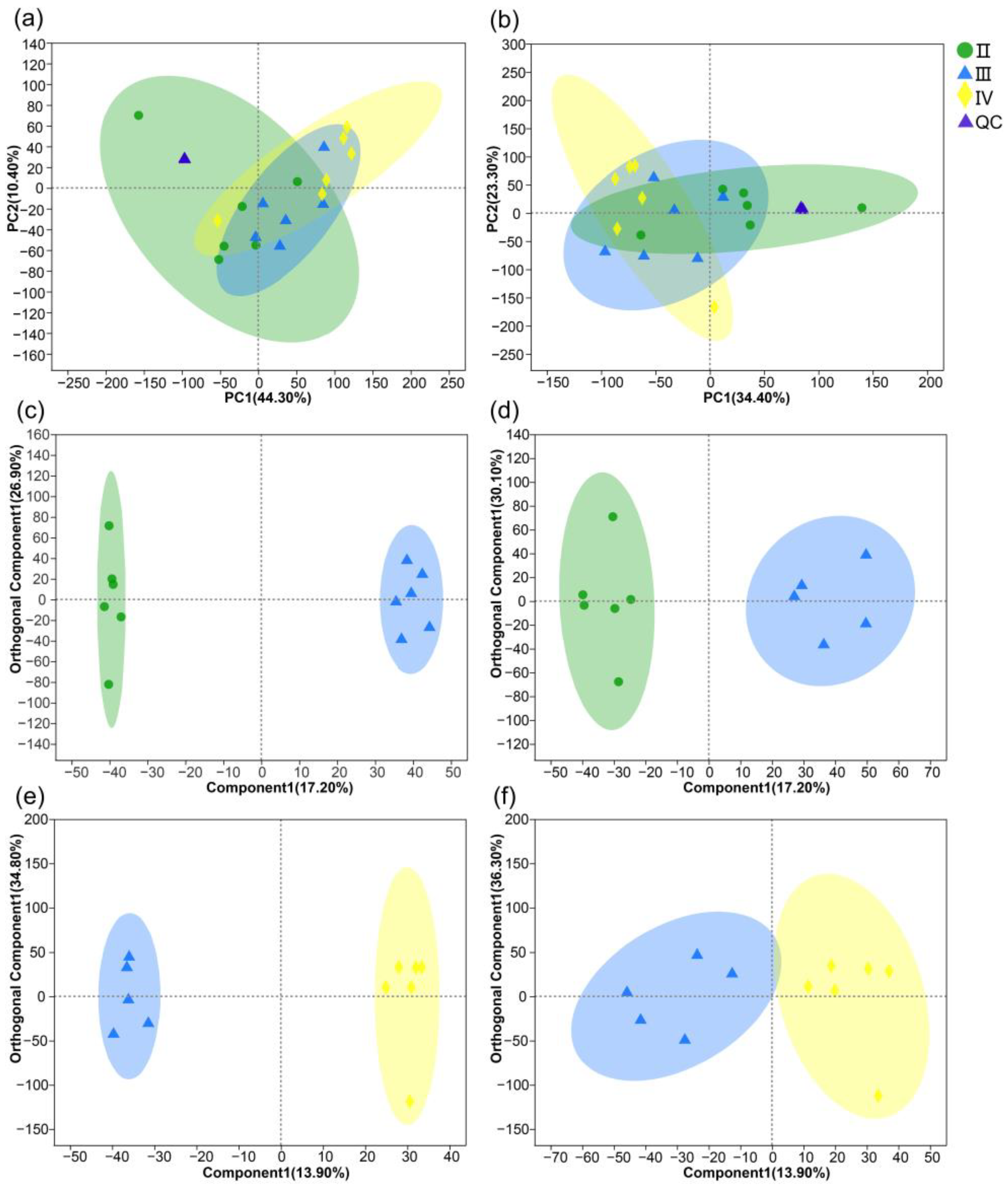
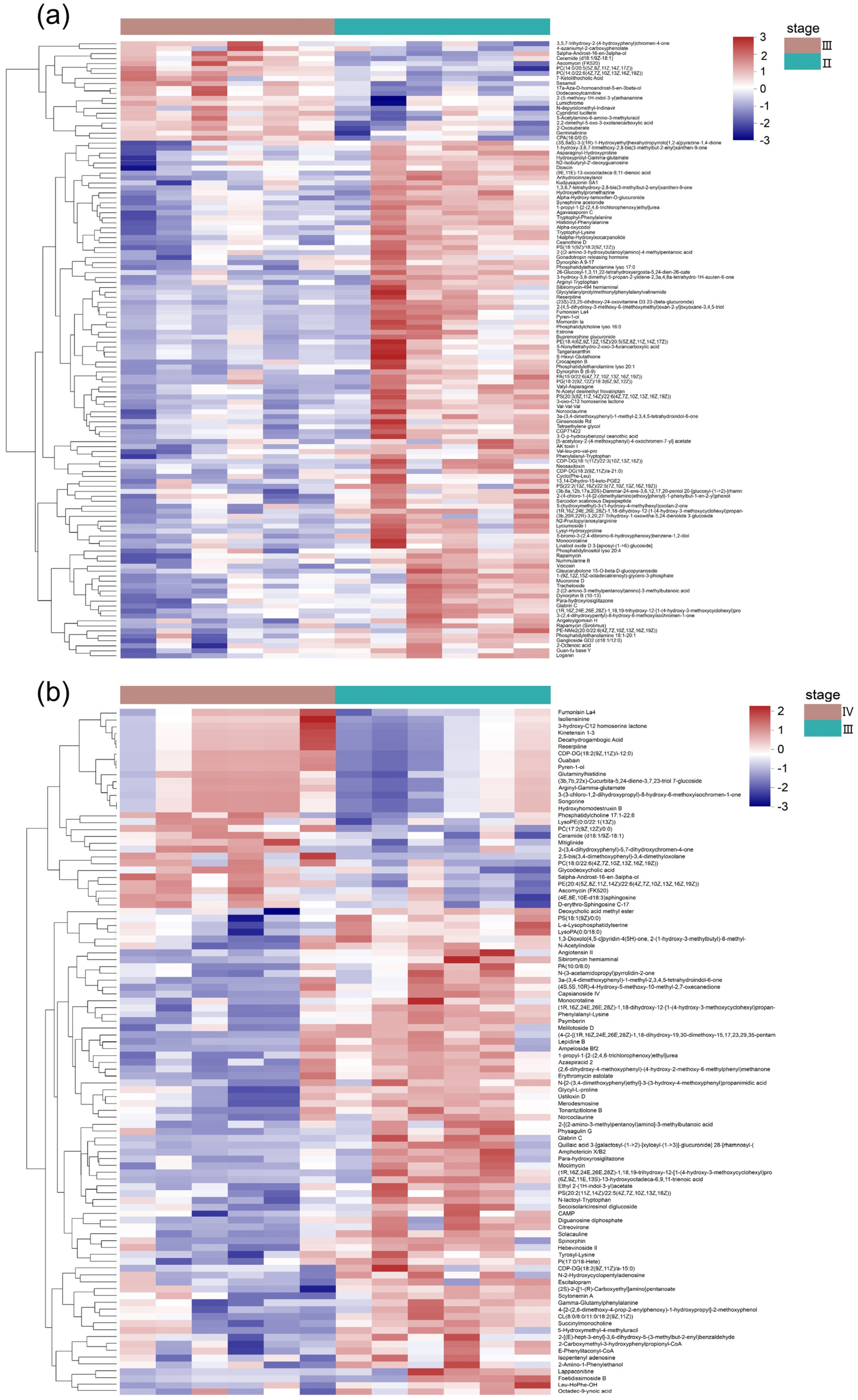
3.2. Metabolite Profiles of the Ovaries at Different Developmental Stages

In total, 6042 variables (3395 peaks in ESI+ mode, and 2647 peaks in ESI− mode) were identified in ovaries from stages II, III, and IV for subsequent analyses. Under positive mode, a total of 745, 721, and 595 annotated metabolites were identified in II, III, and IV, respectively. Under negative mode, a total of 756, 668, and 524 annotated metabolites were identified in II, III, and IV, respectively. The three stages shared 558 and 503 metabolites under positive and negative modes, respectively (Figure 2a,b).
The PCA performed on the whole samples revealed that the QC samples were tightly clustered in PCA score plots (Figure 3a,b), which indicated that the system stability was accommodative for this metabonomic study. PCA results showed that the first components accounted for 44.30% and 34.40% in positive and negative ion modes, respectively, but the discriminations of the three groups were not very distinct. Furthermore, OPLS-DA was performed to maximize the distinction between groups and obtain a global overview of the differences in metabolites between III vs. II and IV vs. III. Positive and negative data revealed clear separation and discrimination between the different developmental stages, indicating the significantly different metabolomic profiles of the three stages (Figure 3c–f). According to the results of OPLS-DA, a total of 124 potential biomarkers between III vs. II and 100 potential biomarkers between IV vs. III were finally screened out based on VIP > 1 and p < 0.05. Heat maps (Figure 4) showed clear differences in the ovarian metabolic profiles of the three developmental stages. Compared with stage II, 20 metabolites were upregulated and 104 metabolites were down-regulated in stage III, while 29 metabolites were upregulated and 71 metabolites were down-regulated in stage IV compared with stage III (Figure 4). The significantly changed metabolites were classified into different classes according to HMDB. The lipids and lipid-like molecules accounted for the largest proportion of the total, in which glycerophospholipid, steroids and steroid derivatives, prenol lipids, and fatty acyls formed the majority of the metabolites. The second largest group comprised amino acids, peptides, and analogues (Figure 5).

3.3. Metabolic Pathways Analysis

The potentially important metabolic pathways during the ovarian quick developmental period were identified using the KEGG database. KEGG enrichment analysis indicated that significantly altered metabolites from stages II to III were enriched in the neurotrophin signaling pathway, adipocytokine signaling pathway, sphingolipid signaling pathway, tryptophan metabolism, steroid hormone biosynthesis, riboflavin metabolism, etc. (Figure 6a), while the most heavily affected pathway (p < 0.05) was the neurotrophin signaling pathway. The significantly altered metabolites from stages III to IV were involved in growth hormone synthesis, oocyte meiosis, progesterone-mediated oocyte maturation, neurotrophin signaling pathway, MAPK signaling pathway, GnRH signaling pathway, adipocytokine signaling pathway, etc. (Figure 6b); the most heavily affected pathways (p < 0.05) were progesterone-mediated oocyte maturation, oocyte meiosis, neurotrophin signaling pathway, and growth hormone synthesis. The pathways that always played roles in ovarian development from stages II to IV were the sphingolipid signaling pathway, adipocytokine signaling pathway, neurotrophin signaling pathway, etc.

4. Discussion

Fish gonad development is a critical biological process that directly affects fish reproduction and population sustainability. The metabolic profiles of T. japonicus’s ovaries at different developmental stages (II–IV) reflect the physiological status of the ovaries before spawning. Metabonomics analysis was performed to detect the potentially important metabolic pathways and metabolites between different ovarian stages, providing fundamental information for further investigations into early maturation mechanisms in wild fish. In the present study, the ovarian metabolites profile of the three stages were separated. The metabolites significantly alerted in each stage were principally lipids and lipid-like molecules and amino acid metabolites.
The lipids and lipid-like molecules formed the majority of significantly altered metabolites during ovarian developmental stages from II to IV in T. japonicus (Figure 5). This result is consistent with other aquatic animal studies that also used metabonomics analysis to investigate ovarian development [30,38,41]. In teleosts, ovarian development is a process of nutrient storage, where all material deposited in an oocyte serves as nutrients for the embryo, primarily consisting of lipoproteins, phosphoproteins, and discrete lipids [42]. The biochemical composition in aquatic animals showed that during maturation process, the fat content in gonads decreased, while the percentage of MUFA acids increased, and fats were actively transferred from muscles and incorporated in gonads [43]. Evidence also implied that part of the deposited fatty acids in the ovary might come from diet, as the effect of dietary fatty acids on reproductive performance and egg quality has been reported in several fishes, such as Siberian sturgeon (Acipenser baeri) [44], tongue sole (Cynoglossus semilaevis) [45], and Atlantic halibut (Hippoglossus hippoglossus) [46]. However, the specific changed metabolites during the ovarian developmental period are stages- and species-specific. For example, significantly changed metabolites during ovarian development from stages III to IV in Coilia nasus were related to the synthesis pathways of steroids, steroid hormones, and arachidonic acid [41]. Serum metabolites of female Chinese sturgeon also indicated that the metabolic pathways related to linoleic acid, α-linolenic acid, and ARAs significantly changed during ovarian development from stages II to IV [30]. In the present study, dominant altered metabolites belonged to glycerophospholipid, steroids and steroid derivatives, prenol lipids, and fatty acyls. As the ovarian samples were used, the ovarian stromal tissue, follicle cells, and non-vitellogenic follicles would contribute some portion of the lipid composition. The quantitative significance of this would vary depending on the degree of maturity of the dominant oocytes [8,47].
The second largest group in significantly altered metabolites comprised amino acids, peptides, and analogues (Figure 5). The study on the changes in serum metabolites during stages II to IV of female Chinese sturgeon also found significant changes in the metabolites related to a large number of different amino acid metabolic pathways [30]. Protein has an important effect on the reproductive performance of aquatic animals, promoting the growth and maturation of ovarian cells, affecting precocious puberty, and benefiting the gonad index, fertility, and larval production (for details, see the review by Shi [48]). Several amino acids such as tryptophan (Try), phenylalanine (Phe), lysine (Lys), leucine (Leu), Valine (Val), alanine (Ala), serine (Ser), glutamic acid (Glu), Arginine (Arg), and aspartic acid (Asp) have been predicted to be important in the reproductive performance of fish [29,38,49,50,51,52]. In the present study, a large number of different incomplete breakdown products of protein catabolism were found in the ovaries of T. japonicus, including oligopeptides, dipeptides, etc. These metabolites consisted of Arg, Val, Glu, Try, Phe, Lys, etc. (Figure 4). Some dipeptides are known to have physiological or cell-signaling effects, although most are simply short-lived intermediates on their way to specific amino acid degradation pathways following further proteolysis [53], which may reveal the important role of amino acids and their related metabolites in ovarian development. The exact functions of these amino acids in species-specific reproduction deserve further investigation. For example, arginine is one of the most versatile amino acids in animal cells and plays a variety of physiological functions, which serves as a precursor for the synthesis not only of proteins but also of nitric oxide, polyamines, proline, glutamate, creatine, and agmatine [54]. It has been shown in mammals that arginine has a strong relationship with reproductive performance [55], but the studies on the effects of arginine on gonad development and reproduction of aquatic animals are still limited; only in crustaceans was it reported that arginine could affect the synthesis and secretion of the vitellogenin-inhibiting hormone, and also could increase the expression of vitellogenin receptor gene in the ovary and promote the deposition of vitellogenin in ootids [56].
Different hormonal signaling initiated by hormones as well as environmental factors plays crucial roles in the various reproductive processes by modulating different signaling pathways [57]. In the present study, a significant decrease was observed in the concentration of estrone during ovarian development from stages II to III (Figure 4). Estrone is a major estrogen and can be converted to estradiol with potent estrogenic properties. In teleosts, estradiol is a key hormone in oocyte growth [58], and in female trout, the process of exogenous vitellogenesis is primarily regulated by estradiol [59]. The cytochrome P450 family, including cholesterol side chain cleavage (P450scc), 17α-hydroxylase/lyase (P450c17), and aromatase (P450arom), is expressed in many tissues, including the ovary, and is the key enzyme in the synthesis of estradiol [60,61,62]. Studies on channel catfish (Ictalurus punctatus) have shown that the transcript abundance for P450c17, P450scc, and P450arom was increased during early vitellogenic growth of the oocytes, and decreased precipitously with the completion of vitellogenesis [60]. In the present study, the cytopigment P450 metabolic pathway was affected during ovarian development from stages Ⅲ to Ⅳ (Figure 6), which was consistent with the observation of fish oocyte growth by transmission electron microscopy [63].
In teleosts, cyclic adenosine monophosphate (cAMP) is considered an important second messenger for estradiol [64]. However, in the present study, the concentration of decreased significantly from stages III to IV (Figure 4). cAMP is a key regulator of oocyte maturation and plays paradoxical roles within the oocyte and cumulus cells to orchestrate oocyte meiotic arrest and resumption [65]. cAMP is acutely and transiently upregulated in the oocyte in response to the luteinizing hormone (LH) surge [66], initiating signaling events that promote oocyte meiotic resumption. cAMP elevation alters adenine nucleotide metabolism and is hydrolyzed to AMP by phosphodiesterases, which increases the AMP/ATP ratio [67]. As 5′AMP-activated protein kinase (AMPK) is sensitive to the AMP-to-ATP ratio, its activation is triggered by an increasing AMP level. This, in turn, leads to the activation of several pathways involved in gonadal steroidogenesis, the proliferation and survival of gonadal cells, and the maturation of oocytes [68].
In addition to hormonal signaling, neuronal signaling has been shown to play a role in oocyte maturation in several aquatic animals [69]. The present study identified the involvement of the tryptophan–serotonin metabolic pathway in ovarian development in in T. japonicus (Figure 6). Specifically, the concentration of 5-methoxytryptamine (5-MT), a nonselective serotonin (5-HT) receptor agonist [70,71], significantly increased from stages II to III (Figure 4). In the hypothalamus, the binding of 5-HT to its receptor stimulates the secretion of gonadotropin-releasing hormone (GnRH), follicle-stimulating hormone (FSH), and LH, which regulate the onset of puberty [72,73]. 5-HT has also been detected in the ovaries [74] and is associated with steroidogenesis, oocyte meiosis, oocyte maturation, and ovulation [75,76,77].
Additionally, the concentration of ceramide increased gradually during gonadal development from stages II to IV (Figure 4). Ceramides are a large family of lipid-signaling molecules that are associated with several biological processes, including cell growth, differentiation, and apoptosis [78,79]. Recent studies have shown that ceramides have prominent metabolic roles as transmitters for the central actions of leptin and ghrelin [80,81], which could regulate puberty onset. An increment in hypothalamic ceramide content could advance puberty [82]. Ceramide has also been detected in the ovary and is speculated to play roles in ovarian development [83]. Considering the existing precocious puberty of T. japonicus, further investigation is needed to explore the relationship between precocious puberty and the activation of ceramide-related metabolic pathways.

5. Conclusions

To better understand the physiological status of the ovary before spawning in T. japonicus, untargeted LC-MS was introduced to analyze the metabolic profiles of ovaries at different developmental stages (II–IV). The findings suggest that the ovarian metabolic profiles are maturation-dependent and reflect the special metabolic demands at each developmental stage. The significantly altered metabolites-enriched pathways showed that the steroid-related pathways were heavily affected during stages II to III, while oocyte-maturation-related pathways played roles from stage III to IV. The results provided basic information on the aspect of metabolomics for further investigation of maturation mechanisms in wild fish.

Supplementary Materials

The following supporting information can be downloaded at: https://www.mdpi.com/article/10.3390/fishes8050262/s1.

Author Contributions

J.-H.C. and Y.J. conceived and designed the research. L.-Y.F., L.-P.Y., and R.-W.L. conducted experiments. S.-F.L. contributed reagents or analytical tools. L.-Y.F. and Y.J. analyzed data. L.-Y.F. wrote the manuscript. All authors have read and agreed to the published version of the manuscript.

Funding

This study was funded by the Shanghai Sailing Program (18YF1429800), Central Public-Interest Scientific Institution Basal Research Fund, East China Sea Fisheries Research Institute, Chinese Academy of Fishery Sciences (L32201921860). And The APC was funded by 18YF1429800.

Institutional Review Board Statement

Ethical review and approval were waived for this study because the samples involved in this study were from fishing operations and did not involve animal protection requirements.

Informed Consent Statement

Not applicable.

Data Availability Statement

The data used to generate the figures of our study are available via https://github.com/fengliuying/LC-MS-Data (accessed on 1 January 2021). Other datasets used and/or analyzed during the current study are available from the corresponding author upon reasonable request.

Acknowledgments

This study was sponsored by the Shanghai Sailing Program (18YF1429800), Central Public-Interest Scientific Institution Basal Research Fund, East China Sea Fisheries Research Institute, Chinese Academy of Fishery Sciences (L32201921860).

Conflicts of Interest

The authors declare no conflict of interest.

References

  1. Nelson, J.S.; Grande, T.C.; Wilson, M.V. Fishes of the World; John Wiley & Sons: Hoboken, NJ, USA, 2016. [Google Scholar] [CrossRef]
  2. Food and Agriculture Organization. The State of World Fisheries and Aquaculture 2022. Towards Blue Transformation; FAO: Rome, Italy, 2022. [Google Scholar] [CrossRef]
  3. Bureau of Fisheries, Ministry of Agriculture and Rural Affairs, China Society of Fisherie. China Fishery Statistical Yearbook; Agriculture Press: Beijing, China, 2000–2020. [Google Scholar]
  4. He, X.B. Spatial Distribution, Population Structure and Trophic Ecology of Common Trichiuridae Species in the Coastal Waters of China. Ph.D. Thesis, JiMei University, Xiamen, China, 2019. [Google Scholar]
  5. Li, C. Annual ovarian changes of Trichiurus haumela in the East China Sea. Oceanol. Limnol. Sin. 1982, 13, 461–472. [Google Scholar]
  6. Kwok, K.; Ni, I.-H. Reproduction of cutlassfishes Trichiurus spp. from the South China Sea. Mar. Ecol. Prog. Ser. 1999, 176, 39–47. [Google Scholar] [CrossRef]
  7. Sun, P.; Chen, Q.; Fu, C.; Zhu, W.; Li, J.; Zhang, C.; Yu, H.; Sun, R.; Xu, Y.; Tian, Y. Daily growth of young-of-the-year largehead hairtail (Trichiurus japonicus) in relation to environmental variables in the East China Sea. J. Mar. Syst. 2020, 201, 103243. [Google Scholar] [CrossRef]
  8. Shin, S.R.; Kim, H.J.; Kim, J.W.; Kwon, D.-H.; Choi, J.; Park, J.J.; Lee, J.S. Sex Ratio, Spawning Period, and Sexual Group Maturity of the Largehead Hairtail Trichiurus japonicus (Teleostei: Trichiuridae) in Korean Waters. Fishes 2023, 8, 194. [Google Scholar] [CrossRef]
  9. Xu, H.X.; Liu, Z.F.; Zhou, Y.D. Variation of Trichiurus haumela productivity and recruitment in the East China Sea. J. Fish. China 2003, 27, 322–327. [Google Scholar]
  10. Chen, Y.L.; Shan, X.J.; Dai, F.Q.; Jin, X.S. Relative stock density and distribution of hairtail Trichiurus lepturus and its spawning stock structure in coastal waters of the East China Sea. Prog. Fish. Sci. 2013, 34, 8–15. [Google Scholar]
  11. Chao, M.; Quan, W.M.; Li, C.H.; Cheng, Y.H. Changes in trophic level of marine catches in the East China Sea region. Mar. Sci. 2005, 29, 54. [Google Scholar]
  12. Hutchings, J.A.; Baum, J.K. Measuring marine fish biodiversity: Temporal changes in abundance, life history and demography. Philos. Trans. R. Soc. B Biol. Sci. 2005, 360, 315–338. [Google Scholar] [CrossRef]
  13. Mu, Y.L.; Liu, Y.; Wang, J.; Yuan, C.Y.; Dong, J. Effects of water temperature and photoperiod on maturation and spawn of flounder (Paralichthys olivaceus). J. Dalian Fish. Univ. 2016, 14, 62–65. [Google Scholar]
  14. Jin, Y.; Liu, Z.L.; Yan, L.P.; Yuan, X.W.; Cheng, J.H. Maturity of Hairtail Varies with Latitude and Environment in the East China Sea. Mar. Coast. Fish. 2020, 12, 395–403. [Google Scholar] [CrossRef]
  15. Targońska, K.; Żarski, D.; Kupren, K.; Palińska-żarska, K.; Mamcarz, A.; Kujawa, R.; Skrzypczak, A.; Furgała-Selezniow, G.; Czarkowski, T.K.; Hakuć-Błażowska, A.; et al. Influence of temperature during four following spawning seasons on the spawning effectiveness of common bream, Abramis brama (L.) under natural and controlled conditions. J. Therm. Biol. 2014, 39, 17–23. [Google Scholar] [CrossRef]
  16. You, H.Z.; Zheng, Y.K.; You, G.C. Research progress on the effects of different salinity on fish farming biology. Hebei Fish. 2013, 6, 47–52. [Google Scholar]
  17. Guo, J.J.; Ch, G.P.; Huang, Z.Y.; Lu, W.Q. Studies on growth and gonadal development of Pseudosciaena crocea in low salinity in door circulating culture system. J. Shanghai Ocean Univ. 2016, 25, 847–852. [Google Scholar] [CrossRef]
  18. Bapary, M.A.J.; Amin, M.N.; Takeuchi, Y.; Takemura, A. The stimulatory effects of long wavelengths of light on the ovarian development in the tropical damselfish, Chrysiptera cyanea. Aquaculture 2011, 314, 188–192. [Google Scholar] [CrossRef]
  19. Leclercq, E.; Taylor, J.; Sprague, M.; Migaud, H. The potential of alternative lighting-systems to suppress pre-harvest sexual maturation of 1+ Atlantic salmon (Salmo salar) post-smolts reared in commercial sea-cages. Aquac. Eng. 2011, 44, 35–47. [Google Scholar] [CrossRef]
  20. Xiao, D.Y. Effects of different levels of dietary vitamin A, C, E on reproductive eperformance and offspringquality of tongue sole (Cynoglossus semilaevis). Ph.D. Thesis, Shanghai Ocean University, Shanghai, China, 2014. [Google Scholar]
  21. Afzal Khan, M.; Jafri, A.; Chadha, N. Effects of varying dietary protein levels on growth, reproductive performance, body and egg composition of rohu, Labeo rohita (Hamilton). Aquac. Nutr. 2005, 11, 11–17. [Google Scholar] [CrossRef]
  22. Shan, X.; Hu, Z.; Shao, C. Progress in the study of fishing-induced evolution of fish biological characteristics. Prog. Fish. Sci. 2020, 41, 165–175. [Google Scholar] [CrossRef]
  23. Taranger, G.L.; Carrillo, M.; Schulz, R.W.; Fontaine, P.; Zanuy, S.; Felip, A.; Weltzien, F.-A.; Dufour, S.; Karlsen, Ø.; Norberg, B. Control of puberty in farmed fish. Gen. Comp. Endocrinol. 2010, 165, 483–515. [Google Scholar] [CrossRef]
  24. Terasawa, E.; Fernandez, D.L. Neurobiological mechanisms of the onset of puberty in primates. Endocr. Rev. 2001, 22, 111–151. [Google Scholar] [CrossRef]
  25. Sisk, C.L.; Foster, D.L. The neural basis of puberty and adolescence. Nat. Neurosci. 2004, 7, 1040–1047. [Google Scholar] [CrossRef]
  26. Yaron, Z.; Gur, G.; Melamed, P.; Rosenfeld, H.; Elizur, A.; Levavi-Sivan, B. Regulation of fish gonadotropins. Int. Rev. Cytol. 2003, 225, 131–185. [Google Scholar] [PubMed]
  27. Zhou, L.-F.; Zhao, B.-W.; Guan, N.-N.; Wang, W.-M.; Gao, Z.-X. Plasma metabolomics profiling for fish maturation in blunt snout bream. Metabolomics 2017, 13, 40. [Google Scholar] [CrossRef]
  28. Zhu, Y.; Wu, J.; Leng, X.; Du, H.; Wu, J.; He, S.; Luo, J.; Liang, X.; Liu, H.; Wei, Q. Metabolomics and gene expressions revealed the metabolic changes of lipid and amino acids and the related energetic mechanism in response to ovary development of Chinese sturgeon (Acipenser sinensis). PLoS ONE 2020, 15, e0235043. [Google Scholar] [CrossRef] [PubMed]
  29. Tamrakar, S.; Huerta, B.; Chung-Davidson, Y.-W.; Li, W. Plasma metabolomic profiles reveal sex-and maturation-dependent metabolic strategies in sea lamprey (Petromyzon marinus). Metabolomics 2022, 18, 90. [Google Scholar] [CrossRef]
  30. Cevallos-Cevallos, J.M.; Reyes-De-Corcuera, J.I.; Etxeberria, E.; Danyluk, M.D.; Rodrick, G.E. Metabolomic analysis in food science: A review. Trends Food Sci. Technol. 2009, 20, 557–566. [Google Scholar] [CrossRef]
  31. Dunn, W.B.; Broadhurst, D.; Begley, P.; Zelena, E.; Francis-McIntyre, S.; Anderson, N.; Brown, M.; Knowles, J.D.; Halsall, A.; Haselden, J.N. Procedures for large-scale metabolic profiling of serum and plasma using gas chromatography and liquid chromatography coupled to mass spectrometry. Nat. Protoc. 2011, 6, 1060–1083. [Google Scholar] [CrossRef]
  32. Zhang, Q.H. Fishery resources and their sustainable use in the East China Sea; Fudan University Press: Shanghai, China, 2007. [Google Scholar]
  33. Yan, L.P.; Hu, F.; Li, J.S.; Liu, Y.; Chen, J.H. Age and growth of Trichiurus haumela in the East China Sea. Mar. Fish. 2005, 27, 139–142. [Google Scholar]
  34. Htun-Han, M. The reproductive biology of the dab Limanda limanda (L.) in the North Sea: Gonosomatic index, hepatosomatic index and condition factor. J. Fish Biol. 1978, 13, 369–378. [Google Scholar] [CrossRef]
  35. Chen, X.J. Fishery Resources and Fishery Oceanography; China Ocean Press: Beijing, China, 2004. [Google Scholar]
  36. Yang, P.; Yao, W.; Wang, Y.; Li, M.; Li, X.; Leng, X. Dietary effects of fish meal substitution with Clostridium autoethanogenum on flesh quality and metabolomics of largemouth bass (Micropterus salmoides). Aquac. Rep. 2022, 23, 101012. [Google Scholar] [CrossRef]
  37. Chen, X.; Hou, X.; Yang, M.; Wang, J.; Wang, C. Differential metabolomic profiles of two closely related Leuciscinae fish, the species Ctenopharyngodon idellus and Leuciscus idus. Aquac. Rep. 2021, 20, 100650. [Google Scholar] [CrossRef]
  38. Zhou, H.; Leng, X.Q.; Tan, Q.S.; Du, H.; Wu, J.P.; Liang, X.F.; Wei, Q.W. Identification of key nutrients for gonadal development by comparative analysis of proximate composition and fatty/amino acid profile in tissues and eggs of Chinese sturgeon (Acipenser sinensis Gray, 1835). J. Appl. Ichthyol. 2017, 33, 885–891. [Google Scholar] [CrossRef]
  39. Duncan, D.B. Multiple range and multiple F tests. Biometrics 1955, 11, 1–42. [Google Scholar] [CrossRef]
  40. Gong, B.; Bao, F.; Wang, Y.; Liu, H.; Xiao, M.; He, J. Metabonomics study on the effect of traditional Chinese medicines feed addition on growth performance and serum metabolic profile of juvenile Chinese softshell turtle (Pelodiscus sinensis Wiegmann). Aquac. Rep. 2021, 20, 100632. [Google Scholar] [CrossRef]
  41. Xu, G.; Du, F.; Li, Y.; Nie, Z.; Xu, P. Integrated application of transcriptomics and metabolomics yields insights into population-asynchronous ovary development in Coilia nasus. Sci. Rep. 2016, 6, 31835. [Google Scholar] [CrossRef] [PubMed]
  42. Mommsen, T.P.; Walsh, P.J. 5 Vitellogenesis and oocyte assembly. In Fish Physiology; Elsevier: Amsterdam, The Netherlands, 1988; Volume 11, pp. 347–406. [Google Scholar] [CrossRef]
  43. Nowosad, J.; Kucharczyk, D.; Łuczyńska, J.; Targońska, K.; Czarkowski, T.K.; Biłas, M.; Krejszeff, S.; Horváth, L.; Müller, T. Changes in European eel ovary development and body and ovary chemistry during stimulated maturation under controlled conditions: Preliminary data. Aquac. Int. 2015, 23, 13–27. [Google Scholar] [CrossRef]
  44. Luo, L.; Ai, L.; Li, T.; Xue, M.; Wang, J.; Li, W.; Liang, X. The impact of dietary DHA/EPA ratio on spawning performance, egg and offspring quality in Siberian sturgeon (Acipenser baeri). Aquaculture 2015, 437, 140–145. [Google Scholar] [CrossRef]
  45. Liang, M.Q.; Lu, Q.K.; Qian, C.; Zheng, K.K.; Wang, X.X. Effects of dietary n-3 to n-6 fatty acid ratios on spawning performance and larval quality in tongue sole Cynoglossus semilaevis. Aquac. Nutr. 2014, 20, 79–89. [Google Scholar] [CrossRef]
  46. Mazorra, C.; Bruce, M.; Bell, J.; Davie, A.; Alorend, E.; Jordan, N.; Rees, J.; Papanikos, N.; Porter, M.; Bromage, N. Dietary lipid enhancement of broodstock reproductive performance and egg and larval quality in Atlantic halibut (Hippoglossus hippoglossus). Aquaculture 2003, 227, 21–33. [Google Scholar] [CrossRef]
  47. Wiegand, M.D. Composition, accumulation and utilization of yolk lipids in teleost fish. Rev. Fish Biol. Fish. 1996, 6, 259–286. [Google Scholar] [CrossRef]
  48. Shi, L.D.; Ren, T.J.; Han, Y.Z. Research progress of nutrients regulating reproductive performance of aquaculture animals: A review. J. Dalian Ocean Univ. 2020, 35, 620–630. [Google Scholar] [CrossRef]
  49. Rønnestad, I.; Thorsen, A.; Finn, R.N. Fish larval nutrition: A review of recent advances in the roles of amino acids. Aquaculture 1999, 177, 201–216. [Google Scholar] [CrossRef]
  50. Chong, A.S.; Hashim, R.; Ali, A.; Hara, K. Amino acid profile of various body tissues and eggs of discus fish, Symphysodon aequifasciata. J. Appl. Aquac. 2004, 16, 157–168. [Google Scholar] [CrossRef]
  51. Lanes, C.F.C.; Bizuayehu, T.T.; Bolla, S.; Martins, C.; de Oliveira Fernandes, J.M.; Bianchini, A.; Kiron, V.; Babiak, I. Biochemical composition and performance of Atlantic cod (Gadus morhua L.) eggs and larvae obtained from farmed and wild broodstocks. Aquaculture 2012, 324, 267–275. [Google Scholar] [CrossRef]
  52. Li, W.; Wei, Q.; Shen, L. Biochemical comparison between eggs from female Chinese sturgeon (Acipenser sinensis Gray, 1835) reconditioned in freshwater and eggs from wild females: Evaluation of female reconditioning as a conservation culture technique. J. Appl. Ichthyol. 2014, 30, 1237–1242. [Google Scholar] [CrossRef]
  53. Ősz, K.; Várnagy, K.; Süli-Vargha, H.; Csámpay, A.; Sanna, D.; Micera, G.; Sóvágó, I. Acid–base properties and copper (II) complexes of dipeptides containing histidine and additional chelating bis (imidazol-2-yl) residues. J. Inorg. Biochem. 2004, 98, 24–32. [Google Scholar] [CrossRef] [PubMed]
  54. Wu, G.; Morris, S.M., Jr. Arginine metabolism: Nitric oxide and beyond. Biochem. J. 1998, 336, 1–17. [Google Scholar] [CrossRef] [PubMed]
  55. Zhou, M.; Shen, S.T. The effect of L-arginine on animal reproductive function. Feed Livest. 2013, 10, 5–6. [Google Scholar]
  56. Qi, C.L. The nutritional and physiological functions of argininein Chinese mitten crab Eriocheir sinensis at juvenile and gonad maturation stage. Ph.D. Thesis, East China Normal University, Shanghai, China, 2020. [Google Scholar]
  57. Jessus, C.; Munro, C.; Houliston, E. Managing the oocyte meiotic arrest—Lessons from frogs and jellyfish. Cells 2020, 9, 1150. [Google Scholar] [CrossRef]
  58. Senthilkumaran, B.; Yoshikuni, M.; Nagahama, Y. A shift in steroidogenesis occurring in ovarian follicles prior to oocyte maturation. Mol. Cell. Endocrinol. 2004, 215, 11–18. [Google Scholar] [CrossRef]
  59. Van Bohemen, C.G.; Lambert, J.; Van Oordt, P. Vitellogenin induction by estradiol in estrone-primed rainbow trout, Salmo gairdneri. Gen. Comp. Endocrinol. 1982, 46, 136–139. [Google Scholar] [CrossRef]
  60. Sampath Kumar, R.; Ijiri, S.; Trant, J.M. Changes in the expression of genes encoding steroidogenic enzymes in the channel catfish (Ictalurus punctatus) ovary throughout a reproductive cycle. Biol. Reprod. 2000, 63, 1676–1682. [Google Scholar] [CrossRef] [PubMed]
  61. Simpson, E.R.; Clyne, C.; Rubin, G.; Boon, W.C. Aromatase—A brief overview. Annu. Rev. Physiol. 2002, 64, 93. [Google Scholar] [CrossRef] [PubMed]
  62. Sreenivasulu, G.; Senthilkumaran, B. A role for cytochrome P450 17α-hydroxylase/c17-20 lyase during shift in steroidogenesis occurring in ovarian follicles prior to oocyte maturation. J. Steroid Biochem. Mol. Biol. 2009, 115, 77–85. [Google Scholar] [CrossRef]
  63. Wang, A.M. Electron microscopic observation on the yolk formation of Oreochomis mossambicus. Acta Hydrobiol. Sinica. 1994, 18, 26–31. [Google Scholar]
  64. Nagahama, Y. Endocrine regulation of gametogenesis in fish. Int. J. Dev. Biol. 2002, 38, 217–229. [Google Scholar] [CrossRef]
  65. Downs, S.M. Regulation of the G2/M transition in rodent oocytes. Mol. Reprod. Dev. 2010, 77, 566–585. [Google Scholar] [CrossRef] [PubMed]
  66. Mattioli, M. Transduction mechanisms for gonadotrophin-induced oocyte maturation in mammals. Zygote 1994, 2, 347–349. [Google Scholar] [CrossRef] [PubMed]
  67. Richani, D.; Lavea, C.F.; Kanakkaparambil, R.; Riepsamen, A.H.; Bertoldo, M.J.; Bustamante, S.; Gilchrist, R.B. Participation of the adenosine salvage pathway and cyclic AMP modulation in oocyte energy metabolism. Sci. Rep. 2019, 9, 18395. [Google Scholar] [CrossRef]
  68. Bertoldo, M.J.; Faure, M.; Dupont, J.; Froment, P. AMPK: A master energy regulator for gonadal function. Front. Neurosci. 2015, 9, 235. [Google Scholar] [CrossRef]
  69. Alhajeri, M.M.; Alkhanjari, R.R.; Hodeify, R.; Khraibi, A.; Hamdan, H. Neurotransmitters, neuropeptides and calcium in oocyte maturation and early development. Front. Cell Dev. Biol. 2022, 10, 980219. [Google Scholar] [CrossRef]
  70. Yamada, J.; Sugimoto, Y.; Yoshikawa, T.; Horisaka, K. Hyperglycemia induced by the 5-HT receptor agonist, 5-methoxytryptamine, in rats: Involvement of the peripheral 5-HT2A receptor. Eur. J. Pharmacol. 1997, 323, 235–240. [Google Scholar] [CrossRef] [PubMed]
  71. Craig, D.; Eglen, R.; Walsh, L.; Perkins, L.; Whiting, R.; Clarke, D. 5-Methoxytryptamine and 2-methyl-5-hydroxytryptamine-induced desensitization as a discriminative tool for the 5-HT3 and putative 5-HT4 receptors in guinea pig ileum. Naunyn-Schmiedebergs Arch. Pharmacol. 1990, 342, 9–16. [Google Scholar] [CrossRef]
  72. Wada, K.; Hu, L.; Mores, N.; Navarro, C.E.; Fuda, H.; Krsmanovic, L.Z.; Catt, K.J. Serotonin (5-HT) receptor subtypes mediate specific modes of 5-HT-induced signaling and regulation of neurosecretion in gonadotropin-releasing hormone neurons. Mol. Endocrinol. 2006, 20, 125–135. [Google Scholar] [CrossRef] [PubMed]
  73. Gouveia, E.M.; Franci, C.R. Involvement of serotonin 5HT1 and 5HT2 receptors and nitric oxide synthase in the medial preoptic area on gonadotropin secretion. Brain Res. Bull. 2004, 63, 243–251. [Google Scholar] [CrossRef] [PubMed]
  74. Amenta, F.; Vega, J.A.; Ricci, A.; Collier, W.L. Localization of 5-hydroxytryptamine-like immunoreactive cells and nerve fibers in the rat female reproductive system. Anat. Rec. -Adv. Integr. Anat. Evol. Biol. 2010, 233, 478–484. [Google Scholar] [CrossRef] [PubMed]
  75. Hirai, S.; Kishimoto, T.; Kadam, A.L.; Kanatani, H.; Koide, S.S. Induction of spawning and oocyte maturation by 5-hydroxytryptamine in the surf clam. J. Exp. Zool. 1988, 245, 318–321. [Google Scholar] [CrossRef]
  76. Tanabe, T.; Osada, M.; Kyozuka, K.; Inaba, K.; Kijima, A. A novel oocyte maturation arresting factor in the central nervous system of scallops inhibits serotonin-induced oocyte maturation and spawning of bivalve mollusks. Gen. Comp. Endocrinol. 2006, 147, 352–361. [Google Scholar] [CrossRef]
  77. Romero-Reyes, J.; Cárdenas, M.; Damián-Matsumura, P.; Domínguez, R.; Ayala, M.E. Inhibition of serotonin reuptake in the prepubertal rat ovary by fluoxetine and effects on ovarian functions. Reprod. Toxicol. 2016, 59, 80–88. [Google Scholar] [CrossRef]
  78. Chaurasia, B.; Summers, S.A. Ceramides–lipotoxic inducers of metabolic disorders. Trends Endocrinol. Metab. 2015, 26, 538–550. [Google Scholar] [CrossRef]
  79. Nikolova-Karakashian, M.N.; Rozenova, K.A. Ceramide in stress response. In Sphingolipids as Signaling and Regulatory Molecules; Springer: Berlin/Heidelberg, Germany, 2010; pp. 86–108. [Google Scholar] [CrossRef]
  80. Gao, S.; Zhu, G.; Gao, X.; Wu, D.; Carrasco, P.; Casals, N.; Hegardt, F.G.; Moran, T.H.; Lopaschuk, G.D. Important roles of brain-specific carnitine palmitoyltransferase and ceramide metabolism in leptin hypothalamic control of feeding. Proc. Natl. Acad. Sci. USA 2011, 108, 9691–9696. [Google Scholar] [CrossRef]
  81. Ramírez, S.; Martins, L.; Jacas, J.; Carrasco, P.; Pozo, M.; Clotet, J.; Serra, D.; Hegardt, F.G.; Diéguez, C.; López, M. Hypothalamic ceramide levels regulated by CPT1C mediate the orexigenic effect of ghrelin. Diabetes 2013, 62, 2329–2337. [Google Scholar] [CrossRef] [PubMed]
  82. Heras, V.; Castellano, J.M.; Fernandois, D.; Velasco, I.; Tena-Sempere, M. Central Ceramide Signaling Mediates Obesity-Induced Precocious Puberty. Cell Metab. 2020, 32, 951–966. [Google Scholar] [CrossRef] [PubMed]
  83. Gao, S.F.; Zhang, J.P.; Yuan, X.P.; Liu, G.Q. Metabonomics analysis of ovaries of Coilia nasus in seawater and freshwater based on liquid chromatography-mass spectrometry. South China Fish. Sci. 2022, 18, 68–75. [Google Scholar]
Figure 1. Basic information of sampled T. japonicus (n = 18), including body weight (a), anal length (b), and GSI (c). GSI = 100 × ovary weight(g)/body weight(g). II = developing, III = maturing, IV = mature. Different letters indicate significant differences using one-way ANOVA followed by Duncan’s multiple range tests (p < 0.05).
Fishes 08 00262 g001
Figure 2. Venn diagram for the number of annotated metabolites in the ovaries of T. japonicus from developmental stages II, III and IV using both positive (a), and negative (b) ion modes. II = developing, III = maturing, IV = mature.
Fishes 08 00262 g002
Figure 3. Global metabolomics profile analysis. PCA score plot of ovaries of T. japonicus at different developmental stages based on the metabolomics data in positive ion mode (a), and negative ion mode (b). QC samples (purple triangle) clustered together tightly in both modes, indicating great QC repeatability and analysis system stability. OPLS−DA score plots of ovaries at different development stages based on the metabolomics data in positive ion mode: (c) II vs. III, R2 = 0.996, Q2 = 0.613; (e) III vs. IV, R2 = 0.91, Q2 = 0.177); and negative ion mode: (d) II vs. III, R2 = 0.977, Q2 = 0.44; (f): III vs. IV, R2 = 0.967, Q2 = 0.268). II = developing, III = maturing, IV = mature.
Fishes 08 00262 g003
Figure 4. Heat map of the differential metabolites in the ovaries of T. japonicus. (a,b) Heat map visualization of differential metabolites in stage III relative to II, and IV relative to III combined positive and negative ion modes, respectively. II = developing, III = maturing, IV = mature. The color scale (right) illustrates the relative expression levels of metabolites across all samples: red represents an expression level above the mean, and blue represents an expression lower than the mean.
Fishes 08 00262 g004
Figure 5. Classification analysis of significantly altered metabolites in the ovaries of T. japonicus combined positive ion mode and negative ion mode between stage III vs. II (a), and stage IV vs. III (b), respectively. II = developing, III = maturing, IV = mature.
Fishes 08 00262 g005
Figure 6. Pathway enrichment analysis of significantly altered metabolites in the ovaries of T. japonicus among different developmental stages according to the KEGG pathway: (a) III vs. II, and (b) IV vs. III. II = developing, III = maturing, IV = mature. The pathways are plotted according to p-values from pathway enrichment analysis and pathway impact values from pathway topology analysis. Color gradient and circle size indicate the significance of the pathway ranked by p-values (blue: higher p-values and red: lower p-values) and the number of altered metabolites in the pathway.
Fishes 08 00262 g006
Disclaimer/Publisher’s Note: The statements, opinions and data contained in all publications are solely those of the individual author(s) and contributor(s) and not of MDPI and/or the editor(s). MDPI and/or the editor(s) disclaim responsibility for any injury to people or property resulting from any ideas, methods, instructions or products referred to in the content.

