Article

Gills of Molly Fish: A Potential Role in Neuro-Immune Interaction

by
Doaa M. Mokhtar
1,2,*,
Marwa M. Hussein
1,
Giacomo Zaccone
3,*,
Alessio Alesci
4,*,
Eugenia Rita Lauriano
4 and
Ramy K. A. Sayed
5
1
Department of Cell and Tissues, Faculty of Veterinary Medicine, Assiut University, Assiut 71526, Egypt
2
Department of Histology and Anatomy, School of Veterinary Medicine, Badr University in Assiut, New Nasser City, Assiut 11829, Egypt
3
Department of Veterinary Sciences, University of Messina, 98168 Messina, Italy
4
Department of Chemical, Biological, Pharmaceutical and Environmental Sciences, University of Messina, 98168 Messina, Italy
5
Department of Anatomy and Embryology, Faculty of Veterinary Medicine, Sohag University, Sohag 82524, Egypt
*
Authors to whom correspondence should be addressed.
Fishes 2023, 8(4), 195; https://doi.org/10.3390/fishes8040195
Submission received: 27 February 2023 / Revised: 3 April 2023 / Accepted: 7 April 2023 / Published: 8 April 2023

Abstract

:
This study identified the cellular compositions of the gills in molly fish and their role in immunity using light-, electron- microscopy, and immunohistochemistry. The molly fish gills consisted of four holobranchs spaced between five branchial slits. Each hemibranch carried many fine primary and secondary gill lamellae. The gill arch was a curved cartilaginous structure, from which radiated the bony supports of the primary lamellae. The gill arch contained the afferent and efferent brachial arteries. The gill arch was covered by epidermal tissue rich with mucous cells. The primary lamella had a central cartilaginous support and efferent and afferent arterioles and was covered with pavement cells (PVC), salt-secreting chloride cells, and pale-staining mucous cells. These chloride cells contained abundant mitochondria and tubulovesicular system and are involved in ionic transport with a potential role in detoxification. The surface of the secondary lamellae (site of gaseous exchange) consisted of overlapping or interdigitating PVC supported and separated by pillar cells. Other cells were found within the gill epithelium and interstitial connective tissues, including lymphocytes, macrophages, monocytes, telocytes, stem cells, astrocytes, and neuroepithelial cells. The immunohistochemical analysis revealed that APG-5, iNOS-2, IL-1β, NF-κB, and TGF-B showed positive immunoreactivity in macrophages. The epithelium of the primary gill lamellae contained positive-GFAP astrocytes and S100 protein—chloride cells. The stem cells expressed SOX9, myostatin, and Nrf2. Neuroendocrine cells expressed S100 protein. In conclusion, the current work suggests that the gills of molly fish are multifunctional organs and are involved in immune reactions.

1. Introduction

Molly fish or Poecilia sphenops is a freshwater fish. It is a natural inhabitant in the freshwater rivers in Mexico and Colombia. It is well-known as an herbivorous fish that can nourish mainly algae and other herbal resources and show a rapid growth rate. The Mollies are registered as one of the most common teleosts that are frequently used in experimental studies [1,2].
The gills play an important role in the oxygenation of the blood. Moreover, the gills are responsible for acid-base balance, osmoregulation and excretion of nitrogenous waste products, primarily ammonia [3,4]. Even slight structural damage can render a fish very vulnerable to loss of osmoregulatory and respiratory difficulties [5].
The gills of most teleosts are composed of a series of arch-like structures extending from the floor to the roof of the buccal cavity [6]. The gill epithelium is thin with a large surface area that allows a high level of exposure of gill capillaries to water. The gaseous exchange occurs across the surface of the secondary lamellae primarily through the countercurrent exchange of blood flowing in the opposite direction from the external water [7].
Chloride-secreting cells or mitochondria-rich cells (MRCs) are the most important cell in gill epithelium. They were characterized by their higher affinity to eosin stain. Since then, some more specific histological stains have been used to identify MRCs. The acid fuchsin stain was used to demonstrate MRCs in the eel. The use of Champy-Maillet’s fixative (ZnIOsO4) has been used by many investigators to quantify and localize MRCs. Watrin and Mayer-Gostan stain reacts specifically with abundant membrane systems of the MRC resulting in its selective blackening [8].
In the respiratory organs of air-breathing fish and in the gills of most teleosts, there are numerous peripheral O2 chemoreceptors or neuroepithelial cells. These receptors are responsible for the initiation of curative measures of molecular O2 level that can be involved in the adjustment of respiratory and cardiovascular reflexes and re-establishing of O2 homeostasis [9,10].
In mammals and in different fish species, the skin and the respiratory organs act as barrier tissues; so that they are heavily innervated by autonomic nerves and contain dense populations of resident immune cells including mast cells, dendritic, and lymphoid cells, resulting in a synchronized response to the numerous harmful stimuli [11,12]. Maina et al. observed a network of immune cells including, eosinophils, monocytes, and mast cells in the gills and the air-breathing organs of the sharp-tooth catfish Clarias gariepinus. They observed that eosinophils expressing 5-HT immunoreactivity were in close contact with mast cells expressing acetylcholine, 5-HT, neuronal nitric oxide synthase, and piscidin 1 and they suggested a possible role of neuro-immune interactions in the gills of fish [13].
Previous studies on molly fish have revealed the presence of a variety of immune cell types with specific functions in the spleen [14], liver [15] and intestinal bulb [16], including dendritic cells, epithelial reticular cells, lymphocytes, and granular leukocytes (eosinophils, basophils, neutrophils), and macrophages.
The gills and air-breathing organs have been extensively studied in several fish species [17,18], However, the origin, distribution, and development of the immune cell populations in these organs are not well investigated in different fish species. Moreover, the physiological role of the gills as mucosal barrier surfaces and the exact mechanism of the neuro-immune interaction in these organs are not well understood. Therefore, this study aimed to identify the cellular organization and distribution in the gills of molly fish focusing on their immunological role using light-, electron- microscopy, and immunohistochemistry.

2. Materials and Methods

The current work was performed in accordance with Egyptian laws and University guidelines for animal care. The National Ethical Committee of the Faculty of Veterinary Medicine, Assiut University, Egypt, has approved all the procedures in this study. The Ethical No is aun/vet/4/0015.

2.1. Sample Collection

The materials employed in this study consisted of randomly obtained 22 adult male specimens of the molly fish (Poecilia sphenops, Valenciennes, 1846). The fish were purchased from an ornamental fish shop. The specimens were 4.00 ± 5.00 cm in standard length and 11.00 ± 1.10 gm in body weight.

2.2. Histological and Histochemical Preparation

Small specimens of gills for the histological technique were dissected and were immediately fixed in a neutral buffered formalin solution of 10% for 24 h. The fixed materials were dehydrated in an ascending series of ethanol, cleared in methyl benzoate, and then embedded in paraffin wax. Tissue specimens were transverse sectioned at 3–5 μm thickness and stained with Harris hematoxylin and eosin, PAS-Ab/HX, and Crossmon’s trichrome [19], and examined under a light microscope.

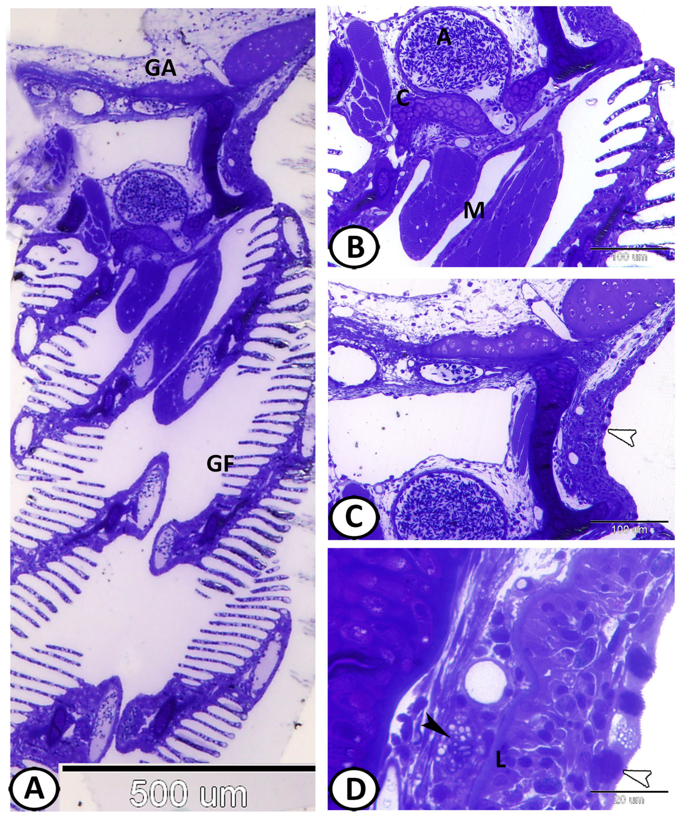
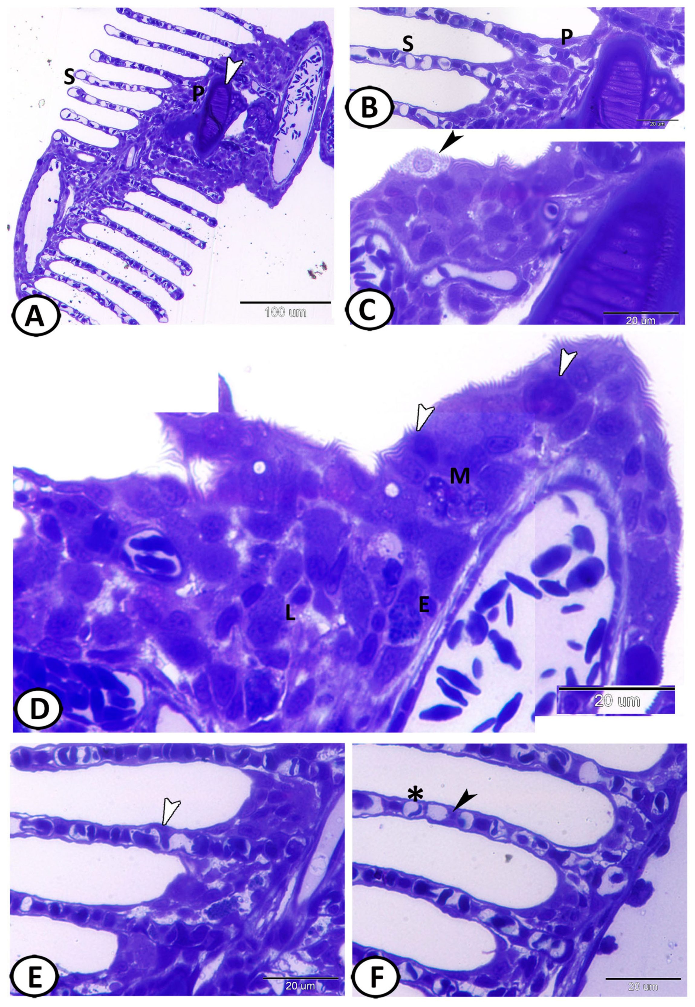
2.3. Semithin Sections and Transmission Electron Microscoy (TEM)

Small specimens of the gills were fixed in a solution of 2.5% paraformaldehyde-glutaraldehyde and left overnight for fixation [20]. Then, they were washed in 0.1 mol/L phosphate buffer and osmicated with 1% osmium tetroxide in 0.1 mol/L sodium-cacodylate buffer at pH 7.3. After that, the specimens were dehydrated by ethanol followed by propylene oxide, and embedded in Araldite. One μm-thick semithin sections were stained with toluidine blue and examined under a light microscope. Ultrathin sections (70 nm) were obtained using Ultrotom-VRV (LKB, Bromma, Germany) and were stained with lead citrate and uranyl acetate. TEM images were captured with a JEOL-100CX II electron microscope (Massachusetts, Boston, MA, USA).

2.4. Scanning Electron Microscopy (SEM)

Small specimens of the gills were immediately washed with 0.1 M Na cacodylate buffer and were fixed in a mixture of 2.5% paraformaldehyde and 2.5% glutaraldehyde in 0.1 M Na-cacodylate buffer (pH 7.3) for 4 h at 4 °C. Samples were washed in the cacodylate buffer and were post-fixed at room temperature in 1% osmic acid in 0.1 M Na-cacodylate buffer for 2 h, followed by dehydration through acetone and isoamyl acetate. The specimens were subjected to a critical point drying method with a polaron apparatus and were finally coated with gold and observed with a JEOL Scanning electron microscope (JSM-5400 LV) at KV 10 at the Electron Microscopy Unit of Assiut University, Egypt.

2.5. Immunohistochemical Analysis

Sections of the gills were prepared for immunohistochemical analysis using Pierce Peroxidase Detection Kit (36,000, Thermo Fisher Scientific, Waltham, MA, USA). The sections were deparaffinized with xylene, rehydrated in graded ethanol, and washed with distilled water. The sections were heated for 15 min in a sodium citrate buffer (0.01 M, pH 6.0) in a microwave to increase epitope exposure. The sections were cooled at room temperature for 30 min, washed with wash buffer (Tris-buffered saline with 0.05% Tween-20 Detergent), and then incubated for 30 min in a peroxidase suppressor to quench the endogenous peroxidase activity. The tissues were washed twice for 3 min with a wash buffer and were blocked with universal blocker™ blocking buffer (36,000, Thermo Fisher Scientific, Waltham, MA, USA) in TBS for 30 at room temperature. The sections were incubated overnight at 4 °C with diluted (1:100) primary antibodies against the polyclonal glial fibrillary acidic protein (GFAP) (PA5-16291, Thermo Fisher Scientific, Waltham, MA, USA), polyclonal Interleukin 1 beta (IL-1β) (sc-7884, Santa Cruz Biotechnology, Heidelberg Germany), monoclonal autophagy protein 5 (APG5) (sc-133158, Santa Cruz Biotechnology, Heidelberg, Germany), mouse monoclonal TGF-β (1:100, MA5-16949, Thermo Fischer Scientific, Warrington, UK), rabbit polyclonal iNOS-2 (RB-1605, Thermo Fisher Scientific, Warrington, UK), rabbit polyclonal nuclear factor kappa B (NF-κB) (10745-1-AP, Proteintech, Rosemont, IL, USA), nuclear factor erythroid 2-related factor 2 (Nrf2) (sc-722, Santa Cruz Biotechnology, Heidelberg, Germany), myostatin (AB3239, Sigma-Aldrich, Madrid, Spain), SRY-Box transcription factor 9 (Sox9) (AB5535, Sigma-Aldrich, Madrid, Spain), and S100 protein (Z0311, Dako, Glostrup, Denmark). In parallel, tissue specimens, in which S100 protein primary antibody was omitted and replaced with buffer, served as negative controls. The slides were washed twice for 3 min with wash buffer and were incubated with diluted (1:1000) goat anti-rabbit IgG (65-6140, Invitrogen, Waltham, MA, USA) and diluted (1:100) goat anti-mouse IgG (31800, Invitrogen, Waltham, MA, USA) secondary antibodies for 30 min at room temperature. Following that, the slides were washed three times for 3 min each with a wash buffer, and the tissues were incubated with the diluted (1:500) Avidin-HRP (43-4423, Invitrogen, Waltham, MA, USA) in a universal blocker blocking buffer for 30 min. The slides were then washed three times for 3 min each with a wash buffer. The tissues were incubated with a 1x metal-enhanced DAB substrate working solution (by adding stable peroxide buffer to the 10x DAB/Metal Concentrate) for 5–15 min until the desired staining was achieved. Finally, the sections were washed twice for 3 min each with a wash buffer, counterstained with Harris-modified hematoxylin, and mounted with mounting media.

2.6. Digitally Colored TEM Images

To increase the visual contrast between many structures on the same electron micrograph, we digitally colored specific cells including; immune cells, chloride cells, neuroepithelial cells, stem cells, pillar cells, mucous cells, squamous PVC, and telocytes. All the elements were carefully hand-colored using Adobe Photoshop software version CS6 (version 6, Adobe, San Jose, CA, USA).

3. Results

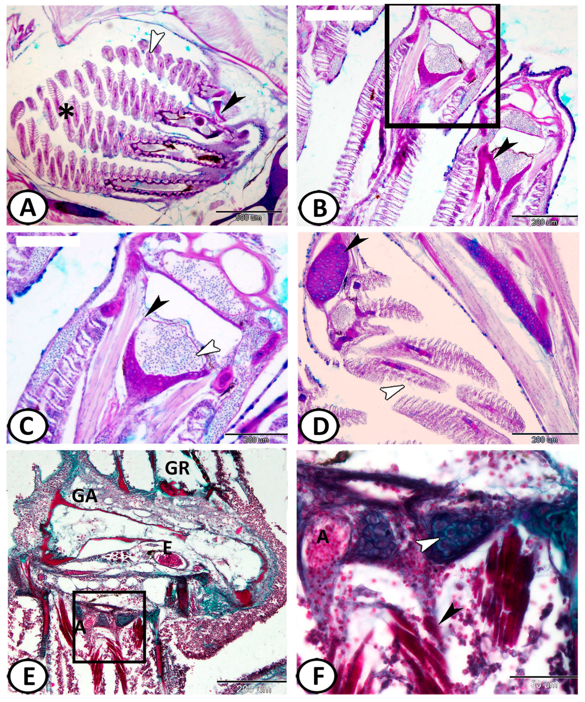
3.1. Histological Analysis

Each gill arch bore regularly spaced free rows of filaments projecting posterolaterally. Each row or stack of filaments constituted a hemibranch, while a set of hemibranchs, one on each side of the gill arch, constituted a holobranch. The gills of molly fishes consisted of four holobranchs spaced between five branchial slits (chambers) (Figure 1A). Each hemibranch carried many fine subdivisions called primary gill lamellae, which projected from the arch like the teeth of a comb (Figure 1B–D). The gill arch was a curved cartilaginous structure, from which radiated the bony supports (the gill rays) of the primary lamellae. The associated striated abductor and adductor muscles were well distinct (Figure 1E,F). The inner surfaces of the gill arches carried one or more rows of stiff strainers called gill rakers (Figure 1E). The gill arch contained the afferent brachial arteries (Figure 1E) from the ventral aorta and the efferent brachial arteries serving the dorsal aorta.
The semithin sections showed that the supported cartilages of the gill arch continued to the primary gill filaments (Figure 2A). The gill arch was supported by skeletal muscle fibers (Figure 2B). The gill arch was covered by epidermal tissue (stratified epithelium) well- endowed with mucous cells. Below the epidermal tissue, there was lymphocytic infiltration. The mucous cells were a distinct feature of the gill filament epithelium that were frequently observed in the afferent and efferent edges, the base of lamellae, and the outer margin of lamellae. Bundles of nerve fibers could be demonstrated in the connective tissue below the epithelium (Figure 2C,D).
The primary lamella was supported by central cartilage and contained afferent and efferent arterioles, and other anastomosing blood vessels (Figure 3A,B). The epithelium of the primary lamellae was thicker than that of the secondary one, typically composed of three or more cell layers (Figure 3C). They were covered by epidermal tissue (stratified epithelium) containing salt-secreting chloride cells. These chloride cells were most numerous in the basal (proximal) part of the lamellae (Figure 3C). At the origin of the primary lamellae, the epithelium was much thicker and contained numerous metachromatic mucous cells. Below the epithelium, there was a varying number of lymphocytes, neuroendocrine cells, and macrophages (Figure 3D).
The secondary lamella appeared as a thin envelope of cells lying on a basement membrane. The secondary lamellae comprised pillar cells separated by blood capillaries that form an air-blood barrier equivalent to that present in mammals. The surface of the secondary lamellae consisted of overlapping or interdigitating squamous epithelial cells called pavement cells (PVC) (Figure 3E,F).

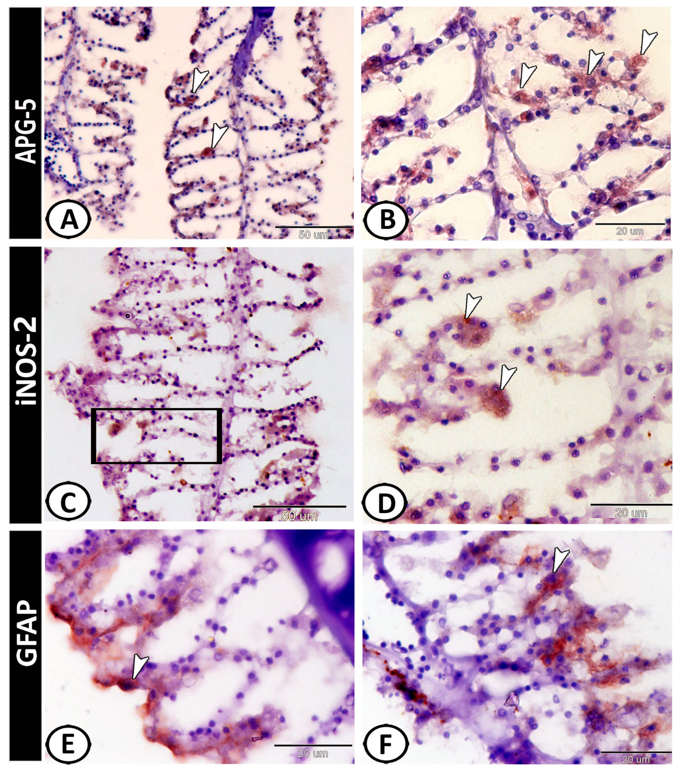
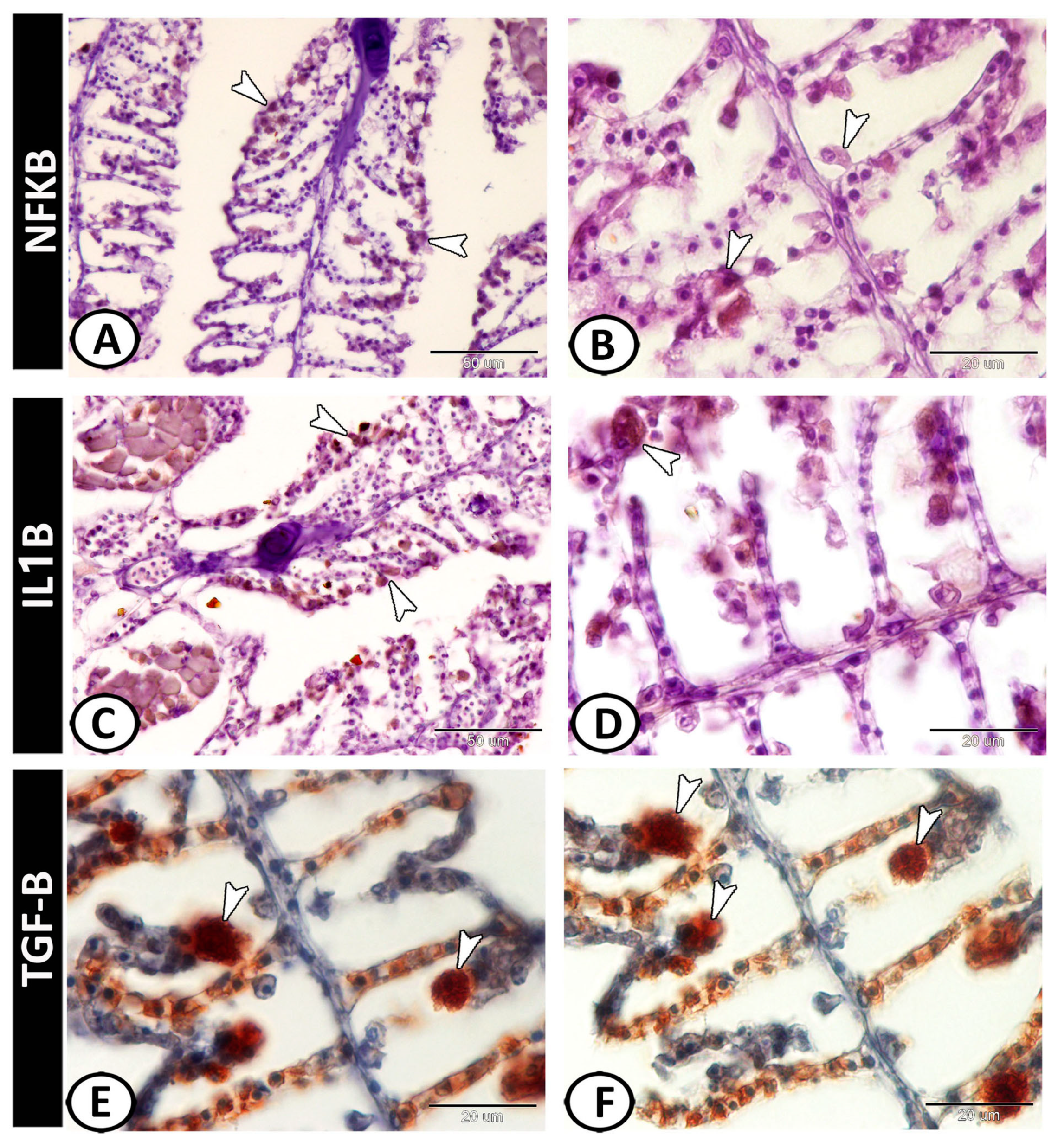
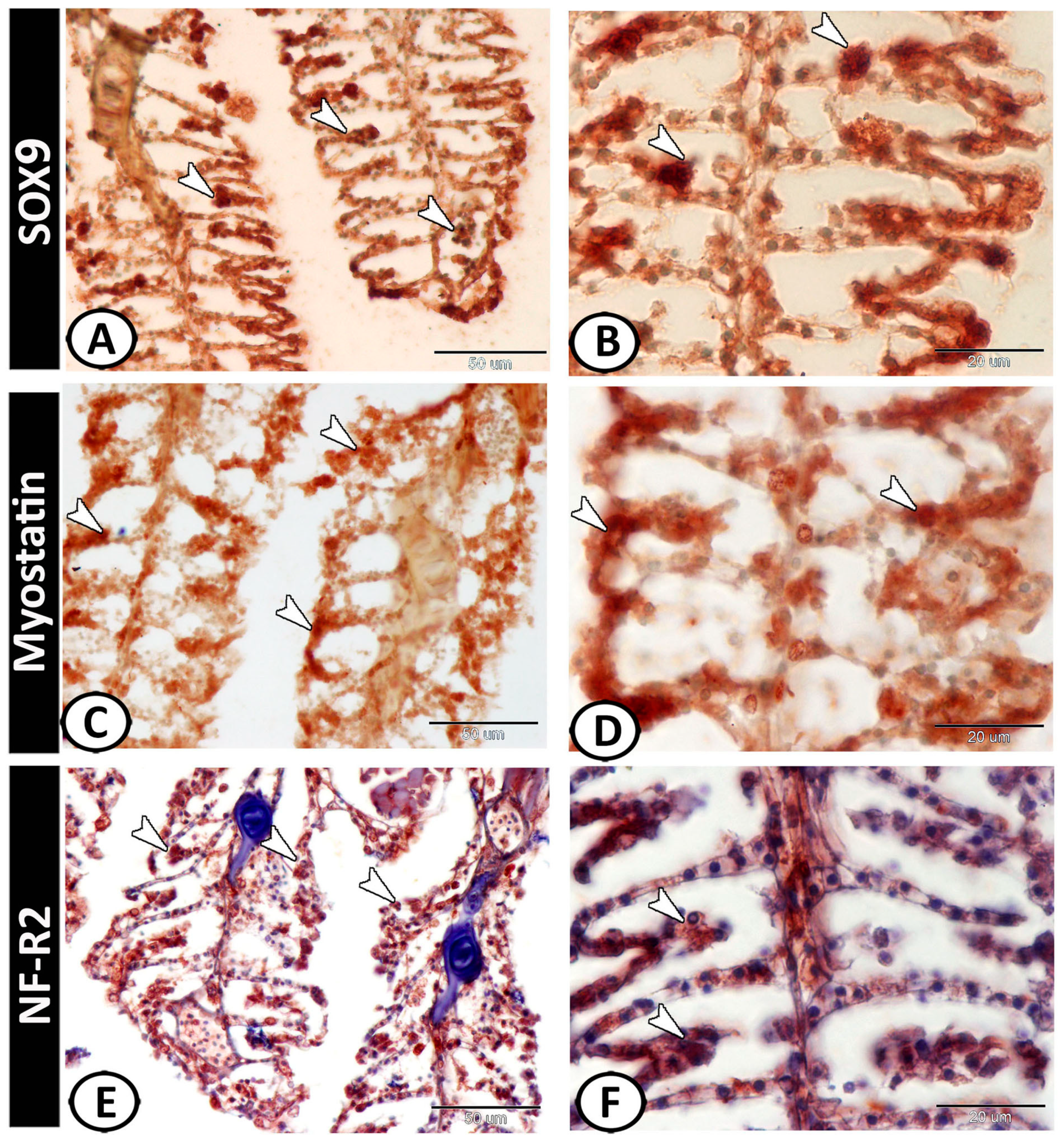
3.2. Immunohistochemical Analysis

The immunohistochemical analysis showed APG-5 positive immunoreactivity of macrophages (Figure 4A,B). Also, iNOS-2 was expressed in macrophages (Figure 4C,D). On the other hand, the epithelium of the primary and secondary gill lamellae contained positive-GFAP astrocytes (Figure 4E,F).
Both IL-1β and NF-κB showed immunoreactivity in the inflammatory cells (including monocytes and macrophages) of the gill filaments (Figure 5A–D), Furthermore, macrophages expressed TGF-B (Figure 5 E,F).
The stem cells expressed SOX9 (Figure 6A,B), myostatin (Figure 6C,D), and Nrf2 (Figure 6E,F). Moreover, neuroendocrine cells expressed S100 protein (Figure 7A–C). Chloride cells showed positive immunoreactivity to S100 protein (Figure 7D).

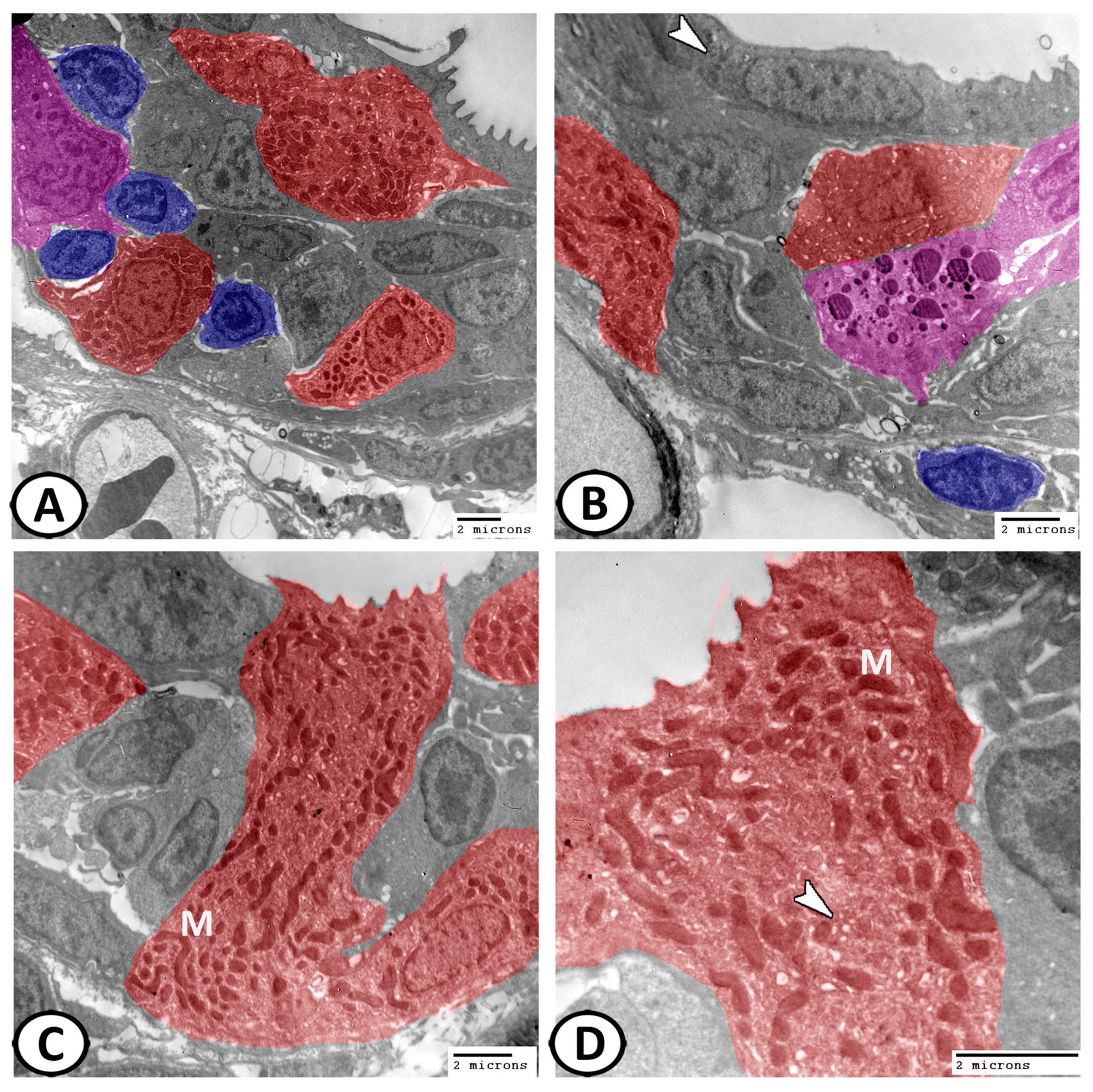
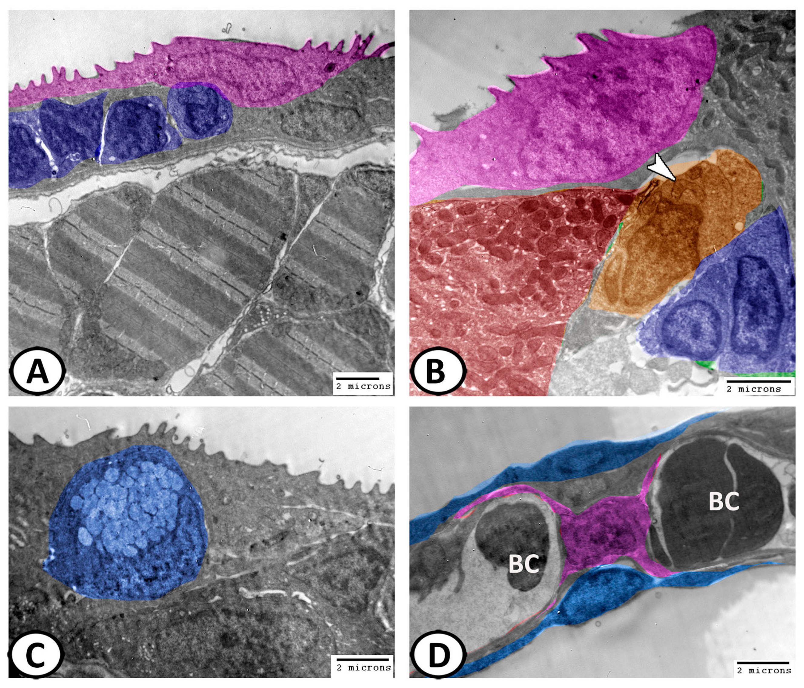
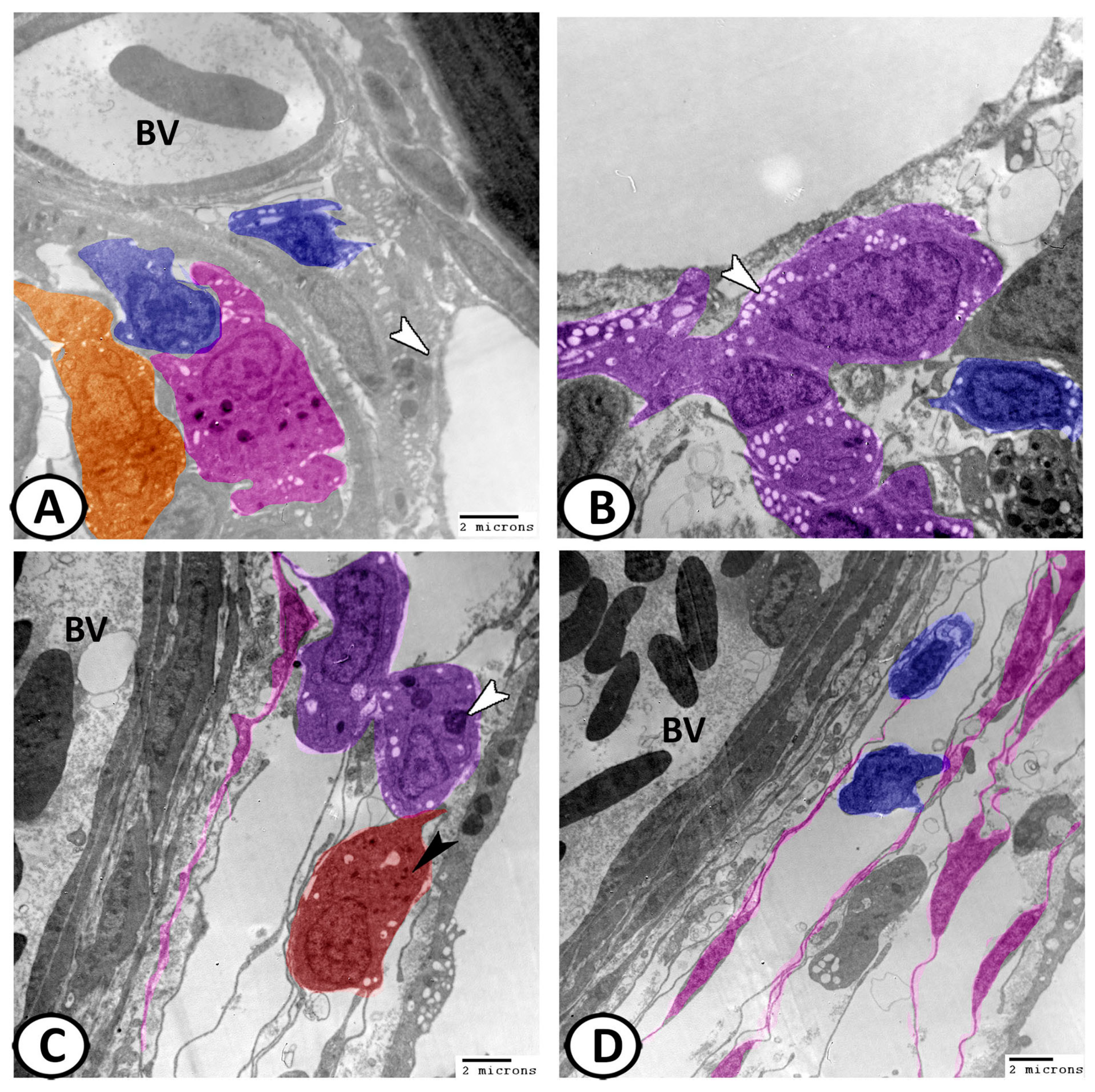
3.3. Transmission Electron Microscopy of Cellular Constituents of the Gill Filaments

3.3.1. Chloride Cells (Mitochondria-Rich Cells, MRCs)

Mitochondria-rich cells (MRCs) were surrounded by flattened PVC (Figure 8A,B). These cells may be elongated or ovoid in shape (Figure 8A–C). The defining characteristic of this cell type was a high mitochondrial density (Figure 8C,D). In the apical region of MRCs was a collection of vesicles and tubules forming a tubulovesicular system. The apical membrane showed shallow microridges (Figure 8D).

3.3.2. Pavement Cells (PVC)

The most abundant cell type covering the epithelium (90% of the surface area) was a squamous to cuboid-shaped cell commonly referred to as the pavement or respiratory cell. The nucleus in squamous PVC was compressed (Figure 9A), while in cuboidal PVC, it was ovoid (Figure 9B). The apical surface of PVC possessed irregular finger-like projection microridges (Figure 9A,B).

3.3.3. Mucous Cells

The mucous cells were a distinct feature of the gill filament epithelium that were frequently observed in the outer margin of the lamellae. They were large ovoid cells that consisted mostly of apical mucous secretory granules. The nucleus was flattened and located in a basal position (Figure 9C).

3.3.4. Pillar Cells

These were modified endothelial cells with a centrally located polymorphic nucleus that supports and defines the lamellar blood spaces. Where the pillar cells impinge on the basement membrane, the blood capillaries spread to form flanges, which coalesce with those of neighboring pillar cells to complete the lining of the lamellar blood channels, which contact the afferent and efferent lamellar arteries. They gave the lamellae the appearance of a string of beads when viewed in cross-section (Figure 9D). The mean thickness of the lamellar blood channel was 5.4 μm, which represented the total diffusion distance for the respiratory exchange.

3.3.5. Other Cells

Other cells found within the filament interstitium include lymphocytes, macrophages, monocytes, neuroepithelial cells, telocytes, and stem cells.
The lymphocytes were characterized by a high nucleus to cytoplasmic ratio and distributed in the epithelium of the primary gill lamellae (Figure 8A,B and Figure 9A,B) and the interstitial connective tissues around the blood vessels (Figure 10A,B,D).
The macrophages were found in the epithelium of the primary lamellae (Figure 8A) and distributed near the blood vessels and capillaries of gill filaments (Figure 10A,C) and their cytoplasm showed pseudopodia, vacuoles, and heterogeneous contents.
Monocytes could also be recorded in the interstitial connective tissue that appeared large with euchromatic nucleus and many clumps of heterochromatin. Their cytoplasm showed few mitochondria and numerous vacuoles (Figure 10B).
The neuroepithelial cells were found deep within the filament epithelium along the full length of the efferent edge, although concentrated near the filament tips and contained electron-dense secretory granules (Figure 8B and Figure 10C).
Telocytes (TCs) were interstitial cells that usually associated with blood vessels and showed telopodes (TPs) that forming a network with immune cells (Figure 10C,D). Stem cells with a dividing nucleus and cytoplasm containing mitochondria could also be identified in the epithelium of the primary gill lamellae (Figure 9B and Figure 10A).

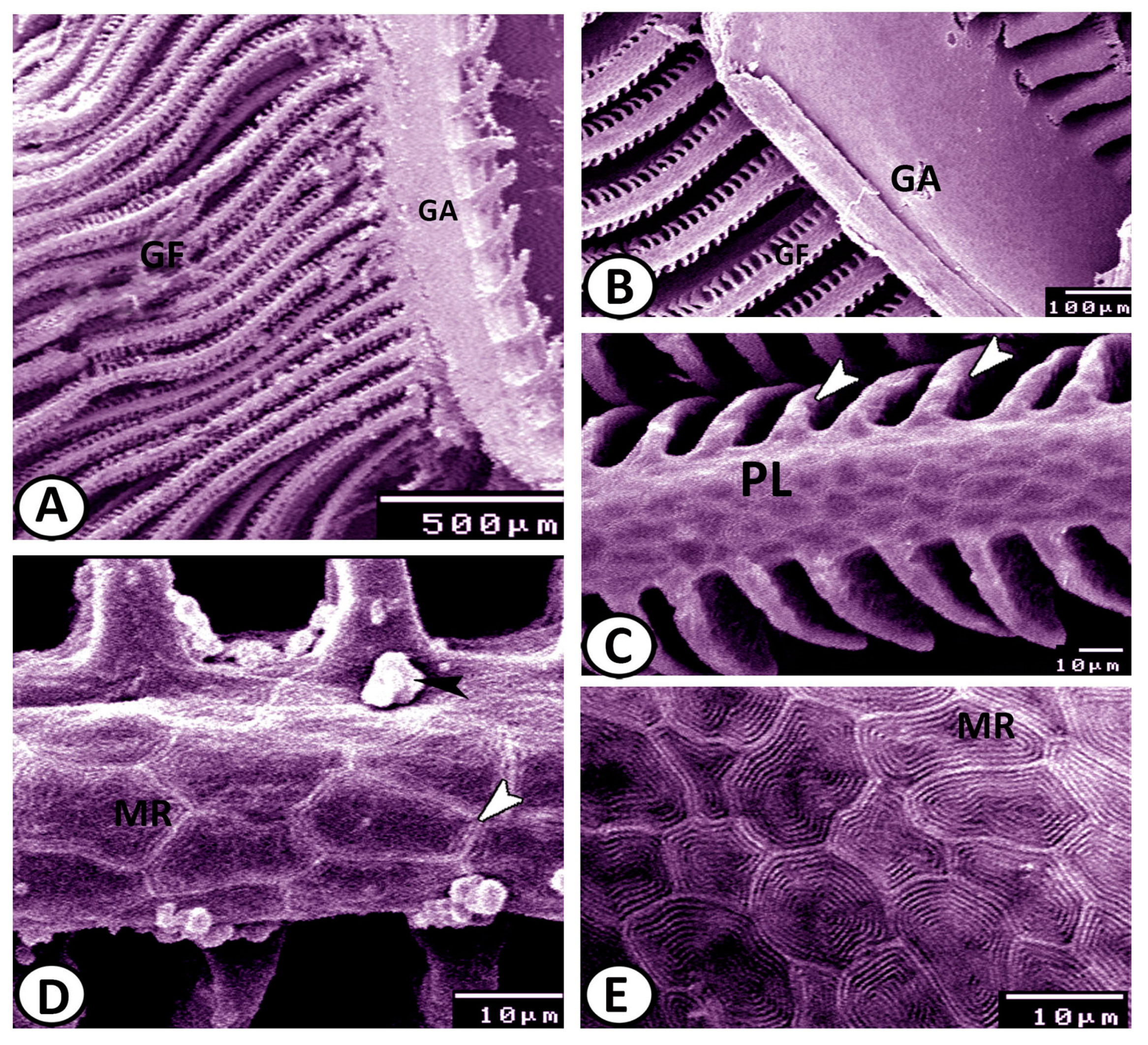
3.4. Scanning Electron Microscopy

The scanning electron microscopy showed that the gill filaments were radiated from the gill arch (Figure 11A,B). The surface of the gill filaments was composed mainly of compactly arranged PVC. The apical surface of PVC possessed concentrically arranged microridges that interwoven to form a finger-print like- pattern. A well-defined double row of microridges demarked the borders of neighboring PVC. Some mucus secretions from the mucous cells were seen on the surface of gill filaments (Figure 11C,D).

4. Discussion

The gill lamellae in molly fish are free and this arrangement provides a larger respiratory surface and the associated striated abductor and adductor muscles in the gill arch facilitate the movement of the gills to favorable respiratory positions [21].
The present study revealed the presence of chloride cells. Chloride cells are acidophilic and are found generally in marine fishes and rarely in freshwater fishes [21]. The chloride cells may be elongated, ovoid, or cuboidal in shape, depending upon the species with granular cytoplasm. MRCs are frequently concentrated in the afferent region of the filament epithelium, although in the interlamellar region, MRCs are also associated with the basal channels of the lamellar arterio-arterial circulation [22].
The term “chloride cell” relates to the main function of the MRC in chloride ion elimination [23]. However, the presence of numerous mitochondria in these cells is thought to supply the adenosine triphosphate (ATP) for ion-transport proteins to drive the vectorial transport of ions as part of ion and acid–base regulation [24]. The function of the chloride cells in ionic transport with a possible role in detoxification was previously reported [25].
The current results indicate that the pillar cells of molly fish form a complex structure similar to the air-blood barrier in mammals. Pillar cells contain columns of contractile protein. Since the blood entering the lamellar blood spaces comes directly from the ventral aorta at high pressure. The presence of contractile elements in the supports of these spaces serves to resist their distension under normal circumstances [26]. Also, the contraction of pillar cells can be used to control the blood flow rate through the gaseous exchange surfaces.
PVCs were abundant in molly fish. These cells were covered by microridges that increase the apical surface area that supports excretory and respiratory functions [27]. These microridges have been supposed to promote protection and mechanical flexibility as well as a firm consistency to the free surface of the epithelial cells [28].
The mucous cells were reported in the epithelium of the gills. These cells function as mechanical and immunological protection in reducing infection and abrasion and have a significant role in regulating the exchange of gas, water, and ions [16]. Goblet cells have been found in all fishes with the exception of the hagfishes [21].
Fish gills are essential for gas exchange, and they are vulnerable to waterborne microorganisms. The gills contain a variety of immune cells, including mast cells that are co-localized with neuroepithelial cells [29]. Peripheral oxygen-sensing chemoreceptors are located mostly in the teleost gills, and are identified as neuroepithelial cells (NECs) [30,31]. The distribution of these NECs has been reported throughout the gill filaments and lamellae of the gill arches in several teleost species, the air-breathing organs of primitive fishes and the gill fans and the suprabranchial chamber of the accessory respiratory organs in the air-breathing catfish [32]. Interestingly, The NECs of Fish gill have been detected to share several morphological features with other peripheral O2 chemoreceptors including the mammalian carotid body and the NEC-like cells of lungfishes’ and bichirs’ lungs [33,34]. Previous studies recorded the occurrence of the NECs in skin surfaces [35]. Many neurotransmitter substances such as nitric oxide, serotonin, acetylcholine, neuropeptides, and catecholamines have been detected in the NECs of teleost and air-breathing fishes [31,35,36,37,38].
The oxygen sensors of the NECs are present in the gill filaments of a wide diversity of teleost and non-teleosts species [31,37,38,39]. The present immunohistochemical analysis revealed that the NECs of molly fish expressed s100 protein. These NECs have a role as sensors of hypoxia changes in the external environments, besides their endocrine nature and secretion of immunomodulatory substances can influence several types of immune cells [40]. NECs also showed immunoreactivity to a variety of both neuronal markers (VAChT, nAChR, GABA-B-R1 receptor, GAD679) and the antimicrobial peptide piscidin, an evolutionary conserved humoral component of the mucosal immune system in fish [41]. Furthermore, these cells are involved in the regulation of blood flow [34]. The fish neuroepithelial cells share common characteristics with the neuroepithelial cells described within the wall of the mammalian respiratory tract [42].
The present study showed a high frequency of immune cells in the gills of molly fish. A network of immune cells (monocytes, eosinophils, and mast cells) was also observed in the gill epithelia of some fish species [43]. The current results showed that the gill macrophages expressed APG5, iNOS2, IL-1B, and NF-kB. APG5 is one of the key players in the autophagy process [44], where it is essential for autophagic vesicle formation, lymphocyte development and proliferation, and apoptosis [45]. NF-κB signaling in epithelial cells was previously reported to have significant roles in immune homeostasis maintenance in barrier tissues [46]. Furthermore, expressions of PTPRC (CD45), CD83, and IL-1B were previously described in antigen-sampling cells in fish gill epithelium [47].
Moreover, macrophages of molly gills also expressed TGF-β. TGF-βs, as multifunctional cytokines, can regulate cell differentiation, proliferation, migration, and apoptosis. The Grass carp (Ctenopharyngodon idellus) TGF-β1 exerts a bidirectional regulatory effect on the proliferation of quiescent and activated peripheral blood leukocytes [48]. Red seabream (Pagrus major) TGF-β can also regulate the migration of head kidney cells and peripheral blood leukocytes in a context-dependent manner. However, there is still a lack of sufficient data to support that TGF-βs are the most ancestral immunomodulatory cytokine in vertebrates [49].
The present study showed distribution of stem cells in the gills of molly fish that expressed SOX9, NFr2, and myostatin. Myostatin is a growth and differentiation factor of the TGF-β superfamily Myostatin precursor was reported to be present in several tissues in teleost fish [50]. The expression of sox9 is detected in gonads, muscles, brains, livers, fins, gills, kidneys, eyes and spleens of Siberian sturgeons (A. baerii), A. schrenckii and stellate sturgeons (A. stellatus), suggesting that sox9 is involved in defining progenitor cells in a variety of tissues in fish. These studies suggest that some sox genes play an important role in physiological and development processes in fish [51].
The current investigation revealed that TCs are interstitial cells found in the connective tissue around the blood vessels that establish relations to various types of immune cells, which indicated their immunological role. A recent study by Huang et al. [52] supposed that TCs are involved in the phagocytosis and differentiation of macrophages. Also, they suggest that TCs may be potential participants in the initiation of inflammation. TCs are also reported in gonads, skin, gills, around the bile ducts and blood vessels of grass carp, silver carp, and tilapia [21].
Some nerve fibers were recorded in the gills of molly fish. The fish gill is a highly complex organ that receives vast innervation from both afferent (sensory) and efferent (motor) fibers. Innervation from the latter source includes autonomic nerve fibers of spinal (sympathetic) and cranial (parasympathetic) origin, whose primary role is to induce vasomotor changes within the respiratory or non-respiratory pathways of the gill vasculature [13].

5. Conclusions

The present study showed for the first time the expression of different immune markers in the gills of molly fish, including APG5, iNOS2, IL-1B, NF-kB, and TGF-β, which indicating the immune role of the gills. Moreover, s100 protein was expressed in the NECs that were found in association with the immune cells. Other epithelial and interstitial cells were recorded in the molly gills, including chloride cells, PVCs, pillar cells, TCs, and mucous cells. The current work suggests that the gills of molly fish is multifunctional organ and are involved in immune reaction.

Author Contributions

Conceptualization—methodology—formal analysis—data curation, D.M.M., R.K.A.S. and M.M.H.; investigation- resources, A.A. and E.R.L.; writing—original draft preparation, D.M.M., R.K.A.S. and M.M.H.; writing—review and editing, G.Z. and A.A.; visualization, E.R.L.; investigation—supervision, D.M.M. and G.Z. All authors have read and agreed to the published version of the manuscript.

Funding

This research received no external funding.

Institutional Review Board Statement

The study protocol was conducted in strict accordance with the relevant guidelines and ethical regulations and was approved by The National Ethical Committee of the Faculty of Veterinary Medicine, Assiut University, Egypt (The Ethical No. is aun/vet/4/0015). This study is reported in accordance with ARRIVE guidelines.

Data Availability Statement

The data presented in this study are available within the article.

Acknowledgments

The authors thank staff members and technicians of the Electron Microscopy Unit of Assiut University, Egypt for their valuable support with transmission electron microscopy.

Conflicts of Interest

The authors declare no conflict of interest.

References

  1. Tembo, R.N. The sublethal effects of low-pH exposure on the chemoreception of Poecilia sphenops. Arch. Environ. Contam. Toxicol. 2009, 57, 157–163. [Google Scholar] [PubMed]
  2. Alda, F.; Reina, R.G.; Doadrio, I.; Bermingham, E. Phylogeny and biogeography of the Poecilia sphenops species complex (Actinopterygii, Poeciliidae) in Central America. Mol. Phylogenetics Evol. 2013, 66, 1011–1026. [Google Scholar]
  3. Goss, G.G.; Perry, S.F.; Fryer, J.N.; Laurent, P. Gill morphology and acid-base regulation in freshwater fishes. Comp. Biochem. Physiol. Part A Mol. Integr. Physiol. 1998, 119, 107–115. [Google Scholar] [CrossRef] [PubMed]
  4. Evans, D.H.; Piermarini, P.M.; Choe, K.P. The multifunctional fish gill: Dominant site of gas exchange, osmoregulation, acid-base regulation, and excretion of nitrogenous waste. Physiol. Rev. 2005, 85, 97–177. [Google Scholar] [PubMed]
  5. Sasai, S.; Kaneko, T.; Hasegawa, S.; Tsukamoto, K. Morphological alteration in two types of gill chloride cells in Japanese eels (Anguilla japonica) during catadromous migration. Can. J. Zool. 1998, 76, 1480–1487. [Google Scholar] [CrossRef]
  6. Elsheikh, E. Scanning electron microscopic studies of gill arches and rakers in relation to feeding habits of some fresh water fishes. J. Basic Appl. Zool. 2013, 66, 121–130. [Google Scholar]
  7. Wilson, J.M.; Laurent, P. Fish gill morphology: Inside out. J. Exp. Zool. 2002, 293, 192–213. [Google Scholar]
  8. Perry, S.F. The chloride cell: Structure and function in the gills of freshwater fishes. Annu. Rev. Physiol. 1997, 59, 325–347. [Google Scholar]
  9. Zaccone, G.; Lauriano, E.R.; Kuciel, M.; Capillo, G.; Pergolizzi, S.; Alesci, A.; Ishimatsu, A.; Ip, Y.K.; Icardo, J.M. Identification and distribution of neuronal nitric oxide synthase and neurochemical markers in the neuroepithelial cells of the gill and the skin in the giant mudskipper, Periophthalmodon schlosseri. Zoology 2017, 125, 41–52. [Google Scholar]
  10. Zaccone, G.; Cupello, C.; Capillo, G.; Kuciel, M.; Nascimento, A.L.; Gopesh, A.; Germana, G.P.; Spanò, N.; Guerrera, M.C.; Aragona, M. Expression of acetylcholine-and G protein coupled muscarinic receptor in the neuroepithelial cells (NECs) of the obligated air-breathing fish, Arapaima gigas (Arapaimatidae: Teleostei). Zoology 2020, 139, 125755. [Google Scholar] [CrossRef]
  11. Blake, K.J.; Jiang, X.R.; Chiu, I.M. Neuronal regulation of immunity in the skin and lungs. Trends Neurosci. 2019, 42, 537–551. [Google Scholar]
  12. Bailly, Y. Serotonergic neuroepithelial cells in fish gills: Cytology and innervation. In Airway Chemoreceptors in the Vertebrates; CRC Press: Boca Raton, FL, USA, 2009; pp. 61–97. [Google Scholar]
  13. Maina, J.N.; Icardo, J.M.; Zaccone, G.; Aragona, M.; Lauriano, E.R.; Alesci, A.; Albano, M.; Guerrera, M.C.; Germana, A.; Fernandes, J.M.O. Immunohistochemical and ultrastructural study of the immune cell system and epithelial surfaces of the respiratory organs in the bimodally breathing African sharptooth catfish (Clarias gariepinus Burchell, 1822). Anat. Rec. 2022, 305, 3212–3229. [Google Scholar] [CrossRef]
  14. Sayed, R.K.; Zaccone, G.; Capillo, G.; Albano, M.; Mokhtar, D.M. Structural and functional aspects of the spleen in molly fish Poecilia sphenops (Valenciennes, 1846): Synergistic interactions of stem cells, neurons, and immune cells. Biology 2022, 11, 779. [Google Scholar]
  15. Hussein, M.M.; Sayed, R.K.; Mokhtar, D.M. Structural and immunohistochemical analysis of the cellular compositions of the liver of molly fish (Poecilia sphenops), focusing on its immune role. Zool. Lett. 2023, 9, 1. [Google Scholar]
  16. Mokhtar, D.M.; Hussein, M.M.; Sayed, R.K. Novel Identification and Microscopy of the Intestinal Bulb of Molly Fish (Poecilia sphenops) with a Focus on Its Role in Immunity. Microsc. Microanal. 2022, 28, 1827–1839. [Google Scholar]
  17. Hirose, S.; Kaneko, T.; Naito, N.; Takei, Y. Molecular biology of major components of chloride cells. Comp. Biochem. Physiol. Part B Biochem. Mol. Biol. 2003, 136, 593–620. [Google Scholar]
  18. Li, J.; Xu, X.; Liu, C. A scanning electron microscopical observation of gill structure of largemouth bass Micropterus salmoides and blue gill Lepomis macrochirus. J. Dalian Fish. Univ. 2009, 24, 231–235. [Google Scholar]
  19. Bancroft, J.D.; Gamble, M. Theory and Practice of Histological Techniques; Elsevier Health Sciences: Amsterdam, The Netherlands, 2008. [Google Scholar]
  20. Karnovsky, M. A formaldehyde-glutaraldehyde fixative of high osmolality for use in electron microscopy. J. Cell Biol. 1965, 27, 137A. [Google Scholar]
  21. Mokhtar, D.M. Fish Histology: From Cells to Organs; Apple Academic Press: Oakville, ON, Canada, 2021. [Google Scholar]
  22. Evans, D.H.; Piermarini, P.M.; Potts, W. Ionic transport in the fish gill epithelium. J. Exp. Zool. 1999, 283, 641–652. [Google Scholar]
  23. Watrin, A.; Mayer-Gostan, N. Simultaneous recognition of ionocytes and mucous cells in the gill epithelium of turbot and in the rat stomach. J. Exp. Zool. 1996, 276, 95–101. [Google Scholar]
  24. Heijden, A.; Verbost, P.; Eygensteyn, J.; Li, J.; Bonga, S.; Flik, G. Mitochondria-rich cells in gills of tilapia (Oreochromis mossambicus) adapted to fresh water or sea water: Quantification by confocal laser scanning microscopy. J. Exp. Biol. 1997, 200, 55–64. [Google Scholar] [CrossRef]
  25. Sturla, M.; Masini, M.; Prato, P.; Grattarola, C.; Uva, B. Mitochondria-rich cells in gills and skin of an African lungfish, Protopterus annectens. Cell Tissue Res. 2001, 303, 351–358. [Google Scholar] [CrossRef]
  26. Bettex-Galland, M.; Hughes, G. Contractile filamentous material in the pillar cells of fish gills. J. Cell Sci. 1973, 13, 359–370. [Google Scholar] [CrossRef] [PubMed]
  27. Carmona, R.; García-Gallego, M.; Sanz, A.; Domezain, A.; Ostos-Garrido, M. Chloride cells and pavement cells in gill epithelia of Acipenser naccarii: Ultrastructural modifications in seawater-acclimated specimens. J. Fish Biol. 2004, 64, 553–566. [Google Scholar] [CrossRef]
  28. Sayed, R.K.; Abd-El Aziz, N.A.; Ibrahim, I.A.; Mokhtar, D.M. Structural, ultrastructural, and functional aspects of the skin of the upper lip of silver carp (Hypophthalmichthys molitrix). Microsc. Res. Tech. 2021, 84, 1821–1833. [Google Scholar] [CrossRef]
  29. Zaccone, G. Immunity and Neuroimmune Interactions at the Mucosal Barriers in Fish. Fishes 2022, 7, 381. [Google Scholar] [CrossRef]
  30. Zaccone, G.; Lauweryns, J.; Fasulo, S.; Tagliafierro, G.; Ainis, L.; Licata, A. Immunocytochemical localization of serotonin and neuropeptides in the neuroendocrine paraneurons of teleost and lungfish gills. Acta Zool. 1992, 73, 177–183. [Google Scholar] [CrossRef]
  31. Zaccone, G.; Fasulo, S.; Ainis, L.; Licata, A. Paraneurons in the skin and gills of fishes. In Ichthyology: Recent Research Advances; Saksena, D.N., Ed.; Science Publishers Inc.: Enfield, NH, USA, 1997; pp. 417–447. [Google Scholar]
  32. Zaccone, G.; Lauriano, E.R.; Capillo, G.; Kuciel, M. Air-breathing in fish: Air-breathing organs and control of respiration: Nerves and neurotransmitters in the air-breathing organs and the skin. Acta Histochem. 2018, 120, 630–641. [Google Scholar] [CrossRef] [PubMed]
  33. Zaccone, G.; Mauceri, A.; Maisano, M.; Giannetto, A.; Parrino, V.; Fasulo, S. Innervation and neurotransmitter localization in the lung of the Nile bichir Polypterus bichir bichir. Anat. Rec. 2007, 290, 1166–1177. [Google Scholar] [CrossRef]
  34. Zachar, P.C.; Jonz, M.G. Neuroepithelial cells of the gill and their role in oxygen sensing. Respir. Physiol. Neurobiol. 2012, 184, 301–308. [Google Scholar] [CrossRef] [PubMed]
  35. Zaccone, D.; Icardo, J.M.; Kuciel, M.; Alesci, A.; Pergolizzi, S.; Satora, L.; Lauriano, E.R.; Zaccone, G. Polymorphous granular cells in the lung of the primitive fish, the bichir P olypterus senegalus. Acta Zool. 2017, 98, 13–19. [Google Scholar] [CrossRef]
  36. Zaccone, G.; Mauceri, A.; Fasulo, S. Neuropeptides and nitric oxide synthase in the gill and the air-breathing organs of fishes. J. Exp. Zool. Part A Comp. Exp. Biol. 2006, 305, 428–439. [Google Scholar] [CrossRef]
  37. Jonz, M.G.; Zaccone, G. Nervous control of the gills. Acta Histochem. 2009, 111, 207–216. [Google Scholar] [CrossRef]
  38. Jonz, M.G.; Buck, L.T.; Perry, S.F.; Schwerte, T.; Zaccone, G. Sensing and surviving hypoxia in vertebrates. Ann. N. Y. Acad. Sci. 2016, 1365, 43–58. [Google Scholar] [CrossRef]
  39. Porteus, C.S.; Pollack, J.; Tzaneva, V.; Kwong, R.W.; Kumai, Y.; Abdallah, S.J.; Zaccone, G.; Lauriano, E.R.; Milsom, W.K.; Perry, S.F. A role for nitric oxide in the control of breathing in zebrafish (Danio rerio). J. Exp. Biol. 2015, 218, 3746–3753. [Google Scholar] [CrossRef] [PubMed] [Green Version]
  40. Zaccone, G.; Capillo, G.; Fernandes, J.M.O.; Kiron, V.; Lauriano, E.R.; Alesci, A.; Lo Cascio, P.; Guerrera, M.C.; Kuciel, M.; Zuwala, K. Expression of the antimicrobial peptide piscidin 1 and neuropeptides in fish gill and skin: A potential participation in neuro-immune interaction. Mar. Drugs 2022, 20, 145. [Google Scholar] [CrossRef] [PubMed]
  41. Lauriano, E.R.; Capillo, G.; Icardo, J.M.; Fernandes, J.M.O.; Kiron, V.; Kuciel, M.; Zuwala, K.; Guerrera, M.C.; Aragona, M.; Zaccone, G. Neuroepithelial cells (NECs) and mucous cells express a variety of neurotransmitters and neurotransmitter receptors in the gill and respiratory air-sac of the catfish Heteropneustes fossilis (Siluriformes, Heteropneustidae): A possible role in local immune defence. Zoology 2021, 148, 125958. [Google Scholar] [PubMed]
  42. Saltys, H.A.; Jonz, M.G.; Nurse, C.A. Comparative study of gill neuroepithelial cells and their innervation in teleosts and Xenopus tadpoles. Cell Tissue Res. 2006, 323, 1–10. [Google Scholar] [CrossRef]
  43. Mokhtar, D.M.; Abdelhafez, E.A. An overview of the structural and functional aspects of immune cells in teleosts. Histol. Histopathol. 2021, 34, 399–414. [Google Scholar]
  44. Yao, Z.; Delorme-Axford, E.; Backues, S.K.; Klionsky, D.J. Atg41/Icy2 regulates autophagosome formation. Autophagy 2015, 11, 2288–2299. [Google Scholar] [CrossRef] [Green Version]
  45. Pierdominici, M.; Vomero, M.; Barbati, C.; Colasanti, T.; Maselli, A.; Vacirca, D.; Giovannetti, A.; Malorni, W.; Ortona, E. Role of autophagy in immunity and autoimmunity, with a special focus on systemic lupus erythematosus. FASEB J. 2012, 26, 1400–1412. [Google Scholar] [CrossRef]
  46. Pasparakis, M. Role of NF-κB in epithelial biology. Immunol. Rev. 2012, 246, 346–358. [Google Scholar] [CrossRef]
  47. Kato, G.; Miyazawa, H.; Nakayama, Y.; Ikari, Y.; Kondo, H.; Yamaguchi, T.; Sano, M.; Fischer, U. A novel antigen-sampling cell in the teleost gill epithelium with the potential for direct antigen presentation in mucosal tissue. Front. Immunol. 2018, 9, 2116. [Google Scholar] [CrossRef] [PubMed]
  48. Huminiecki, L.; Goldovsky, L.; Freilich, S.; Moustakas, A.; Ouzounis, C.; Heldin, C.-H. Emergence, development and diversification of the TGF-β signalling pathway within the animal kingdom. BMC Evol. Biol. 2009, 9, 28. [Google Scholar] [CrossRef] [Green Version]
  49. Liu, S.; Guo, J.; Cheng, X.; Li, W.; Lyu, S.; Chen, X.; Li, Q.; Wang, H. Molecular Evolution of Transforming Growth Factor-β (TGF-β) Gene Family and the Functional Characterization of Lamprey TGF-β2. Front. Immunol. 2022, 13, 836226. [Google Scholar] [CrossRef] [PubMed]
  50. Radaelli, G.; Rowlerson, A.; Mascarello, F.; Patruno, M.; Funkenstein, B. Myostatin precursor is present in several tissues in teleost fish: A comparative immunolocalization study. Cell Tissue Res. 2003, 311, 239–250. [Google Scholar] [CrossRef]
  51. Hu, Y.; Wang, B.; Du, H. A review on sox genes in fish. Rev. Aquac. 2021, 13, 1986–2003. [Google Scholar] [CrossRef]
  52. Huang, Y.-L.; Zhang, F.-L.; Tang, X.-L.; Yang, X.-J. Telocytes enhances M1 differentiation and phagocytosis while inhibits mitochondria-mediated apoptosis via activation of NF-κB in macrophages. Cell Transplant. 2021, 30, 09636897211002762. [Google Scholar] [CrossRef] [PubMed]
Figure 1. The general structure of the gills of molly fish by light microscopy. (A,B) The gills of molly fish consisted of four holobranchs (white arrowhead) that extend from the floor to the roof of the buccal cavity with four pairs of cartilaginous gill arches (black arrowheads), onto which two rows of gill filaments (asterisk) are attached. (PAS-AB/HX). (C) Higher magnification of the boxed area in (B) shows the gill arch (black arrowhead) and afferent blood vessels (white arrowhead). (D) Gills stained by PAS-AB/HX showing gill filaments (white arrowhead) radiating from the gill arch (black arrowhead). (E) The gill rakers (GR) are bony projections, which point forward and inward from the gill (or branchial) arch (GA). Note the afferent (A) and efferent (E) blood vessels (Crossmon’s trichrome). (F) Higher magnification of the gill arch shows cartilaginous supports (white arrowheads) and branchial muscles (black arrowheads). Note the afferent (A) blood vessels.
Fishes 08 00195 g001
Figure 2. Semithin sections stained with toluidine blue. (A) The cartilaginous core of the gill arch (GA) extends to the gill filaments (GF). (B) Higher magnification of (A) shows the presence of afferent artery (A) with supported cartilage (C) and skeletal muscle fibers (M). (C,D) The covering epithelium of the gill arch consisted of stratified epithelial cells with apical mucous cells (white arrowhead) and basal lymphocytes (L). Note the presence of a bundle of nerve fibers (black arrowhead).
Fishes 08 00195 g002
Figure 3. Semithin sections stained with toluidine blue. (A,B) the gill filaments consist of primary (P) and secondary (S) gill lamellae. (C,D) Higher magnification of one primary gill lamella shows its covering stratified epithelium with chloride cells (black arrowhead), mucous cells (white arrowheads), macrophages (M), and lymphocytes (L). Note the presence of endocrine cells (E) with metachromatic granules in the vicinity of the blood vessels. (E,F) The secondary lamellae consisted of pillar cells (black arrowhead) separated by blood capillaries (asterisk). The surface of the secondary lamellae is covered by pavement cells (PVC, white arrowhead).
Fishes 08 00195 g003
Figure 4. Immunohistochemistry of APG-5, iNOS-2, and GFAP. (A,B) The immunohistochemical analysis revealed APG-5 positive immunoreactivity of macrophages (arrowheads). (C,D) iNOS-2 was expressed in macrophages (arrowheads). (E,F) The epithelium of the primary and secondary gill lamellae contained positive-GFAP astrocytes (arrowheads).
Fishes 08 00195 g004
Figure 5. Immunohistochemistry of IL-1β, NF-κB, and TGF-B. (A,B) IL-1β showed immunoreactivity in the monocytes and macrophages of the gill filaments (arrowheads). (C,D) NF-κB showed immunoreactivity in the inflammatory cells (including monocytes and macrophages) of the gill filaments (arrowheads). (E,F) Macrophages expressed TGF-B (arrowheads).
Fishes 08 00195 g005
Figure 6. Immunohistochemistry of SOX9, myostatin, and Nrf2. (A,B) The stem cells expressed SOX9 (arrowheads). (C,D) The stem cells expressed myostatin (arrowheads). (E,F) The stem cells expressed Nrf2 (arrowheads).
Fishes 08 00195 g006
Figure 7. Immunohistochemistry of S100protein. (AC) Neuroendocrine cells expressed S100 protein (arrowheads, boxed areas). (D) Chloride cells showed positive immunoreactivity to S100protein (arrowheads).
Fishes 08 00195 g007
Figure 8. Digital colored TEM images of gills. (A) Primary gill lamellae contained chloride cells (red), lymphocytes (blue), and macrophages (pink). (B) Primary gill lamellae showed apical PVC (arrowhead), chloride cell (red), lymphocytes (blue), and neuroepithelial cells with electron-dense granules (pink). (C,D) Chloride cells (red) contained mitochondria (M), and a tubulovesicular system (arrowhead).
Fishes 08 00195 g008
Figure 9. Digital colored TEM images of gills. (A,B) Apical PVC with microridges (pink) with many underlining lymphocytes (blue), chloride cells (red), and stem cells (orange) with dividing nucleus and cytoplasm contained mitochondria (arrowhead). (C) Mucous cells (blue) with apical mucous droplets are seen on the surface of gill filaments. (D) lamellar blood channel showing blood capillaries (BC), pillar cells (pink), and squamous PVC (blue).
Fishes 08 00195 g009
Figure 10. Digital colored TEM images of gills. (A) Lymphocytes (blue), macrophages (pink), and stem cells (orange) around the blood vessels (BV) in the interstitial connective tissue between gill lamellae. (B) Lymphocytes (blue) and monocytes (violet) with many vacuoles (arrowhead). (C) Macrophages (violet) contained heterogeneous contents (white arrowhead) and neuroepithelial cells (red) contained electron-dense granules (black arrowhead). Note the distribution of telocytes (pink) around the blood vessels (BV). (D) Many lymphocytes (blue) and telocytes (pink) are around the blood vessels (BV).
Fishes 08 00195 g010
Figure 11. SEM of the gill of molly fish. (A,B) The gill arches (GA) radiated many gill filaments (GF). (C) The primary gill lamellae (PL) showed many secondary lamellae (arrowheads). (D,E) The fingerprint-like microridges (MR) of the apical epithelial PVC cover the gill surface. Note the double ridge marks the borders (white arrowhead) between the neighboring cells. Some mucus secretion (black arrowhead) for the mucous cells could be observed on the surface of the gills.
Fishes 08 00195 g011
Disclaimer/Publisher’s Note: The statements, opinions and data contained in all publications are solely those of the individual author(s) and contributor(s) and not of MDPI and/or the editor(s). MDPI and/or the editor(s) disclaim responsibility for any injury to people or property resulting from any ideas, methods, instructions or products referred to in the content.

