Article

Different Animal Metabolism Markers for Artemia Nauplii in Crude Protein Digestibility Assay for Lophiosilurus alexandri Larvae

by
Jaqueline Simões Saliba
1,
Fabio Aremil Costa dos Santos
1,
Eloísa de Oliveira Simões Saliba
2 and
Ronald Kennedy Luz
1,*
1
Departamento de Zootecnia, Laboratório de Aquacultura, Universidade Federal de Minas Gerais, Av. Presidente Antônio Carlos, n° 6627, Belo Horizonte 30161-970, MG, Brazil
2
Departamento de Zootecnia, Laboratório de Nutrição, Universidade Federal de Minas Gerais, Av. Presidente Antônio Carlos, n° 6627, Belo Horizonte 30161-970, MG, Brazil
*
Author to whom correspondence should be addressed.
Fishes 2023, 8(2), 110; https://doi.org/10.3390/fishes8020110
Submission received: 19 December 2022 / Revised: 5 February 2023 / Accepted: 10 February 2023 / Published: 14 February 2023
(This article belongs to the Special Issue Fish Nutrition and Physiology)

Abstract

:
This work aimed to develop a methodology for marking Artemia with different markers and evaluating their passage rate and digestibility in Lophiosilurus alexandri larvae of different ages. In the first phase, Artemia nauplii were marked 28 h after hatching with chromium oxide, titanium dioxide and NANOLIPE®. Contact times with the markers were 30, 60, 90 min at a concentration of 500 mg/L. Titanium dioxide had a higher concentration in Artemia than the other markers. The passage rate did not differ significantly between titanium and NANOLIPE®. In the second phase, the apparent digestibility of the protein in Artemia nauplii was evaluated for L. alexandri larvae at three ages. Average real protein digestibility at the three ages was 95.08%, demonstrating Artemia as a good quality food. All ages exhibited the same final time of fecal excretion of 30 min after feeding. Artemia nauplii presented satisfactory results for assimilation for all markers tested. In determining the digestibility of Artemia protein, we can conclude that chromium oxide and NANOLIPE® were good markers of animal metabolism and titanium dioxide was the worst marker of Artemia digestibility for the initial stages of L. alexandri.

1. Introduction

The production of larvae is one of the most complex stages of fish farming due to the lack of information about their nutrition and best rearing conditions [1]. Thus, research into nutritional requirements at this stage, through methods that can assess the use of living organisms and how they are used by the larvae, is essential.
Artemia is considered a remarkable living organism because, in addition to being widely used as an experimental model, it is an important element in aquaculture [2,3,4]. Artemia is considered essential in marine fish larviculture [5]. Despite being a saltwater organism, it has also been used successfully to feed freshwater fish larvae, such as those of the yellow mandi, Pimelodus maculatus [6]; piracanjuba, Bycon orbignyanus [7], oscar, Astronotus ocellatus; pacu, Piaractus mesopotamicus, pintado, Pseudoplatystoma corruscans [8]; pacamã, Lophiosilurus alexandri [9]; matrinxã Brycon amazonicus; piau, Leporinus macrocephalus [10]; trairão, Hoplias lacerdae [11]; and tambaqui, Colossoma macropomum [12,13], among others. However, little is known about how these species can actually take advantage of the nutrients from this food. This micro-crustacean has some interesting characteristics such as non-selective filter feeding [14] and the ability to assimilate nanoparticles and store them in its intestine [15].
The use of markers in diets is an old but quick and inexpensive alternative that is still commonly used in research to determine the needs of fish [16,17,18,19,20]. However, to use markers with live food, it is necessary to determine whether they can be incorporated, as well as their passage time through the digestive system of the live food to be fed to larvae. Chromic oxide is a commonly used marker in fish nutrition studies [21,22,23,24,25,26], although it may have limitations in terms of its quantification through laboratory analysis [27]. Another relevant marker is titanium dioxide, which showed good results when tested on rainbow trout, Oncorhynchus mykiss [28,29]; pintado, Pseudoplatystoma sp. [30]; Arctic charr, Salvelinus alpinus; Eurasian perch, Perca fluviatilis [31]; and carp, Cyprinus carpio [32]. However, there are some restrictions on the use of titanium dioxide as a marker regarding its analytical determination since the method used and described by [33] is labor-intensive and uses many highly toxic substances. NANOLIPE® has also been studied as a marker in fish, with results similar to those of titanium dioxide for Nile tilapia, Oreochromis niloticus [34]. NANOLIPE® is a modified and enriched hydroxyphenylpropane containing nanosized particles.
In addition to determining whether live food can assimilate markers, it is essential to assess retention rates. Several techniques have been described in the literature for estimating the passage rate (k) or average retention time (1/k) of particles passing through an animal’s gastrointestinal tract [35]. Passage rate has been evaluated through direct and indirect methods. Although direct methods have some technical limitations, as in vivo studies require complete excretion from the digestive tract, they are more accurate estimates for determining particle retention time [36], but this is not a consensus [37]. Indirect methods use non-absorbable markers by ingestion or infusion in a single dose, followed by concentration analyses at pre-established time intervals. The marker concentration curve is subsequently adjusted to a mathematical model as a function of the time elapsed since administration, aiming to determine the parameters related to the dynamics of the passage of the particles in the gastrointestinal tract [38].
Pacamã, Lophiosilurus alexandri, is a carnivorous fish species that has been studied for the development of technology for juvenile production. Artemia has been widely used in the larviculture of this species with success in studies of different water salinities [1,9,39], stocking densities [40], prey concentrations [1,41], water temperatures [42], feeding frequencies [43], culture tank water flow rates [44], culture tank drainage systems [45], water filtration systems [46] and, when larvae are so classified, of different personalities [47]. However, despite the importance of Artemia as food, its digestibility for L. alexandri larvae is still unknown.
Thus, the present work aimed to develop a method for marking Artemia with chromic oxide, titanium dioxide and NANOLIPE®, and evaluate marker passage time through the gastrointestinal tract of Artemia using an age-dependent mathematical model. This study also evaluated the behavior of the markers for the apparent digestibility assay of the protein of Artemia nauplii offered to L. alexandri larvae of different ages.

2. Materials and Methods

The experiment was carried out in the Laboratório de Aquacultura (LAQUA) and Laboratório de Nutrição da Escola de Veterinária at the Universidade Federal de Minas Gerais (UFMG, Brazil). All procedures described herein were approved by the Committee for Ethics in Animals Use (CEUA/UFMG—n° 39/2021). The experiment was divided into two phases.

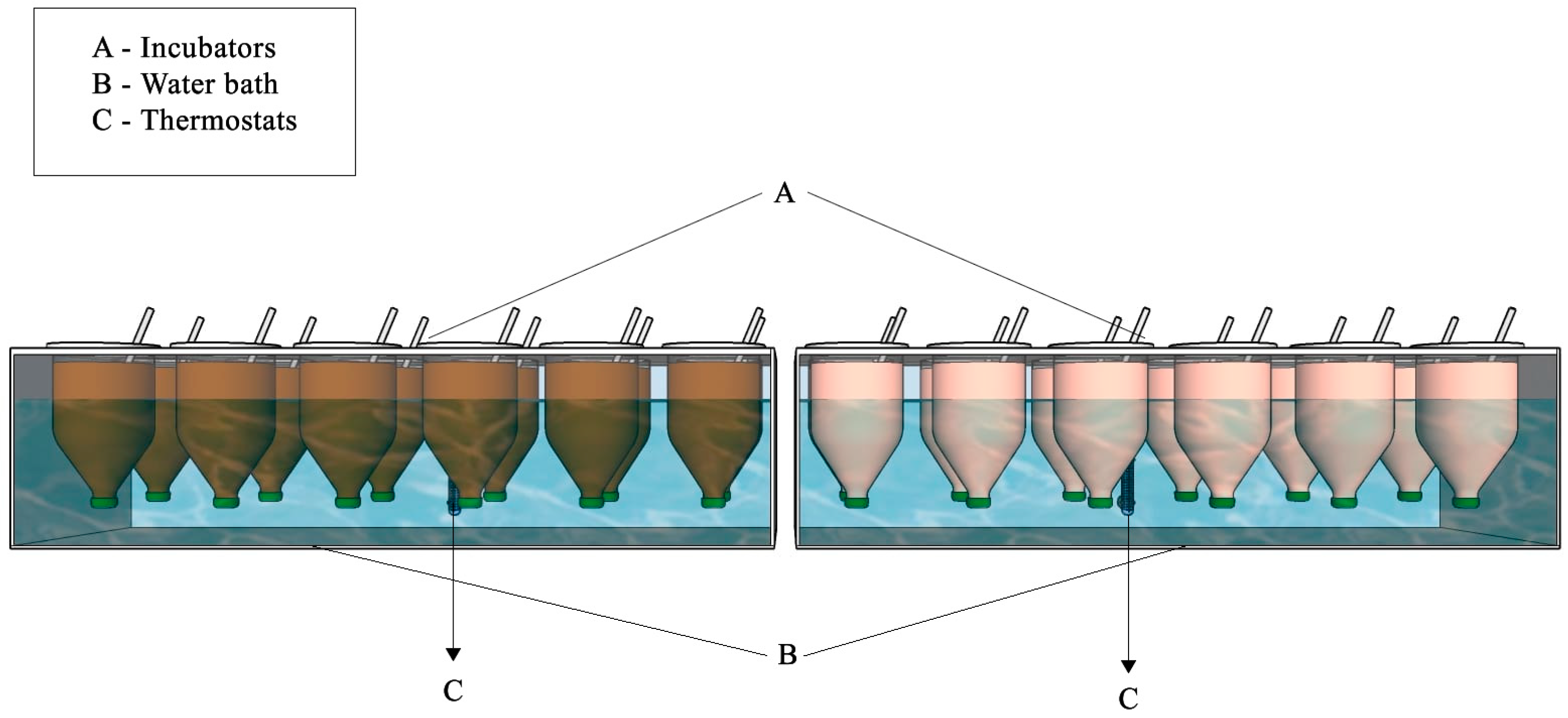
2.1. Phase I—Ingestion and Passage Time of Different Makers in Artemia

2.1.1. Obtainment of Colloidal Particles of the Markers Chromic Oxide and Titanium Dioxide

Based on preliminary tests, and due to the non-homogenization of particles in the water column, the markers chromic oxide and titanium dioxide were reduced to the colloidal size of 2.5 × 10⁻⁷ min diameter using a Partica LA-950 Laser Diffraction Particle Size Distribution Analyzer (Horiba Instruments, Inc., Irvine, CA). This technique avoids contamination, since it does not use chemical reagents. The analysis was performed in the Laboratório de Nutrição da Escola de Veterinária da UFMG. Five grams of each marker was used after pre-maceration in a quartz mortar. The obtained powder was then put in laser diffraction equipment using a 2.5 × 10⁻⁷ m diameter mesh. The particle separation run took 15 min until the desired size was obtained. The marker NANOLIPE® needed no transformation since it has a particle size of 10 nanometers. Thus, all markers were of an appropriate size for being ingested by Artemia.

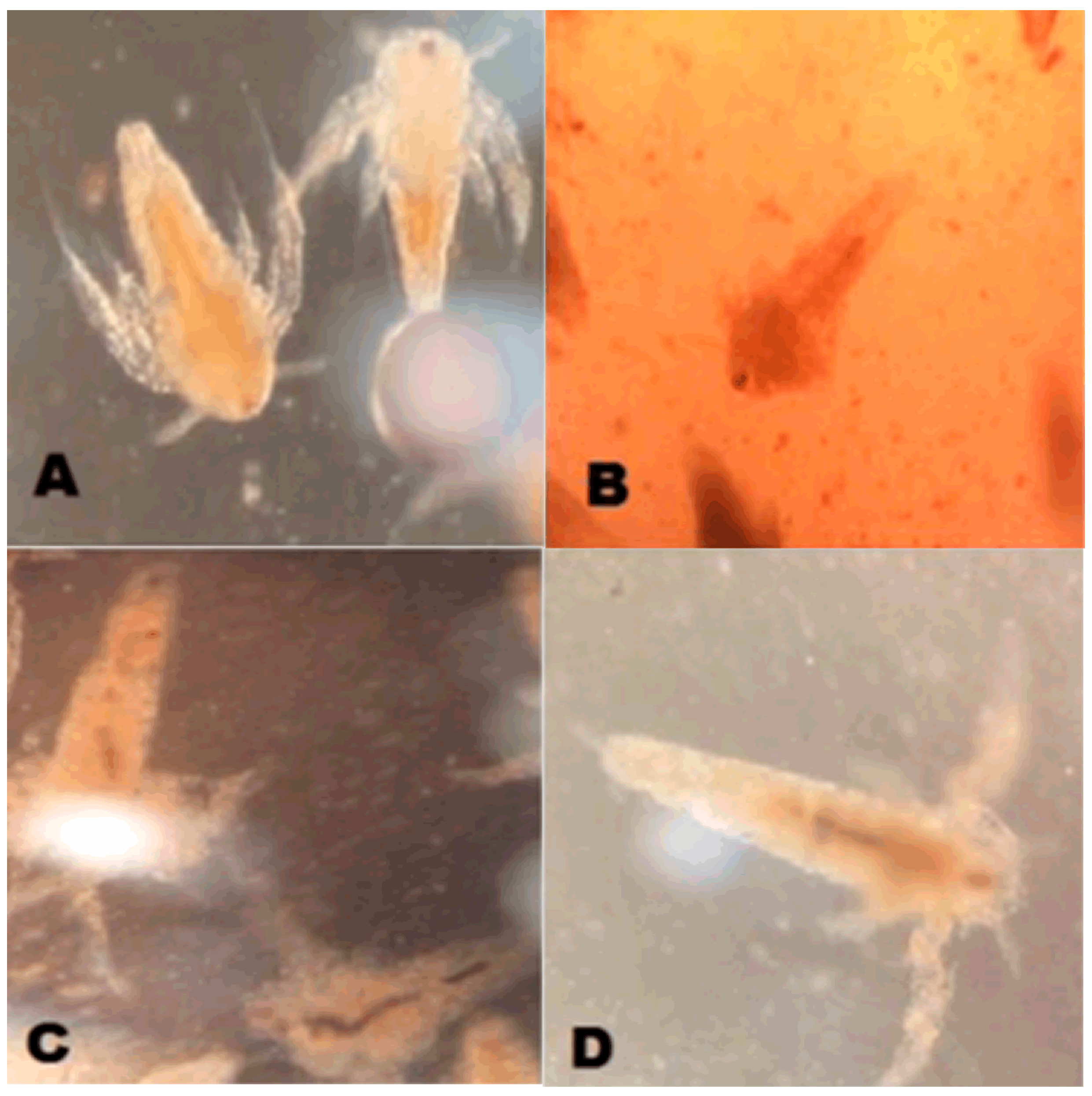
2.1.2. Marking Artemia

The hatching of Artemia cysts (BioArtemia, Rio Grande do Norte, Brazil) was conducted according to the manufacturer’s guidelines, as was the separation of the newly hatched Artemia from cysts. After quantification, the Artemia were stocked in 24 funnel-shaped incubators (Figure 1) with 1 L of net volume, at a density of 300 nauplii/mL (FAO 1996). Incubators were set in a thermostatically controlled water bath and water conditions were kept the same in all incubators (temperature at 30 °C, salinity at 30 g salt/L and dissolved oxygen at 6 mg/L). Supplementary aeration in each incubator helped nauplii moving through the water column as well as homogenizing the markers.
The design consisted of four treatment groups in sextuplicate: T1—control group (no marker), T2—chromic oxide, T3—titanium dioxide and T4—NANOLIPE® (with 6 replicates each). Based on the work of [48], who successfully marked Artemia with some oxides at 50 mg/L and 250 mg/L, the present study evaluated the non-previously tested concentration of 500 mg/L for all oxides. The markers were randomly and individually added into the incubators by slow placement onto the water surface up to the formation of a homogeneous solution. This procedure was performed each 4 h after nauplii separation, that is, 28 h after the start of hatching. Nauplii were collected at 30, 60 and 90 min after marker addition. The maximum time of 90 min was stipulated according to [48]. A Bioval light microscope (Hexasystems Group, São Paulo, Brazil) with a 40/065 (160/017) objective lens was used to verify ingestion of the markers by Artemia. Thus, the experimental design was a 4 × 3 factorial (four treatments and three collection times), with six replicates each.
Collection was performed in a standardized manner at all times of sampling. The entire content of the incubator was strained through a 50 μm nylon mesh and washed with freshwater, followed by distilled water and finally with deionized water, to expunge excess markers. The washed samples were placed into 250 mL plastic flasks, with small portions also being placed into Eppendorf tubes. Both were immediately cryopreserved in liquid nitrogen (−196 °C) to grant sample integrity. All materials were kept frozen at −40 °C until chemical and microscopy analyses.

2.1.3. Chemical Analyses of Markers

All samples were thawed and placed into a glass decanting funnel with deionized water to remove any possible contamination with cysts or markers that remained after washing.
For chromic oxide and titanium dioxide analyses, samples were pre-dried in a hot air drier at 55 °C and ground through a 1 mm sieve. Chromic oxide was analyzed using an atomic absorption technique [49], while titanium dioxide was analyzed using the ultraviolet method [50]. NANOLIPE® was analyzed using thawed samples and infrared spectroscopy [51].

2.1.4. Mathematical Model for Passage Time in Artemia: age-dependent

The use of non-linear models is suggested for the study of flow kinetics [52]. Previous studies have mainly resorted to three classes of non-linear models for adjusting fecal concentration data for markers administrated in a single dose. The classes are: (1) models with mixing compartments independent of time (age-independent); (2) models with mixing compartments dependent of time (age-dependent); (3) multi-compartment models. Age-dependent models assume that the system works with principles of probability or has random elements, which cannot be certainly known. This model uses gamma functions (non-exponential distributions of permanence times) to describe the passage of materials through the several segments of the gastrointestinal tract. Therefore, the age-dependent model implies that the probability for passage increases over time [53]. The flexibility inherent to age-dependent models allows their use with fecal excretion data of diverse animal categories [54].
Based on this finding, we decided to examine all the parameters described in the kinetics model of [55], which was proven to be appropriate to the present study.
The following equation was employed for the model Faichney [55]:
Y = Ae-k1 × (tTT) − Ae-k2 × (tTT) fort ≥ TT (transit time)
Y = 0 for t < TT
where “Y” is fecal concentration of marker at time “t”.
The mean retention time (MRT) in the gastrointestinal tract (GIT) can be defined as TRR (retention time up to the GIT) + TRPOS (retention time from GIT + TT (total time) [56].
Model of Faichney
MRT = ∑ [mi [(ti − 1 + (ti − ti − 1)/2)]∑ mi
where mi expresses the amount of marker.
The definition of ti varies among authors: mean time (h) for sampling [57], the total amount of marker excreted at defecation “i” at time “ti”, after the intake of the marker.

2.2. Phase II—Apparent Digestibility of Artemia Nauplii for Lophiosilurus alexandri Larvae of Different Ages

2.2.1. Animals and Conditions

The larvae of L. alexandri were all derived from breeding stock of the Laboratório de Aquacultura of UFMG. Fertilized eggs were collected from natural spawning and incubated at 28 °C with dissolved oxygen maintained above 5 mg/L. Larvae 8 days post-hatching were cultivated in a recirculating aquaculture system (RAS), in tanks with a useful volume of 28 L maintained at an average temperature of 28 °C, with dissolved oxygen greater than 5 mg/L (measured with a YSI 6920VZ2 multiparameter probe) and pH 8.0 ± 0.2 (measured with a Hanna HI98130 portable multiparameter probe) and fed three times a day. The larvae were stocked at a density of 15 larvae/L and fed with newly hatched Artemia nauplii following the daily feeding protocol: 1300 nauplii/larva from the first to the fifth day, 1950 nauplii/larva from the sixth to the tenth day, 2600 nauplii/larva from the eleventh to the fifteenth day, 3250 nauplii/larva from the sixteenth to the twentieth day and 3900 nauplii/larva from the twenty-first to the twenty-fifth day (adapted from [41])
Larvae at three different ages were used for the digestibility tests: after 3 (0.012 g ± 0.02 g), 15 (0.175 g ± 0.03 g) and 25 (0.622 g ± 0.03 g) days of exogenous feeding, corresponding to 11, 23 and 33 days post-hatching, respectively.

2.2.2. Marking Artemia

Artemia cysts (Bio-artemia from Rio Grande do Norte, Brazil) were hatched according to the manufacturer’s recommendation. Newly hatched Artemia nauplii were separated from unhatched cysts and, after quantification, were stored in 24 funnel-type incubators (n = 6 replicates each treatment) with 1 L useful volume at a density of 300 nauplii/mL [58]. The incubators were installed in a thermostatically controlled water bath, maintaining a temperature of 28 °C, salinity of 30 g of salt/L (refined salt from Mossoró, Rio Grande do Norte, Brazil. Ingredients: sodium chloride and sodium ferrocyanide) and dissolved oxygen of 6 mg/L. Individual supplemental aeration of each incubator served both to keep the nauplii moving in the water column and to aid in the homogenization of digestibility and consumption markers.
The markers (chromium oxide, titanium dioxide, NANOLIPE®—500 mg/L for each) were individually and randomly added to the incubators. Six incubators were also maintained with only Artemia (Control). The markers were slowly deposited on the surface of the water until a homogeneous solution was formed. This procedure was performed 4 h after the separation of the nauplii, that is, 28 h after the beginning of the hatching process. The nauplii were in contact with the markers for 90 min. After the 90 min, the entire contents of each incubator were filtered through a 50 μm nylon screen and washed with water under the same incubation conditions. The Artemia retained on the screen were then offered to the L. alexandri larvae as the only food for carrying out the digestibility experiment.

2.2.3. Digestibility Test

For the digestibility test, 360 larvae of each age (after 3, 15 and 25 days of exogenous feeding) were stored in 24 beakers (n = 6 replicates each treatment) with 1 L of water at a density of 15 larvae/L and kept fasting for 24 h under the same limnological and cultivation conditions. After this period, the bottom was cleaned and 80% of the water volume was changed. The larvae were then fed with Artemia previously marked with the markers composing the following treatments: T1—control group (no marker), T2—chromic oxide, T3—titanium dioxide and T4—NANOLIPE®.
After 15 min of feeding, the larvae were captured with a traditional sieve and immediately placed in funnel-shaped incubators with 1 L of water under the same conditions, for the collection of feces. Continuous observations were then carried out to identify the beginning of fecal production for each treatment, until the moment when fecal production was no longer detected, and then for another hour to guarantee that there would be no more production of feces. After this period, the larvae were removed from the incubators, euthanized with a 285 mg/L eugenol solution [59] and frozen at −40 °C for subsequent chemical analysis of their body composition and markers to certify that their gastrointestinal tract was completely emptied. The water with the feces from each incubator was also frozen individually at −40 °C for later collection and analysis of the feces.
Digestibility was calculated using the internal marker formula suggested by Burns et al., 1994 [60]:
Dig = 100 − [100 × (CIdiet/CIfeces)]
where:
Dig: Digestibility;
CIdiet: Concentration of marker in diet;
CIfeces: Concentration of marker in feces.

2.2.4. Analyses

All analyses were carried out in the Nutrition Laboratório de Nutrição of the Escola de Veterinária of UFMG, Brazil. The water with the feces was thawed, pre-dried and ground in a 1 mm sieve [50]. The nutrients analyzed were Crude Protein (CP) by the Kjeldahl method and Dry Matter (DM) at 105 °C [50]. Chromic oxide was analyzed using the atomic absorption technique [49]. Titanium dioxide was analyzed according to [33] by the ultraviolet technique. NANOLIPE® analysis was performed according to the methodology described by [51] through infrared spectroscopy in NirsLab N-200 (MCS 100) equipment.
The larvae were analyzed using the same methodology as the feces and diet to determine the presence or absence of the marker in their gastrointestinal tract and thus assess whether or not there was total collection of feces.
The nutritional composition of Artemia nauplii (Table 1) was analyzed using benchtop techniques according to [50], to characterize the food offered to L. alexandri larvae.

2.3. Data Analysis

Statistical analyses, whether of marker concentration or passage time, were performed using InfoStat software [61]. All data were analyzed for normality using the Shapiro–Wilk test and homogeneity of variance using the Cochran test. The results for the variable concentration of markers were log-transformed and analyzed by factorial ANOVA followed by Tukey’s test with a significance level of 5%. For parametric data, ANOVA was used followed by Tukey’s test at 5% probability, while the Kruskal–Wallis test at 5% was used for non-parametric data.

3. Results

3.1. Phase I—Ingestion and Passage Time of Different Markers in Artemia

The tested markers were ingested by Artemia nauplii (Figure 2).
Table 2 shows the results for the concentrations of the three markers found in Artemia. There was a significant effect of marker (p < 0.001) and exposure period (p < 0.05) on concentration but not for their interaction (marker × period; p > 0.05). The greatest marker concentration found in Artemia was for T3, followed by T4 and lastly T2.
The highest concentrations were seen when nauplii remained in contact with the markers for 90 min, followed by 60 min and then, with the lowest concentrations, 30 min of exposure, independent of the tested marker (Table 2).
Table 3 contains the transit times observed through the gastric emptying of Artemia during the marking process with the three markers. Both T2 and T4 had a total transit time of 30 min, while T3 took more time for the complete departure of the digestive tract. However, after 90 min no markers were detected in the digestive tract of Artemia.
The marker chromium oxide (T2) had greater MRT than the other markers (T4 and T3; Table 4), but lower average passage time, with T4 and T3 having similar passage values. The transit time (TT) of the markers through the digestive tract of Artemia corroborates the data found for MRT, since it was greater for T2, compared to T3 and T4.

3.2. Phase II—Apparent Digestibility of Artemia nauplii for Lophiosilurus alexandri Larvae of Different Ages

Real digestibility for larvae after 3 days of feeding. T1 was similar between T4 and T2; it was not possible to determine it for T3 (p < 0.05) (Table 5). The comparison of T4 and T2 revealed they differed from each other (p < 0.05). The initial time of fecal production was lower for T1, T2 and T4 than for T3 (p < 0.05), with T2 and T4 being similar to T1 (Real time). The final fecal production time did not differ among all treatments (p > 0.05).
Real digestibility after 15 days of exogenous feeding was similar between T4 and T2 and T1 (p > 0.05), with T3 differing, underestimating real digestibility (p < 0.05) (Table 5). The same was true for the initial time of fecal production, with T3 being different and superior to the others (p < 0.05). The final time of fecal production did not differ among treatments (p > 0.05).
Estimated and real digestibility did not differ for T4 after 25 days of feeding (Table 5). Estimated digestibility for T4 was similar to that of T2, but different from that of T3, with and T2 and T3 underestimating real digestibility (p < 0.05). As for the previous ages, initial fecal production time was similar for T1, T2 and T4 and greater for T3 (p > 0.05) and final fecal production time did not differ among treatments (p > 0.05).
Larvae from all treatments did not show readings for the three markers studied, confirming complete gastric emptying.

4. Discussion

Research into fish larvae nutrition and feeding is known to be very laborious; thus, simpler and more accurate methods are desired [62]. The present work demonstrated that the three studied markers could be assimilated by Artemia and quantified through the methods employed. These findings will allow future studies to use this technique to evaluate nutritional trials involving fish larvae of the diversity of species that use this live feed. The results also allow for comparisons to be made with other techniques, such as incubation with trypsin in vitro [63], the marking of microdiets with FluoSpheres® [64], or analyses with stable carbon isotopes (δ13C) [65].
The markers chromium oxide and titanium dioxide used in the present study were obtained at the colloidal size of 2.5 × 10⁻⁷ m in diameter, to achieve nanometric scale. NANOLIPE® also consists of extremely small particles at nanometric scale, with a diameter of 10−9 m [66]. According to [15], Artemia is able to assimilate nanoparticles, which was demonstrated in the present work by the conducted analyses and micrography, showing the adequate size of the markers for Artemia. It should also be highlighted that the washing of nauplii assures that the analyzed markers were inside Artemia, and not on their surface. Thus, the use of this technique in future assays of larval nutrition might assure that the quality of the marker could be quantified more precisely, since if it was adhered to their surface it would probably be more easily lost when offered to larvae as live feed in rearing tanks.
The formation of colloids of chromium oxide and titanium dioxide was also important for obtaining a homogeneous suspension of these markers in the water column, ensuring better contact with Artemia. On the other hand, in another study, the animals needed to go through intense aeration when testing the markers yttrium oxide, ytterbium oxide, lanthanum oxide and dysprosium oxide, to keep them in solution [48]. Thus, preparation of the colloidal suspension of the markers has proven itself more efficient. For NANOLIPE®, however, direct use was efficient in proportioning a homogeneous suspension, as the markers that were obtained for forming the colloidal suspension.
Among the evaluated markers, titanium dioxide was found at the highest concentrations in Artemia. It is known that, for some animal species, up to 99% of the provided marker may be recovered, depending on the time when the trial is conducted [67,68]. This knowledge, as well as the present work, suggest that this marker is good for future studies in fish larval nutrition. The second most recovered marker was NANOLIPE®, followed by chromium oxide. Due to their nanometric size, NANOLIPE® particles have faster dispersion through the digestive tract [66], which can explain the higher concentrations recovered compared to chromium oxide. According to [69], chromium oxide needs an adaptation period of 6 to 7 days to reach a constant concentration in the animal digestive tract. The longest contact time of Artemia with chromic oxide was 90 min. Thus, future studies are needed to undertake this kind of evaluation, regarding marking with this marker, and to better understand the behavior of markers in the digestive tract of Artemia.
The highest marker concentrations resulted when nauplii remained in contact with them for 90 min, the longest exposure duration evaluated. These results imply the need to evaluate longer exposure durations. The lower concentrations found at 30 and 60 min can be explained by the heterogeneity of Artemia development stages in the samples, since multi-stage nauplii were seen under microscopy. This implies the need for a better definition of the ideal moment to start marking Artemia, which is difficult to set since even with a standardized technique of cyst hatching, it does not occur in all cysts at the same time.
The passage rate of these markers in Artemia is also important. On the basis of our data, many trials in fish nutrition will be able to be developed, addressing the digestibility and intake of nutrients in the diets of the larvae of several fish species. The main point should be to employ an experimental design that ensures that fish will ingest the marked Artemia.
The nutritional quality of Artemia varies considerably, which may be related to geographic origin [14], differences among different batches of cysts from the same origin and to methods of analysis. This variation makes it important to determine the biochemical composition of live food used in nutritional studies with fish larvae [70]. It is noteworthy that L. alexandri larvae have a low capacity to digest artificial food at the beginning of exogenous feeding due to the lack of digestive enzymes, and that live food would provide complementary enzymes for effective protolytic activity [46], thus facilitating digestion.
The average real digestibility of the protein of Artemia at the three ages was 95.08%, demonstrating it to be an excellent quality food for L. alexandri larvae. The average digestibility for Herring larvae (Clupea harengus), was 60% of the protein of Artemia nauplii (marked by the addition of a protein hydrolysate), with 20% found in the body and 39% catabolized by the larvae, 24 h after feeding [71]. The average digestibility of Artemia nauplii by Senegalese sole (Solea senegalensis) ranged between 83.08 ± 4.37% (6 days after hatching—DAH) and 69.04 ± 5.45% (15 DAH) [72]. Both works showed lower values than the present study. However, in another study, the authors found digestibility of up to 86%, comparing the digestibility of Artemia and microencapsulated diets for Atlantic cod larvae (Gadus morhua) testing the rare earth oxides yttrium oxide (Y₂O3) and dysprosium oxide (III) (Dy2O3) [73].
The protein digestibility of the different live feeds used in larviculture was also studied through the in vitro technique of incubation in trypsin, a different technique from the one used in the present study [63]. The authors concluded that there is relative susceptibility to proteolytic digestion among different organisms and prey and that there is a natural orchestration of several enzymes that participate in larval digestion, increasing or not the efficiency of enzymatic degradation. These factors may explain the previously reported differences in digestibility among larvae of different species.
In a study of the digestibility of L. alexandri juveniles fed inert diets, testing different ingredients of animal and vegetable origin [74], the authors found digestibility results between 82.38% and 91.49%, the latter being for purified fish meal (an ingredient of animal origin). These values were also lower than those verified in the present study for larvae, suggesting an effect of animal age on the use of food, as well as the type of food used.
The present experiment found the same final time of fecal excretion for all ages, demonstrating that ingested Artemia is completely excreted in 30 min under the conditions of this study. This was the time used in the experiment as the feeding interval for Atlantic cod (G. morhua) larvae [73]. All markers showed the same final fecal excretion profile, demonstrating that none of the markers influenced the rate of passage of Artemia through the digestive tract of L. alexandri larvae, thus fulfilling one of the requirements of an ideal marker [75].

5. Conclusions

Colloidal particles of metallic markers and nanoparticles of an organic marker were shown to be efficient for Artemia intake. The exposure duration of 30 min was, for all markers, enough for both ingestion and quantification; however, higher concentrations were found for 90 min. The three tested markers showed potential, regarding passage rate and mean retention time, for use in fish larvae nutrition.
The digestibility of Artemia for L. alexandri larvae at the three studied ages can be determined using NANOLIPE® or chromic oxide at 3 and 15 days of feeding. Titanium was the worst marker of Artemia digestibility at the three ages evaluated for L. alexandri larvae.

Author Contributions

J.S.S.—Conceptualization, Methodology, Validation, Formal analysis, Investigation, Writing—Original Draft, Writing—Review and Editing, Visualization. F.A.C.d.S.—Conceptualization, Methodology, Validation, Formal analysis, Investigation, Writing—Original Draft, Writing—Review and Editing, Visualization. E.d.O.S.S.—Conceptualization, Methodology, Validation, Formal analysis, Investigation. R.K.L.—Conceptualization, Methodology, Validation, Formal analysis, Investigation, Resources, Data curation, Investigation, Writing—Original Draft, Writing—Review and Editing, Visualization, Supervision, Project Administration, Funding Acquisition. All authors have read and agreed to the published version of the manuscript.

Funding

This research was funded by Conselho Nacional de Desenvolvimento Científico e Tecnológico (CNPq-Brazil), Coordenação de Aperfeiçoamento de Pessoal de Nível Superior (CAPES-Brazil) and Fundação de Amparo à Pesquisa do Estado de Minas Gerais (FAPEMIG-Brazil). LUZ, R.K. received a research grant from the Conselho Nacional de Desenvolvimento Científico e Tecnológico (CNPq Process 308547/2018-7).

Institutional Review Board Statement

The study was conducted according to the guidelines of the Declaration of Helsinki and approved by the Institutional Review Board (or Ethics Committee) of the Committee for Ethics in Animals Use (CEUA/UFMG—n° 39/2021).

Data Availability Statement

The data that support the findings of this study are available from the author, Jaqueline Simões Saliba upon reasonable request.

Conflicts of Interest

The authors declare no conflict of interest.

References

  1. Santos, J.C.E.; Luz, R.K. Effect of Salinity and Prey Concentrations on Pseudoplatystoma corruscans, Prochilodus costatus and Lophiosilurus alexandri Larviculture. Aquaculture 2009, 287, 324–328. [Google Scholar] [CrossRef]
  2. Podrabsky, J.E.; Hand, S.C. Physiological Strategies during Animal Diapause: Lessons from Brine Shrimp and Annual Killifish. J. Exp. Biol. 2015, 218, 1897–1906. [Google Scholar] [CrossRef]
  3. Takeuchi, T. Progress on Larval and Juvenile Nutrition to Improve the Quality and Health of Seawater Fish: A Review. Fish. Sci. 2014, 80, 389–403. [Google Scholar] [CrossRef]
  4. Talens-perales, D.; Marín-Navarro, J.; Garrido, D.; Almansa, E.; Polaina, J. Fixation of Bioactive Compounds to the Cuticle of Artemia. Aquaculture 2017, 474, 95–100. [Google Scholar] [CrossRef]
  5. Kolkovski, S.; Curnow, J.; King, J. Intensive Rearing System for Fish Larvae Research II Artemia Hatching and Enriching System. Aquac. Eng. 2004, 31, 309–317. [Google Scholar] [CrossRef]
  6. Luz, R.K.; Zaniboni, E.F. Utilização de Diferentes Dietas Na Primeira Alimentação Do Mandi-Amarelo (Pimelodus maculatus, Lacépéde). Acta Sci. Biol. Sci. 2001, 23, 483–489. [Google Scholar]
  7. Reynalte-tataje, D.; Luz, R.K.; Meurer, S.; Zaniboni-filho, E.; Pires, A.; Nuñer, D.O. Influência Do Fotoperíodo No Crescimento e Sobrevivência de Pós—Larvas de Piracanjuba Brycon orbignyanus (Valenciennes, 1849). Acta Sci. Biol. Sci. 2002, 24, 439–443. [Google Scholar]
  8. Luz, R.K. Resistência Ao Estresse e Crescimento de Larvas de Peixes Neotropicais Alimentadas Com Diferentes Dietas. Pesqui. Agropecuária Bras. 2007, 42, 65–72. [Google Scholar] [CrossRef]
  9. Luz, R.K.; Dos Santos, J.C.E. Densidade de Estocagem e Salinidade Da Água Na Larvicultura Do Pacamã. Pesqui. Agropecuária Bras. 2008, 43, 903–909. [Google Scholar] [CrossRef]
  10. Jomori, R.K.; Luz, R.K.; Takata, R.; Perez Fabregat, T.E.H.; Portella, M.C. Água Levemente Salinizada Aumenta a Eficiência Da Larvicultura De Peixes Neotropicais. Pesqui. Agropecuária Bras. 2013, 48, 809–815. [Google Scholar] [CrossRef]
  11. Luz, R.K.; Portella, M.C. Effect of Prey Concentrations and Feed Training on Production of Hoplias lacerdae Juvenile. An. Da Acad. Bras. De Ciências 2015, 87, 1125–1132. [Google Scholar] [CrossRef]
  12. Santos, F.A.C.; Gustavo, S.; Julio, C.; Luz, R.K. Stocking Density in Colossoma macropomum Larviculture, a Freshwater Fish, in Recirculating Aquaculture System. Aquac. Res. 2020, 52, 1185–1191. [Google Scholar] [CrossRef]
  13. Santos, F.A.C.; Julio, G.S.C.; Batista, F.S.; Miranda, N.L.; Pedras, P.P.C.; Luz, R.K. High Stocking Densities in the Larviculture of Colossoma macropomum in a Recirculating Aquaculture System: Performance, Survival and Economic Viability. Aquaculture 2022, 552, 738016. [Google Scholar] [CrossRef]
  14. Léger, P.; Bengtson, D.A.; Simpson, K.L.; Sorgeloos, P. The use and nutritional value of Artemia as a food source. Oceanogr. Mar. Biol. Annu. Rev. 1986, 24, 521–623. [Google Scholar]
  15. Mesarič, T.; Gambardella, C.; Milivojević, T.; Faimali, M.; Drobne, D.; Falugi, C.; Sepčić, K. High Surface Adsorption Properties of Carbon-Based Nanomaterials Are Responsible for Mortality, Swimming Inhibition, and Biochemical Responses in Artemia Salina Larvae. Aquat. Toxicol. 2015, 163, 121–129. [Google Scholar] [CrossRef]
  16. Hamzah, M.; Shaik, M.I.; Sarbon, N.M. Effect of Fish Protein Hydrolysate on Physicochemical Properties and Oxidative Stability of Shortfin Scad (Decapterus macrosoma) Emulsion Sausage. Food Res. 2021, 5, 225–235. [Google Scholar] [CrossRef]
  17. Siddik, M.A.B.; Pham, H.D.; Francis, D.S.; Vo, B. Van Dietary Supplementation of Fish Protein Hydrolysate in High Plant Protein Diets Modulates Growth, Liver and Kidney Health, and Immunity of Barramundi (Lates calcarifer). Aquac. Nutr. 2021, 27, 86–98. [Google Scholar] [CrossRef]
  18. Miranda, A.; Altiery, D.S.; Daniela, S.; Bacconi, F.; Fernando, J.; Melo, B.; Thiago, A.; Luiz, S.; Oliveira, V. Corn Substitution by Mesquite Bean Flour (Prosopis juliflora) Maintains Growth and Improves Protein Metabolism of Nile Tilapia Juveniles (Oreochromis niloticus). Trop. Anim. Health Prod. 2021, 53, 1–15. [Google Scholar] [CrossRef]
  19. Mohseni, M.; Najjar, S.; Younes, L.; Sarameh, S.P. Effects of Different Dietary Canola and Fish Oil Levels on Overall Performance, Fatty Acid Profile, Haemato—Biochemical Responses, and Digestibility of Macronutrients of Caspian Brown Trout (Salmo trutta caspius Kessler) Fingerling. J. Appl. Ichthyol. 2022, 38, 212–222. [Google Scholar] [CrossRef]
  20. Zaretabar, A.; Ouraji, H.; Abedian, A.; Yeganeh, S.; Esmaeili, M.; Keramat, A. One Step toward Aquaculture Sustainability of a Carnivorous Species: Fish Meal Replacement with Barley Protein Concentrate plus Wheat Gluten Meal in Caspian Brown Trout (Salmo trutta caspius). Aquac. Rep. 2021, 20, 100714. [Google Scholar] [CrossRef]
  21. Takakuwa, F.; Sato, H.; Mineyama, N.; Yamada, S.; Biswas, A.; Tanaka, H. Bioavailability of Porcine Blood Meal as a Fish Meal Substitute in the Diet for Red Sea Bream (Pagrus major, Temminck & Schlegel) Fingerling. Aquac. Res. 2022, 53, 4616–4626. [Google Scholar] [CrossRef]
  22. Abedi, S.Z. The Influence of Probiotic (Isolated Based on Phytase Activity) on Growth Performance, Body Composition, and Digestibility of Rainbow Trout, Oncorhynchus mykiss. J. World Aquac. Soc. 2022, 53, 1006–1030. [Google Scholar] [CrossRef]
  23. Marcos, A.; Ramos, J.; Machado, D.; Eduardo, L.; De Freitas, L.; Rodrigues, V.; Paula, A.; Rodrigues, O. Optimizing Methodological Aspects of Stool Collection by Sedimentation in Digestibility Studies with Pirarucu (Arapaima gigas). Aquac. Res. 2022, 53, 1456–1467. [Google Scholar] [CrossRef]
  24. Hamilton, L.; Yadav, R.; Kumar, N.; Ved, C.; Saini, P.; Banerjee, P.; Manohar, S.; Ojha, L.; Jayant, M.; Raj, N. Substitution of De-Oiled Rice Bran with Khejri Pod Meal and Groundnut Oilcake with Khejri Seed Meal in the Diet Of. Aquac. Res. 2022, 53, 642–656. [Google Scholar] [CrossRef]
  25. Major, P.; Shadrack, R.S.; Manabu, I.; Koshio, S.; Yokoyama, S.; Zhang, Y.; Mzengereza, K.; Fouad, M.; Basuini, E.; Dawood, M.A.O. Effects of Single and Mixture Probiotic Supplements on Growth, Digestive Activity, Antioxidative Status, Immune and Growth-Related Genes, and Stress Response of Juvenile Red Sea Bream. Aquac. Nutr. 2022, 2022, 1–17. [Google Scholar]
  26. Mzengereza, K.; Ishikawa, M.; Koshio, S.; Yokoyama, S.; Yukun, Z.; Shadrack, R.S.; Seo, S.; Kotani, T.; Dossou, S.; El Basuini, M.F.; et al. Growth Performance, Growth-Related Genes, Digestibility, Digestive Enzyme Activity, Immune and Stress Responses of de novo Camelina Meal in Diets of Red Seabream (Pagrus major). Animals 2021, 11, 3118. [Google Scholar] [CrossRef]
  27. Bowen, S.H. Chromic Acid in Assimilation Studies—A Caution. Trans. Am. Fish. Soc. 1978, 107, 755–756. [Google Scholar] [CrossRef]
  28. Vandenberg, G.W.; Nou, J.D.E.L.A. Apparent Digestibility Comparison in Rainbow Trout (Oncorhynchus mykiss) Assessed Using Three Methods of Faeces Collection and Three Digestibility Markers. Aquac. Nutr. 2021, 7, 237–245. [Google Scholar] [CrossRef]
  29. Dumas, V.; Labbé, L.; Pelissier, P. Rainbow trout (Oncorhynchus mykiss) freshwater recirculating aquaculture system: Nutrient flow management with aquaponics. In Proceedings of the World Aquaculture 2018, #We R Aquaculture, Montpellier, France, 25–29 August 2018. [Google Scholar]
  30. Mayumeoshiro, F.; Fraga, T.; Honorato, C. Tempo de trânsito gastrointestinal do pintado (Pseudoplatystoma sp.). J. Agron. Sci. 2012, 1, 128–138. [Google Scholar]
  31. Langeland, M.; Vidakovic, A.; Vielma, J.; Lindberg, J.E.; Kiessling, A.; Lundh, T. Digestibility of microbial and mussel meal for Arctic charr (Salvelinus alpinus) and Eurasian perch (Perca fluviatilis). Aquac. Nutr. 2016, 22, 485–495. [Google Scholar] [CrossRef]
  32. Heinitz, M.C.; Lemme, A.; Schulz, C. Measurement of Digestibility in Agastric Fish Based on Stripping Method—Apparent Nutrient, Energy and Amino Acid Digestibilities of Common Feed Ingredients for Carp Diets (Cyprinus carpio). Aquac. Nutr. 2016, 22, 1065–1078. [Google Scholar] [CrossRef]
  33. Myers, W.D.; Ludden, P.A.; V Nayigihugu, V.; Hess, B.W. Technical Note: A procedure for the preparation and quantitative analysis of samples for titanium dioxide. J. Anim. Sci. 2004, 82, 179–183. [Google Scholar] [CrossRef] [PubMed]
  34. Evans, O.; Jones, E.; Teixeira, A.E. Validation of the external marker Nanolipe as an indicator of apparent nutrient and energy digestible in juvenile Nile Tilápia (Oreochromos niloticus). J. Anim. Sci. 2012, 90, 558–559. [Google Scholar]
  35. Rosenfeld, I.; Austbø, D.; Volden, H. Models for Estimating Digesta Passage Kinetics in the Gastrointestinal Tract of the Horse. J. Anim. Sci. 2006, 84, 3321–3328. [Google Scholar] [CrossRef]
  36. Sciences, A.; Stensig, T.; Martin, R. Evaluation of Different Methods for the Determination of Digestion and Passage Rates of Fibre in the Rumen of Dairy Cows. Acta Agric. Scand. Part A 1998, 48, 141–154. [Google Scholar]
  37. Firkins, J.L. Effects of Feeding Nonforage Fiber Sources on Site of Fiber Digestion 1. J. Dairy Sci. 1997, 80, 1426–1437. [Google Scholar] [CrossRef]
  38. Lascano, C.; Quiroz, R. Metodologia para estimar la dinámica de la digestión en rumiantes. In Nutrición de Rumiantes: Guía Metodológica de Investigación; Ruiz, M.E., Ruiz, A., Eds.; IICA: San Jose, Costa Rica, 1990; pp. 89–104. [Google Scholar]
  39. Luz, R.K.; dos Santos, J.C.E. Evaluation of the Tolerance of “Pacamã” Lophiosilurus alexandri Steindachner, 1877 (Pisces: Siluriformes) Larvae to Different Salinities. Acta Scientiarum. Biol. Sci. 2008, 30, 345–350. [Google Scholar] [CrossRef] [Green Version]
  40. Cordeiro, B.N.I.S.; Costa, D.C.; Silva, W.D.S.; Takata, R.; Luz, R.K. High Stocking Density during Larviculture and Effect of Size and Diet on Production of Juvenile Lophiosilurus alexandri Steindachner, 1876 (Siluriformes: Pseudopimelodidae). J. Appl. Ichthyol. 2016, 32, 61–66. [Google Scholar] [CrossRef]
  41. Santos, J.C.E.; Correia, E.; Luz, R.K. Effect of Daily Artemia Nauplii Concentrations during Juvenile Production of Lophiosilurus alexandri. Bol. do Inst. de Pesca 2015, 41, 771–776. [Google Scholar] [CrossRef]
  42. Takata, R.; Silva, W.; Costa, D.C.; Filho, R.M.; Luz, R.K. Effect of Water Temperature and Prey Concentrations on Initial Development of Lophiosilurus alexandri Steindachner, 1876 (Siluriformes: Pseudopimelodidae), a Freshwater Fish. Neotrop. Ichthyol. 2014, 12, 853–860. [Google Scholar] [CrossRef]
  43. Claudio, J.; Dos, E.; Pedreira, M.M.; Luz, R.K. Feeding Frequency in pacamã larviculture. Revista Caatinga 2016, 2125, 512–518. [Google Scholar]
  44. Luz, R.K.; Santos, J.C.E.; Pedreira, M.M.; Teixeira, E.A. Effect of Water Flow Rate and Feed Training on “Pacamã” (Siluriforme: Pseudopimelodidae) Juvenile Production. Arq. Bras. De Med. Vet. E Zootec. 2011, 63, 973–979. [Google Scholar] [CrossRef]
  45. Melillo Filho, R.; Takata, R.; Santos, E.H.; De Souza, W.; Luz, R.K.; Ana, L. Draining System and Feeding Rate during the Initial Development of Lophiosilurus alexandri (Steindachner, 1877), a Carnivorous Freshwater Fish. Aquac. Res. 2014, 45, 1913–1920. [Google Scholar] [CrossRef]
  46. Pedreira, M.M.; Luz, R.K.; Cláudio, J. Biofiltração Da Água e Tipos de Substrato Na Larvicultura Do Pacamã. Pesqui. Agropecuária Bras. 2009, 44, 511–518. [Google Scholar] [CrossRef]
  47. Torres, I.F.A.; Júlio, G.S.C.; Figueiredo, L.G.; De Lima, N.L.C.; Soares, A.P.N.; Luz, R.K. Larviculture of a Carnivorous Freshwater Catfish, Lophiosilurus alexandri, Screened by Personality Type. Behav. Process. 2017, 145, 44–47. [Google Scholar] [CrossRef]
  48. Cook, M.A.; Johnson, R.B.; Nicklason, P.; Barnett, H.; Rust, M.B. Marking Live Feeds with Inert Metal Oxides for Fish Larvae Feeding and Nutrition Studies. Aquac. Res. 2008, 39, 347–353. [Google Scholar] [CrossRef]
  49. Silva, J.D.; Queiroz, A.C. Análise de Alimentos—Métodos Químicos e Biológicos, 3rd ed.; UFV: Viçosa, Brazil, 2009; p. 235. [Google Scholar]
  50. Detmann, E.; Souza, M.A.; Valadares Filho, S.C.; Rocha, G.C.; Palma, M.N.N.; Rodrigues, J.P.P. Métodos Para análise de Alimentos; Suprema: Visconde do Rio Branco, MG, Brazil, 2012; p. 214. [Google Scholar]
  51. Saliba, E.; Moreira, G.R.; Federal, U.; De Pernambuco, R.; Borges, I.; Costa, A.L. Use of Infrared Spectroscopy to Estimate Fecal Output with Marker Lipe®. Int. J. Food Sci. Nutr. Diet. 2015, 4, 1–10. [Google Scholar] [CrossRef]
  52. Ellis, W.C.; Matis, J.H.; Hill, T.M.; Murphy, M.R. Methodology for estimation digestion and passage kinetics of forages. In Forage Quality, Evaluation, and Utilization; Fahey, G.C., Jr., Collins, M., Mertens, D.R., Moser, L.E., Eds.; American Society of Agronomy: Madison, WI, USA, 1994; pp. 682–756. [Google Scholar]
  53. Quiroz, R.A.; Pond, K.R.; Tolley, E.A.; Johnson, W.L. Selection among nonlinear models for rate of passage studies in ruminants. J. Anim. Sci. 1988, 66, 2977–2986. [Google Scholar] [CrossRef] [Green Version]
  54. Pond, K.R.; Ellis, W.; Matis, C. Compartment models for estimating attributes of digesta flow in cattle. Br. J. Nutr. 1988, 60, 571–595. [Google Scholar] [CrossRef]
  55. Faichney, G.J. The use of markers to partition digestion within the gastrointestinal tract. In Digestion and Metabolism in the Ruminant; Macdonald, I.W., Warner, A.A.I., Eds.; University of New England Publishing Unit: Armidale, NSW, Australia, 1975; pp. 277–291. [Google Scholar]
  56. Beauchemin, K.A.; Buchanan-Smith, J.G. Evaluation of markers, sampling sites and models for estimating rates of passage of silage or hay in dairy cows. Anim. Feed Sci. Technol. 1989, 27, 59–75. [Google Scholar] [CrossRef]
  57. Uden, P. The influence of leaf and stem particle size in vitro and sample size in sacco on neutral detergent fiber fermentation kinetics. Anim. Feed Sci. Technol. 1992, 37, 85. [Google Scholar] [CrossRef]
  58. Lavens, P.; Sorgeloos, P.; FAO. Manual on the Production and Use of Live Food for Aquaculture; Fisheries Technical Paper; FAO: Rome, Italy, 1996; p. 295. [Google Scholar]
  59. Mattioli, C.C.; Takata, R.; De Oliveira, F.; Leme, P.; Cristina, D.; Melillo, R.; de Souza, W.; Kennedy, R. The Effects of Acute and Chronic Exposure to Water Salinity on Juveniles of the Carnivorous Freshwater Catfish Lophiosilurus alexandri. Aquaculture 2017, 481, 255–266. [Google Scholar] [CrossRef]
  60. Burns, J.C.; Pond, K.R.; FIisher, D.S. Measurement of forage intake. In Forage Quality, Evoluatin, and Utilization; Fahey, J.R., Ed.; American Society of Agronomy: Madison, WI, USA, 1994; pp. 494–532. [Google Scholar]
  61. Di Rienzo, J.A.; Casanoves, F.; Balzarini, M.G.; Gonzalez, L.A.; Tablada, M.; Robledo, C.W. InfoStat Version. Grupo InfoStat, FCA, Universidad Nacional de Cordoba, Argentina. 2015. Available online: http://www.infostat.com.ar (accessed on 1 December 2022).
  62. Teshima, T.; Ishikawa, M.; Koshio, S. Nutritional assessment and feed intake of microparticulate diets in crustaceans and fish. Aquat. Res. 2000, 31, 691–702. [Google Scholar]
  63. Arndt, C.; Sommer, U.; Ueberschär, B. A comparative in-vitro-test on the digestibility of live prey for fish larvae under specific consideration of trypsin. Aquaculture 2015, 446, 12–16. [Google Scholar] [CrossRef]
  64. Hansen, J.M.; Lazo, J.P.; Kling, L.J. A method to determine protein digestibility of microdiets for larval and early juvenile fish. Aquac. Nutr. 2009, 15, 615–626. [Google Scholar] [CrossRef]
  65. Gamboa-delgado, J.; Cañavate, J.P.; Zerolo, R.; Le Vay, L. Natural carbon stable isotope ratios as indicators of the relative contribution of live and inert diets to growth in larval Senegalese sole (Solea senegalensis). Aquaculture 2008, 280, 190–197. [Google Scholar] [CrossRef]
  66. Gonçalves, N.C. Validação Do Nanolipe Como Indicador Para Estimativa Do Consumo Em Bovinos Leiteiros; UFMG: Belo Horizonte, MG, Brazil, 2012; p. 40. [Google Scholar]
  67. Hafez, S.; Junge, W.; Kalm, E. Schatzung der verdaulichkeit mit einer indikatormethode bei milchku¨hen im vergleich zum hohenheimer-futterwert-test. Arch. Für Tierernaehrung 1988, 38, 929–945. [Google Scholar] [CrossRef]
  68. Titgemeyer, E.C.; Armendariz, C.K.; Bindel, D.J.; Greenwood, R.H.; Löest, C.A. Evaluation of titanium dioxide as a digestibility marker for cattle. J. Anim. Sci. 2001, 79, 1059–1063. [Google Scholar] [CrossRef]
  69. Rodriguez, N.M.; Saliba, E.O.S.; Guimarães-Junior, R. Uso de indicadores para estimar consumo y digestibilidad de pasto. LIPE, lignina purificada y enriquecida. Rev. Colomb. De Cienc. Pecu. 2007, 20, 518–525. [Google Scholar]
  70. Coutteau, P.; Segner, H.; Huisman, E.A.; Sorgeloos, P. Biochemical and Enzymatic Characterization of Decapsulated Cysts and Nauplii of the Brine Shrimp Artemia at Different Developmental Stages. Aquaculture 1998, 161, 501–514. [Google Scholar]
  71. Conceicão, L.E.C.; Teresa, M.; Rønnestad, I. A Method for Radiolabeling Artemia with Applications in Studies of Food Intake, Digestibility, Protein and Amino Acid Metabolism in Larval Fish. Aquaculture 2004, 231, 469–487. [Google Scholar] [CrossRef]
  72. Engrola, S.; Dinis, M.T.; Conceição, L.E.C. Senegalese sole larvae growth and protein utilization is depressed when co-fed high levels of inert diet and Artemia since first feeding. Aquac. Nutr. 2010, 16, 457–465. [Google Scholar] [CrossRef]
  73. Johnson, R.B.; Cook, A.M.; Nicklason, P.M.; Rust, M.B. Determination of apparent protein digestibility of live Artemia and a microparticulate diet in 8- week-old Atlantic cod Gadus morhua larvae. Aquaculture 2009, 288, 290–298. [Google Scholar] [CrossRef]
  74. Melo, K.D.M.; Oliveira, G.R.; Brito, T.S.; Soares, D.R.P.; Tessitore, A.J.D.A.; Alvarenga, É.R.D.; Teixeira, E.D.A. Digestibilidade de ingredientes em dietas para juvenis de pacamã (Lophiosilurus alexandri). Pesqui. Agropecuária Bras. 2016, 51, 785–788. [Google Scholar] [CrossRef]
  75. Owens, F.N.; Hanson, C.F. External and internal markers for appraising site and extent of digestion in ruminants. J. Dairy Sci. 1992, 75, 2605–2617. [Google Scholar] [CrossRef]
Figure 1. Incubators with both Artemia and markers.
Fishes 08 00110 g001
Figure 2. Digestive tract of Artemia after the marking step. (A)—No marker control; (B)—Titanium Dioxide; (C)—Chromic Oxide; (D)—NANOLIPE®.
Fishes 08 00110 g002
Table 1. Nutritional composition of Artemia nauplii used in the experiment.
CompositionValue
Total dry matter (%)57.65
Dry matter 105 °C (%)82.98
Crude protein (%)16.37
Ethereal extract (%)2.42
Crude fiber (%)6.67
Calcium (%)8.90
Phosphor (%)2.15
Gross energy (cal/g)4615.15
Table 2. Concentration of the markers Chromium Oxide (T2), Titanium Dioxide (T3) and NANOLIPE® (T4) (mean ± standard deviation) found in Artemia at the exposure periods of 30, 60 and 90 min.
ANOVAF (Value)
Marker (M)<0.0001 *
Duration (D)0.0286 *
M × D0.4066 ns
FactorsConcentration (ppm)
Marker
T29.53 ± 0.74 c
T367.79 ± 6.59 a
T425.08 ± 0.62 b
Duration (minutes)
3029.82 ± 6.01 b
6033.74 ± 6.86 ab
9038.85 ± 8.75 a
ns not significant F test result (p < 0.05). * significant F test result (p < 0.01). Means followed by distinct letters differ in Tukey’s test (p < 0.01).
Table 3. Transit time (min) of the different markers in Artemia.
Time (min)
Marker0102030
T216.7 c10.6 c5.34 c0
T375.4 a42.03 a20.75 a0.06
T443.8 b24.6 b12.05 b0
Standard error0.80.80.80.8
p-value<0.05<0.05<0.050.9983
Means followed by distinct letters differ in Tukey’s test (p < 0.01). T2—chromic oxid, T3—titanium dioxide and T4—NANOLIPE®.
Table 4. Mean retention time (MRT), passage rate (Tx %/min) and total passage time (TT) of the markers in Artemia, after Faichney’s model.
MRTTx %/minTT
T26.515.3419.5
T36.116.5315.5
T46.116.5315.5
T2—chromic oxid, T3—titanium dioxide and T4—NANOLIPE®.
Table 5. Values (mean ± standard deviation) of digestibility, initial time of fecal production and final time of fecal production of Lophiosilurus alexandri larvae of different ages fed with Artemia nauplii with different markers.
Treatments
T1T2T3T4p-Value
L. alexandri larvae after 3 days of exogenous feeding
Digestibility % *94.38 ± 0.94 ab93.53 ± 1.09 bSR95.50 ± 0.97 a0.0152
Ti (min.) **38.83 ± 1.32 b40.17 ± 2.71 b56.83 ± 3.76 a40.17 ± 1.72 b0.0015
Tf (min.) **29.83 ± 0.7530.33 ± 0.5129.17 ± 1.4730.6 7 ± 0.810.1229
L. alexandri larvae after 15 days of exogenous feeding
Digestibility % *96.65 ± 1.00 a96.53 ± 0.90 a94.75 ± 1.34 b96.62 ± 0.81 a0.0245
Ti (min.) **39.50 ± 1.04 b40.00 ± 0.89 b59.83 ± 2.04 a40.50 ± 0.54 b0.0015
Tf (min.) **29.83 ± 1.1631.17 ± 0.7530.17 ± 1.6030.50 ± 0.830.2341
L. alexandri larvae after 25 days of exogenous feeding
Digestibility % *97.22 ± 0.44 a96.27 ± 0.61 bc95.43 ± 0.68 c96.62 ± 0.52 ab0.0391
Ti (min.) **40.16 ± 1.47 b40.17 ± 1.32 b59.83 ± 0.75 a40.50 ± 1.22 b0.0034
Tf (min.) **29.67 ± 0.8130.50 ± 0.8330.33 ± 0.8129.50 ± 1.040.1724
Ti—initial fecal production time in minutes. Tf—final fecal production time in minutes after the beginning of fecal production. SI—Artemia without marker. SF—without fecal recovery. Artemia without marking (T1), Artemia with Chromium oxide (T2), Artemia with Titanium dioxide (T3) and Artemia with NANOLIPE® (T4). * Different letters in line indicate difference by Tukey’s test at 5%. ** Different letters in line indicate difference by the Kruskal–Walllis test at 5% probability.
Disclaimer/Publisher’s Note: The statements, opinions and data contained in all publications are solely those of the individual author(s) and contributor(s) and not of MDPI and/or the editor(s). MDPI and/or the editor(s) disclaim responsibility for any injury to people or property resulting from any ideas, methods, instructions or products referred to in the content.

