Article

Effects of Ulva prolifera Degradation on Growth Performance and Antioxidant Capacity of Japanese Flounder (Paralichthys olivaceus) Family

1
State Key Laboratory of Mariculture Biobreeding and Sustainable Goods, Yellow Sea Fisheries Research Institute, Chinese Academy of Fishery Sciences, Qingdao 266071, China
2
Laboratory for Marine Fisheries Science and Food Production Processes, Qingdao Laoshan Laboratory for Marine Science and Technology, Qingdao 266235, China
3
School of Ocean, Yantai University, Yantai 264005, China
*
Author to whom correspondence should be addressed.
Fishes 2023, 8(12), 598; https://doi.org/10.3390/fishes8120598
Submission received: 22 October 2023 / Revised: 1 December 2023 / Accepted: 1 December 2023 / Published: 5 December 2023
(This article belongs to the Section Environment and Climate Change)

Abstract

:
Massive macroalgae blooms, primarily caused by the overgrowth of Ulva prolifera (U. prolifera) in the Yellow Sea of China, pose a severe risk to both marine organisms and the aquaculture industry. This study’s aim was to evaluate the impact of U. prolifera degradation on the growth performance and antioxidant capacity of Japanese flounder (Paralichthys olivaceus) and select some potential Japanese flounder families (labeled 2101–2108, established by crossbreeding) tolerating U. prolifera degradation conditions. Thus, a 60-day U. prolifera exposure experiment was conducted. The results showed that the contents of Na, K, Mg, and Fe elements in the U. prolifera degradation water were significantly increased. The specific growth rate and survival rate of flounder were significantly decreased under the U. prolifera degradation condition, while the 2101 and 2103 flounder families showed a better growth performance compared with the positive control (PC) group. Moreover, the results showed that activities of total antioxidant capacity (T-AOC), transaminases, and alkaline phosphatase (AKP) in serum were significantly decreased, while the 2103 flounder family showed higher activities. Furthermore, U. prolifera degradation significantly increased superoxide dismutase (SOD) activity and glutathione (GSH) content while decreasing catalase (CAT) activity and malondialdehyde (MDA) content in the liver. Specifically, SOD and CAT activities of the 2103 flounder family were higher than the 2101 flounder family and PC group. In addition, the gill SOD and CAT activities of the 2103 flounder family were significantly higher than the PC group. Similarly, the antioxidant-related gene (sod and cat) expressions were synchronously upregulated or downregulated in the liver and gills in response to U. prolifera degradation. These results revealed that U. prolifera degradation decreased the growth performance and influenced the antioxidant capacity of Japanese flounder, while the 2103 flounder family had better advantages in the U. prolifera degradation condition. Therefore, the 2103 flounder family could be regarded as the potential flounder family tolerating U. prolifera degradation. The increased Fe content in the U. prolifera degradation water may be one of the main causes of the physiological alterations observed in Japanese flounder.
Key Contribution: U. prolifera degradation decreased the growth performance and influenced antioxidant capacity of Japanese flounder, while the 2103 flounder family had better advantages in the U. prolifera degradation conditions.

Graphical Abstract

1. Introduction

In recent years, coastal areas worldwide have seen an upsurge in macroalgal blooms driven by water eutrophication and global climate change. For the past 15 years (2007–2021), the extensive proliferation of macroalgae, primarily attributed to the excessive growth of Ulva prolifera (U. Prolifera), has consistently plagued the Yellow Sea coastline [1,2]. The proliferation of U. prolifera fronds has a detrimental impact on the aquaculture industry and marine fisheries, leading to substantial economic losses, estimated at 1.3 billion RMB [3]. The natural decomposition of U. prolifera releases substantial quantities of biogenic elements, such as carbon (C), nitrogen (N), phosphorus (P), sulfur (S), and metallic elements, such as iron (Fe). These released elements alter the chemical properties of the local seawater, resulting in hypoxia and increased metal ion content. Such changes further foster microbial growth and consequently disrupt the survival of marine organisms [4,5,6]. Hence, understanding the effects of U. prolifera blooms on marine fish is of paramount importance.
The potential Impact of U. prolifera degradation on biological pathways that contribute to various pathogenic infections in marine organisms has been observed [7,8]. A common characteristic observed in these diseases is an increase in oxidative stress levels [9]. Oxidative stress arises from an imbalance in the production and detoxification of reactive oxygen species (ROS) within the cells. Furthermore, the elevated ROS levels can cause irreversible damage to lipids, proteins, and nucleic acids. Specifically, lipid peroxidation leads to the formation of malondialdehyde (MDA), which is a primary contributor to cellular damage and mortality. Concurrently, antioxidant enzymes, such as superoxide dismutase (SOD), catalase (CAT), and glutathione peroxidase (GPx), along with intracellular thiols, such as glutathione (GSH), play a crucial role in maintaining cellular redox balance by eliminating excess ROS [10,11,12]. The modulation of oxidation-related biomarker activities represents an initial response to counteract oxidative stress and can be utilized to evaluate the impact of pollution on fish [13]. However, further investigation is required to explore the effects and mechanisms of U. prolifera degradation on the antioxidant capacity of marine fish.
Japanese flounder (Paralichthys olivaceus), a prevalent benthic economic species distributed in the Yellow Sea [14], is highly susceptible to diverse environmental pollutants, such as cadmium, arsenic, and macroalgae [15,16,17]. The degradation of U. prolifera, including metallic element Fe, was discharged into coastal seawaters, which could influence the spawning and nursery grounds of marine fish [18]. Thus, it is important to breed flounder families to tolerate U. prolifera degradation. This study aimed to investigate the effects of U. prolifera degradation on the growth performance and antioxidant capacity of Japanese flounder and select some potential Japanese flounder families with an enhanced tolerance of U. prolifera degradation. This study advanced the understanding of the effects of U. prolifera degradation on the marine fish, Japanese flounder, and promoted the sustainable development of the factory farming of aquaculture.

2. Materials and Methods

The experimental design adhered to the guidelines presented in the Regulations of the Laboratory Animal—Guideline for Ethical Review of Animal Welfare (Standardization Administration of China, 2018). Additionally, the animal care and use committee of the Yellow Sea Fisheries Research Institute thoroughly reviewed and granted approval for the experimental protocol (YSFRI-2023021). Throughout the experiment, we took great care to uphold the principles of reduction, replacement, and refinement.

2.1. Experimental Design

The normal Japanese flounder (initial average weight of 2.5 ± 0.02 g) cultured in normal seawater without U. prolifera degradation was considered as the negative control group (NC), while the normal Japanese flounder exposed in the U. prolifera degradation conditions was designated as the positive control group (PC). Eight experimental Japanese flounder families derived from the offspring of anti-vibrio breeders were generated in our lab using partial mating scheme and raised in Yellow Sea Aquatic Product Co., Ltd. (Yantai, China) [19]. The families (2101–2108) were named based on the breeding cultivation time and were established through crossbreeding by the way of artificial insemination to select and breed the better variety of Japanese flounder. Before the main experiment, a total of 150 individuals per treatment were randomly selected and separated into three replicates. Each individual was tagged with the visible implant elastomer (obtained from Qingdao starfish Instruments Co., Ltd., Qingdao, China).
The water environment of U. prolifera degradation was collected from Haiyang Beach (the Yellow Sea, Yantai, China) during the bloomed period. Then, the water was filtered through a quartz filtration system to remove remaining U. prolifera particulates and was stored in 6 tanks (d = 250 cm, h = 120 cm, volume = 6000 L) for further exposure experiments. All Japanese flounder were acclimated in normal seawater (salinity: 28–33‰, dissolved oxygen: 5.5–7.0 mg/L, water temperature: 24–28 °C, and pH: 7.5–7.7) for 7 days to adapt to the aquaculture environment. After acclimation, the selected 8 experimental Japanese flounder families and PC group were housed in a 4m3 opaque tank (450 fish per tank) with 2 m3 of U. prolifera degradation seawater provided by water circulating for 60 days (salinity: 28–33‰, dissolved oxygen: 5.5–7.0 mg/L, water temperature: 24–28 °C, and pH: 7.30–7.45). During the trial, Japanese flounder were fed with suited commercial feed (Crude protein 52%, Crude lipid 8%; Santong Chubu Feed Co. Ltd., Weifang, China). On a daily basis, the physical condition of the fish was monitored, and the rate of mortality was documented.

2.2. Sampling Collection

At the end of the experiment, the flounder were conducted through a 24 h period of starvation followed by anesthesia using MS222 (1:10,000 diluted in water; Sigma, St. Louis, MO, USA). Subsequently, the survival rate (SR) was determined by assessing the fish count in each fiberglass tank and measuring their weight and total length. Additionally, for serum collection, blood samples were collected from six fish per treatment (enough to obtain the 80% chance of detecting a difference in a parameter) [20], which were later subjected to centrifugation. The serum was then stored at −80 °C for future biochemical analysis purposes. Moreover, nine fish from each family were randomly selected and had samples of liver and gill obtained from them to conduct antioxidant capacity and gene expression analysis. The samples were thoroughly rinsed with phosphate buffer before being stored in 1.5 mL Eppendorf tubes on ice. To preserve their integrity, the tubes were subsequently frozen using liquid nitrogen and stored at −80 °C.

2.3. Chemical Analysis

In order to evaluate the concentration of major metal ions in the experimental solutions, 100 mL of water were extracted from two different test conditions. The water samples were filtered using a mixed fiber filter membrane with a pore size of 0.45 μM. After filtration, the samples were then transferred to polyethylene bottles and stored at 4 °C. In order to ascertain the level of iron ions, the water samples were approached for the application of inductively coupled plasma mass spectrometry (ICP-MS) using the Elan DRC II instrument (Perkin Elmer, Waltham, MA, USA). The levels of Fe in the degradation water of U. prolifera were detected through an oxidizer reaction involving Dichloroisocyanuric acid and Trichloroisocyanuric acid.

2.4. Biochemical and Antioxidant Capacity Analysis

Serum samples were analyzed for total antioxidant capacity (T-AOC), glutamic-pyruvic transaminase (GPT), glutamic-oxalacetic transaminase (GOT), and alkaline phosphatase (AKP) using assay kits from Nanjing Jiancheng Corp., Nanjing, China. Liver and gill tissues were also assessed for malondialdehyde (MDA), catalase (CAT), glutathione (GSH), and superoxide dismutase (SOD) levels. Before the analysis of enzyme activity, 0.1 g of frozen tissue samples were ground in a solution of 0.9% NaCl in a ratio of 1:9. Then, the homogenates were centrifugated at 4000 rpm for 15 min at 4 °C. The resulting supernatants were then transferred to a fresh centrifuge tube. These analyses were performed following the manufacturer’s instructions provided by Nanjing Jiancheng Corp., Nanjing, China. The enzyme activities were finally corrected with the protein concentration of samples.

2.5. Real-Time Quantitative PCR (RT-qPCR) Analysis

To extract the total RNA, tissues were first pulverized using liquid nitrogen and then followed by the addition of Trizol reagent (Takara, Tokyo, Japan). The quality of RNA was evaluated by utilizing a 1.2% denaturing agarose gel, and the concentration of total RNA was measured using the NanoDrop spectrophotometer (Thermo Scientific, Wilmington, DE, USA). The RNA underwent a DNA removal process to eliminate any potential DNA contamination, and subsequently, it was reverse transcribed into cDNA with the PrimeScript™ RT reagent Kit (Takara, Tokyo, Japan) following the manufacturer’s instructions. The primers utilized in the experiment were available in Table 1. The RT-qPCR reactions were conducted three times, with a total volume of 20 μL (10 μL SYBR mix, 2 μL cDNA, 1 μL each primer, and 6 μL RNase-free H2O). The RT-qPCR program included an initial denaturation step at 95 °C for 30 s followed by 40 cycles of denaturation at 95 °C for 5 s and annealing/extension at 60 °C for 31 s. These reactions were performed using an ABI PRISM 7500 Sequence Detection System (Applied Biosystems, Foster, Waltham, MA, USA). The level of gene transcription was determined using the CT values of each selected gene based on the 2−ΔΔCT method, which was then normalized with β-actin [21].

2.6. Calculations and Statistical Analysis
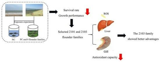
Survival rate (SR%) = Nt × 100/N0
Specific growth rate (SGR, % day−1) = (Ln Wt − Ln W0) × 100/t
Nt and N0 were final and initial fish number, respectively; Wt and W0 were final and initial fish weight, respectively; t was period of experimental days.
For statistical analysis, all data underwent evaluation in SPSS 23.0, a software developed by IBM in America. Various statistical tests, including independent sample t-tests or one-way analysis of variance (ANOVA), were performed, followed by Tukey’s multiple-range test to determine differences between groups. Statistical significance was considered at a level of p < 0.05. Results were presented as means ± S.E.M (standard error of the mean).

3. Results

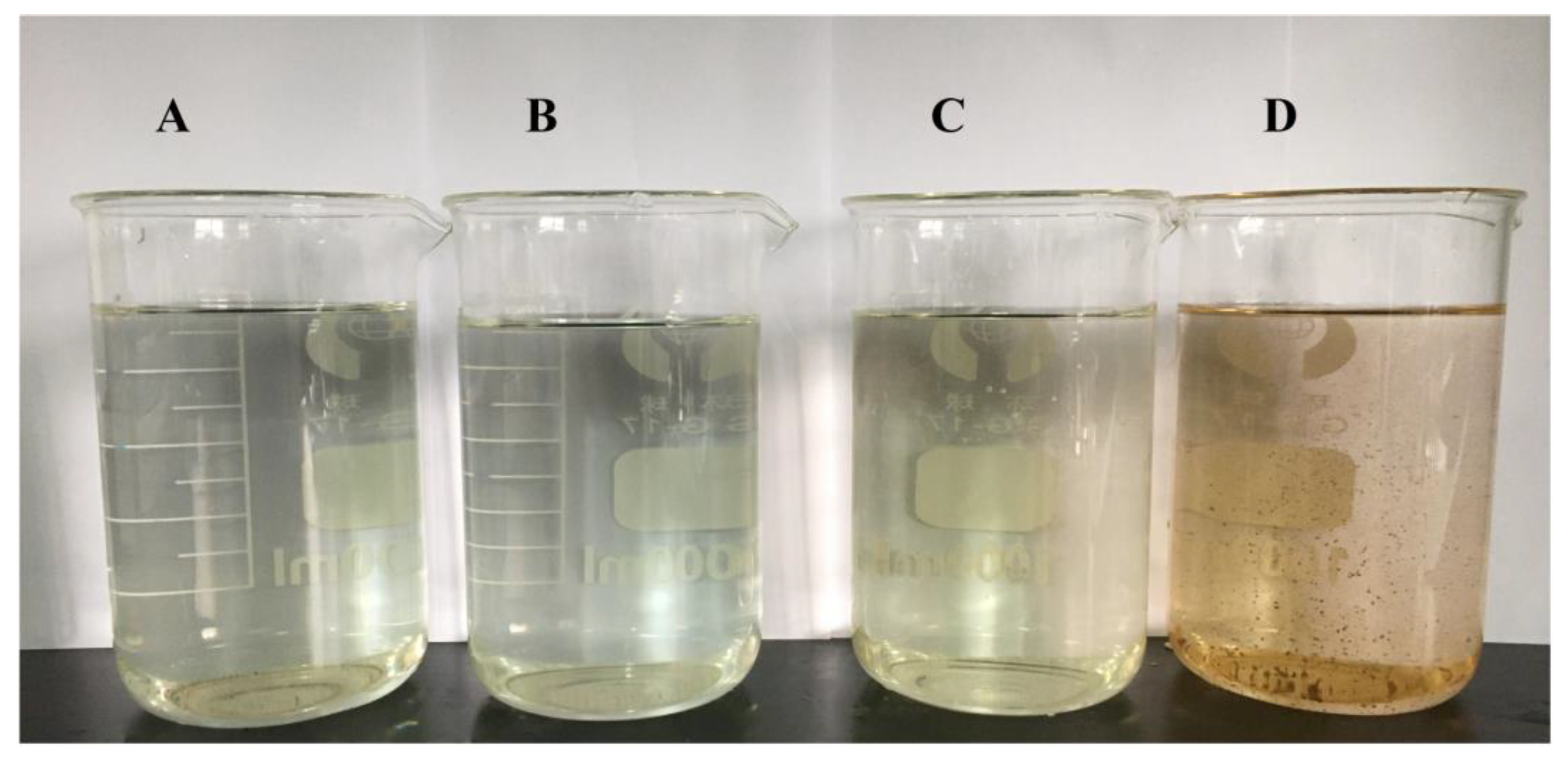
3.1. Metal Element Parameters of the Water Solution

First, the metal element parameters of U. prolifera degradation water were detected. In Table 2, the contents of Na, K, Mg, and Fe elements in the U. prolifera degradation water were significantly increased compared with the normal seawater (p < 0.05), respectively, while the Ca content showed no significant difference (p > 0.05). In addition, the chemical reaction test of the Fe element in the water solution was carried out. The results showed that the color of U. prolifera degradation water sample was turned to yellow (Figure 1).

3.2. Survival Rate and Growth Performance

In Table 3, U. prolifera degradation condition significantly decreased the survival rate (SR) and specific growth rate (SGR) compared with the NC group (p < 0.05), while SR of eight flounder families were higher than the PC group (48.0 ± 3.46%), especially 2101 (77.3 ± 9.24%) and 2103 (75.3 ± 1.15%) flounder families (p < 0.05). However, the SGR of eight flounder families had no significant differences compared with the PC group (p > 0.05).

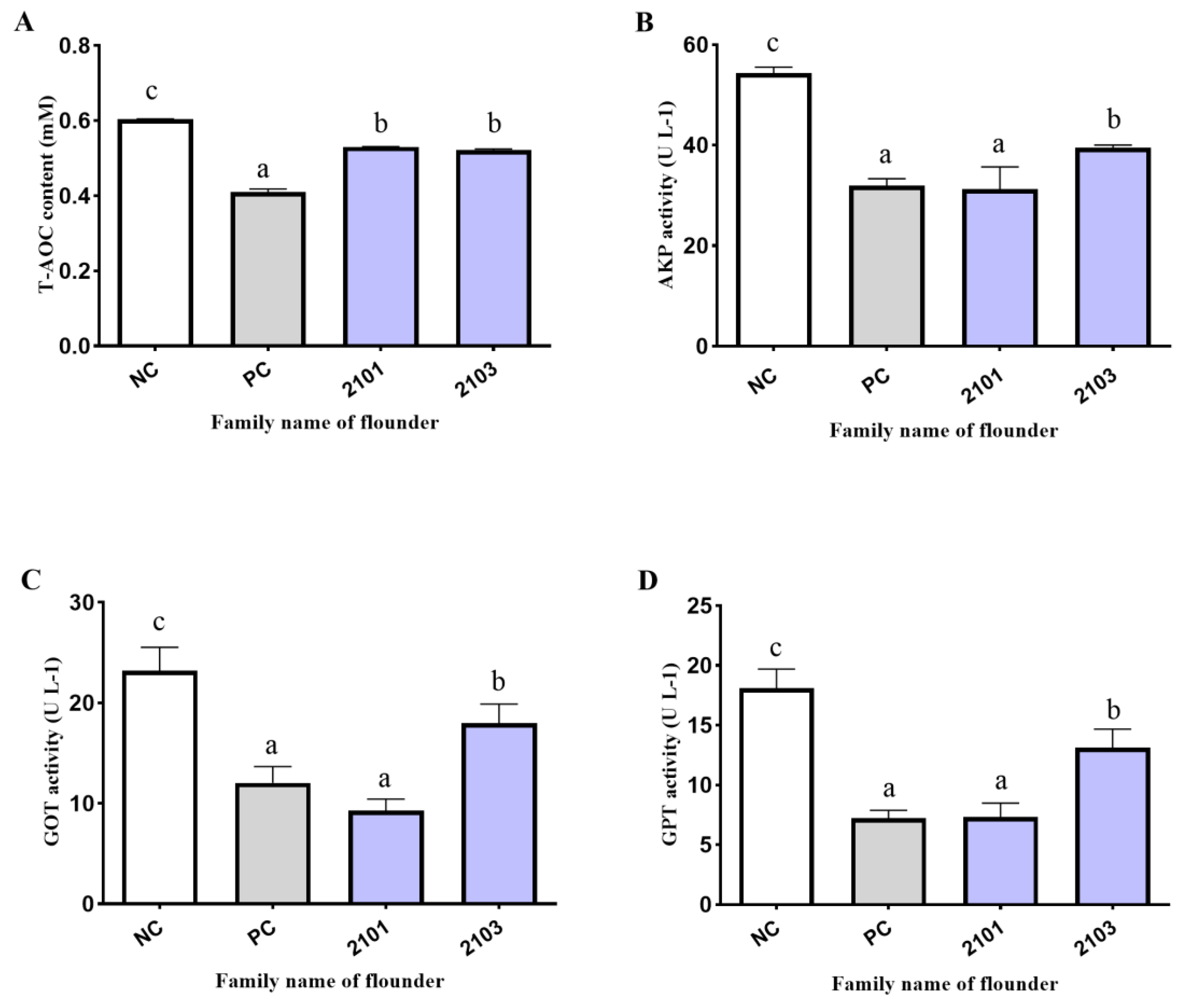
3.3. Serum Biochemical Indexes

In the present study, a significant decrease in T-AOC in the serum was observed compared with the NC group (p < 0.05) in U. prolifera degradation conditions, while the T-AOC content of 2101 and 2103 flounder families was significantly higher than the PC group (p < 0.05) (Figure 2A). In addition, U. prolifera degradation also significantly decreased GPT, GOT, and AKP activities compared with the NC group (p < 0.05), while these enzyme activities of 2103 flounder family were significantly higher than the PC group (p < 0.05) (Figure 2B–D). The 2101 flounder family had no significant difference compared with the PC group (p > 0.05) (Figure 2B–D).

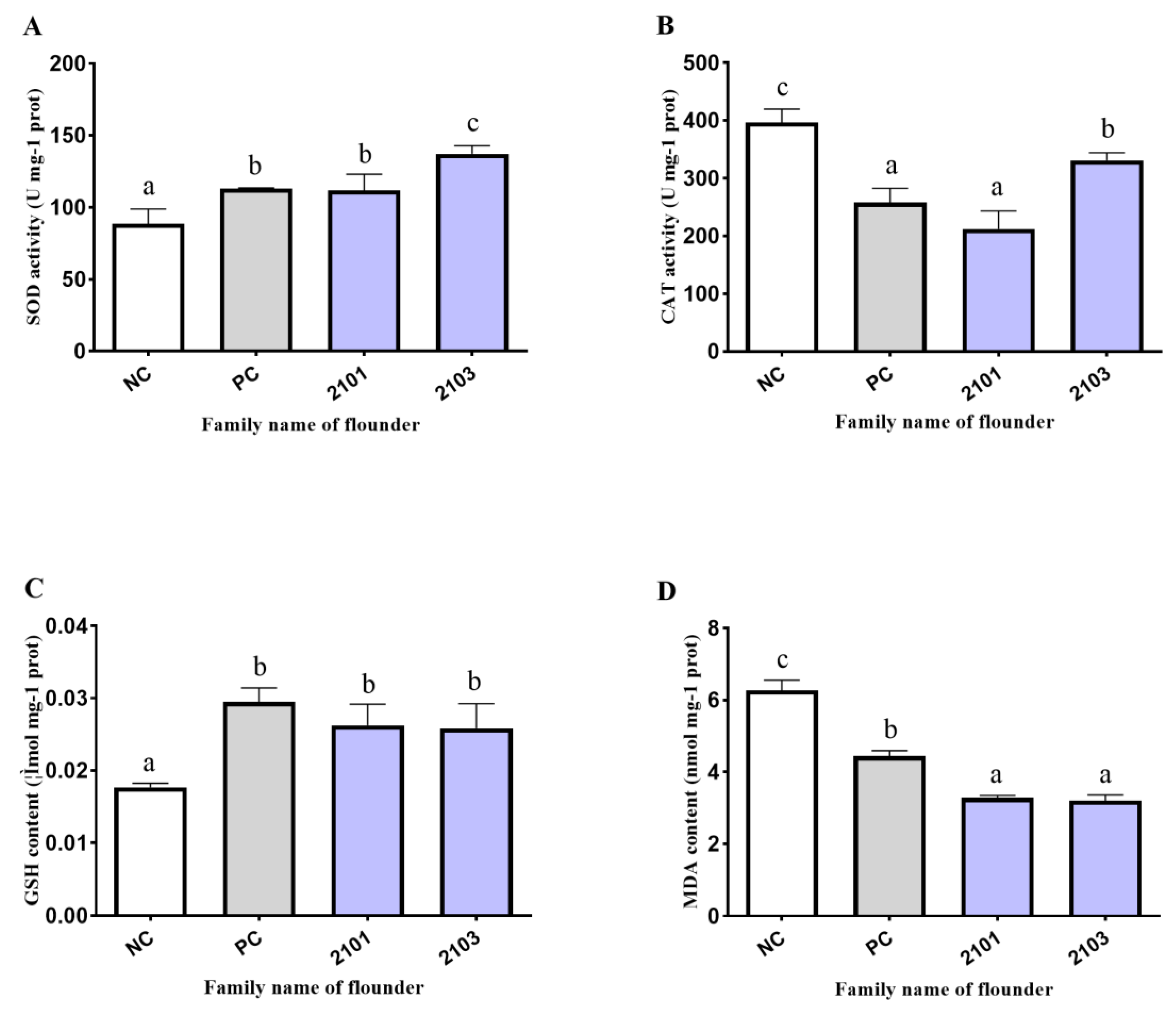
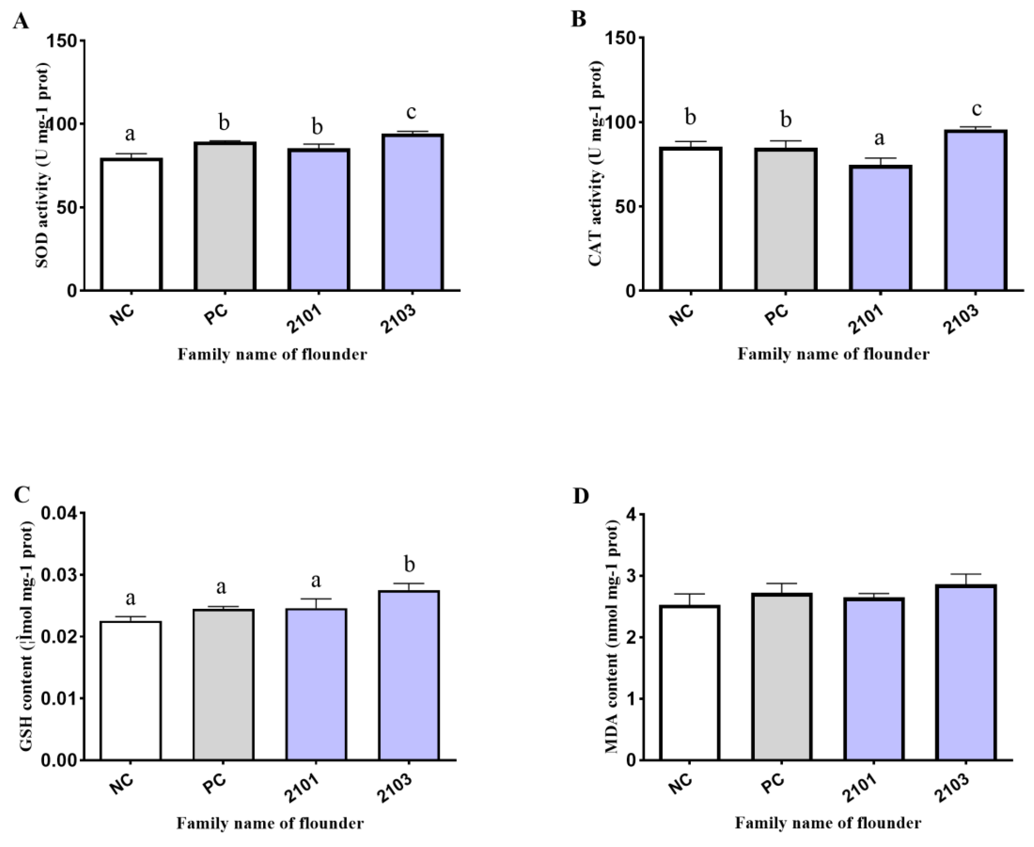
3.4. Antioxidant Enzyme Activity

The effects of U. prolifera degradation on the antioxidant capacity in the liver and gills of Japanese flounder were detected. The results showed that the SOD activity was significantly increased in the liver compared with the NC group (p < 0.05) (Figure 3A). Meanwhile, the SOD activity of the 2103 flounder family was significantly higher than the 2101 flounder family and PC group in the liver (p < 0.05), while the SOD activity of the 2101 flounder family had no significant difference compared with PC group (p > 0.05) (Figure 3A). In the liver, the CAT activity was notably reduced compared to the NC group (p < 0.05), while the CAT activity of the 2103 flounder family was significantly higher than the 2101 flounder family and PC group (p < 0.05) (Figure 3B). Moreover, U. prolifera degradation also significantly increased liver GSH content compared with the NC group (p < 0.05), while the GSH content of the 2101 and 2103 flounder families had no significant difference compared with the PC group, respectively (p > 0.05) (Figure 3C). In addition, U. prolifera degradation significantly decreased the liver MDA content compared with the NC group (p < 0.05), and the MDA content of the 2101 and 2103 flounder families were significantly lower than the PC group (p < 0.05) (Figure 3D).
In the gills, U. prolifera degradation significantly increased the SOD activity compared with the NC group (p < 0.05), and the SOD activity of the 2103 flounder family was significantly higher than the 2101 flounder family and PC group (p < 0.05) (Figure 4A). In addition, U. prolifera degradation significantly increased the CAT activity of the 2103 flounder family in the gills, while the CAT activity of the 2101 flounder family was significantly decreased (p < 0.05) (Figure 4B). In addition, the GSH content of the 2103 flounder family was significantly increased in the gills compared with the NC group (p < 0.05) and higher than the 2101 flounder family and PC group (p < 0.05) (Figure 4C). The GSH content of the 2101 flounder family had no significant difference compared with the NC and PC group (p > 0.05) (Figure 4C). However, U. prolifera degradation has no significant effect on the MDA content of Japanese flounder (p > 0.05) (Figure 4D)

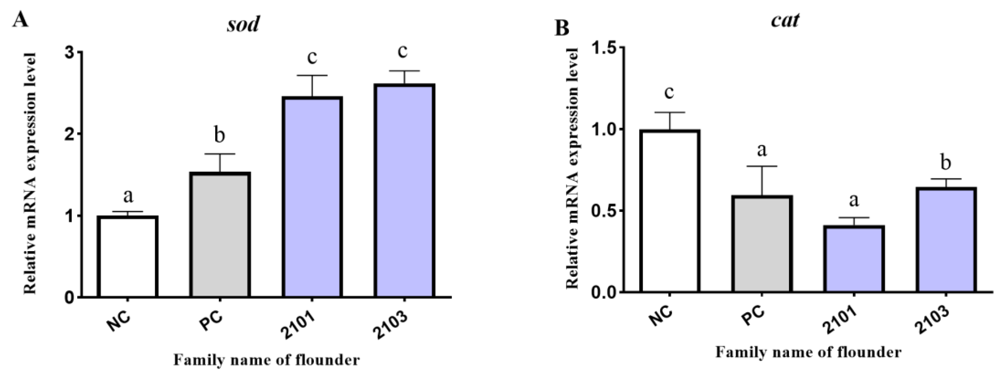
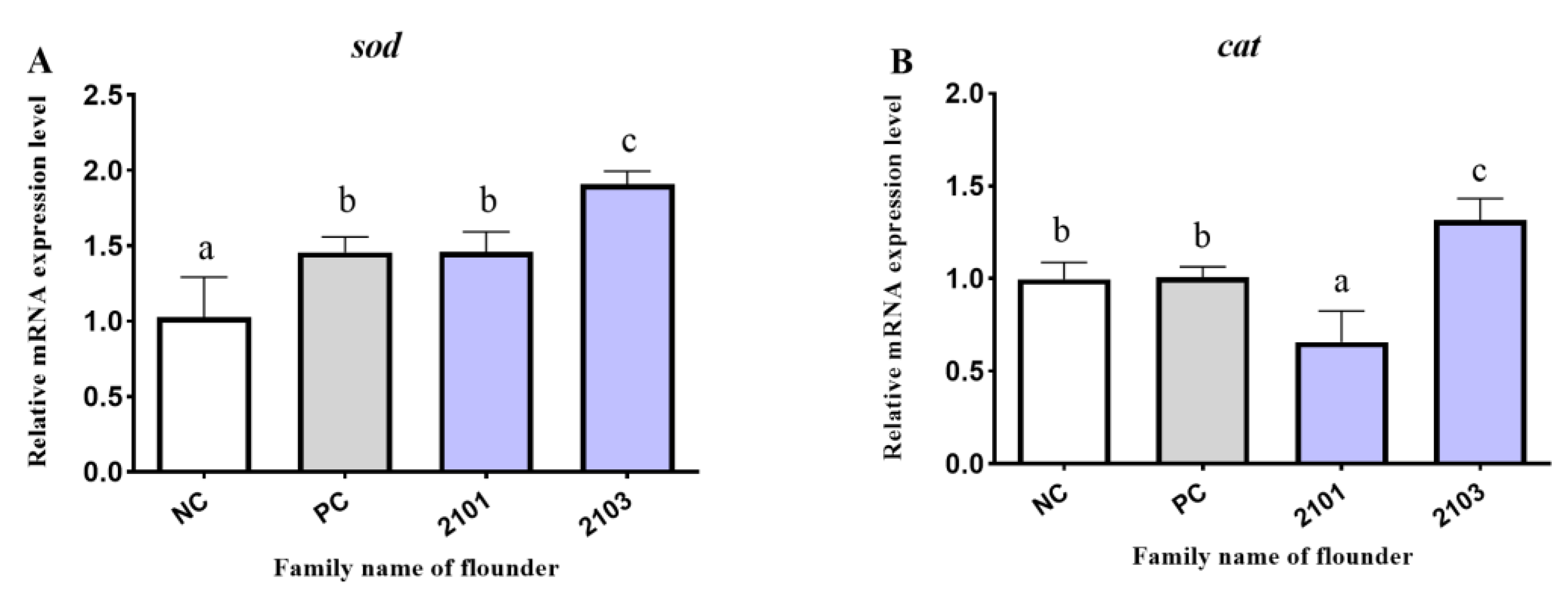
3.5. Antioxidant-Related Genes Expression

The effects of U. prolifera degradation on mRNA expression level of antioxidant-related genes in the liver and gills were further detected. The results showed that U. prolifera degradation significantly upregulated sod mRNA expression compared with the NC group in the liver (p < 0.05), and the sod mRNA expression of the 2101 and 2103 flounder families were significantly higher than the PC group (p < 0.05) (Figure 5A). In addition, U. prolifera degradation significantly downregulated the cat mRNA expression compared with the NC group in the liver (p < 0.05), while the cat mRNA expression of the 2103 flounder family was significantly higher than the 2101 flounder family and PC group (p < 0.05) (Figure 5B).
In the gills, U. prolifera degradation significantly upregulated the sod mRNA expression compared with the NC group (p < 0.05), while the sod mRNA expression of the 2103 flounder family was significantly higher than the 2101 flounder family and PC group (p < 0.05) (Figure 6A). The sod mRNA expression of the 2101 flounder family had no significant difference compared with the PC group (p > 0.05) (Figure 6A). Meanwhile, the cat mRNA expression of the 2103 flounder family was significantly higher than the 2101 flounder family, PC group, and NC group in the gills (p < 0.05) (Figure 6B), while the cat mRNA expression of 2101 flounder family was significantly lower than PC group and NC group in gill (p < 0.05) (Figure 6B).

4. Discussion

Driven by water eutrophication and global climate change, U. prolifera blooms in coastal areas of the Yellow Sea of China have become increasingly common in recent years [1]. The degradation of U. prolifera not only disrupts marine ecosystems, but also significantly impacts aquaculture [22,23,24]. Therefore, it is crucially important to detect the effects of U. prolifera degradation on the growth performance and antioxidant capacity of Japanese flounder (Paralichthys olivaceus). In the present study, we first detected the element composition of the U. prolifera degradation water sample. The total Na, K, Mg, and Fe contents of U. prolifera degradation water were markedly higher than that of the normal seawater, with noticeable changes in color during chemical tests. Previous studies have shown that the total macro-, micro-, and trace-element contents of Enteromorpha species were higher than the values reported for some edible land vegetables and seaweeds, especially micronutrients (Fe, Zn, Mn, and Cu) [25]. However, no studies reported the metal contents of U. prolifera degradation water. In mammals, the possibility of excess iron promoting oxidative stress and inducing multiple disorders due to its redox reactivity has been proven [26,27,28]. In addition, previous studies in fish also have demonstrated that excess iron promoted oxidative stress and mitochondrial dysfunction [29,30]. Therefore, the abnormal increase in Fe and other metal elements in the water environment and whether it affects the growth performance and antioxidant capacity of Japanese flounder need more investigation.
After determining the elements composition of U. prolifera degradation water, we investigated the effects of U. prolifera degradation on growth performance. Due to the U. prolifera degradation stress, the growth index and SR were significantly decreased, which was consistent with the previous findings that massive macroalgal blooms have caused the sea cucumber and shellfish death in the Yellow Sea [31]. Two flounder families (2101 and 2103) exhibited better survival and growth appearance among eight flounder families compared with the flounder of the PC group under the U. prolifera degradation stress, and they are considered to be the potential families for tolerance to the U. prolifera degradation conditions.
In addition to the growth performance, the effects of oxidative stress caused by the U. prolifera degradation on the antioxidant capacity of Japanese flounder were further explored. The previous study has shown that enzymatic activities of the transaminase enzymes (GOT and GPT) and alkaline phosphatase (AKP) are common indicators of hepatic damage [32]. The research demonstrated a noteworthy reduction in the activities of GOT, GPT, and AKP in the serum as a result of U. prolifera degradation. The findings indicated substantial damage to the liver, aligning with the cytotoxic effects observed in a rainbow trout hepatocyte analysis [33].
Additionally, SOD and CAT play a crucial role in the primary defense against oxidative stress. SOD primarily facilitates the conversion of O2 and H+ into less reactive compounds, such as H2O2, while CAT effectively neutralizes H2O2 into harmless substances H2O and O2 [34,35]. GSH, a key non-protein thiol in cells, serves as an integral component in the cellular defense mechanism against toxic effects triggered by xenobiotics, oxyradicals, and metal cations [36]. MDA, emerges as a byproduct of lipid peroxidation, predominantly induced by excessive ROS attacking the polyunsaturated fatty acids (PUFAs) within the cellular membrane. This process can severely impair the structure and functionality of cells [37]. The current investigation revealed that U. prolifera degradation significantly elevated the levels of SOD and GSH while reducing CAT activity and MDA content within the liver. This suggests that U. prolifera degradation actively facilitates the elimination of O2- and prevents damage caused by oxidative stress. This effect is likely attributable to the polysaccharide found in U. prolifera degradation, which enhances the overall antioxidant capacity in flounder organisms. Similarly, previous studies have shown polysaccharides [38] from the U. prolifera degradation has a positive effect on antioxidant defense system in Carassius auratus [39,40]. The observed decrease in CAT activity in the liver could be attributed to iron accumulation, which generated excessive ROS through the Fenton reaction [41]. Combining the results of antioxidant enzyme activities in the serum and liver, the function and antioxidant capacity of the liver could be damaged under U. prolifera degradation. In addition, the 2103 flounder family displayed significantly higher activities of SOD and CAT than the 2101 and PC group in the liver, suggesting that the 2103 flounder family has greater antioxidant capacity under the U. prolifera degradation conditions. In the gills, the SOD and CAT activities and the GSH content of the 2103 flounder family was significantly higher than the 2101 flounder family and PC group, suggesting that the antioxidant defense system of the 2103 flounder family was influenced and higher than the 2101 flounder family in U. prolifera degradation conditions.
In this study, we further explored the effects of U. prolifera degradation on the mRNA expression of antioxidant-related genes in aquatic organisms. Our findings revealed that the degradation of U. prolifera had a significant impact, resulting in increased sod mRNA expression and decreased cat mRNA expression in the liver. Furthermore, in the gills of the 2103 flounder family, there was a marked rise in sod mRNA expression. These results were consistent with the changes observed in the activities of SOD and CAT in both the liver and gills. It is important to note that these findings contradict a previous study, which reported that Alexandrium affine significantly affects the immunity and antioxidant capacity of red seabream gills [42]. The probable reason was that the flounder was not directly attached to the floccules of U. prolifera. To sum up, although the T-AOC activity of Japanese flounder was significantly decreased compared to the NC group under the U. prolifera degradation stress, the antioxidant ability of the liver and gills of the 2103 family was higher compared with the PC group. Therefore, the selected flounder family (2103) could be regarded as a potential flounder family tolerating U. prolifera degradation stress.

5. Conclusions

In conclusion, this study revealed that U. prolifera degradation influenced the growth performance and antioxidant capacity of Japanese flounder, while the 2103 flounder family was more resilient on the growth performance and antioxidant defense in U. prolifera degradation conditions. Thereby, the 2103 flounder family could be regarded as the potential family tolerating U. prolifera degradation stress. Furthermore, the metal element content of U. prolifera degradation water was detected to understand its impact on ecological environment and marine fish. The increased Fe content in the U. prolifera degradation water may be one of the main causes of the physiological alterations observed in Japanese flounder. Therefore, this study could advance the understanding of the effects of U. prolifera degradation on marine fish, specifically regarding the growth performance and antioxidant capacity, and provide scientific evidence for excellent germplasm screening.

Author Contributions

Y.Y.: conceptualization, data curation and writing - review and editing; W.L. and R.W.: methodology and visualization; D.X.: software and analyze; Y.C. and Z.C.: methodology and editing; S.C.: project administration and funding acquisition. All authors have read and agreed to the published version of the manuscript.

Funding

This work was supported by the Central Public-interest Scientific Institution Basal Research Fund, CAFS (2020TD20), the Key Research and Development Project of Shandong Province (2021LZGC028), Taishan Scholar Climbing Project Fund of Shandong, China.

Institutional Review Board Statement

The experimental protocol was designed in accordance with the recommendations of the Regulations of the Laboratory Animal—Guideline for Ethical Review of Animal Welfare (Standardization Administration of China, 2018) and reviewed and approved by the animal care and use committee of the Yellow Sea Fisheries Research Institute (YSFRI-2023021).

Data Availability Statement

Data are contained within the article.

Acknowledgments

We thank Wenteng Xu, Yang Liu, Sheng Lu, Yu Song, and Yellow Sea Aquatic Product Co., Ltd. for their experimental assistance.

Conflicts of Interest

The authors declare no conflict of interest.

References

  1. Hallegraeff, G.; Enevoldsen, H.; Zingone, A. Global harmful algal bloom status reporting. Harmful Algae 2021, 102, 101992. [Google Scholar] [CrossRef] [PubMed]
  2. Liu, X.; Wang, Z.; Zhang, X. A review of the green tides in the Yellow Sea, China. Mar. Environ. Res. 2016, 119, 189–196. [Google Scholar] [CrossRef] [PubMed]
  3. Ye, N.; Zhang, X.; Mao, Y.; Liang, C.; Xu, D.; Zou, J.; Zhuang, Z.; Wang, Q. ‘Green tides’ are overwhelming the coastline of our blue planet: Taking the world’s largest example. Ecol. Res. 2011, 26, 477–485. [Google Scholar] [CrossRef]
  4. Van Alstyne, K.L.; Nelson, T.A.; Ridgway, R.L. Environmental Chemistry and Chemical Ecology of “Green Tide” Seaweed Blooms. Integr. Comp. Biol. 2015, 55, 518–532. [Google Scholar] [CrossRef] [PubMed]
  5. Yang, X.; Lin, K.; Tan, L.; Wang, J. Utilization and release of biogenic elements by macroalgae Ulva prolifera: A mesocosm experiment off the coast of Qingdao, China. Mar. Pollut. Bull. 2021, 170, 112612. [Google Scholar] [CrossRef] [PubMed]
  6. Zhang, T.; Wang, X. Release and microbial degradation of dissolved organic matter (DOM) from the macroalgae Ulva prolifera. Mar. Pollut. Bull. 2017, 125, 192–198. [Google Scholar] [CrossRef] [PubMed]
  7. Arisekar, U.; Shakila, R.J.; Shalini, R.; Jeyasekaran, G.; Padmavathy, P.; Hari, M.S.; Sudhan, C. Accumulation potential of heavy metals at different growth stages of Pacific white leg shrimp, Penaeus vannamei farmed along the Southeast coast of Peninsular India: A report on ecotoxicology and human health risk assessment. Environ. Res. 2022, 212, 113105. [Google Scholar] [CrossRef]
  8. Talmage, S.C.; Gobler, C.J. Effects of past, present, and future ocean carbon dioxide concentrations on the growth and survival of larval shellfish. Proc. Natl. Acad. Sci. USA 2010, 107, 17246–17251. [Google Scholar] [CrossRef]
  9. Gu, P.; Li, Q.; Zhang, W.; Gao, Y.; Sun, K.; Zhou, L.; Zheng, Z. Biological toxicity of fresh and rotten algae on freshwater fish: LC(50), organ damage and antioxidant response. J. Hazard. Mater. 2021, 407, 124620. [Google Scholar] [CrossRef]
  10. Ezraty, B.; Gennaris, A.; Barras, F.; Collet, J.F. Oxidative stress, protein damage and repair in bacteria. Nat. Rev. Microbiol. 2017, 15, 385–396. [Google Scholar] [CrossRef]
  11. Latifi, A.; Ruiz, M.; Zhang, C.C. Oxidative stress in cyanobacteria. FEMS Microbiol. Rev. 2009, 33, 258–278. [Google Scholar] [CrossRef] [PubMed]
  12. Sies, H.; Berndt, C.; Jones, D.P. Oxidative Stress. Annu. Rev. Biochem. 2017, 86, 715–748. [Google Scholar] [CrossRef]
  13. Cui, W.; Cao, L.; Liu, J.; Ren, Z.; Zhao, B.; Dou, S. Effects of seawater acidification and cadmium on the antioxidant defense of flounder Paralichthys olivaceus larvae. Sci. Total Environ. 2020, 718, 137234. [Google Scholar] [CrossRef]
  14. Zhu, Y.; Li, S.; Su, B.; Xue, T.; Cao, M.; Li, C. Genome-wide identification, characterization, and expression of the Toll-like receptors in Japanese flounder (Paralichthys olivaceus). Aquaculture 2021, 545, 737127. [Google Scholar] [CrossRef]
  15. Fan, Q.; Shi, K.; Zhan, M.; Xu, Q.; Liu, X.; Li, Z.; Liu, H.; Xia, Y.; Chen, Y.; Shi, X.; et al. Acute damage from the degradation of Ulva prolifera on the environmental microbiota, intestinal microbiota and transcriptome of Japanese flounder Paralichthys olivaceus. Environ. Pollut. 2022, 302, 119022. [Google Scholar] [CrossRef] [PubMed]
  16. Kim, J.; Choi, H.; Sung, G.; Seo, S.; Kim, K.; Kang, Y.; Kang, J. Toxic effects on hematological parameters and oxidative stress in juvenile olive flounder, Paralichthys olivaceus exposed to waterborne zinc. Aquacult. Rep. 2019, 15, 100225. [Google Scholar] [CrossRef]
  17. Lu, Z.; Wang, S.; Ji, C.; Li, F.; Cong, M.; Shan, X.; Wu, H. iTRAQ-based proteomic analysis on the mitochondrial responses in gill tissues of juvenile olive flounder Paralichthys olivaceus exposed to cadmium. Environ. Pollut. 2020, 257, 113591. [Google Scholar] [CrossRef] [PubMed]
  18. Nelson, T.A.; Lee, D.J.; Smith, B.C. Are “green tides” harmful algal blooms? Toxic properties of water-soluble extracts from two bloom-forming macroalgae, Ulva fenestrata and Ulvaria obscura (Ulvophyceae). J. Phycol. 2003, 39, 874–879. [Google Scholar] [CrossRef]
  19. Li, Y.; Zhang, B.; Lu, S.; Tian, Y.; Yang, Y.; Chen, S. Genetic parameters estimates for growth performance traits at harvest in Japanese flounder (Paralichthys olivaceus). Aquaculture 2018, 489, 56–61. [Google Scholar] [CrossRef]
  20. Gagnon, M.M.; Hodson, P. Field studies using fish biomarkers–How many fish are enough? Mar. Pollut. Bull. 2012, 64, 2871–2876. [Google Scholar] [CrossRef]
  21. Livak, K.J.; Schmittgen, T.D. Analysis of relative gene expression data using real-time quantitative PCR and the 2−ΔΔCT Method. Methods 2001, 25, 402–408. [Google Scholar] [CrossRef] [PubMed]
  22. Andersen, N.G.; Hansen, P.J.; Engell-Sorensen, K.; Norremark, L.H.; Andersen, P.; Lorenzen, E.; Lorenzen, N. Ichthyotoxicity of the microalga Pseudochattonella farcimen under laboratory and field conditions in Danish waters. Dis. Aquat. Organ. 2015, 116, 165–172. [Google Scholar] [CrossRef] [PubMed]
  23. John, U.; Supraha, L.; Gran-Stadniczenko, S.; Bunse, C.; Cembella, A.; Eikrem, W.; Janouskovec, J.; Klemm, K.; Kuhne, N.; Naustvoll, L. Spatial and biological oceanographic insights into the massive fish-killing bloom of the haptophyte Chrysochromulina leadbeateri in northern Norway. Harmful Algae 2022, 118, 102287. [Google Scholar] [CrossRef] [PubMed]
  24. Sakamoto, S.; Lim, W.A.; Lu, D.; Dai, X.; Orlova, T.; Iwataki, M. Harmful algal blooms and associated fisheries damage in East Asia: Current status and trends in China, Japan, Korea and Russia. Harmful Algae 2021, 102, 101787. [Google Scholar] [CrossRef] [PubMed]
  25. Ganesan, K.; Suresh Kumar, K.; Subba Rao, P.V.; Tsukui, Y.; Bhaskar, N.; Hosokawa, M.; Miyashita, K. Studies on chemical composition of three species of Enteromorpha. Biomed. Prev. Nutr. 2014, 4, 365–369. [Google Scholar] [CrossRef]
  26. Ganz, T. Systemic iron homeostasis. Physiol. Rev. 2013, 93, 1721–1741. [Google Scholar] [CrossRef] [PubMed]
  27. Kim, C.H.; Leitch, H.A. Iron overload-induced oxidative stress in myelodysplastic syndromes and its cellular sequelae. Crit. Rev. Oncol. Hematol. 2021, 163, 103367. [Google Scholar] [CrossRef]
  28. Wang, J.; Pantopoulos, K. Regulation of cellular iron metabolism. Biochem. J. 2011, 434, 365–381. [Google Scholar] [CrossRef]
  29. Ahmed, U.; Latham, P.S.; Oates, P.S. Interactions between hepatic iron and lipid metabolism with possible relevance to steatohepatitis. World J. Gastroenterol. 2012, 18, 4651–4658. [Google Scholar] [CrossRef]
  30. Luo, Z.; Zou, G.Y.; Gao, Y.; Ye, H.M.; Xi, W.Q.; Liu, X. Effect of dietary iron (Fe) levels on growth performance, hepatic lipid metabolism and antioxidant responses in juvenile yellow catfish Pelteobagrus fulvidraco. Aquacult. Nutr. 2017, 23, 1475–1482. [Google Scholar] [CrossRef]
  31. Zhang, X.; Song, Y.; Liu, D.; Keesing, J.K.; Gong, J. Macroalgal blooms favor heterotrophic diazotrophic bacteria in nitrogen-rich and phosphorus-limited coastal surface waters in the Yellow Sea. Estuar. Coast. Shelf Sci. 2015, 163, 75–81. [Google Scholar] [CrossRef]
  32. Abdel-Wahhab, M.A.; Aly, S.E. Antioxidant property of Nigella sativa (black cumin) and Syzygium aromaticum (clove) in rats during aflatoxicosis. J. Appl. Toxicol. 2005, 25, 218–223. [Google Scholar] [CrossRef]
  33. Shahraki, J.; Motallebi, A.; Pourahmad, J. Oxidative mechanisms of fish hepatocyte toxicity by the harmful dinoflagellate Cochlodinium polykrikoides. Mar. Environ. Res. 2013, 87–88, 52–60. [Google Scholar] [CrossRef] [PubMed]
  34. Huang, X.; Liu, Y.; Liu, Z.; Zhao, Z.; Dupont, S.; Wu, F.; Huang, W.; Chen, J.; Hu, M.; Lu, W.; et al. Impact of zinc oxide nanoparticles and ocean acidification on antioxidant responses of Mytilus coruscus. Chemosphere 2018, 196, 182–195. [Google Scholar] [CrossRef] [PubMed]
  35. Qu, R.; Wang, X.; Wang, Z.; Wei, Z.; Wang, L. Metal accumulation and antioxidant defenses in the freshwater fish Carassius auratus in response to single and combined exposure to cadmium and hydroxylated multi-walled carbon nanotubes. J. Hazard. Mater. 2014, 275, 89–98. [Google Scholar] [CrossRef] [PubMed]
  36. Srikanth, K.; Pereira, E.; Duarte, A.C.; Ahmad, I. Glutathione and its dependent enzymes’ modulatory responses to toxic metals and metalloids in fish—A review. Environ. Sci. Pollut. Res. 2013, 20, 2133–2149. [Google Scholar] [CrossRef] [PubMed]
  37. Freitas, R.; Pires, A.; Velez, C.; Almeida, Â.; Moreira, A.; Wrona, F.J.; Soares, A.M.V.M.; Figueira, E. Effects of seawater acidification on Diopatra neapolitana (Polychaete, Onuphidae): Biochemical and regenerative capacity responses. Ecol. Indic. 2016, 60, 152–161. [Google Scholar] [CrossRef]
  38. Wassie, T.; Niu, K.; Xie, C.; Wang, H.; Xin, W. Extraction Techniques, Biological Activities and Health Benefits of Marine Algae Enteromorpha prolifera Polysaccharide. Front. Nutr. 2021, 8, 747928. [Google Scholar] [CrossRef]
  39. Garcia, D.; Lima, D.; da Silva, D.G.H.; de Almeida, E.A. Decreased malondialdehyde levels in fish (Astyanax altiparanae) exposed to diesel: Evidence of metabolism by aldehyde dehydrogenase in the liver and excretion in water. Ecotoxicol. Environ. Saf. 2020, 190, 110107. [Google Scholar] [CrossRef]
  40. Zhou, Z.; Pan, S.; Wu, S. Modulation of the growth performance, body composition and nonspecific immunity of crucian carp Carassius auratus upon Enteromorpha prolifera polysaccharide. Int. J. Biol. Macromol. 2020, 147, 29–33. [Google Scholar] [CrossRef]
  41. Dixon, S.J.; Lemberg, K.M.; Lamprecht, M.R.; Skouta, R.; Zaitsev, E.M.; Gleason, C.E.; Patel, D.N.; Bauer, A.J.; Cantley, A.M.; Yang, W.S.; et al. Ferroptosis: An iron-dependent form of nonapoptotic cell death. Cell 2012, 149, 1060–1072. [Google Scholar] [CrossRef] [PubMed]
  42. Haque, M.N.; Nam, S.E.; Shin, Y.K.; Rhee, J.S. The dinoflagellate Alexandrium affine acutely induces significant modulations on innate immunity, hepatic function, and antioxidant defense system in the gill and liver tissues of red seabream. Aquat. Toxicol. 2021, 240, 105985. [Google Scholar] [CrossRef] [PubMed]
Figure 1. Chemical reaction result of Fe ion in normal seawater and U. prolifera degradation water environments. (A): normal seawater; (B): U. prolifera degradation water; (C): chemical reaction result of Fe ion in normal seawater; (D): chemical reaction result of Fe ion in U. prolifera degradation water.
Fishes 08 00598 g001
Figure 2. Effects of U. prolifera degradation on the serum biochemical indexes and enzyme activities ((A): T-AOC, (B): AKP, (C): GOT, and (D): GPT) of Japanese flounder. NC group: negative control group; PC group: positive control group; 2101 and 2103: 2 Japanese flounder families. Data are presented as means ± S.E.M. and are analyzed using one-way ANOVA followed by Tukey’s test (n = 6). Data labeled with the same letters are not significantly different (p > 0.05).
Fishes 08 00598 g002
Figure 3. Effects of U. prolifera degradation on antioxidant capacity in liver of Japanese flounder. (A,B): enzyme activities of SOD and CAT. (C,D): contents of GSH and MDA. NC group: negative control group; PC group: positive control group; 2101 and 2103: 2 Japanese flounder families. Data are presented as means ± S.E.M. and are analyzed using one-way ANOVA followed by Tukey’s test (n = 6). Data labeled the same letters are not significantly different (p > 0.05).
Fishes 08 00598 g003
Figure 4. Effects of U. prolifera degradation on antioxidant capacity in gills of Japanese flounder. (A,B): enzyme activities of SOD and CAT. (C,D): contents of GSH and MDA. NC group: negative control group; PC group: positive control group; 2101 and 2103: 2 Japanese flounder families. Data are presented as means ± S.E.M. and are analyzed using one-way ANOVA followed by Tukey’s test (n = 6). Data labeled the same letters and no letters are not significantly different (p > 0.05).
Fishes 08 00598 g004
Figure 5. Effects of U. prolifera degradation on the mRNA expression of antioxidant-related genes ((A): sod and (B): cat) in liver of Japanese flounder. NC group: negative control group; PC group: positive control group; 2101 and 2103: 2 Japanese flounder families. Data are presented as means ± S.E.M. and are analyzed using one-way ANOVA followed by Tukey’s test (n = 6). Data labeled the same letters are not significantly different (p > 0.05).
Fishes 08 00598 g005
Figure 6. Effects of U. prolifera degradation on the mRNA expression of antioxidant-related genes ((A): sod and (B): cat) in gills of Japanese flounder. NC group: negative control group; PC group: positive control group; 2101 and 2103: 2 Japanese flounder families. Data are presented as means ± S.E.M. and are analyzed using one-way ANOVA followed by Tukey’s test (n = 6). Data labeled the same letters are not significantly different (p > 0.05).
Fishes 08 00598 g006
Table 1. Primer information used for RT-qPCR in this study.
GeneSequence (5′–3′)GenBank Accession Number
β-actin-FGGAAATCGTGCGTGACATTAAGXM_020109620.1
β-actin-RCCTCTGGACAACGGAACCTCT
sod-FCGTTGGAGACCTGGGGAATGTGEF681883.1
sod-RATCGTCAGCCTTCTCGTGGATC
cat-FCACGGACCAGATGAAGCAGTGXM_020079314.1
cat-RCCTTGGAGTAGCGGGTAATGTC
Table 2. The content of five metal elements in two water environments.
IndexTypes
UGNCp Value
Fe (μg/L)7.4 ± 0.79 *5.4 ± 0.170.013
Ca (mg/L)330.3 ± 1.16375.0 ± 29.460.12
K (mg/L)439.0 ± 3.67 **378.7 ± 18.560.005
Mg (mg/L)1075.7 ± 4.04 **970.7 ± 25.540.002
Na (mg/L)9904.7 ± 65.07 **8953.7 ± 258.720.000
Notes: Data are presented as means ± S.E.M. and are analyzed using independent t-tests. * p < 0.05 and ** p < 0.01 indicate significant differences compared with the control group. UG: U. prolifera degradation treatment group; NC: negative control group.
Table 3. Growth performance parameters and survival rate of the Japanese flounder cultivated in different environments.
GroupIBW (g)FBW (g)SR (%)SGR (%/day)
NC2.5 ± 0.0252.9 ± 0.87 a88.7 ± 1.16 a5.1 ± 0.04 a
PC2.5 ± 0.0140.8 ± 0.64 bc48.0 ± 3.46 c4.6 ± 0.01 bc
21012.5 ± 0.0142.7 ± 1.83 bc77.3 ± 9.24 ab4.8 ± 0.07 b
21022.5 ± 0.0144.6 ± 3.1 b53.3 ± 9.87 c4.8 ± 0.12 b
21032.5 ± 0.0343.2 ± 2.29 bc75.3 ± 1.15 ab4.8 ± 0.04 b
21042.5 ± 0.0143.6 ± 1.89 b58.7 ± 4.16 bc4.8 ± 0.7 b
21052.5 ± 0.0142.2 ± 3.32 bc56.0 ± 15.62 bc4.7 ± 0.14 bc
21062.5 ± 0.0139.9 ± 4.68 bc63.3 ± 8.08 bc4.6 ± 0.19 bc
21072.5 ± 0.0236.6 ± 1.04 c61.3 ± 10.07 bc4.5 ± 0.04 c
21082.5 ± 0.0238.3 ± 1.87 bc52.7 ± 4.16 c4.6 ± 0.08 bc
Notes: Data are presented as means ± S.E.M. and are analyzed using one-way ANOVA followed by Tukey’s test (n = 6). Data labeled the same letters are not significantly different (p > 0.05). NC group: negative control group; PC group: positive control group; 2101–2108: 8 Japanese flounder families; IBW: initial body weight; FBW: final body weight; SR: survival rate; SGR: specific growth rate.
Disclaimer/Publisher’s Note: The statements, opinions and data contained in all publications are solely those of the individual author(s) and contributor(s) and not of MDPI and/or the editor(s). MDPI and/or the editor(s) disclaim responsibility for any injury to people or property resulting from any ideas, methods, instructions or products referred to in the content.

