Article

Identification and Characterization of the Stimulator of Interferon Genes (STING) in Chinese Giant Salamander Andrias davidianus

1
Hunan Provincial Key Laboratory for Molecular Immunity Technology of Aquatic Animal Diseases, Hunan Provincial Key Laboratory for Health Aquaculture and Product Processing in Dongting Lake Area, Changde Research Center for Agricultural Biomacromolecule, Innovation Team of Microbial Technology, College of Life and Environmental Sciences, Hunan University of Arts and Science, Changde 415000, China
2
Key Laboratory of Biology and Genetic Resources of Tropical Crops, Ministry of Agriculture and Rural Affairs, Institute of Tropical Bioscience and Biotechnology, Chinese Academy of Tropical Agricultural Sciences, Haikou 571101, China
*
Authors to whom correspondence should be addressed.
Fishes 2023, 8(12), 597; https://doi.org/10.3390/fishes8120597
Submission received: 8 October 2023 / Revised: 30 November 2023 / Accepted: 30 November 2023 / Published: 4 December 2023
(This article belongs to the Special Issue Advances in Aquatic Diseases and Immunity in Aquaculture)

Abstract

:
The stimulator of interferon genes (STING) is a central and multifaceted mediator in innate immunity, and plays vital roles in defending against pathogen infection. In this study, we identified and functionally characterized the STING homolog from the Chinese giant salamander Andrias davidianus (AdSTING) for the first time. The open reading frame of AdSTING encodes a 362 amino acid protein with a predicted molecular mass of 41.6 kDa, which shares 31.1–46.7% of its sequence identity with STING homologs in other vertebrates. Structural analysis revealed that AdSTING possesses four predicted transmembrane domains (TMs) at the N-terminal, and a C-terminal domain (CTD) featuring a dimerization domain (DD), a c-di-GMP-binding domain (CBD), and a short C-terminal tail (CTT). Tissue distribution analysis showed that AdSTING mRNA was ubiquitously expressed in all examined tissues, with abundant expression in muscles, intestine, and thymus. During Andrias davidianus ranavirus (ADRV) infection, significant up-regulation of AdSTING expression was observed in the thymus, spleen, and kidney. Upon different stimuli in vitro, the expression of AdSTING was significantly induced by ADRV infection or polyinosin-polycytidylic acid (poly I:C) stimulation, but no obvious changes were observed during lipopolysaccharide (LPS) stimulation. Subcellular localization analysis revealed that AdSTING mainly localized in the cytoplasm in the Chinese giant salamander thymus cell line (GSTC) and co-localized with the endoplasmic reticulum (ER). Luciferase reporter assays confirmed the ability of AdSTING to activate the interferon-stimulated response element (ISRE) and interferon (IFN) promoter. Furthermore, overexpression of AdSTING effectively decreased ADRV infection, as evidenced by the reduction of virus titers and viral gene expression. Collectively, our findings underscore the pivotal role of AdSTING in the antiviral innate immunity of the Chinese giant salamander, offering insights into the functional evolution of STING in amphibians.
Key Contribution: AdSTING shared 46.7% identity with that from Xenopus tropicalis and showed different distribution patterns in the tissues of Chinese giant salamanders. Overexpression of AdSTING significantly decreased the replication of ADRV.

1. Introduction

Innate immunity serves as the host’s initial defense mechanism against microbial pathogens. Cytosolic DNA derived from invading pathogens triggers signaling events that result in the induction of downstream anti-microbial effector genes [1]. The stimulator of interferon genes (STING), also known as the mediator of IRF3 (IRF3) activation (MITA), endoplasmic reticulum interferon stimulator (ERIS), membrane tetraspanner (MPYS), and transmembrane protein 173 (TMEM173), has been identified as a central adaptor in the innate immune response to cytosolic DNA [2,3,4,5]. In mammals, STING functions downstream of the mitochondrial antiviral signaling protein (MAVS) and upstream of TANK-binding kinase 1 (TBK1) and interferon regulatory factors 3 in the retinoic acid inducible gene-I (RIG-I)-like receptors (RLRs)-mediated signaling pathway. It recruits and activates TBK1, which in turn phosphorylates IRF3 and the transcription factor nuclear factor κB (NF-κB), thereby resulting in the production of type I interferons (IFNs) and pro-inflammatory cytokines [2,6,7]. Moreover, extensive studies have indicated that STING can directly recognize cyclic dinucleotides (e.g., cyclic di-GMP, cyclic di-AMP and cyclic GAMP) as well as the exposed dsDNA and ssDNA in the cytoplasm to initiate an IFN response [8,9,10]. While the functional aspects of STING have been studied in various aquatic vertebrates such as crucian carp, grass carp, black carp, grouper, mandarin fish, and tilapia [11,12,13,14,15,16], its role in amphibians remains largely unexplored.
Chinese giant salamanders, Andrias davidianus, belongs to order Caudata, family Cryptobranchidae. It is the largest existing amphibian species and renowned as a living fossil from more than 350 million years ago [17]. Serving as a transitional species between aquatic and terrestrial life, the Chinese giant salamander holds significant scientific value for research in vertebrate evolution and comparative immunology. The wild population of the Chinese giant salamander has declined dramatically in the past five decades [18], and it has now been farmed in many locations across China for scientific conservation and commercial purposes. However, infectious diseases, especially caused by Andrias davidianus ranavirus (ADRV), have caused major impacts to Chinese giant salamander cultures and resulted in a great threat to the conservation of wild Chinese giant salamanders [19,20,21]. Consequently, there is an urgent need to understand the immune system of this species and the pathogenesis of ADRV infections. The genome structure and pathogenicity of ADRV and host-virus interactions have been well-characterized [19,22,23,24,25,26,27]. Several immune genes from Chinese giant salamanders have also been identified and characterized to show their functional aspects [28,29,30,31,32]. However, the role of STING in the antiviral immune response of Chinese giant salamanders remains to be elucidated.
In this study, the STING homolog from the Chinese giant salamander (AdSTING) was cloned and characterized. The expression profiles and subcellular localization of AdSTING as well as its role in ADRV infection were investigated further.

2. Materials and Methods

2.1. Animals, Cell, and Virus

Chinese giant salamanders, each weighing approximately 150 g, were purchased from a farm in Hunan Province, China. All the salamanders were maintained in 626 L fiberglass tanks with a recirculating water system at 20–22 °C and fed with fresh fish meal twice daily for two weeks before use in experiments. The Chinese giant salamander thymus cell line (GSTC) was cultured at 25 °C in medium 199 supplemented with 10% fetal bovine serum (FBS). Andrias davidianus ranavirus (ADRV) was originally isolated from diseased Chinese giant salamanders in our laboratory and propagated in GSTC cells as described previously [23].

2.2. Cloning and Sequence Analysis of AdSTING

Based on the expressed sequence tag (EST) sequences of AdSTING obtained from the Chinese giant salamander spleen transcriptome [25], specific primers were designed (Table 1), and the full length of the AdSTING cDNA was amplified with a SMARTer® RACE 5′/3′ Kit (Clontech, Mountain View, CA, USA) following the manufacturer’s protocol. Subsequently, the resulting PCR products were cloned into the pMD18-T vector (Takara, Kusatsu, Japan) and verified through sequencing.
The obtained nucleotide sequences were assembled using DNAMAN software (Version 6.0). Homology searches were conducted using the BLAST program (http://blast.ncbi.nlm.nih.gov/Blast.cgi, accessed on 15 March 2023). The functional domains, motifs, and features of AdSTING were analyzed using the SMART program (http://smart.embl-heidelberg.de/, accessed on 18 March 2023). Multiple alignments of amino acid sequences were carried out using the ClustalX 1.83 program. A phylogenetic tree was constructed using the neighbor-joining method with MEGA 7.0 [33].

2.3. Expression Profiles Analyses of AdSTING

For tissue distribution analysis of AdSTING, three healthy Chinese giant salamanders were randomly selected and anesthetized with MS-222 (150 mg/L) (Sigma, Steinheim am Albuch, Germany). After dissection, 0.2 g of kidney, heart, liver, spleen, thymus, intestine, stomach, muscle, lung, and skin were taken from each salamander and frozen in liquid nitrogen. Total RNA was exacted from each tissue using TRIzol reagent (Invitrogen, Waltham, MA, USA) separately, then reverse transcribed into cDNA using the PrimScript™ RT reagent Kit with gDNA Eraser (Takara, Kusatsu, Japan). Quantitative real-time PCR (qRT-PCR) was carried out using Fast SYBR Green Master Mix with the LightCycler 480 System (Roche Diagnostics, Mannheim, Germany), as described previously [34]. All primers are listed in Table 1. The relative expression levels of AdSTING genes were determined using β-actin as an internal control with the comparative Ct (2−ΔΔCt) method.
For the in vivo stimulation experiment, Chinese giant salamanders were intraperitoneally injected with 5 × 10⁶ 50% tissue culture infectious does (TCID50) of ADRV or sterile phosphate-buffered saline (PBS) as the control. At 6 h, 12 h, 24 h, 48 h, and 72 h post-infection, tissue samples from the thymus, spleen and kidney were collected from three individuals. RNA isolation and qRT-PCR analysis were performed as described above.
For the in vitro stimulation experiment, GSTC cells cultured in 24-well plates were transfected with 1 μg/mL polyinosinicepolycytidylic acid (poly I:C) (Sigma, Steinheim am Albuch, Germany) using Lipofectamine 3000 (Invitrogen, Waltham, MA, USA) or incubated with 250 μg/mL lipopolysaccharide (LPS) (Sigma, Steinheim am Albuch, Germany). Cells were harvested for RNA extraction at various times (6 h, 12 h, 24 h, 48 h and 72 h) post-treatment. For the virus challenge, GSTC cells were infected with ADRV at a multiplicity of infection (MOI) of 0.1. At 6 h, 12 h, 24 h, 48 h, and 72 h post-infection, the cellular RNA was extracted for further analysis. The cells treated with FBS-free 199 were used as the control in parallel. The expression pattern of AdSTING upon different stimuli was examined using qRT-PCR, as described above.

2.4. Plasmid Construction

The full length of the open reading frame (ORF) of AdSTING was amplified from the Chinese giant salamander spleen cDNA, and then subcloned into the pcDNA 3.1 and pEGFP-N3 to obtain plasmids pcDNA-AdSTING and pEGFP-AdSTING, respectively. Because there are four predicted transmembrane domains in the N terminal region of AdSTING, a fragment encoding the C-terminal region (amino acids 186-362) of AdSTING was amplified and ligated into prokaryotic vector pET-32a to give plasmid pET-AdSTING. The primers were shown in Table 1. All constructed plasmids were confirmed through DNA sequencing.

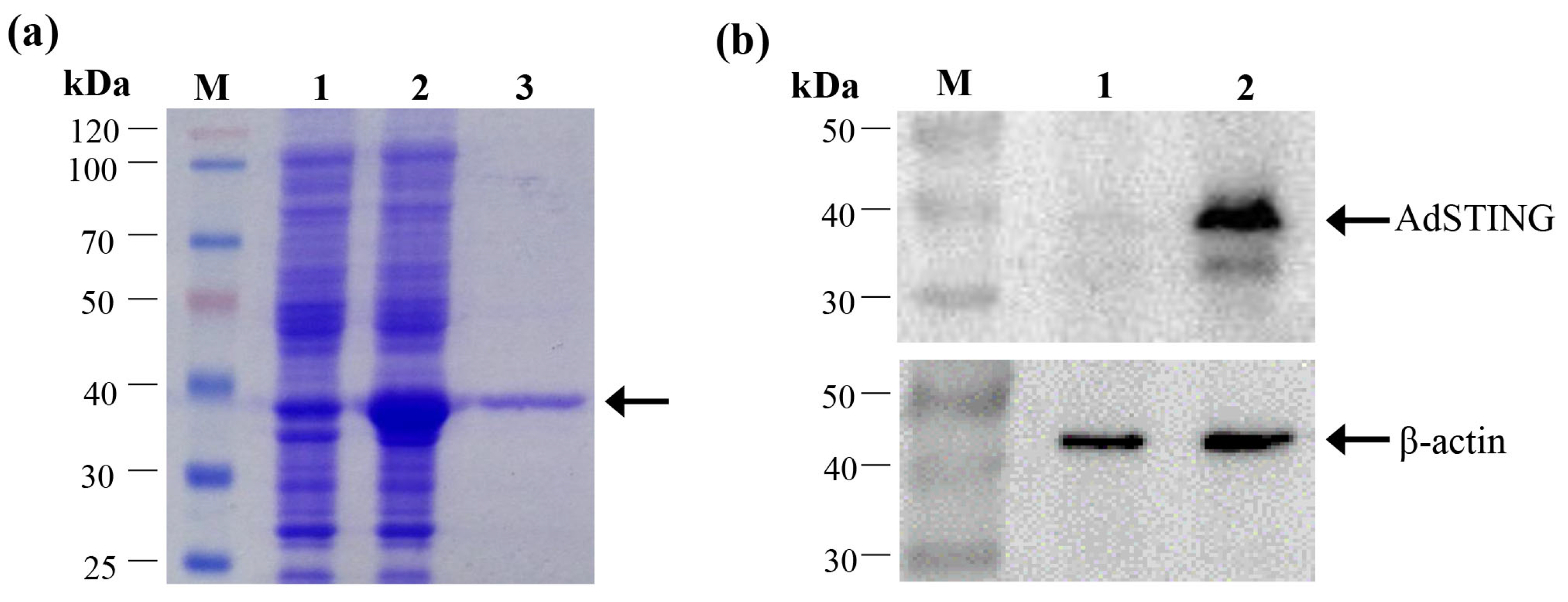
2.5. Prokaryotic Expression, Protein Purification and Antibody Preparation

The recombinant plasmid pET-AdSTING was transformed into Escherichia coli BL21 (DE3), and protein expression was induced by incubating the bacterial culture with 1 mM isopropyl-β-D-thiogalactopyranoside (IPTG) at 37 °C for 6 h. The resulting fusion protein was purified using the HisBind purification kit (Novagen, Darmstadt, Germany) and subsequently analyzed by sodium dodecyl sulfate-polyacrylamide gel electro-phoresis (SDS-PAGE). Mouse anti-AdSTING polyclonal antibodies were prepared following previously described methods [35].

2.6. Western Blot Analysis

GSTC cells transfected with pcDNA-AdSTING or empty vector were collected and subjected to Western blot analysis as described previously [26]. Anti-AdSTING serum served as the primary antibody at a 1:1000 dilution, while a horseradish peroxidase-conjugated goat anti-mouse IgG antibody was used as the secondary antibody at a 1:2000 dilution (Vector Laboratories, Newark, CA, USA). Protein bands were visualized using an enhanced chemiluminescence (ECL) kit (Millopore, Burlington, MA, USA).

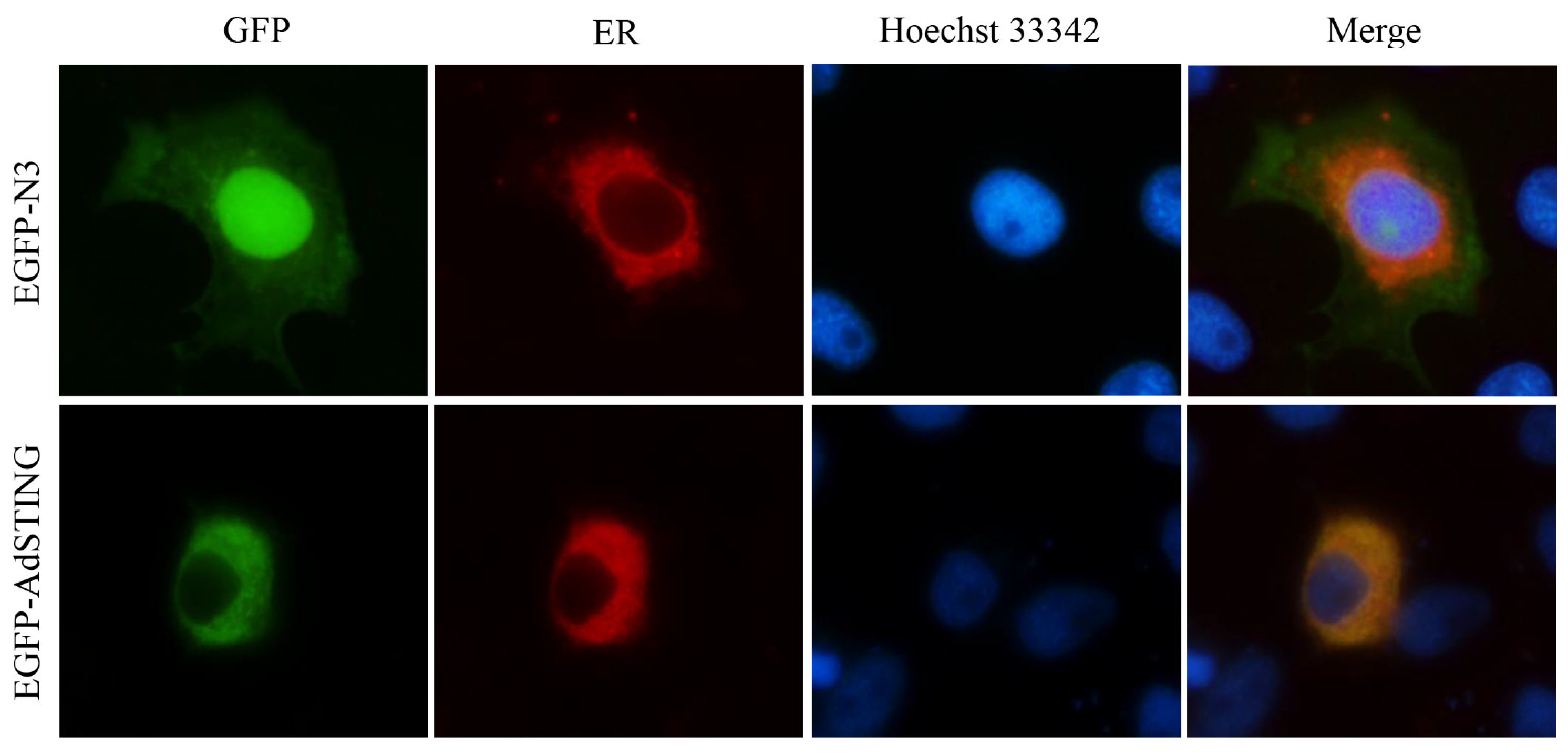
2.7. Fluorescence Microscopy Observation

GSTC cells grown on coverslips in six-well plates were co-transfected with the pEGFP-AdSTING and pDsRed2-ER, an endoplasmic reticulum (ER)-specific marker (Clontech, Mountain View, CA, USA). After 48 h post-transfection, the cells were fixed with 4% paraformaldehyde, permeabilized with 0.2% Triton X-100, stained with Hoechst 33342 (Sigma, Steinheim am Albuch, Germany), and then examined under a Leica DM IRB fluorescence microscope (objective 100×, Leica, Wetzlar, Germany).

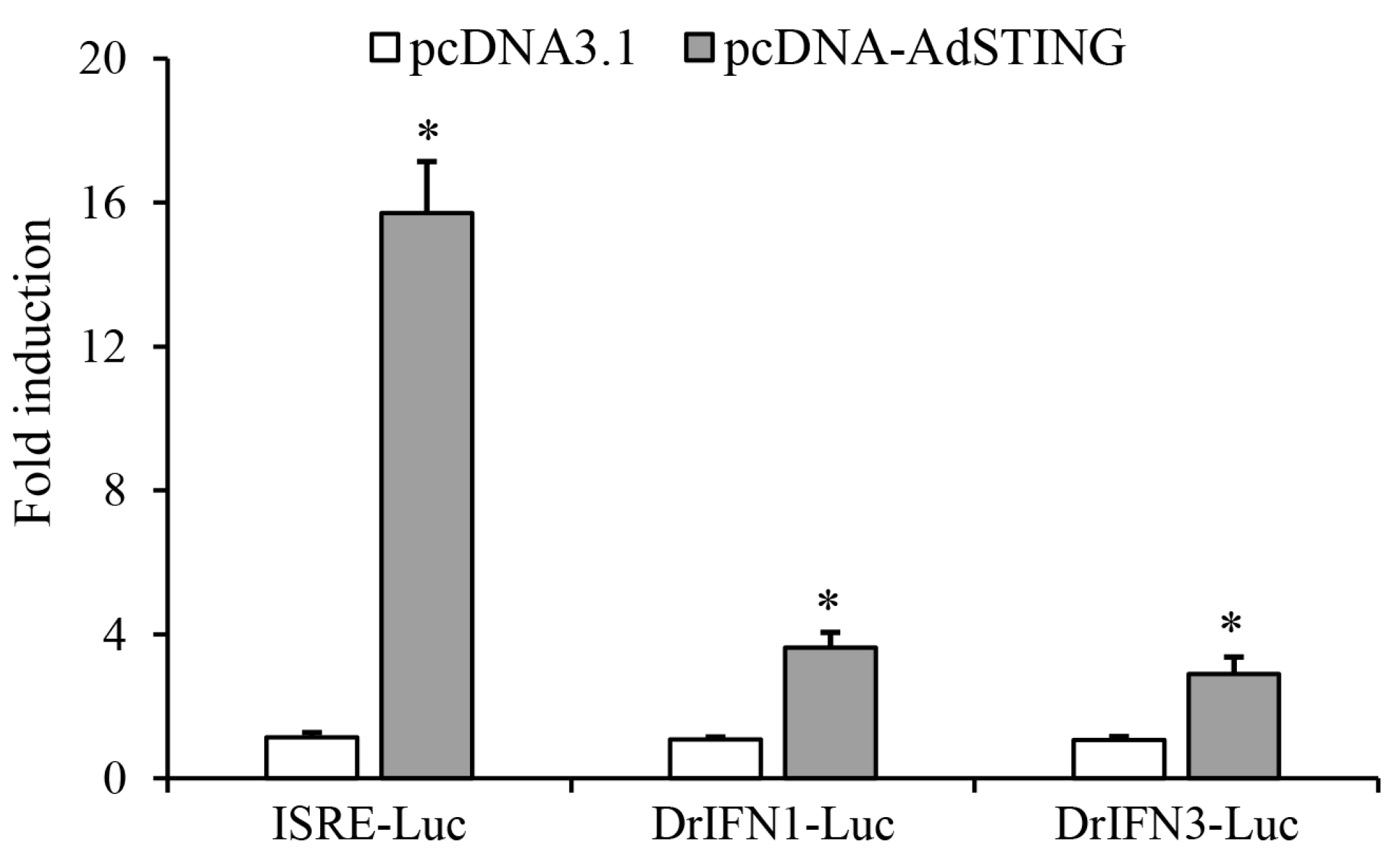
2.8. Luciferase Reporter Assay

To evaluate the effects of AdSTING on the promoter activity of the interferon-stimulated response element (ISRE) and zebrafish IFN, luciferase activity assays were carried out as described previously [13]. The ISRE promoter-driven luciferase vector (ISRE-Luc) containing five ISRE motifs in series was purchased from Stratagene. The DrIFN1-Luc (for analyzing the activity of zebrafish IFN1 promoter) and DrIFN3-Luc (for analyzing the activity of zebrafish IFN3 promoter) were kindly provided by Dr. Shun Li at the Institute of Hydrobiology, Chinese Academy of Sciences. GSTC cells in 24-well plates were co-transfected with pcDNA-AdSTING (250 ng/well), pRL-TK (25 ng/well), and either ISRE-Luc, DrIFN1-Luc, or DrIFN3-Luc (250 ng/well). At 48 h post-transfection, cells were harvested and lysed to examine the luciferase activity with a dual-luciferase reporter assay system (Promega, Madison, WI, USA) according to the manufacturer’s instructions. The luciferase activities were normalized and expressed as fold changes relative to cells transfected with the empty vector.

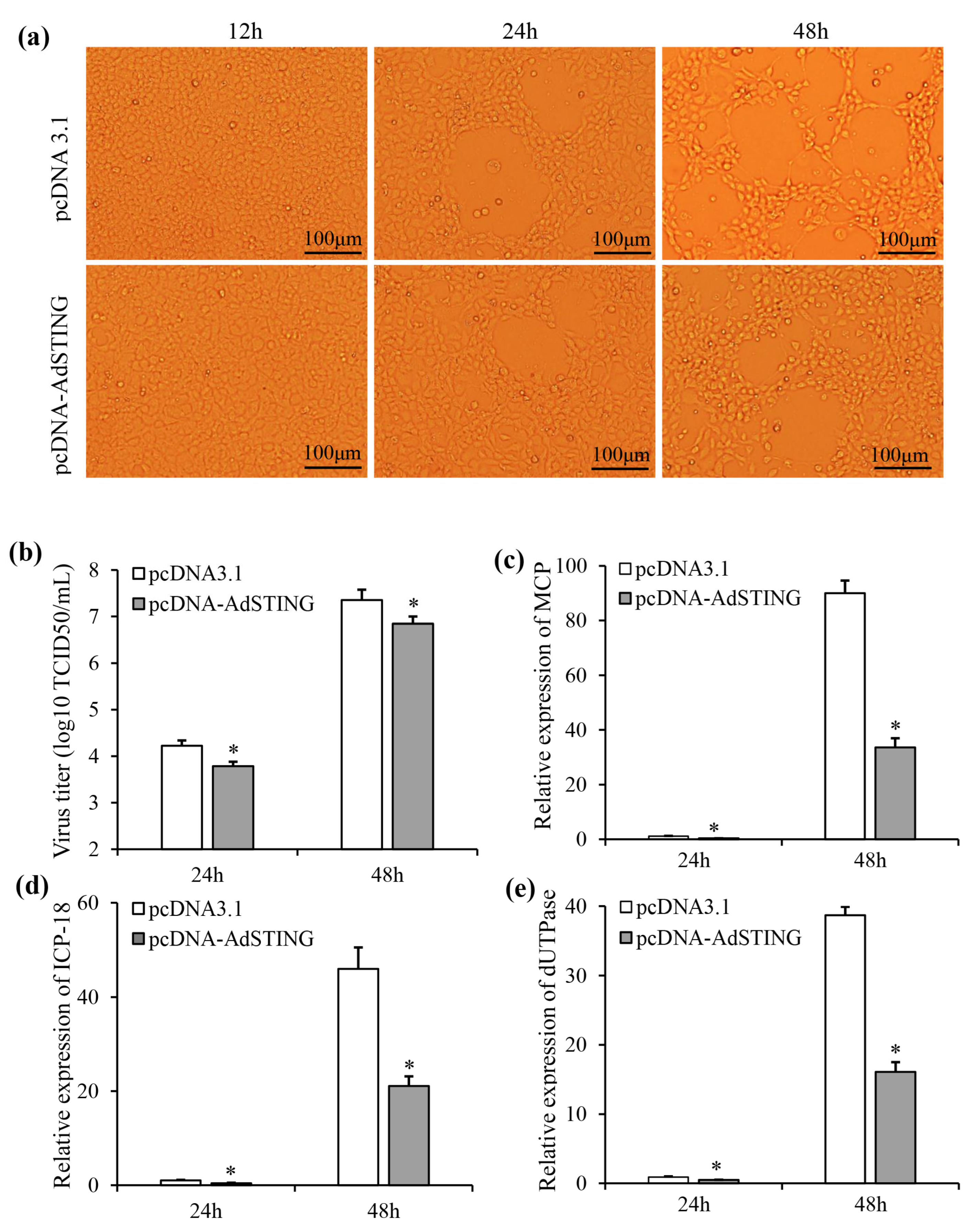
2.9. Effects of AdSTING Overexpression on ADRV Infection

To examine the effects of AdSTING on virus infection, GSTC cells transfected with pcDNA-AdSTING or empty vector were infected with ADRV at a MOI of 0.1. Cell morphology was imaged under inverted light microscopy at 12 h, 24 h, and 48 h post-infection. The culture supernatants were collected at 24 h and 48 h post-infection, and the viral titer was determined by a TCID50 assay on GSTC cells, as described previously [19]. Cell monolayers were harvested for RNA extraction. The relative expression levels of viral genes, including ADRV major capsid protein (MCP), immediate-early protein 18 (ICP-18), and deoxyuridine 5’-triphosphate nucleotidohydrolase (dUTPase) genes, were analyzed by qRT-PCR as described above.

3. Results

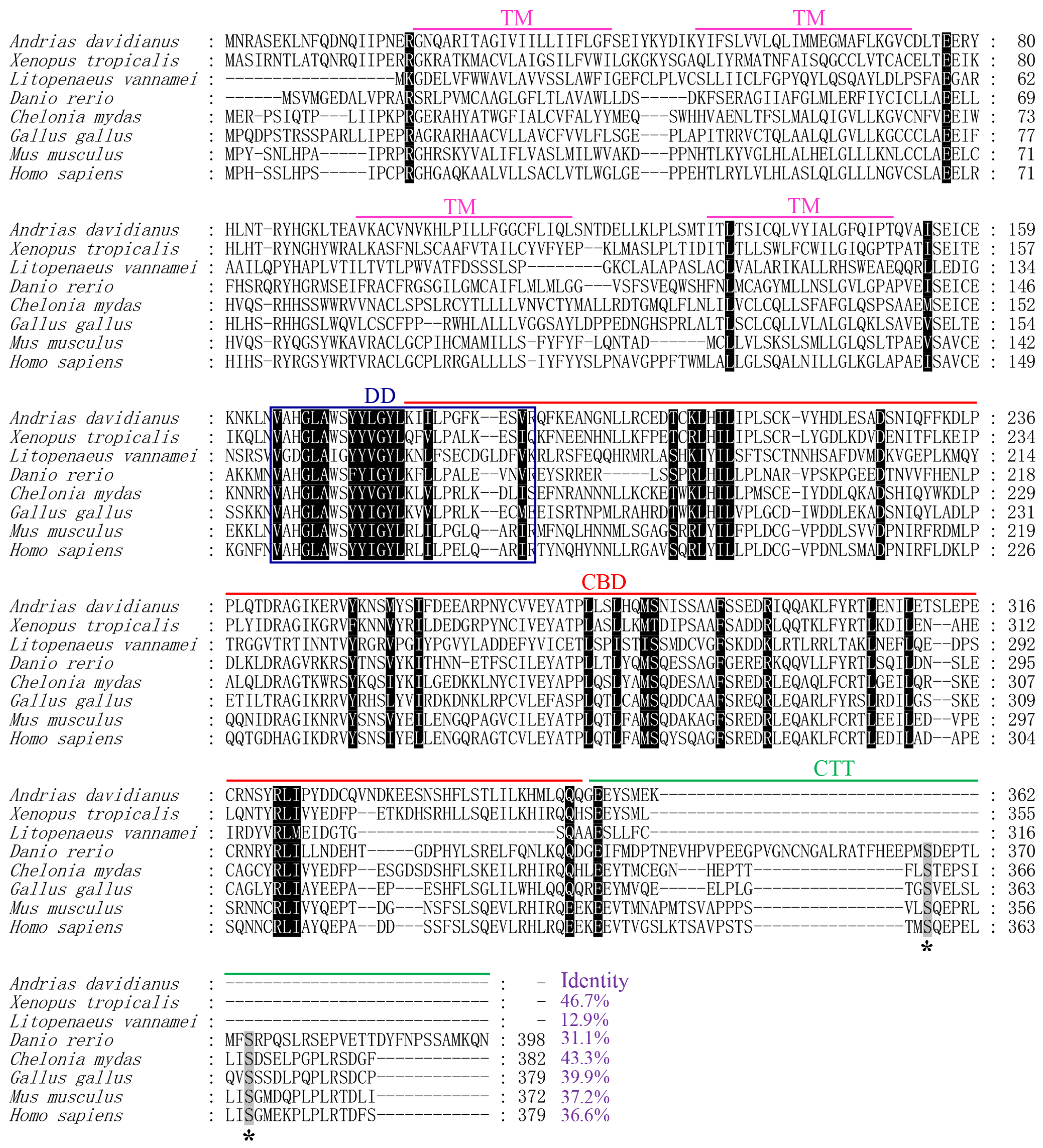
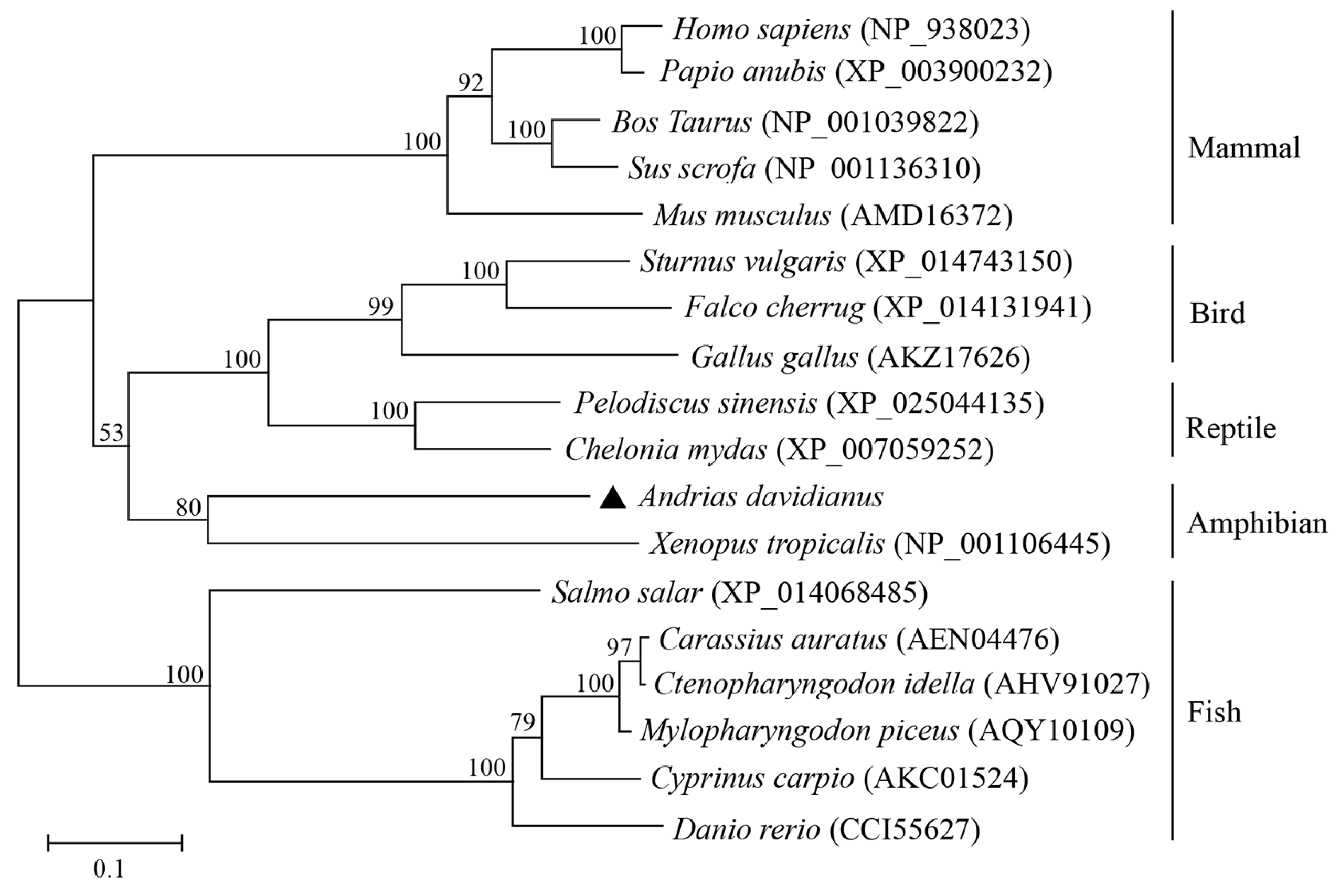
3.1. Identification and Sequence Characterization of AdSTING

The full-length cDNA of AdSTING is 2450 bp, comprising a 5′ untranslated region (UTR) of 136 bp, an open reading frame (ORF) of 1089 bp, and a 3′ UTR of 1225 bp with a poly-A tail (Genbank accession no. OR577306). The AdSTING cDNA encodes a protein comprising 362 amino acids, with a calculated isoelectric point of 6.11 and an estimated molecular weight of 41.6 kDa. Multiple sequence alignments revealed that AdSTING contains four predicted transmembrane domains (TMs) at the N terminal region, and a C-terminal domain (CTD) region consisting of a dimerization domain (DD), cdi-GMP-binding domain (CBD), and a short C-terminal tail (CTT). Notably, serine residues S358 and S366 in human STING were conserved in that of fish, reptiles, birds, and mammals, but were absent in AdSTING and the STING of another amphibian, Xenopus tropicalis (Figure 1).
Sequence comparison showed that AdSTING shares sequence identities of 46.7%, 43.3%, 39.9%, 37.2%, 36.6%, 31.1%, and 12.9% with the STING proteins of Xenopus tropicalis, Chelonia mydas, Gallus gallus, Mus musculus, Homo sapiens, Danio rerio, and Litopenaeus vannamei, respectively (Figure 1). Phylogenetic analysis further corroborated these sequence similarities (Figure 2). The phylogenetic tree indicated that STING sequences from vertebrate species are grouped into two distinct branches. One branch was from fish, and the other branch was from amphibians, reptiles, birds, and mammals. Notably, AdSTING exhibited the closest relationship with the STING from Xenopus tropicalis.

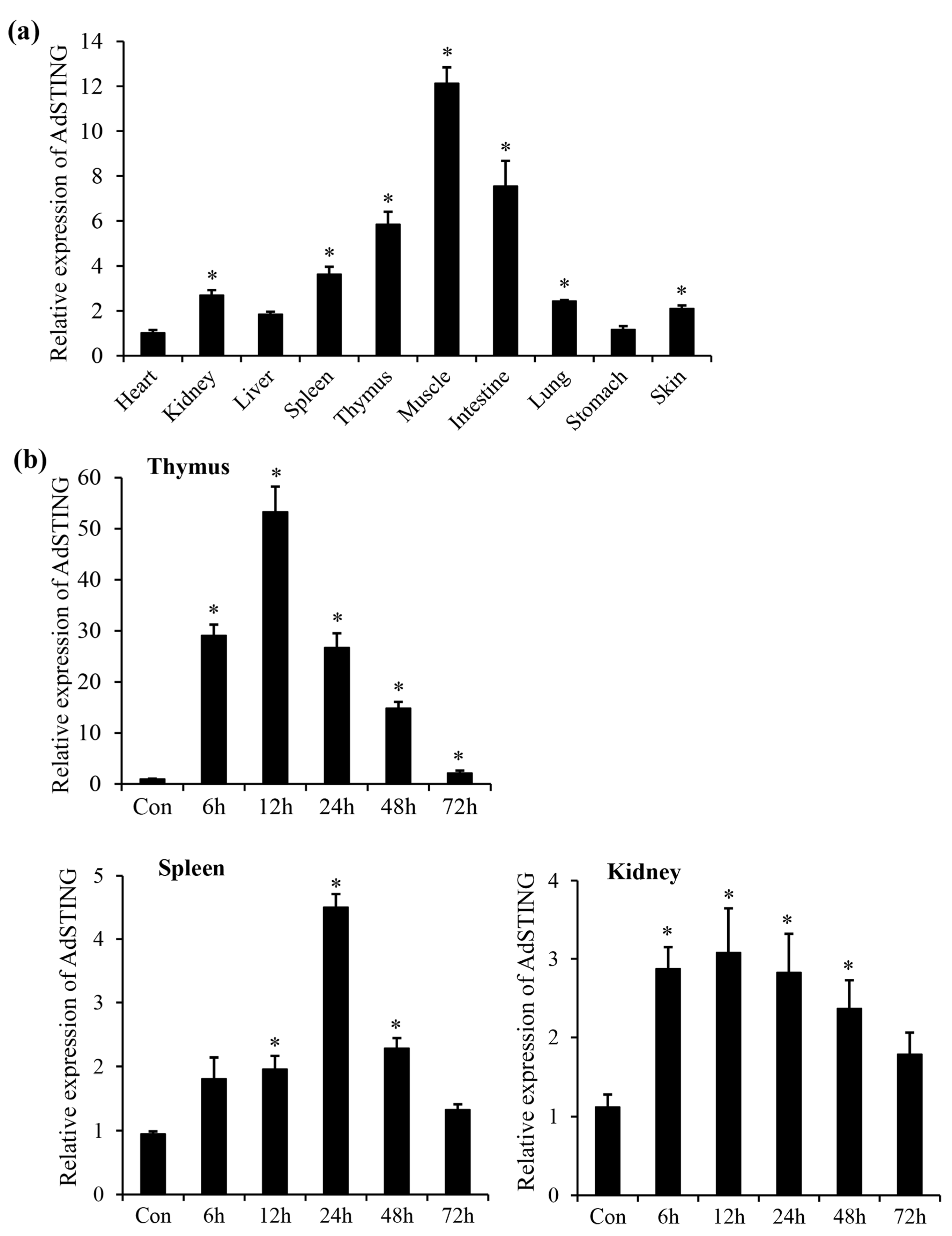
3.2. Tissue Expression Profile of AdSTING

The expression patterns of AdSTING were investigated across various tissues in healthy Chinese giant salamanders. As shown in Figure 3a, AdSTING was ubiquitously expressed in all ten examined tissues, with high expression in the muscle, intestine, and thymus; moderate expression in the spleen, kidney, lung and skin; and low expression in the liver, stomach, and heart.
Likewise, the expression profiles of AdSTING were assessed in the ADRV-infected thymus, spleen, and kidney at different time points (Figure 3b). In the thymus, the expression of AdSTING rapidly increased at 6 h (29.1-fold), reached the peak at 12 h (53.3-fold), followed by a decrease at 24 h and 48 h, and kept a significant up-regulation level at 72 h (2.1-fold). In the spleen, mRNA expression of AdSTING was significantly up-regulated at 12 h (2.0-fold), reached the peak at 24 h (4.5-fold), and recovered to the normal level at 72 h. In the kidney, AdSTING mRNA increased to the peak at 12 h (3.1-fold), then gradually decreased with a slight increment at 72 h (1.8-fold).

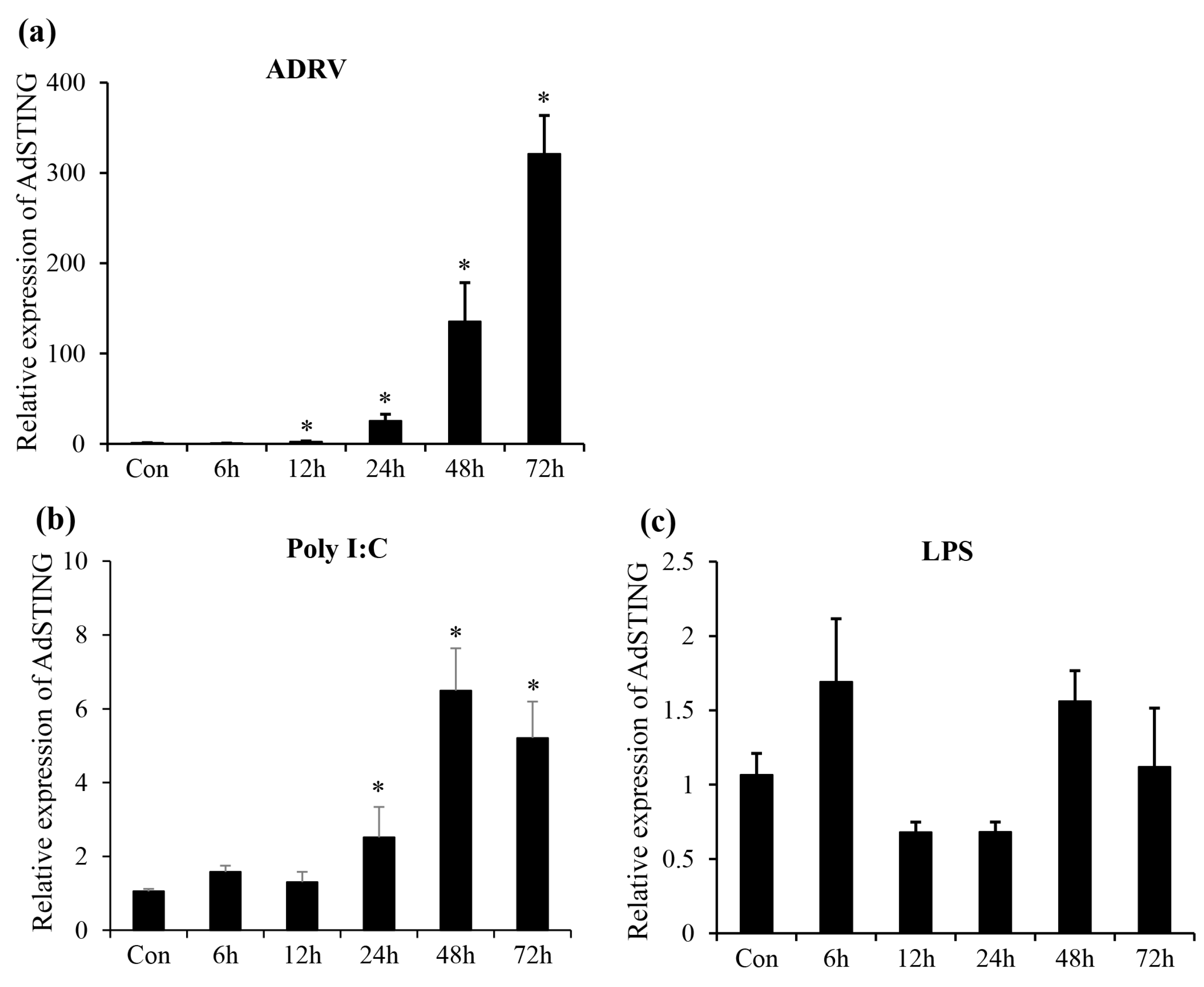
3.3. Expression Profile of AdSTING after In Vitro Stimulation

The expression changes of AdSTING in response to ADRV, poly I:C, and LPS stimulations were evaluated in GSTC cells (Figure 4). Following ADRV infection, the transcription of AdSTING was initially down-regulated at 6 h, then gradually increased from 12 to 72 h, reaching the peak at 72 h (321.2-fold). After stimulation of poly I:C, the expression of AdSTING was significantly increased at 24 h, reached the peak at 48 h (6.5-fold), and kept the level at 72 h. Interestingly, no statistically significant change of STING mRNA level was observed during LPS stimulation.

3.4. Expression and Confirmation of AdSTING

The plasmid pET-AdSTING was transformed into E. coli BL21 (DE3), and expression of the fusion protein was subsequently induced. As shown in Figure 5a, the molecular weight of the recombinant AdSTING fusion protein was approximately 38.3 kDa. The fusion protein was purified and used for anti-AsSTING serum preparation in mice. Expression of pcDNA-AdSTING in transfected GSTC cells were further confirmed through Western blot analysis. As shown in Figure 5b, the anti-AdSTING antibody specifically recognized a 41.6 kDa protein band from lysates of cells transfected with pcDNA-AdSTING, aligning with the predicted molecular weight of AdSTING. No specific protein band was detected in the lysates of cells transfected with the empty pcDNA3.1 vector.

3.5. Subcellular Localization of AdSTING

To investigate the subcellular localization of AdSTING in vitro, the pEGFP-AdSTING was co-transfected with pDsRed2-ER into GSTC cells. Fluorescence microscopy observation revealed that AdSTING-GFP was predominantly localized in the cytoplasm and exhibited co-localization with the ER marker (Figure 6). In contrast, the green fluorescence was distributed throughout the cytoplasm and nucleus of the cells transfected with pEGFP-N3. The results suggested that AdSTING is primarily localized in the ER.

3.6. AdSTING Induced ISRE and IFN Promoter Activity

Using transfection and a luciferase reporter assay system, the effect of AdSTING overexpression on ISRE and IFN promoter activity was examined in GSTC cells. As shown in Figure 7, overexpression of AdSTING significantly induced the promoter activities of ISRE, DrIFN1, and DrIFN3, showing increases of up to 15.7-, 3.6-, and 2.7-fold compared to the empty vector pcDNA3.1, respectively. The results indicated that AdSTING was effective in activating an IFN response.

3.7. Overexpression of AdSTING-Reduced ADRV Replication

To determine the effects of AdSTING on virus infection, we evaluated the severity of the cytopathic effect (CPE) induced by ADRV infection and measured the virus titers and transcription levels of viral genes in pcDNA-AdSTING and empty-vector transfected GSTC cells, respectively. As shown in Figure 8a, the CPE induced by ADRV infection in AdSTING overexpressing cells appeared to be not significantly impacted in comparison with those in the empty-vector transfected cells. The viral titer results are shown in Figure 8b. The average viral titer in the AdSTING overexpressing cells was 7.01 × 10⁶ TCID50/mL at 48 h post-infection, which was significantly lower than that in the cells transfected with the empty vector (2.25 × 10⁷ TCID50/mL). Consistently, the overexpression of AdSTING led to a significant reduction in the transcription levels of key viral genes, including ADRV MCP, ICP-18, and dUTPase (Figure 8c–e). The results indicated that overexpression of AdSTING effectively decreased ADRV replication and the production of infected progeny ADRV virions.

4. Discussion

Amphibians serve as a crucial evolutionary link between aquatic and terrestrial vertebrates. The global populations of amphibians have significantly declined, and infectious diseases such viral infections by ranaviruses (Iridoviridae family) are now considered important [36,37]. The Chinese giant salamander, as a primitive and endangered amphibian species, has suffered from ranavirus diseases in both natural habitats and farming environments [19,21,38]. Despite this, there is limited understanding of its immune responses to viral infections. STING is an essential adaptor molecule that activates the type I interferon (IFN) signaling pathway and plays a pivotal role in innate antiviral immunity in vertebrates [7]. In this study, we identified and functionally analyzed the STING in Chinese giant salamanders. To our knowledge, this is the first report on the functions of STING in amphibian species.
Bioinformatics analysis revealed that AdSTING contains four TM domains in its N-terminal region and a DD, CBD, and short CTT domain in its C-terminal region. Previous studies have reported that STING is an ER-associated transmembrane protein, and it facilitates the production of viral signaling molecules, such as IRF3 and IFNs [2,6]. The TM domains of STING are essential for its localization with the ER and are required for its interaction with MAVS to activate IRF3 and induce IFNs [2,8,9]. Consistent with this, subcellular localization analysis showed that AdSTING primarily co-localizes with the ER, suggesting that the TM domains in AdSTING protein may be involved in these kinds of activation related to the antiviral response. The DD is the most conserved region, and plays a vital role in the function of STING [39]. Recent study has indicated that the DD of black carp STING negatively regulates the STING-mediated antiviral immunity [40]. The DD is highly conserved across all species examined in this study, suggesting an important and conserved function for this region of AdSTING. Moreover, the CTT domain of AdSTING is similar to Xenopus tropicalis STING, but it is significantly shorter than that of other vertebrates. This suggests that amphibian STINGs may have lost most of this structural domain during evolution. The CTT domain has been proposed to be important for STING to recruit the critical downstream TBK1 and IRF3 signaling components [41,42]. In Penaeus monodon, PmSTING lacks the CTT domain, but still plays a vital role in the innate antiviral response against white spot syndrome virus (WSSV) infection, possibly through the PmDDX41/PmSTING/PmIRF signaling cascade [43]. Similarly, AdSTING may activate antiviral immunity in Chinese giant salamanders through a comparable signaling pathway. Multiple amino acid alignments showed that AdSTING shares low sequence identities (31.1–46.7%) with STING from other vertebrate species, having the highest identity with Xenopus tropicalis STING. Phylogenetic analysis further supports this, showing that STINGs from amphibians, reptiles, birds, and mammals form a distinct cluster separate from fish species, aligning with traditional classifications [15,44].
The distribution of STING across tissues and cells is crucial for its function, as it affects the ability to detect various microorganisms during their entry and proliferation in different tissues [44]. The AdSTING mRNA was constitutively expressed in all the tissues examined, similar to those reported in other vertebrate species [13,15,44]. Notably, high levels of AdSTING expression were observed in the muscle, intestine, and thymus. In amphibians, the intestine serves as a key site for mucosal immunity, while the thymus is thought to play capital roles in both cell-mediated and humoral immunity [45,46]. This expression pattern might allow AdSTING to respond early to invading pathogens. Moreover, AdSTING mRNA expression was significantly up-regulated in the thymus, spleen, and kidney tissues after ADRV infection. In GSTC cells, the expression of AdSTING was significantly increased at 24 h, reached the peak at 48 h (6.5-fold), and kept the level at 72 h after poly I:C stimulation. The mRNA expression of AdSTING was significantly up-regulated at 12 h and reached the peak at 72 h (321.2-fold) after ADRV infection. In contrast, the AdSTING mRNA level was not significantly undulated after LPS treatment. The expression pattern of AdSTING after viral infection and poly I:C stimulation were consistent to those reported in fish STINGs [13,15]. Moreover, several studies have shown that the induced expression of fish STING after stimulation in vitro was influenced by the dose of virus and the concentrations of poly (I:C) and LPS [12,14]. Thus, this could be the reasons for the differences in the expression profile of AdSTING following the treatments of ADRV, poly (I:C), and LPS, respectively. However, the up-regulation of AdSTING transcription after ADRV infection and poly I:C stimulation could play a key positive role in response to dsDNA and dsRNA synthetic analogues.
Extensive studies have demonstrated that STING can trigger a type I IFN response through activation of the IRF3-dependent pathway, playing a pivotal role in innate immunity against virus infections [9,47]. Luciferase reporter assays confirmed that overexpression of AdSTING could activate both the ISRE promoter and fish IFN promoters, aligning with the behavior of STING in humans and fish. This implied that AdSTING had the ability to mediate IFN immune response like its mammalian counterpart. However, whether AdSTING activated IFN signaling dependent on the IRF3 pathway needs further investigation. Furthermore, the ectopic expression of AdSTING significantly suppressed the replication of ADRV in GSTC cells, as evidenced by the reduction of viral titers and viral gene transcriptions. These results suggest that AdSTING likely activates the IFN response and exerts an antiviral effect against viral infections.

5. Conclusions

In summary, we have identified and characterized a STING homolog from the Chinese giant salamander, referred to as AdSTING. This protein is localized in the ER, and its expression was significantly up-regulated after ADRV infection both in vivo and in vitro. Moreover, the overexpression of AdSTING could activate the interferon immune response and exhibit antiviral activity against ADRV infection. Taken together, these results provide the first evidence that amphibian STING plays an important role in antiviral innate immunity.

Author Contributions

Conceptualization, Z.C. and P.Y.; methodology, N.Y. and R.W. (Ronghua Wang); software, R.W. (Ruisong Wang); validation, Z.C. and R.W. (Ronghua Wang); formal analysis, Z.C.; investigation and resources, R.W. (Ronghua Wang) and N.Y.; data curation, Z.C.; writing—original draft preparation, Z.C.; writing—review and editing, R.W. (Ruisong Wang), Z.C. and P.Y.; supervision, P.Y.; project administration, Z.C.; funding acquisition, Z.C. and R.W. (Ronghua Wang). All authors have read and agreed to the published version of the manuscript.

Funding

This research was funded by the National Natural Science Foundation of China (Grant no. 31972835), the Natural Science Foundation of Hunan Province (Grant no. 2022JJ30420), the Research Project of Education Department of Hunan Province (Grant no. 22B0691 and 22B0701) and the Research Project of Hunan University of Arts and Science (Grant no. 19BSQD28).

Institutional Review Board Statement

The animal study protocol was approved by the Institutional Animal Care and Use Committee of Hunan University of Arts and Science (JSDX-2019-012, 15 October 2019).

Informed Consent Statement

Not applicable.

Data Availability Statement

Data are contained within the article.

Acknowledgments

The authors sincerely thank Qiya Zhang from Institute of Hydrobiology, Chinese Academy of Sciences for providing the Chinese giant salamander thymus cell line (GSTC) and relevant technical support.

Conflicts of Interest

The authors declare no conflict of interest. The funders had no role in the design of the study; in the collection, analyses, or interpretation of data; in the writing of the manuscript; or in the decision to publish the results.

References

  1. Chan, Y.K.; Gack, M.U. Viral evasion of intracellular DNA and RNA sensing. Nat. Rev. Microbiol. 2016, 14, 360–373. [Google Scholar] [CrossRef]
  2. Zhong, B.; Yang, Y.; Li, S.; Wang, Y.Y.; Li, Y.; Diao, F.; Lei, C.; He, X.; Zhang, L.; Tien, P.; et al. The adaptor protein MITA links virus-sensing receptors to IRF3 transcription factor activation. Immunity 2008, 29, 538–550. [Google Scholar] [CrossRef]
  3. Ishikawa, H.; Ma, Z.; Barber, G.N. STING regulates intracellular DNA-mediated, type I interferon-dependent innate immunity. Nature 2009, 461, 788–792. [Google Scholar] [CrossRef]
  4. Barber, G.N. STING-dependent cytosolic DNA sensing pathways. Trends Immunol. 2014, 35, 88–93. [Google Scholar] [CrossRef]
  5. Chauvin, S.; Stinson, W.A.; Platt, D.J.; Poddar, S.; Miner, J.J. Regulation of cGAS and STING signaling during inflammation and infection. J. Biol. Chem. 2023, 299, 104866. [Google Scholar] [CrossRef] [PubMed]
  6. Zevini, A.; Olagnier, D.; Hiscott, J. Crosstalk between cytoplasmic RIG-I and STING sensing pathways. Trends Immunol. 2017, 38, 194–205. [Google Scholar] [CrossRef] [PubMed]
  7. Du, Y.; Hu, Z.Q.; Luo, Y.; Wang, H.Y.; Yu, X.; Wang, R.F. Function and regulation of cGAS-STING signaling in infectious diseases. Front. Immunol. 2023, 14, 1130423. [Google Scholar] [CrossRef] [PubMed]
  8. Abe, T.; Harashima, A.; Xia, T.; Konno, H.; Konno, K.; Morales, A.; Ahn, J.; Gutman, D.; Barber, G.N. STING recognition of cytoplasmic DNA instigates cellular defense. Mol. Cell 2013, 50, 5–15. [Google Scholar] [CrossRef]
  9. Hopfner, K.P.; Hornung, V. Molecular mechanisms and cellular functions of cGAS-STING signalling. Nat. Rev. Mol. Cell Biol. 2020, 21, 501–521. [Google Scholar] [CrossRef] [PubMed]
  10. Zhang, X.W.; Bai, X.C.; Chen, Z.J. Structures and mechanisms in the cGAS-STING innate immunity pathway. Immunity 2020, 53, 43–53. [Google Scholar] [CrossRef]
  11. Sun, F.; Zhang, Y.B.; Liu, T.K.; Shi, J.; Wang, B.; Gui, J.F. Fish MITA serves as a mediator for distinct fish IFN gene activation dependent on IRF3 or IRF7. J. Immunol. 2011, 187, 2531–2539. [Google Scholar] [CrossRef] [PubMed]
  12. Feng, X.L.; Yang, C.R.; Zhang, Y.X.; Peng, L.M.; Chen, X.H.; Rao, Y.L.; Gu, T.L.; Su, J.G. Identification, characterization and immunological response analysis of stimulator of interferon gene (STING) from grass carp Ctenopharyngodon idella. Dev. Comp. Immunol. 2014, 45, 163–176. [Google Scholar] [CrossRef]
  13. Huang, Y.H.; Ouyang, Z.L.; Wang, W.; Yu, Y.P.; Li, P.F.; Zhou, S.; Wei, S.N.; Wei, J.G.; Huang, X.H.; Qin, Q.W. Antiviral role of grouper STING against iridovirus infection. Fish Shellfish Immunol. 2015, 47, 157–167. [Google Scholar] [CrossRef] [PubMed]
  14. Lu, L.; Wang, X.; Wu, S.Z.; Song, X.J.; Zou, Z.Q.; Xie, X.C.; Xiao, J.; Chen, S.; Feng, H. Black carp STING functions importantly in innate immune defense against RNA virus. Fish Shellfish Immunol. 2017, 70, 13–24. [Google Scholar] [CrossRef]
  15. Qin, X.W.; He, J.; Yu, Y.; Liu, C.; Luo, Z.Y.; Li, Z.M.; Weng, S.P.; Guo, C.J.; He, J.G. The roles of mandarin fish STING in innate immune defense against infectious spleen and kidney necrosis virus infections. Fish Shellfish Immunol. 2020, 100, 80–89. [Google Scholar] [CrossRef]
  16. Gao, F.Y.; Lu, M.X.; Wang, M.; Liu, Z.G.; Ke, X.L.; Zhang, D.F.; Cao, J.M. Nile tilapia DNA sensor STING is involved in the IFN-β and AP-1 signaling pathways in the immune response dependent on DDX41. Int. J. Biol. Macromol. 2023, 225, 27–39. [Google Scholar]
  17. Zhang, P.; Chen, Y.Q.; Liu, Y.F.; Zhou, H.; Qu, L.H. The complete mitochondrial genome of the Chinese giant salamander, Andrias davidianus (Amphibia: Caudata). Gene 2003, 311, 93–98. [Google Scholar] [CrossRef]
  18. Turvey, S.T.; Chen, S.; Tapley, B.; Wei, G.; Xie, F.; Yan, F.; Yang, J.; Liang, Z.; Tian, H.; Wu, M.; et al. Imminent extinction in the wild of the world’s largest amphibian. Curr. Biol. 2018, 28, R592–R594. [Google Scholar] [CrossRef]
  19. Chen, Z.Y.; Gui, J.F.; Gao, X.C.; Pei, C.; Hong, Y.J.; Zhang, Q.Y. Genome architecture changes and major gene variations of Andrias davidianus ranavirus (ADRV). Vet. Res. 2013, 44, 101. [Google Scholar] [CrossRef]
  20. Zhou, Z.Y.; Geng, Y.; Liu, X.X.; Ren, S.Y.; Zhou, Y.; Wang, K.Y.; Huang, X.L.; Chen, D.F.; Peng, X.; Lai, W.M. Characterization of a ranavirus isolated from the Chinese giant salamander (Andrias davidianus, Blanchard, 1871) in China. Aquaculture 2013, 384, 66–73. [Google Scholar] [CrossRef]
  21. Herath, J.; Ellepola, G.; Meegaskumbura, M. Patterns of infection, origins, and transmission of ranaviruses among the ectothermic vertebrates of Asia. Ecol. Evol. 2021, 11, 15498–15519. [Google Scholar] [CrossRef]
  22. Li, W.; Zhang, X.; Weng, S.P.; Zhao, G.X.; He, J.G.; Dong, C.F. Virion-associated viral proteins of a Chinese giant salamander (Andrias davidianus) iridovirus (genus Ranavirus) and functional study of the major capsid protein (MCP). Vet. Microbiol. 2014, 172, 129–319. [Google Scholar] [CrossRef] [PubMed]
  23. Yuan, J.D.; Chen, Z.Y.; Huang, X.; Gao, X.C.; Zhang, Q.Y. Establishment of three cell lines from Chinese giant salamander and their sensitivities to the wild-type and recombinant ranavirus. Vet. Res. 2015, 46, 58. [Google Scholar] [CrossRef] [PubMed]
  24. Jiang, N.; Fan, Y.D.; Zhou, Y.; Liu, W.Z.; Ma, J.; Meng, Y.; Xie, C.X.; Zeng, L.B. Characterization of Chinese giant salamander iridovirus tissue tropism and inflammatory response after infection. Dis. Aquat. Organ. 2015, 114, 229–237. [Google Scholar] [CrossRef] [PubMed]
  25. Ke, F.; Gui, J.F.; Chen, Z.Y.; Li, T.; Lei, C.K.; Wang, Z.H.; Zhang, Q.Y. Divergent transcriptomic responses underlying the ranaviruses-amphibian interaction processes on interspecies infection of Chinese giant salamander. BMC Genomics 2018, 19, 211. [Google Scholar] [CrossRef] [PubMed]
  26. Chen, Z.Y.; Li, T.; Gao, X.C.; Wang, C.F.; Zhang, Q.Y. Protective immunity induced by DNA vaccination against ranavirus infection in Chinese giant salamander Andrias davidianus. Viruses 2018, 10, 52. [Google Scholar] [CrossRef] [PubMed]
  27. Ke, F.; Yu, X.D.; Wang, Z.H.; Gui, J.F.; Zhang, Q.Y. Replication and transcription machinery for ranaviruses: Components, correlation, and functional architecture. Cell Biosci. 2022, 12, 6. [Google Scholar] [CrossRef] [PubMed]
  28. Chen, Q.; Ma, J.; Fan, Y.D.; Meng, Y.; Xu, J.; Zhou, Y.; Liu, W.Z.; Zeng, X.H.; Zeng, L.B. Identification of type I IFN in Chinese giant salamander (Andrias davidianus) and the response to an iridovirus infection. Mol. Immunol. 2015, 65, 350–359. [Google Scholar] [CrossRef]
  29. Huang, L.; Fan, Y.D.; Zhou, Y.; Jiang, N.; Liu, W.Z.; Meng, Y.; Zeng, L.B. Cloning, sequence analysis and expression profiles of Toll-like receptor 7 from Chinese giantsalamander Andrias davidianus. Comp. Biochem. Physiol. B Biochem. Mol. Biol. 2015, 184, 52–57. [Google Scholar] [CrossRef]
  30. Yang, H.; Lan, Q.J.; Liu, R.R.; Cui, D.; Liu, H.X.; Xiong, D.M.; Li, F.G.; Liu, X.L.; Wang, L.X. Characterization of galectin-1 from Chinese giant salamanders Andrias davidianus and its involvements during immune response. Dev. Comp. Immunol. 2017, 70, 59–68. [Google Scholar] [CrossRef]
  31. Jiang, N.; Fan, Y.D.; Zhou, Y.; Meng, Y.; Liu, W.Z.; Li, Y.Q.; Xue, M.Y.; Robert, J.; Zeng, L.B. The immune system and the antiviral responses in Chinese giant salamander, Andrias davidianus. Front Immunol. 2021, 12, 718627. [Google Scholar] [CrossRef]
  32. Meng, Y.; Fan, Y.D.; Jiang, N.; Xue, M.Y.; Li, Y.Q.; Liu, W.Z.; Zeng, L.B.; Zhou, Y. Four Mx genes identified in Andrias davidianus and characterization of their response to Chinese giant salamander iridovirus infection. Animals 2022, 12, 2147. [Google Scholar] [CrossRef] [PubMed]
  33. Kumar, S.; Stecher, G.; Tamura, K. MEGA7: Molecular evolutionary genetics analysis version 7.0 for bigger datasets. Mol. Biol. Evol. 2016, 33, 1870–1874. [Google Scholar] [CrossRef] [PubMed]
  34. Xia, H.; Yang, P.H.; Chen, Z.Y.; Zhang, Y.S.; Liu, L.G.; Luo, Y.S.; Meng, S.Y.; Fang, X.; Liu, Y.J.; Liu, W.; et al. Polymeric immunoglobulin receptor in dongtingking crucian carp (Carassius auratus indigentiaus): Molecular characterization and expression analysis in response to Aeromonas hydrophila challenge. Aquac. Res. 2022, 53, 3818–3827. [Google Scholar] [CrossRef]
  35. He, L.B.; Ke, F.; Wang, J.; Gao, X.C.; Zhang, Q.Y. Rana grylio virus (RGV) envelope protein 2L: Subcellular localization and essential roles in virus infectivity revealed by conditional lethal mutant. J. Gen. Virol. 2014, 95, 679–690. [Google Scholar] [CrossRef]
  36. Earl, J.E.; Chaney, J.C.; Sutton, W.B.; Lillard, C.E.; Kouba, A.J.; Langhorne, C.; Krebs, J.; Wilkes, R.P.; Hill, R.D.; Miller, D.L.; et al. Ranavirus could facilitate local extinction of rare amphibian species. Oecologia 2016, 182, 611–623. [Google Scholar] [CrossRef] [PubMed]
  37. Snyder, P.W.; Ramsay, C.T.; Harjoe, C.C.; Khazan, E.S.; Briggs, C.J.; Hoverman, J.T.; Johnson, P.T.J.; Preston, D.; Rohr, J.R.; Blaustein, A.R. Experimental evidence that host species composition alters host-pathogen dynamics in a ranavirus-amphibian assemblage. Ecology 2023, 104, e3885. [Google Scholar] [CrossRef]
  38. Gui, L.; Chinchar, V.G.; Zhang, Q.Y. Molecular basis of pathogenesis of emerging viruses infecting aquatic animals. Aquac. Fish. 2018, 3, 1–5. [Google Scholar] [CrossRef]
  39. Ouyang, S.Y.; Song, X.Q.; Wan, Y.Y.; Ru, H.; Shaw, N.; Jiang, Y.; Niu, F.F.; Zhu, Y.P.; Qiu, W.C.; Parvatiyar, K.; et al. Structural analysis of the STING adaptor protein reveals a hydrophobic dimer interface and mode of cyclic di-GMP binding. Immunity 2012, 36, 1073–1086. [Google Scholar] [CrossRef]
  40. Yan, J.; Qiao, G.X.; Chang, H.Y.; Wu, H.; Liu, M.L.; Tu, J.G.; Zhang, Y.G.; Feng, H. Role of the dimerization domain of black carp STING during the antiviral innate immunity. Reprod. Breed. 2023, 3, 59–65. [Google Scholar] [CrossRef]
  41. Tanaka, Y.; Chen, Z.J. STING specifies IRF3 phosphorylation by TBK1 in the cytosolic DNA signaling pathway. Sci. Signal. 2012, 5, ra20. [Google Scholar] [CrossRef]
  42. Zhao, B.Y.; Du, F.L.; Xu, P.B.; Shu, C.; Sankaran, B.; Bell, S.L.; Liu, M.M.; Lei, Y.J.; Gao, X.S.; Fu, X.F.; et al. A conserved PLPLRT/SD motif of STING mediates the recruitment and activation of TBK1. Nature 2019, 569, 718–722. [Google Scholar] [CrossRef]
  43. Amparyup, P.; Charoensapsri, W.; Soponpong, S.; Jearaphunt, M.; Wongpanya, R.; Tassanakajon, A. Stimulator of interferon gene (STING) and interferon regulatory factor (IRF) are crucial for shrimp antiviral defense against WSSV infection. Fish Shellfish Immunol. 2021, 117, 240–247. [Google Scholar] [CrossRef]
  44. Cheng, Y.Q.; Sun, Y.J.; Wang, H.G.; Yan, Y.X.; Ding, C.; Sun, J.H. Chicken STING mediates activation of the IFN gene independently of the RIG-I gene. J. Immunol. 2015, 195, 3922–3936. [Google Scholar] [CrossRef] [PubMed]
  45. Colombo, B.M.; Scalvenzi, T.; Benlamara, S.; Pollet, N. Microbiota and mucosal immunity in amphibians. Front. Immunol. 2015, 6, 111. [Google Scholar] [CrossRef] [PubMed]
  46. Ruiz, V.L.; Robert, J. The amphibian immune system. Phil. Trans. R. Soc. B 2023, 378, 20220123. [Google Scholar] [CrossRef] [PubMed]
  47. Liu, S.; Cai, X.; Wu, J.; Cong, Q.; Chen, X.; Li, T.; Du, F.; Ren, J.; Wu, Y.T.; Grishin, N.V.; et al. Phosphorylation of innate immune adaptor proteins MAVS, STING, and TRIF induces IRF3 activation. Science 2015, 347, aaa2630. [Google Scholar] [CrossRef]
Figure 1. Multiple sequence alignment of STING homologs from different species. The transmembrane domains (TMs), c-di-GMP-binding domain (CBD), and C-terminal tail (CTT) are labeled above the sequences, and the dimerization domain (DD) is shown with a rectangle. The conserved serine residues (human S358 and S366) are indicted with asterisks. The intensity of the background color indicates the conservation of amino acids among these sequences.
Fishes 08 00597 g001
Figure 2. Phylogenetic analysis of AdSTING and other STING homologs. The phylogenetic tree was conducted by the neighbor-joining method in MEGA 7.0 with 1000 bootstrap replications. AdSTING was marked with a triangle. GenBank accession numbers are indicated in parentheses after each sequence name.
Fishes 08 00597 g002
Figure 3. Expression profiles of AdSTING in healthy and challenged Chinese giant salamanders. (a) The expression patterns of AdSTING in different tissues from healthy Chinese giant salamanders. Values were normalized against that of the heart tissue and expressed as mean ± SD (n = 3). (b) The expression levels of AdSTING in the thymus, spleen and kidney after ADRV infection. The Chinese giant salamanders were intraperitoneally injected with 5 × 10⁶ TCID50 of ADRV or PBS as control. At 6 h, 12 h, 24 h, 48 h, and 72 h post-infection, the tissue samples from thymus, spleen and kidney were collected from three individuals, and the expression level of AdSTING was examined by qRT-PCR analysis. Values were normalized against that of the PBS-injected group (Con) and expressed as mean ± SD (n = 3). Statistical analysis was performed using Student’ t-test. *, p < 0.05.
Fishes 08 00597 g003
Figure 4. Expression profiles of AdSTING in GSTC cells in response to different stimulus. GSTC cells were infected with ADRV (a) or stimulated with poly I:C (b) or LPS (c) for the indicated length of time. Cells were collected for RNA extraction and qRT-PCR analysis. All data were normalized relative to β-actin and represented by means ± SD (n = 3). *, p < 0.05.
Fishes 08 00597 g004
Figure 5. Prokaryotic expression of AdSTING C-terminal peptide and western blot analysis of AdSTING using anti-AdSTING serum. (a) SDS-PAGE of expressed and purified tagged fusion protein. Lanes: 1, pET-AdSTING, non-induced; 2, pET-AdSTING, induced; 3, purified fusion protein. The arrow indicates the purified AdSTING protein. (b) Western blot detection of AdSTING protein. Lanes: 1, lysate from GSTC cells transfected with empty vector; 2, lysate from GSTC cells transfected with pcDNA-AdSTING. The β-actin was detected under the same conditions as an internal control.
Fishes 08 00597 g005
Figure 6. Subcellular localization of AdSTING in GSTC cells. GSTC cells were co-transfected with pDsRed2-ER and an empty vector (upper panel) or pEGFP-AdSTING (lower panel). At 36 h post transfection, the cells were fixed, permeabilized, and stained with Hoechst 33342. The yellow staining in the overlay image (right) indicates that AdSTING is localized to the ER.
Fishes 08 00597 g006
Figure 7. Overexpression of AdSTING-induced ISRE and IFN promoter activity. GSTC cells were co-transfected with pcDNA-AdSTING, pRL-TK, ISRE-Luc, DrIFN1-Luc, or DrIFN3-Luc. The cells were harvested and used for a luciferase reporter assay at 48 h post transfection. The relative fold induction of the ISRE and IFN promoter activity was normalized to that of the control cells transfected with the empty vector and represented by means ± SD (n = 3). *, p < 0.05.
Fishes 08 00597 g007
Figure 8. Overexpression of AdSTING reduced virus replication. (a) Cytopathic effects progression induced by ADRV infection in AdSTING and vector overexpressing GSTC cells. (b) Viral titers in AdSTING and vector overexpressing GSTC cells at 24 h and 48 h after ADRV infection. (ce) Real-time PCR analysis of ADRV MCP, ICP-18, and dUTPbase transcripts in AdSTING or vector overexpressing GSTC cells at 24 h and 48 h after ADRV infection. All data were normalized relative to β-actin and represented by means ± SD (n = 3). *, p < 0.05.
Fishes 08 00597 g008
Table 1. Primers used in this study.
Primer NameSequence (5′-3′)Usage
AdSTING-F1GAAATCAAGCACGTATAACTGCTGGGene cloning
AdSTING-R1CTTTCTTCTTTGTCATTCACCTGGC
NUPAAGCAGTGGTATCAACGCAGAGTRACE-PCR
UPMCTAATACGACTCACTATAGGGCAAGCAGTGGTATCAACGCAGAGT
5′GSP1CATTAACACATGCCTTTACAGCCTCAG
5′GSP2TTTCTTCTGTTAGGTCGCACACTCC
3′GSP1GTAGCGAAGATCGCATTCAACAAGC
3′GSP2CGTTCTCTATTTACATGGCAAATTCC
AdSTING-32-FGGGATCCATGAAAGAGTCTGTACGCCAATPlasmid construction
AdSTING-32-RCCCTCGAGTTTCTCCATAGAATATTCC
AdSTING-3.1-FCGGGATCCAAGATGAATCGTGCATC
AdSTING-3.1-RGGAATTCTTATTTCTCCATAGAATATTCC
AdSTING-N3-FGGAATTCATGAATCGTGCATCAGA
AdSTING-N3-RCGGGATCCTTTCTCCATAGAATATTCC
AdSTING-RT-FTGCCATTGAGCATGACCATAACReal-time PCR
AdSTING-RT-RTGTCTTCACATCTCAGGAGGTTC
MCP-RT-FCAGTCAGGGACATGGTCGTG
MCP-RT-RGGGAGTGACGCAGGTGTAAT
ICP18-RT-FCAGGTCAGGATCACCATGACTC
ICP18-RT-FTCCAGGTTGTCGTACAGGCAT
DUT-RT-FACGGCAGGGTGGCTCCCAGG
DUT-RT-RCGGTAGAGTCCAGGCTGTCCA
β-Actin-FCCACTGCTGCCTCCTCTT
β-Actin-RGCAATGCCTGGGTACATG
Disclaimer/Publisher’s Note: The statements, opinions and data contained in all publications are solely those of the individual author(s) and contributor(s) and not of MDPI and/or the editor(s). MDPI and/or the editor(s) disclaim responsibility for any injury to people or property resulting from any ideas, methods, instructions or products referred to in the content.

