Article

Effects of Stocking Density on Intestinal Health of Juvenile Micropterus salmoides in Industrial Aquaponics

1
Key Laboratory of Smart Agricultural Technology in the Southwest Mountains, Ministry of Agriculture and Rural Affairs (Co-Construction by Ministry and Province), Chongqing Academy of Agricultural Sciences, Chongqing 400715, China
2
Research Center of Fishery Resources and Environment, Conservation and Research Center for Aquatic Biodiversity in the Upper Reaches of Yangtze River Ministry of Agriculture and Rural Affairs, College of Fisheries, Southwest University, Chongqing 400715, China
3
Chongqing Fisheries Technical Extension Center, Chongqing 400715, China
*
Author to whom correspondence should be addressed.
These authors contributed equally to this work.
Fishes 2023, 8(11), 555; https://doi.org/10.3390/fishes8110555
Submission received: 18 October 2023 / Revised: 15 November 2023 / Accepted: 16 November 2023 / Published: 17 November 2023
(This article belongs to the Section Physiology and Biochemistry)

Abstract

:
Stocking density is a key factor affecting the health of aquatic organisms in industrial aquaponics. In this study, Micropterus salmoides were assigned to one of two density groups (160 and 120 fish/m3) for 40 days. The growth performance, intestinal morphology, enzyme activity, and microbial communities were compared between the two density groups. The findings revealed that the higher stocking density condition exhibited an increased weight gain rate and specific growth rate during the developmental phase of the juvenile M. salmoides. Moreover, remarkable increases in villi height, villi width, and muscular layer thickness were observed. Additionally, this elevated stocking density condition also enhanced the activity of intestinal antioxidant enzymes, consequently improving the structural integrity of the intestine and augmenting the digestive and absorptive capacities of the juvenile M. salmoides. With regard to the intestinal microbial community, the dominant phyla detected were Firmicutes and Proteobacteria. However, under the higher stocking density condition, there was a significant upsurge in the abundance of Mycoplasma. Consequently, it is advised to mitigate the abundance of such pathogenic microorganisms through the regulation of the water environment during the aquacultural process. Drawing from recent investigations on the impact of various factors on the intestinal microbiota of M. salmoides, it can be deduced that the composition of the intestinal microbiota is closely intertwined with factors including aquaculture practices, feed composition, water environment, and developmental stage. In summary, the aforementioned research findings possess noteworthy implications for the control of stocking density in the cultivation of juvenile M. salmoides.
Key Contribution: This study shows that a stocking density of 160 fish/m3 can promote the growth and intestinal structure of juvenile Micropterus salmoides, thereby improving their digestive and absorption capacity. In addition, a significant increase in Mycoplasma abundance may lead to disease outbreaks. Drawing from recent investigations of the impact of various factors on the intestinal microbiota of M. salmoides, it can be deduced that the composition of the intestinal microbiota is closely intertwined with factors including aquaculture practices, feed composition, water environment, and developmental stage.

Graphical Abstract

1. Introduction

The maintenance of optimal intestinal health is crucial for promoting fish growth and immune functionality, as any perturbation to intestinal health can have a direct impact on their productive performance [1]. Currently, the assessment of intestinal tissue morphology [2], intestinal enzyme activity [3], and the structure and functionality of microbial communities [4] can provide valuable insights into fish intestinal health. Serving as a vital immune organ, the fish intestine represents the largest and most intricate secretory organ and tissue, playing a pivotal role in nutrient digestion and absorption within the piscine organism [5,6]. Furthermore, it serves as a biological, physical, and chemical barrier, safeguarding against bacterial infiltration and the uptake of toxic substances [7,8]. Consequently, intestinal morphology and health status unavoidably exert a profound influence on critical physiological processes in fish, including growth, development, and reproduction [9]. Previous studies have unveiled that different stocking densities can elicit distinct effects on the height of columnar cells, thickness of the muscular layer, and dimensions of goblet cells in the anterior, middle, and posterior segments of Clarias leather intestines [10], hence exerting an impact on intestinal health.
The digestive enzymes present in fish facilitate the breakdown of macromolecules into smaller, bioavailable compounds, thereby augmenting their nutrient absorption capacity and growth performance [11]. Consequently, the assessment of intestinal digestive enzyme activity has emerged as a pivotal determinant influencing organismal survival and growth [12]. Furthermore, during cellular stress, mitochondria exhibit heightened production of reactive oxygen species. To counterbalance this oxidative burden, the body’s antioxidant system orchestrates an upregulation in the activity of antioxidant enzymes and antioxidants, effectively scavenging excessive oxygen free radicals within cells and safeguarding the integrity of the intestine against oxidative damage [13]. Additionally, the structure and functionality of the intestinal microbiota indirectly mirror the state of host intestinal health in the intestinal microecosystem of fish [14]. In line with advancements in molecular biology techniques, the application of high-throughput sequencing of 16S rRNA and other cutting-edge technologies has gained momentum in assessing the intestinal microbiota of fish. This shift in focus from solely analyzing microbial community structure to investigating microbial community function has paved the way for a more comprehensive understanding [15,16]. However, the intestinal microbiota is not static and can vary according to environment, diet, host behavior, differentiation stage, and genotype. Prior studies have demonstrated significant alterations in enzyme activity and microbial diversity within the intestines of Sparus aurata [17] and Oncorhynchus mykiss [18] under varying stocking densities.
The aquaponics system represents an intricately integrated aquaculture model that harmoniously amalgamates recirculating aquaculture systems with soil-less cultivation techniques, thereby embracing a holistic and interdisciplinary approach [19]. Prior studies have unveiled the profound merits of aquaponics in augmenting water quality and bolstering aquaculture productivity [20,21]. However, extant scholarship predominantly fixates on optimizing the overall system dynamics, encompassing topics such as water quality, vegetable assortment, quality, and yield, while regrettably overlooking the meticulous examination of fish intestinal microbiota within this paradigm. Furthermore, stocking density assumes a pivotal role in shaping the productivity of aquaculture water and engenders a multifaceted mechanism, involving intricate interplays among various contextual factors contingent upon divergent circumstances [22]. Hence, this study endeavors to meticulously evaluate the ramifications of distinct stocking densities on the intestinal tissue morphology, digestive enzyme activity, antioxidant enzyme activity, and microbial community of M. salmoides inhabiting an industrial aquaponics system. By unearthing the underlying mechanisms underscoring the influence of stocking density on the intestinal health of M. salmoides, this work strives to furnish a solid theoretical foundation for expediting the adoption of salubrious pisciculture technologies.

2. Materials and Methods

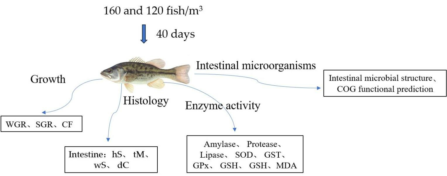
2.1. Experimental Material

The feed was procured from Chongqing Haida Feed Co., Ltd. (particle size: 2.0, crude protein ≥ 49.0%, crude fiber ≤ 3.5%, crude ash ≤ 18.0%, calcium: 0.80–4.0%, total phosphorus ≥ 1.2%, crude fat ≥ 5.0%, lysine ≥ 3.0%, moisture ≤ 10.0%). Juvenile M. salmoides bass were acquired from a piscicultural facility in Guangdong and nurtured at Liangping Shugu Farm in Chongqing City until they attained an average body length of 10.13 ± 0.42 cm and an average body weight of 27.59 ± 3.34 g. Preceding the commencement of the experiment, the juvenile M. salmoides bass underwent a fortnight of transient rearing within a circular recirculating aquaculture tank encompassing a volumetric capacity of 100 m3 (D × H: 8 × 2 m). Concurrently, lettuce cultivation was initiated within the proximate vegetable area interconnected with the piscine reservoir, thereby establishing an integrated aquaponics system. The corresponding planting area of the whole system was 300 m2 and the planting density was 18 trees/m2. Throughout the temporary rearing phase, optimal aeration techniques were employed to ensure sufficient oxygenation within all tanks, while vital physicochemical parameters such as dissolved oxygen levels, water temperature, and pH were meticulously assessed daily. The concentration of dissolved oxygen within the experimental water was meticulously maintained above 8 mg/L, whereas the water temperature was strictly upheld between 23–28 °C, and the pH was meticulously regulated within the range of 7.0–8.0. Regular volumetric water replacements were meticulously executed, constituting 1% of the entire culture tank volume. The system mainly consisted of six tanks, microfilters, fluidized beds, ultraviolet lamps, and other auxiliary facilities. After treatment through the filters, fluidized beds, and UV lamps, the water flowed to the lettuce growing area and then returned to the fish pond.

2.2. Experimental Methods

The experimentation was carried out at the Aquaponics AI Factory situated in Liangping District, Chongqing City. The experimental setup encompassed two meticulously designed culture tanks, wherein the juvenile M. salmoides bass were judiciously stocked at densities of 160 fish/m3 (HSD) and 120 fish/m3 (LSD), respectively. Three replicates were set for each treatment group. The duration of the culture phase spanned a comprehensive 40-day timeframe, wherein the piscine inhabitants received a carefully calculated daily dietary intake equivalent to 2% of their body mass. Fish weight was measured every 7 days and the 2% dietary intake re-calculated. Feeding regimens were meticulously adhered to, with nutrient provision occurring twice daily at precise timings of 9:00 a.m. and 18:00 p.m. The morning feed comprised 1% of the overall ration, followed by an equivalent portion being tendered during the evening hours. Continuous aeration with liquid oxygen was faithfully maintained throughout the entire experimental period to optimize the oxygen saturation levels within the culture tanks. An orchestrated water replacement procedure was diligently executed once, entailing the meticulous substitution of 1% of the total volume of water. The requisite parameters pertaining to water quality were scrupulously gauged and judiciously upheld in strict accordance with the standards established during the temporary rearing phase, commencing 1 h subsequent to each feeding event.
Upon the culmination of the aquaculture undertaking, a meticulous sampling procedure was undertaken. The harvested juvenile M. salmoides specimens were rendered inert through the administration of 100 mg/L MS-222, subsequently transferred onto an ice tray. To ensure the elimination of any extraneous impurities, the piscine subjects underwent a thorough cleansing process involving 70% ethanol and sterile water solutions. In an environment characterized by sterility, the intact intestinal tracts of each fish were delicately extracted. The procured intestinal tissue samples were then segregated into two discrete portions. The first component was suitably preserved in a solution comprising 4% paraformaldehyde, while the second fraction was carefully placed within a centrifuge tube and stored at an ultra-low temperature of −80 °C within a freezer facility. Furthermore, the contents of the intestines from three representative fish in each treatment group were amalgamated to generate a unified composite sample. Three parallel samples were collected for each respective treatment group and diligently preserved at −80 °C until such time that microbial sequencing analysis could be performed.

2.3. Growth Performance Analysis

Prior to the conclusion of the aquaculture trial, a 24 h fasting period was imposed. Eleven fish were randomly selected from each treatment group, and their body weight and length were measured to calculate morphometric indices. The formulas for calculation are as follows:
Weight gain ratio (WGR, %) = (Final average body weight − Initial average body weight)/Initial average body weight × 100%
Specific growth rate (SGR, %) = (ln(Final average body weight) − ln(Initial average body weight))/Experimental duration × 100%
Condition factor (CF, %) = 100 × Body weight/(Body length)3

2.4. Histological Analysis

The anterior intestine, middle intestine, and posterior intestine (obtained by dividing the intestine into three equal parts) of the juvenile M. salmoides were processed into paraffin sections for HE staining. The sections were observed using an Olympus BX 51 (Olympus, Tokyo, Japan) microscope, and five fields of view were selected for each slide. The measurements of villus height (hS), muscular layer thickness (tM), villus width (wS), and crypt depth (dC) were performed using Image-Pro Plus 6.0 software (Media Cybernetics, Inc., Rockville, MD, USA).

2.5. Determination of Enzyme Activity

In this study, the activity of intestinal amylase, protease, lipase, superoxide dismutase (SOD), glutathione-S-transferase (GST), glutathione peroxidase (GPx), and glutathione (GSH), and the content of glutathione (GSH) and malondialdehyde (MDA), were determined with kits from Nanjing Constructive Biotechnology Institute (Nanjing, China).

2.6. Extraction of DNA from the Sample

The DNA of microbial contents in the gastrointestinal tract was extracted using the Stool DNA Kit (OMEGA, Norcross, GA, USA). The DNA extraction steps for all samples followed the instructions provided with the Stool DNA Kit, and the quality of the extracted genomic DNA was evaluated using 1% agarose gel electrophoresis.

2.7. High-Throughput Sequencing Analysis

The specific primers 338F/806R containing a barcode were designed for the V3 + V4 region of the bacterial 16S rRNA gene. The primer sequences were 338F:5′–ACTCCTACGGGAGGCAGCA–3′ and 806R:5′–GGAC-TACHVGGGTWTCTAAT–3′. PCR amplification was performed using an rTaqDNA Polymerase-20 μL reaction system, 10 × Buffer 2 μL, 2.5 mmoL·L−1 DNTPS 2 μL, primer 338F/806R 0.8 μL, Taq enzyme 0.2 μL, BSA 0.2 μL, template DNA 10 ng, and ddH2O supplemented to 20 μL. The PCR conditions were denaturated at 95 °C for 3 min, 27 cycles of 95 °C for 30 s, 55 °C for 30 s, and 72 °C for 45 s. The final extension was 72 °C for 10 min (PCR instrument ABI Gene-Amp 9700, Thermo Fisher Scientific, Waltham, MA, USA).
The PCR products were subjected to 2% agarose gel electrophoresis, resulting in clear bands with the appropriate fragment size (430 bp). The gel-extracted PCR products were assessed using the AxyPrepDNA Gel Recovery Kit (Axygen, Saint Louis, MO, USA), followed by ligation of “Y” adapters after Tris-HCl elution. Magnetic bead selection was employed to remove adapter-dimer fragments. To enrich the PCR products, a standard enrichment PCR was conducted with 1 ng of PCR product for each sample, and subsequently denatured with a 0.1 M NaOH solution to obtain single-stranded DNA fragments. The sequencing library was constructed, and high-throughput sequencing was performed on the 16S-338F-806R Miseq platform (Shanghai Meiji Biotechnology Co., Ltd., Shanghai, China).

2.8. Data Analysis

The experimental data are presented as mean ± standard error (mean ± SE). One-way analysis of variance (ANOVA) was performed using SPSS 23 (IBM, USA) on the aforementioned data, followed by Tukey’s honestly significant difference (HSD) test for multiple comparisons. The significance level (p-value) was set at 0.05, indicating a significant difference when p ≤ 0.05, and a highly significant difference when p ≤ 0.01.

3. Results and Analysis

3.1. Growth and Intestinal Morphology of Juvenile M. salmoides Cultured at Different Densities

At the end of the experiment, the average body weight, weight gain rate, and specific growth rate of the HSD group were significantly higher than those of the LSD group (p ≤ 0.05). However, there was no significant difference in condition factor (p > 0.05) among the groups (Table 1).
After the completion of the aquaculture experiment, morphological analysis was conducted on the intestinal tract of juvenile M. salmoides (Figure 1). The structure of the intestine appeared relatively intact, with well-organized intestinal villi and a smooth mucosal surface. Measurements were taken for villus height (Vh), villus width (Vw), crypt depth (Cd), and muscle layer thickness (Mt) in different sections of the intestine, and the results are presented in Table 2. For the anterior intestine, the villus width of HSD groups was significantly higher than that of LSD groups (p ≤ 0.05). In the middle intestine, both villus height and muscle layer thickness of HSD groups were significantly higher than those of LSD groups (p ≤ 0.01). As for the posterior intestine, the villus height of HSD groups was significantly higher than that of LSD groups (p ≤ 0.01). There were no significant differences in crypt depth among the treatment groups and in the anterior, middle, and posterior sections of the intestine (p > 0.05).

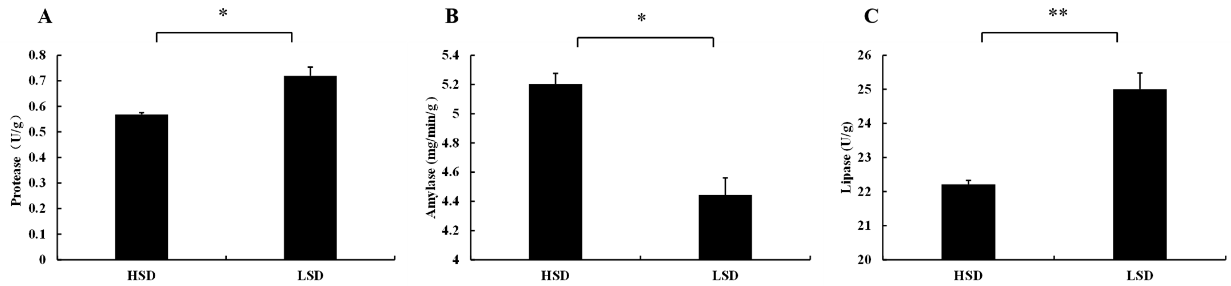
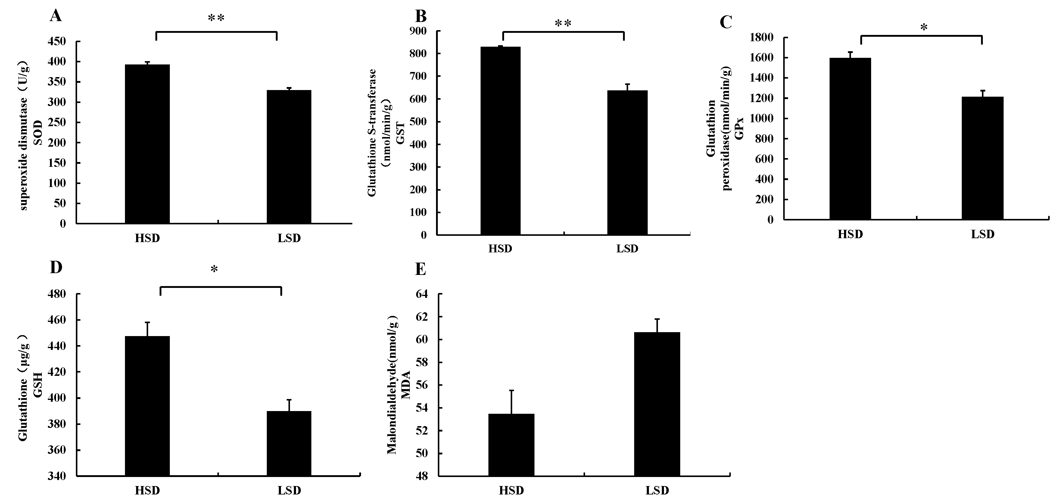
3.2. Changes in Enzyme Activities in Intestinal Tissues of Juvenile M. salmoides Cultured at Different Densities

After the completion of the aquaculture experiment, significant changes were observed in the digestive enzyme activities of juvenile M. salmoides. Specifically, the protease activity (p ≤ 0.05) (Figure 2A) and lipase activity (p ≤ 0.01) (Figure 2C) of the LSD group were significantly higher than those of the HSD group, while the amylase activity was significantly lower in the LSD group compared to the HSD group (p ≤ 0.05) (Figure 2B).
After the conclusion of the aquaculture experiment, significant changes were observed in the antioxidant enzyme activities of the intestinal tract in juvenile M. salmoides. Specifically, the SOD activity (p ≤ 0.01) (Figure 3A), GST activity (p ≤ 0.01) (Figure 3B), and GPx activity (p ≤ 0.05) (Figure 3C) were significantly higher in the HSD group compared to the LSD group. Moreover, the GSH content in the HSD group was significantly higher than that in the LSD group (p ≤ 0.05) (Figure 3D). However, there were no significant differences in the MDA content between the LSD group and the HSD group (p > 0.05) (Figure 3E).

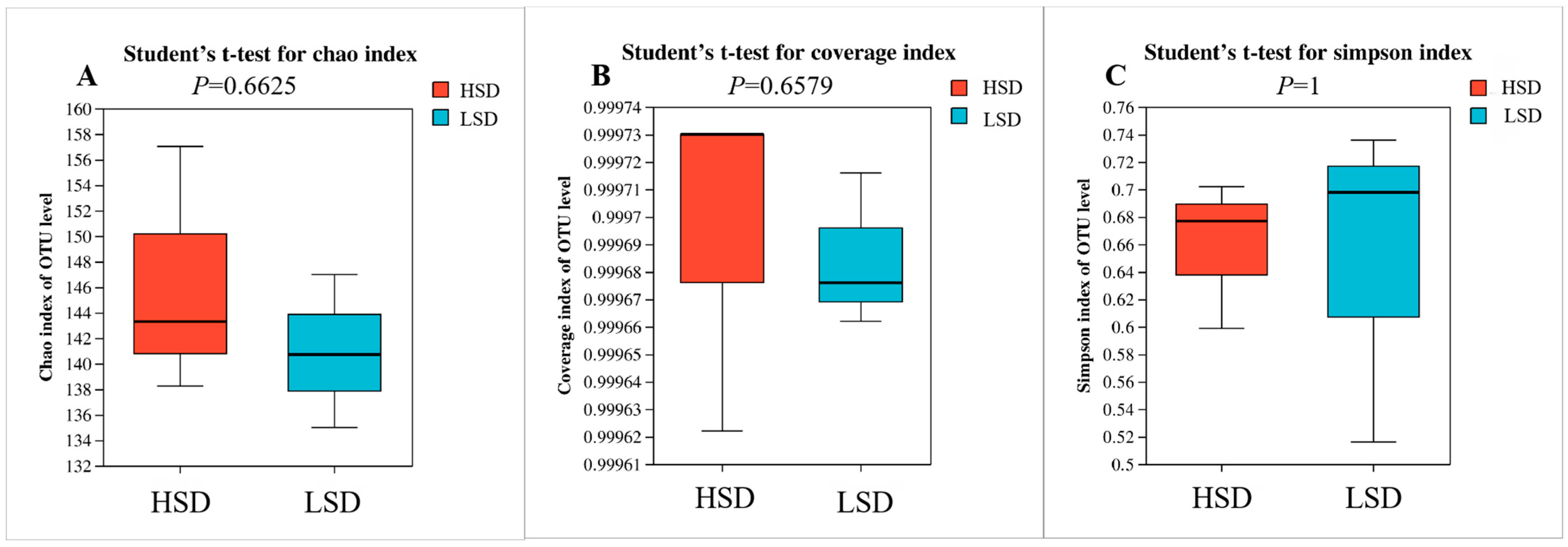
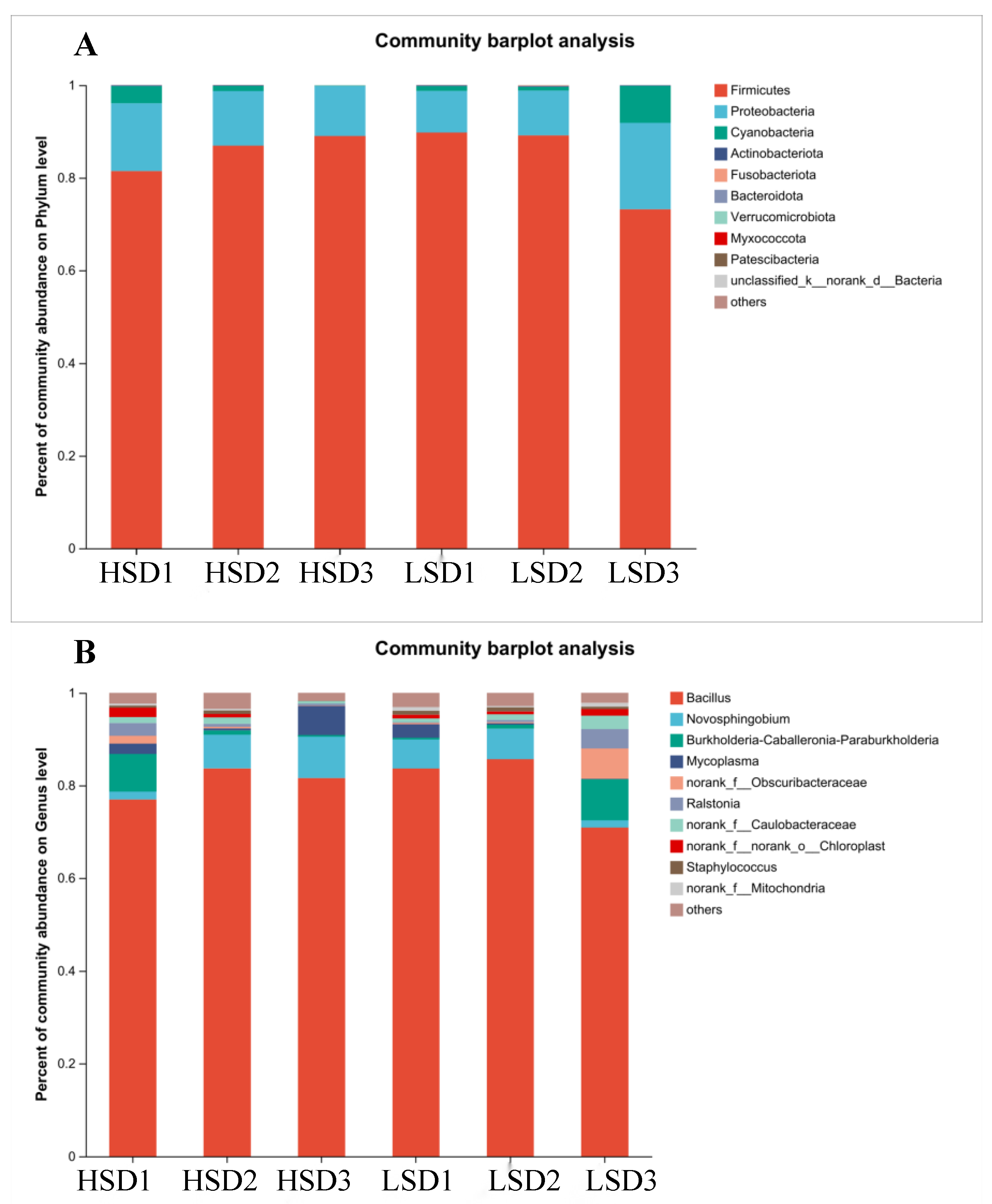
3.3. Intestinal Microbial Structure of Juvenile M. salmoides at Different Breeding Densities

Statistical analysis was conducted on the Alpha diversity of intestinal samples from juvenile M. salmoides. The Chao1, Good’s coverage, and Simpson indices obtained in this experiment are presented in the figure below, aiming to explore richness, diversity, and coverage characterization. The results revealed that the community coverage of each sample exceeded 99.97%, indicating a low probability of unsequenced samples. There were no significant differences in the Chao1, Coverage, and Simpson indices between each sample of the two groups of fish (p > 0.05) (Figure 4).
In the HSD and LSD treatment groups, a total of 11 phyla were detected in the intestinal tract of juvenile M. salmoides. These phyla mainly consisted of Firmicutes, Proteobacteria, Cyanobacteria, Actinobacteriota, and Fusobacteriota, although their proportions varied (Figure 5A). In the LSD group, Firmicutes (84.04%), Proteobacteria (12.44%), and Cyanobacteria (3.28%) were predominant, accounting for 99.76% of the total abundance. Similarly, in the HSD group, Firmicutes (85.80%), Proteobacteria (12.42%), and Cyanobacteria (1.64%) were the major phyla, representing 99.86% of the total abundance. Firmicutes were the dominant phylum in all groups, but there were no significant differences between the groups (p > 0.05).
In the stacked bar plot of bacterial genus-level relative abundance, a total of 239 genera were identified in the intestinal tract of juvenile M. salmoides, with the top 10 genera accounting for over 96% of the abundance in each group. In both density groups, the dominant genera in terms of abundance were Bacillus, Novosphingobium, and Burkholderia, in that order (Figure 5B). In the LSD group, the dominant genera were Bacillus (79.83%), Novosphingobium (4.77%), Burkholderia (3.42%), and Mycoplasma (1.07%), together representing 89.09% of the total abundance. In the HSD group, the dominant genera were Bacillus (80.58%), Novosphingobium (5.96%), Burkholderia (3.13%), and Mycoplasma (3.00%), accounting for 92.67% of the total abundance. Bacillus was the overwhelmingly dominant genus in all groups, but there were no significant differences between the groups (p > 0.05).
The microbial communities in the intestinal tract of juvenile M. salmoides in the HSD and LSD treatment groups were subjected to COG functional prediction and classification analysis using the PICRUST tool. The results showed that both groups had 22 functional clusters, with relatively higher abundance in the categories of amino acid transport and metabolism, transcription, and inorganic ion transport and metabolism. There were no significant differences in functional classification between the different density treatment groups (p > 0.05) (Figure 6).

4. Discussion

4.1. Effects of Culture Density on Growth and Intestinal Morphology of Juvenile M. salmoides

In the realm of aquaculture, stocking density has long been recognized as a pivotal factor exerting influence over growth dynamics [23]. Scientific studies from the past have elucidated the profound impact of high stocking density on some species, instigating a cascade of physiological impairments and, in severe cases, mortality [24,25]. Conversely, reduced stocking density fails to optimize the utilization of aquacultural waters, leading to diminished production yields. Moreover, it curtails interindividual interactions among fish, thereby compromising their growth potential and physiological capacities [26]. Hence, each fish species possesses an optimal stocking density range capable of eliciting peak growth performance [27]. The findings of this empirical endeavor elucidate a conspicuous disparity between the HSD and LSD experimental groups in terms of average body weight, weight gain, and specific growth rate of juvenile M. salmoides. The results of this study indicated that the average body weight, weight gain, and specific growth rate of juvenile M. salmoides in the HSD group were significantly higher than those in the LSD group. It suggests that, in this particular mode, the stocking density in the HSD group can enhance the growth performance of juvenile M. salmoides. This observation bears striking resemblance to the outcomes documented by Zhu et al. [26], who expounded upon the impact of density variations on the growth of juvenile Pseudobagrus ussuriensis. In summary, judiciously augmenting the stocking density of juvenile M. salmoides holds the potential to ameliorate both production yield and aquacultural efficiency.
The intestinal tract of fish is commonly partitioned into three distinct segments: the foregut, midgut, and hindgut [28]. Functionally, the foregut primarily assumes responsibility for digestion and absorption, whereas the midgut and hindgut play a pivotal role in immune-related processes [29]. Notably, the digestive and absorptive efficiency of the fish intestine is intricately linked to the condition of its mucosal surface structure [30]. Morphological parameters such as villus height, villus width, muscular layer thickness, and crypt depth are frequently employed as metrics to assess intestinal development, health status, and digestive absorptive capacity [31,32]. Heightened villus height and width signify an increased presence of epithelial cells, which, in turn, indicates a heightened nutrient absorption potential within the intestine. Conversely, reduced villus dimensions point toward an inverse scenario [33]. In the present study, notable disparities in villus height and width within the foregut, midgut, and hindgut of juvenile M. salmoides were observed between the HSD and LSD experimental groups. This suggests that rearing juvenile M. salmoides at a higher density in the HSD group can increase the absorptive surface area of their intestines, enhance their digestive and absorptive capacity, and improve intestinal structure, thereby maximizing their growth potential. Importantly, the thickening of the intestinal muscular layer expedites peristaltic contractions, consequently amplifying intestinal absorption rates [34]. The substantially augmented muscular layer thickness observed within the foregut of the HSD group, relative to the LSD group, further corroborates this notion. In sum, the divergences in intestinal morphology provide tangible evidence of variances in the intestinal architecture of juvenile M. salmoides across distinct stocking densities. Fish specimens reared within the HSD group exhibit heightened digestive and absorptive capacities, alongside a healthier intestinal milieu.

4.2. Effects of Different Culture Densities on Enzyme Activities in Intestinal Tissues of Juvenile M. salmoides

The level of digestive enzyme activity serves as an indicator of the alimentary capability and absorptive prowess of piscine organisms with respect to nutrient assimilation [35]. In addition to environmental factors such as temperature [36], pH [37], salinity [38], and dissolved oxygen [39], stocking density also has a significant impact on digestive enzyme activity [40]. Previous studies have shown that cultured fish exhibit optimal digestive physiological status and higher activity of digestive enzymes in their tissues within an appropriate stocking density [41]. However, when the density exceeds the carrying capacity of the culture system, the digestive enzyme activity in the fish is actually inhibited. Exemplifying this, augmentation in stocking density prompts a concomitant decrement in the activities of five digestive enzymes residing within the gastric and intestinal compartments of Larimichthys crocea [42]. Ergo, the extent of digestive enzyme activity within cultured fish directly corresponds to stocking density. In this study, the protease and lipase activities in the juvenile M. salmoides were significantly higher in the LSD group than in the HSD group. This discrepancy intimates that diminished stocking density engenders heightened digestive enzyme activity within juvenile M. salmoides, thereby concomitantly propelling the process of nutrient digestion and absorption enacted by the enteral tissues. This finding aligns with the observations imparted by Ezhilmathi et al., who expounded upon the ramifications of stocking density on digestive enzyme activity within Lates calcarifer [24]. However, the amylase activity in the HSD group of juvenile M. salmoides was significantly higher than that in the LSD group. This divergence likely stems from the adaptive capacity exhibited by the juvenile M. salmoides towards elevated stocking densities, whereby an augmentation in amylase activity facilitates efficacious digestion and utilization of residual sustenance, consequently sharpening their survival aptitude within the milieu. This parallels the conspicuous surge in amylase activity documented in Cyprinus carpio subsequent to bouts of nutrient deprivation [43].
The intestinal tract of fish serves not only as a site for digestion but also actively participates in immune responses, metabolic processes, and various stress reactions [44]. Extensive research has revealed that high stocking density induces stress responses in fish intestinal cells, leading to an excessive generation of reactive oxygen species (ROS) that can inflict damage upon cells, tissues, and other vital components within the fish [45]. However, throughout the course of evolution, organisms have evolved a sophisticated antioxidant defense system to effectively eliminate superfluous free radicals and safeguard cellular integrity [46]. This antioxidant system primarily comprises enzymatic and non-enzymatic components, with the enzymatic system encompassing key enzymes such as superoxide dismutase, glutathione transferase, and glutathione peroxidase, all instrumental in the elimination of ROS [47,48]. On the other hand, the non-enzymatic system chiefly involves reduced glutathione, which serves as an adept scavenger of oxygen free radicals and acts as a detoxifying agent against electrophiles, thereby maintaining the equilibrium of thiol-disulfide bonds and facilitating signal transduction processes [49]. Malondialdehyde (MDA) is the end product of lipid peroxidation, and its content combined with changes in various antioxidant enzymes can collectively reflect the oxidative stress encountered by fish [50]. Under conditions of high stocking density, free radicals directly attack polyunsaturated fatty acids within membranes, thus initiating lipid peroxidation reactions [51]. In this study, the activities of superoxide dismutase (SOD), glutathione peroxidase (GPx), and glutathione transferase (GST), as well as the content of reduced glutathione (GSH) within the intestinal tract of juvenile M. salmoides, were found to be significantly higher in the HSD group compared to the LSD group. There was no discernible disparity observed in malondialdehyde (MDA) levels, indicating that a stocking density of 160 fish/m3 only increases the antioxidant enzyme activity in the intestinal tract of juvenile M. salmoides. However, through the upregulation of their endogenous antioxidant enzymes, juvenile M. salmoides can effectively eliminate excessive ROS and superoxide radicals, thereby circumventing oxidative damage. Consequently, based on the comprehensive alterations in digestive enzyme and antioxidant enzyme activities within the intestinal tract of juvenile M. salmoides, it can be inferred that the stocking density employed in the HSD group is relatively suitable during the juvenile stage.

4.3. Effects of Different Culture Densities on Intestinal Microorganisms in Juvenile M. salmoides

The intestinal microbiota in fish exhibits a vast assemblage of species and a substantial population [52]. It fulfills an indispensable role in the decomposition and synthesis of essential nutrients required by the host, augmentation of nutrient absorption within the intestinal milieu, production of digestive enzymes, and regulation of protein expression associated with metabolic processes [53]. Studies have demonstrated that stocking density exerts a significant influence on the intestinal microbiota of fish, thereby influencing their growth by modulating the activity of digestive enzymes and antioxidant enzymes [54]. The present study scrutinized the configuration of the intestinal microbial community in juvenile M. salmoides reared within an aquaponics system at distinct density levels. Alpha diversity indices divulged no noteworthy disparities in the diversity of bacterial communities among the various groups, which is consistent with the findings of Xu et al. on the impact of dissolved oxygen on the intestinal microbiota of Salmo salar [55]. Additionally, this study verified that the prevailing bacteria residing within the intestinal microbiota of juvenile M. salmoides primarily belong to the phyla Firmicutes and Proteobacteria [56]. Proteobacteria are known to be abundant in stable (healthy) gut microbiota [57], while Firmicutes, particularly the Bacillus genus, are major bacteria involved in the metabolism of food residues [58]. These bacteria foster digestion and enhance nutrient assimilation efficacy while simultaneously acting as pivotal microorganisms engaged in disease prevention and control via the production of antimicrobial proteins or enzymes eliciting host resistance against diseases [59]. However, a significantly heightened abundance of Chlamydia was observed in the HSD group relative to the LSD group. Chlamydia, a member of the Chlamydiae phylum, represents one of the smallest self-replicating free-living microorganisms known to date [60]. It is also a potential pathogenic bacterium that can cause intestinal-related diseases such as enteritis [61]. Based on these findings, it is speculated that elevated stocking density may augment the abundance of potentially pathogenic bacteria, consequently instigating intestinal damage, which aligns with prior research outcomes concerning M. salmoides [62]. Thus, it is advisable to exercise heightened vigilance pertaining to pathogenic microorganisms such as Chlamydia during the farming process to preclude disease outbreaks and mitigate fish mortality.
The intestinal microbiota of organisms possesses the capacity to ferment dietary amino acids, resulting in the production of diverse metabolites [63]. Through the modulation of gut microbiota, the host can derive advantageous outcomes by fine-tuning its own metabolic processes, thereby enabling superior adaptation to novel nutritional and environmental exigencies [64]. Hence, delving into the functional transformations occurring within the gut microbiota yields a more comprehensive comprehension of the profound impact imposed upon the host by perturbations in its intestinal microbial milieu. Functional predictions have unearthed that the intestinal microbial community of juvenile M. salmoides primarily engages in pivotal activities encompassing amino acid transportation and metabolism, transcription, as well as inorganic ion transportation and metabolism. These functions are beneficial for the utilization of nutrients by the fish’s body and the proper functioning of cellular activities, ultimately promoting the fish’s adaptability to the environment. Nonetheless, it is imperative to acknowledge that prevailing research outcomes predominantly hinge upon sequencing data and functional pathway prognostications, with restricted validation data at hand. Thus, further investigations are warranted to unravel the underlying mechanisms at play.

4.4. Comparison of the Dominant Intestinal Microflora of M. salmoides under Different Factors

The composition of the intestinal microbiota in fish is shaped by the intricate interplay between host physiological traits and external environmental factors [65]. Fish reside within a dynamic ecological system, wherein fluctuations in endogenous or exogenous elements wield the potential to impact both the structure and diversity of their intestinal microbiota [66]. Comprising archaea, bacteria, protists, fungi, and viruses [49], the fish intestinal microbiota is predominantly characterized by the bacterial contingent [67]. Notably, within the realm of bacteria, notable representatives from the phyla Proteobacteria, Firmicutes, and Bacteroidetes have been identified across diverse fish species, constituting a substantial proportion—upwards of 90%—of the investigated fish intestinal microbiota. This observation not only signifies the putative significance of these bacterial cohorts in shaping fish intestinal function but also underscores their pivotal contributions [68]. Detailed insights into the factors influencing the intestinal microbiota of M. salmoides are presented in Table 3, underscoring the preponderance of Firmicutes, Proteobacteria, and Bacteroidetes at the phylum level. At the genus level, dominant bacterial taxa encompass Mycoplasma, Leptothrix, and Aeromonas. Moreover, distinctions in the structure and diversity of the gut microbiota in M. salmoides may be ascribed, in part, to the employed farming methodologies, including pond culture, laboratory recirculating water culture, pond recirculating water culture, factory recirculating water culture, and cage culture. Intriguingly, feed composition incorporating nutritional constituents and additives, the aquatic milieu encompassing dissolved oxygen levels and environmental pollutants, as well as the developmental stage of the host individual, all emerge as influential determinants in orchestrating the establishment of intestinal microbiota in juvenile M. salmoides. Ultimately, these factors culminate in the composition of a relatively stable microbiota within adult individuals. In conclusion, to ensure a stable intestinal microbiota structure in M. salmoides during cultivation, it is necessary to adjust the nutritional strategies according to different environmental conditions and growth stages, aiming to achieve efficient and healthy growth.

5. Conclusions

This study presents the inaugural exposition of the repercussions of divergent population densities upon intestinal tissue morphology, digestive enzyme activity, antioxidant enzyme activity, and microbial community in juvenile M. salmoides. Our findings elucidate that fish density within HSD groups engenders an augmented weight gain rate, and specific growth rate, as well as heightened dimensions of intestinal villi, muscle layer thickness, and intestinal antioxidant enzyme activity in juvenile M. salmoides. Such advancements in intestinal architecture concomitantly augment the digestive and absorptive prowess of the piscine subjects. Furthermore, Firmicutes and Proteobacteria constitute the predominant bacterial phyla within the intestinal microbiota. Nevertheless, the heightened prevalence of Mycoplasma within HSD groups may precipitate pathogenic occurrences, necessitating due regulation of the aquatic milieu to attenuate the presence of such pathogenic microorganisms during the cultivation process. By summarizing the research on the influence of various factors on the intestinal microbiota of M. salmoides in recent years, it is evident that the intestinal microbiota is closely related to different farming modes, feed composition, water environment, and developmental stage. Therefore, adjusting the nutritional strategy based on specific conditions is necessary. These research findings have important implications for the healthy cultivation of juvenile M. salmoides with respect to fish density.

Author Contributions

J.Z.: Data collection and curation; funding acquisition; writing—original draft; writing—review and editing. Z.W.: Data collection and curation; Analysis; writing—original draft; writing—review and editing. D.P.: Writing—original draft; writing—review and editing. P.L.: Investigation; analysis. X.W.: Project administration. M.L.: Data collection and curation. D.L.: Data collection and curation. L.G.: Funding acquisition; resources. X.Z.: Methodology; supervision. All authors have read and agreed to the published version of the manuscript.

Funding

This research was funded by the Research and Application of Plant-based Balanced Production Technology of Aquaponics for Epidemic Preservation (No. cstc2022jxjl80023), the Key Technology and Equipment Research of Aquaponics Intelligent Factory (No. CSTB2022TIAD-ZXX0053), Key Research and Development Plan of the Ministry of Science and Technology (No. 2022YFD2001700), the toxic effects of ammonia nitrogen on M. salmoides in high density Aquaponics Industrialized Recirculating Aquaculture (No. cqaas2023sjczqn036), Chongqing Modern·Agricultural Industry Technology·System (No. CQMAITS202315), and Study on key technologies of intelligent breeding plant for Micropterus salmoides (No. CSTB2022TIAD-LDX0008).

Institutional Review Board Statement

This study was carried out without sacrificing organisms unnecessarily to reduce and perfect the use of animals used for scientific purposes. The proposed methods, use of animals, and research practice (approval number: IACUC-20181015-12) were examined and approved by the Animal Ethics Committee of Southwest University. This research received no external funding.

Data Availability Statement

Data are contained within the article.

Conflicts of Interest

The authors declare no conflict of interest.

Abbreviations

AbbreviationsFull name in English
HSD160 fish/m3, high stocking density
LSD120 fish/m3, low stocking density
IBWinitial average body weight
FBWfinal average body weight
WGRweight gain ratio
SGRspecific growth rate
CFcondition factor
hSvillus height
tMmuscular layer thickness
wSvillus width
dCcrypt depth
SODsuperoxide dismutase
GSTglutathione-S-transferase
GPxglutathione peroxidase
GSHglutathione
MDAmalondialdehyde

References

  1. Liu, C.; Zhao, L.P.; Shen, Y.Q. A systematic review of advances in intestinal microflora of fish. Fish Physiol. Biochem. 2021, 47, 2041–2053. [Google Scholar] [CrossRef]
  2. Liu, S.; Yu, H.; Li, P.; Wang, C.; Liu, G.; Zhang, X.; Zhang, C.; Qi, M.; Ji, H. Dietary nano-selenium alleviated intestinal damage of juvenile grass carp (Ctenopharyngodon idella) induced by high-fat diet: Insight from intestinal morphology, tight junction, inflammation, anti-oxidization and intestinal microbiota. Anim. Nutr. 2022, 8, 235–248. [Google Scholar] [CrossRef]
  3. Huang, B.; Zhang, S.; Dong, X.; Chi, S.; Yang, Q.; Liu, H.; Tan, B.P.; Xie, S.W. Effects of fishmeal replacement by black soldier fly on growth performance, digestive enzyme activity, intestine morphology, intestinal flora and immune response of pearl gentian grouper (Epinephelus fuscoguttatus female × Epinephelus lanceolatus male). Fish Shellfish. Immunol. 2022, 120, 497–506. [Google Scholar] [CrossRef]
  4. Zhang, F.L.; Hao, Q.; Zhang, Q.S.; Lv, H.Y.; Yang, Y.L.; Chao, R.; Zhang, Z.; Zhou, Z. Influences of dietary Eucommia ulmoides leaf extract on the hepatic lipid metabolism, inflammation response, intestinal antioxidant capacity, intestinal microbiota, and disease resistance of the channel catfish (Ictalurus punctatus). Fish Shellfish. Immunol. 2022, 123, 75–84. [Google Scholar] [CrossRef]
  5. Wang, C.; Wang, X.; Xiao, S.; Bu, X.; Lin, Z.; Qi, C.; Qin, J.G.; Chen, L.Q. T-2 toxin in the diet suppresses growth and induces immunotoxicity in juvenile Chinese mitten crab (Eriocheir sinensis). Fish Shellfish. Immunol. 2020, 97, 593–601. [Google Scholar] [CrossRef] [PubMed]
  6. Mi, H.F.; Sun, R.J.; Zhang, L.; Li, B.S.; Wang, W.G.; Wu, Y.Y.; Wang, Y.L. Research progress of fish intestinal health. China Feed. 2015, 15, 19–22. [Google Scholar] [CrossRef]
  7. Øivind, B. Bacteria associated with early life stages of halibut, Hippoglossus hippoglossus L., inhibit growth of a pathogenic Vibrio sp. J. Fish Dis. 2010, 18, 31–40. [Google Scholar] [CrossRef]
  8. Chen, X.M.; Wang, G.Q.; Shan, X.F. Research Progress on the Relationship between Intestinal Barrier Damage and Intestinal Inflammation Development in Fish. J. Henan Agric. Sci. 2022, 51, 1–9. [Google Scholar] [CrossRef]
  9. Xu, G.F.; Chen, X.J.; Du, J.; Mou, Z.B. Fish Digestive System: It’s Structure, Function and The Distributions and Characteristics of Digestive Enzymes. Chin. J. Fish. 2009, 22, 49–55. [Google Scholar]
  10. Ji, Y.B.; Zhang, M.Y.; Wang, Y.; Wang, X.M.; Dai, W.; Wang, C.; Sun, X.L. Quantitative Histology Study on the Stomach and Intestines of Clarias gariepinus at Different Culture Densities. J. Anhui Agri Sci. 2013, 41, 5399–5401. [Google Scholar]
  11. Su, X. Study on Muscle Growth and Digestive Performance of Micropterus salmoides during Early Development. Master’s Thesis, Suzhou University, Suzhou, China, 2022. [Google Scholar]
  12. Comabella, Y.; Mendoza, R.; Aguilera, C.; Carrillo, O.; Hurtado, A.; García-Galano, T. Digestive enzyme activity during early larval development of the Cuban gar Atractosteus tristoechus. Fish Physiol. Biochem. 2006, 32, 147–157. [Google Scholar] [CrossRef]
  13. Lushchak, V.I.; Bagnyukova, T.V.; Lushchak, O.V.; Storey, J.M.; Storey, K.B. Hypoxia and recovery perturb free radical processes and antioxidant potential in common carp (Cyprinus carpio) tissues. Int. J. Biochem. Cell Biol. 2005, 37, 1319–1330. [Google Scholar] [CrossRef]
  14. Feng, Z.D.; Zhong, Y.F.; He, G.L.; Sun, H.; Chen, Y.J.; Zhou, W.H.; Lin, S.M. Yeast culture improved the growth performance, liver function, intestinal barrier and microbiota of juvenile largemouth bass (Micropterus salmoides) fed high-starch diet. Fish Shellfish Immunol. 2022, 120, 706–715. [Google Scholar] [CrossRef] [PubMed]
  15. Miyuki, M.; Taiga, A.; Kenji, S.; Tomofumi, Y.; Kazuhisa, T.; Jun, K. Intestinal microbiota composition is altered according to nutritional biorhythms in the leopard coral grouper (Plectropomus leopardus). PLoS ONE 2018, 13, e0197256. [Google Scholar] [CrossRef]
  16. Li, J.Z.; Hou, J.; Zhang, P.F. Comparative study of intestinal microbial community structure in different species of carp in aquaponics system. South China Fish. Sci. 2016, 12, 42–50. [Google Scholar] [CrossRef]
  17. Parma, L.; Pelusio, N.F.; Gisbert, E.; Esteban, M.A.; D’Amico, F.; Soverini, M.; Candela, M.; Dondi, F.; Gatta, P.P.; Bonaldo, A. Effects of rearing density on growth, digestive conditions, welfare indicators and gut bacterial community of gilthead sea bream (Sparus aurata, L. 1758) fed different fishmeal and fish oil dietary levels. Aquaculture 2020, 518, 734854. [Google Scholar] [CrossRef]
  18. Wong, S.; Waldrop, T.; Summerfelt, S.; Davidson, J.; Barrows, F.; Kenney, P.B.; Welch, T.; Wiens, G.D.; Snekvik, K.; Rawls, J.F.; et al. Aquacultured rainbow trout (Oncorhynchus mykiss) possess a large core intestinal microbiota that is resistant to variation in diet and rearing density. Appl. Environ. Microbiol. 2013, 79, 4974–4984. [Google Scholar] [CrossRef]
  19. Yep, B.; Zheng, Y. Aquaponic trends and challenges—A review. J. Clean. Prod. 2019, 228, 1586–1599. [Google Scholar] [CrossRef]
  20. Krastanova, M.; Sirakov, I.; Ivanova-Kirilova, S.; Yarkov, D.; Orozova, P. Aquaponic systems: Biological and technological parameters. Biotechnol. Biotechnol. Equip. 2022, 36, 305–316. [Google Scholar] [CrossRef]
  21. Spradlin, A.; Saha, S. Saline aquaponics: A review of challenges, opportunities, components, and system design. Aquaculture 2022, 555, 738173. [Google Scholar] [CrossRef]
  22. Ashley, P.J. Fish welfare: Current issues in aquaculture. Appl. Anim. Behav. Sci. 2007, 104, 199–235. [Google Scholar] [CrossRef]
  23. Debnath, S.; Ahmed, M.U.; Parvez, M.S.; Karmokar, A.K.; Nazmul, M. Effect of stocking density on growth performance and body composition of climbing perch (Anabas testudineus) in biofloc system. Aquacult Int. 2021, 30, 1089–1100. [Google Scholar] [CrossRef]
  24. Ezhilmathi, S.; Ahilan, B.; Uma, A.; Felix, N.; Cheryl, A.; Somu Sunder Lingam, R. Effect of stocking density on growth performance, digestive enzyme activity, body composition and gene expression of Asian seabass reared in recirculating aquaculture system. Aquac. Res. 2022, 53, 1963–1972. [Google Scholar] [CrossRef]
  25. Liu, B.L.; Lei, J.L.; Jia, R. A review: The influence of stocking density on fish welfare. Eng. Sci. China 2014, 16, 100–105. [Google Scholar]
  26. Zhu, C.K.; Jiang, F.F.; Zhai, W.J.; Chen, C.R.; Wei, M.Y.; Wu, M.R.; Wang, L.J.; Xu, J.R.; Xi, X.C.; Zhao, J.; et al. Effects of Stocking Density and Water Volume on Growth of Juvenile Ussuri Bullhead Pseudobagrus ussuriensis. Fish. Sci. 2019, 38, 682–687. [Google Scholar] [CrossRef]
  27. Liao, R.; Ou, Y.J.; Gou, X.W. A review: Influence of stocking density on fish welfare I mortality, growth, feeding and stress response. South China Fish. Sci. 2006, 6, 76–80. [Google Scholar]
  28. Liu, J.; Yan, Z.H.; Yao, B.L.; Li, Y.H. Research progress on the tissue structure, function, influencing factors and protective substances of fish intestine. Fish. Sci. Technol. Inf. 2023, 50, 121–127. [Google Scholar] [CrossRef]
  29. Noaillac-Depeyre, J.; Gas, N. Structure and function of the intestinal epithelial cells in the perch (Perca fluviatillis L.). Anat. Rec. 1979, 195, 621–627. [Google Scholar] [CrossRef]
  30. Sun, Y.X.; Dong, H.B.; Duan, Y.F.; Li, H.; Liu, Q.; Zhang, J.; Wang, W. Progresses in Stress Damage and Protection Studies on Fish Intestine. Trans. Oceanol. Limnol. 2019, 3, 174–183. [Google Scholar] [CrossRef]
  31. Farhangi, M.; Carter, C.G. Growth, physiological and immunological responses of rainbow trout (Oncorhynchus mykiss) to different dietary inclusion levels of dehulled lupin (Lupinus angustifolius). Aquac. Res. 2001, 32, 329–340. [Google Scholar] [CrossRef]
  32. Nie, G.X.; Wang, J.L.; Zhu, M.W.; Zhou, H.Q. The influences of xylanse added in wheat basal diet on intestine chymeviscosity and the development of villi and microvilli of Tilapia nilotica. J. Fish. China 2007, 1, 54–61. [Google Scholar]
  33. Zhong, Q.; Xu, D.D.; Wang, X.; Liu, C.d.; Zhou, H.H.; Mai, K.S.; He, K. Effects of Dietary Leucine and Glutamine on Growth and Intestinal Health of Juvenile Turbot (Scophthalmus maximus L.). Period. Ocean. Univ. China 2023, 53, 008–017. [Google Scholar] [CrossRef]
  34. Zhou, F.L. Analysis of Difference in Morphological Structure and Enzyme Activity on Different Parts of Black Carp Intestine. Master’s Thesis, Shanghai Ocean University, Shanghai, China, 2020. [Google Scholar]
  35. Lundstedt, L.M.; José Fernando Bibiano, M.; Moraes, G. Digestive enzymes and metabolic profile of Pseudoplatystoma corruscans (Teleostei: Siluriformes) in response to diet composition. Comp. Biochem. Physiol. Part B Biochem. Mol. Biol. 2004, 137, 331–339. [Google Scholar] [CrossRef] [PubMed]
  36. Volkoff, H.; Rønnestad, I. Effects of temperature on feeding and digestive processes in fish. Temperature 2020, 7, 307–320. [Google Scholar] [CrossRef]
  37. Zhang, T.; Cai, S.L.; Ju, C.X. Effects of temperature and pH on activity of digestive enzymes in Amphiprion ocellaris juvenile. Guangdong Agric. Sci. 2014, 41, 131–135. [Google Scholar] [CrossRef]
  38. Mozanzadeh, M.T.; Safari, O.; Oosooli, R.; Mehrjooyan, S.; Najafabadi, M.Z.; Hoseini, S.J.; Saghavi, H.; Monem, J. The effect of salinity on growth performance, digestive and antioxidant enzymes, humoral immunity and stress indices in two euryhaline fish species: Yellowfin seabream (Acanthopagrus latus) and Asian seabass (Lates calcarifer). Aquaculture 2021, 534, 736329. [Google Scholar] [CrossRef]
  39. Bowyer, J.N.; Booth, M.A.; Qin, J.G.; D'Antignana, T.; Thomson, M.J.S.; Stone, D.A.J. Temperature and dissolved oxygen influence growth and digestive enzyme activities of yellowtail kingfish Seriola lalandi (Valenciennes, 1833). Aquac. Res. 2014, 45, 2010–2020. [Google Scholar] [CrossRef]
  40. Ni, M.; Liu, M.; Lou, J.; Mi, G.; Yuan, J.; Gu, Z. Stocking density alters growth performance, serum biochemistry, digestive enzymes, immune response, and muscle quality of largemouth bass (Micropterus salmoides) in in-pond raceway system. Fish Physiol. Biochem. 2021, 47, 1243–1255. [Google Scholar] [CrossRef]
  41. Peng, S.M.; Lin, S.Z.; Shi, Z.H.; Gao, Q.X.; Wang, J.G.; Sun, P.; Yin, F. Effects of rearing density on growth rate and digestive enzyme activity of juvenile Pampus argenteus. Mar. Fish. 2013, 35, 72–76. [Google Scholar] [CrossRef]
  42. Yu, Y.B.; Huang, W.Y.; Cui, M.C. Effects of stocking densities on growth performance, nutrient composition serum biochemical, digestive and metabolic enzymes activities of large yellow croaker (Larimichthys crocea). Fish. Mod. 2023, 50, 64–71. [Google Scholar]
  43. Wang, Y.N.; Zhang, Z.R.; Zheng, S.M. The carp compensate the influence of growth and hunger of amylase. Reserv. Fish. 2001, 5, 6–7. [Google Scholar] [CrossRef]
  44. Wu, W.Q.; Yao, J.M.; Zhao, J.; Chen, X.M.; Xia, C.G.; Niu, X.T.; Wang, G.Q. Effects of Astaxanthin on Growth, Intestinal Digestive Enzymes and Antioxidant Indexes of Juvenile Paramisgurnus dabryanus. J. Jilin Agric. Univ. 2021, 43, 482–487. [Google Scholar] [CrossRef]
  45. Wang, Y.; Chen, C.G.; Zhang, J.R.; Gao, Y.J. Effects of stocking density on growth, feed utilization and intestinal oxidative stress resistance in juvenile Megalobrama pellegrini (Tchang, 1930). Prog. Fish. Sci. 2022, 43, 106–114. [Google Scholar] [CrossRef]
  46. Lushchak, V.I. Environmentally induced oxidative stress in aquatic animals. Aquat. Toxicol. 2011, 101, 13–30. [Google Scholar] [CrossRef]
  47. Ighodaro, O.M.; Akinloye, O.A. First line defence antioxidants-superoxide dismutase (SOD), catalase (CAT) and glutathione peroxidase (GPX): Their fundamental role in the entire antioxidant defence grid. Alex. J. Med. 2018, 54, 287–293. [Google Scholar] [CrossRef]
  48. Qu, J.H.; Chen, J.C.; Hu, G.D.; Wu, W. Dynamic changes of catalase and glutathione-s-transferase in the different tissues of tilapia exposed to phenol. Ecol. Environ. 2006, 15, 687–692. [Google Scholar] [CrossRef]
  49. Zang, L.X.; Ma, Y.; Huang, W.H.; Ling, Y.H.; Sun, L.M.; Wang, X.D.; Zeng, A.B.; Dahlgren, R.A.; Wang, C.H.; Wang, H.L. Dietary Lactobacillus plantarum ST-III alleviates the toxic effects of triclosan on zebrafish (Danio rerio) via gut microbiota modulation. Fish Shellfish Immunol. 2019, 84, 1157–1169. [Google Scholar] [CrossRef] [PubMed]
  50. Scandalios, J.G. Oxygen Stress and Superoxide Dismutases. Plant Physiol. 1993, 101, 7–12. [Google Scholar] [CrossRef]
  51. Sahin, K.; Yazlak, H.; Orhan, C.; Tuzcu, M.; Akdemir, F.; Sahin, N. The effect of lycopene on antioxidant status in rainbow trout (Oncorhynchus mykiss) reared under high stocking density. Aquaculture 2014, 418–419, 132–138. [Google Scholar] [CrossRef]
  52. Cantas, L.; Sørby, J.R.; Aleström, P.; Sørum, H. Culturable gut microbiota diversity in zebrafish. Zebrafish 2012, 9, 26–37. [Google Scholar] [CrossRef]
  53. Lyons, P.P.; Turnbull, J.F.; Dawson, K.A.; Crumlish, M. Phylogenetic and functional characterization of the distal intestinal microbiome of rainbow trout Oncorhynchus mykiss from both farm and aquarium settings. J. Appl. Microbiol. 2017, 122, 347–363. [Google Scholar] [CrossRef] [PubMed]
  54. Li, Q.; Sun, L.H.; Jiang, J.H. Stocking densities on growth, antioxidant enzyme activities and intestinal microbiota of hybrid of Culter alburnus(♀) × Megalobrama terminalis(♂) in IPRA system. Acta Hydrobiol. Sin. 2023, 47, 479–487. [Google Scholar]
  55. Xu, S.H. The Growth and Physiological Response of Atlantic Salmon (Salmo salar) under the Conditions of Different Rearing Densities and Dissolved Oxygen. Ph.D. Thesis, Huazhong Agricultural University, Wuhan, China, 2021. [Google Scholar]
  56. Zhou, M.; Liang, R.; Mo, J.; Yang, S.; Gu, N.; Wu, Z.; Badu, V.S.; Li, J.; Huang, Y.; Lin, L. Effects of brewer’s yeast hydrolysate on the growth performance and the intestinal bacterial diversity of largemouth bass (Micropterus salmoides). Aquaculture 2018, 484, 139–144. [Google Scholar] [CrossRef]
  57. Wang, C.H.; Zhou, Y.F.; Fang, D.A.; Zhang, X.Z.; Zhang, L.; Liu, J.Y.; Zheng, Z.; You, Y. Effects of starvation and refeeding on intestinal microflora of chinese mitten crab (Eriocheir sinensis). Acta Hydrobiol. Sin. 2019, 43, 748–756. [Google Scholar] [CrossRef]
  58. Zhang, Y.J.; Li, S.; Gan, R.Y.; Zhou, T.; Xu, D.P.; Li, H.B. Impacts of Gut Bacteria on Human Health and Diseases. Int. J. Mol. Sci. 2015, 16, 7493–7519. [Google Scholar] [CrossRef]
  59. Karl, J.P.; Margolis, L.M.; Madslien, E.H.; Murphy, N.E.; Castellani, J.W.; Gundersen, Y.; Hoke, A.V.; Levangie, M.W.; Kumar, R.; Chakraborty, N.; et al. Changes in intestinal microbiota composition and metabolism coincide with increased intestinal permeability in young adults under prolonged physiological stress. Am. J. Physiol. Gastrointest. Liver Physiol. 2017, 312, G559–G571. [Google Scholar] [CrossRef]
  60. Sellyei, B.; Varga, Z.; Cech, G.; Varga, Á.; Székely, C. Mycoplasma infections in freshwater carnivorous fishes in Hungary. J. Fish Dis. 2021, 44, 297–304. [Google Scholar] [CrossRef]
  61. Gnanadurai, R.; Fifer, H. Mycoplasma genitalium: A Review. Microbiology 2020, 166, 21–29. [Google Scholar] [CrossRef] [PubMed]
  62. Song, Z.; Ye, W.; Tao, Y.; Zheng, T.; Qiang, J.; Li, Y.; Li, Y.; Liu, W.T.; Xu, P. Transcriptome and 16S rRNA Analyses Reveal That Hypoxic Stress Affects the Antioxidant Capacity of Largemouth Bass (Micropterus salmoides), Resulting in Intestinal Tissue Damage and Structural Changes in Microflora. Antioxidants 2022, 12, 1. [Google Scholar] [CrossRef] [PubMed]
  63. Wei, X.B.; Zhang, L.L.; Si, D.Y. Decomposition of amino acids by intestinal flora and the effect of the metabolites on the host. Siliao Gongye 2017, 38, 41–44. [Google Scholar] [CrossRef]
  64. Lin, K.T. Comparison of Intestinal Microbes between Feeding Broad Bead and Common Feed in Grass Carps. Master’s Thesis, South China Agricultural University, Guangzhou, China, 2019. [Google Scholar]
  65. Amato, K.R. Co-evolution in context: The importance of studying gut microbiomes in wild animals. Microbiome Sci. Med. 2013, 1, 10–29. [Google Scholar] [CrossRef]
  66. Ghanbari, M.; Kneifel, W.; Domig, K.J. A new view of the fish gut microbiome: Advances from next-generation sequencing. Aquaculture 2015, 448, 464–475. [Google Scholar] [CrossRef]
  67. Wang, Q. Effect of Low Temperature on the Gut Microbiome Composition of Fish. Master’s Thesis, Shanghai Ocean University, Shanghai, China, 2021. [Google Scholar]
  68. Zhai, W.Y.; Guo, A.N. Research progress on the fish intestinal microbiota. Henan Shuichan 2016, 4, 18–21+40. [Google Scholar]
  69. Fan, Z.; Wu, D.; Li, J.N.; Zhang, Y.Y.; Cui, Z.Y.; Li, T.B.; Zheng, X.H.; Liu, H.B.; Wang, L.S.; Li, H.Q. Assessment of fish protein hydrolysates in juvenile largemouth bass (Micropterus salmoides) diets: Effect on growth, intestinal antioxidant status, immunity, and microflora. Front. Nutr. 2022, 9, 816341. [Google Scholar] [CrossRef]
  70. Meng, X.L.; Cai, H.M.; Li, H.; You, F.; Jiang, A.X.; Hu, W.P.; Li, K.K.; Zhang, X.D.; Zhang, Y.M.; Chang, X.L.; et al. Clostridium butyricum-fermented Chinese herbal medicine enhances the immunity by modulating the intestinal microflora of largemouth bass (Micropterus salmoides). Aquaculture 2023, 562, 738768. [Google Scholar] [CrossRef]
  71. Zhao, Y.; Yang, C.; Zhu, X.X.; Feng, L.; Liu, Y.; Jiang, W.D.; Wu, P.; Huang, X.L.; Chen, D.F.; Yang, S.Y.; et al. Dietary methionine hydroxy analogue supplementation benefits on growth, intestinal antioxidant status and microbiota in juvenile largemouth bass Micropterus salmoides. Aquaculture 2022, 556, 738279. [Google Scholar] [CrossRef]
  72. Zhang, G.G. Effects of Different Lipid Sources and Protein Sources on Lipid Metabolism and Protein Metabolism of Largemouth Bass (Micropterus Salmoides). Master’s Thesis, Shanghai Ocean University, Shanghai, China, 2019. [Google Scholar]
  73. Lu, Z.Y. Effect of Lysophospholipids or Alpha-Lipoic Acid Added to Diets with Different Lipid Levels on Lipid Metabolism in Largemouth Bass (Micropterus salmoides). Master’s Thesis, Guangdong Ocean University, Zhenjiang, China, 2022. [Google Scholar]
  74. Chang, K.; Gao, S.Y.; Tian, R.R. Effect of dietary sodium butyrate supplementation on intestinal microbiota composition of largemouth bass (Micropterus salmoides) fed high-soybean meal diets. China Feed. 2023, 1–7. [Google Scholar] [CrossRef]
  75. Bian, Y.R. Study on Partial Replacement of Fish Meal with Fermented Silkworm Pupae Meal in Feeding Largemouth Bass, Micropterus salmoides. Master’s Thesis, Jiangsu University of Science and Technology, Zhenjiang, China, 2021. [Google Scholar] [CrossRef]
  76. Huang, H. Fermented soybean meal instead of fish meal on growth, lipid Metabolism, serum non-specific immunity and intestinal flora of juvenile largemouth bass. Acta Hydrobiol. Sin. 2022, 46, 466–477. [Google Scholar] [CrossRef]
  77. Du, R.Y. Evaluation of Yeast Hydrolysate and Bacillus subtilis as Feed Additives in Largemouth Bass Micropterus salmoides. Master’s Thesis, Southwest University, Chongqing, China, 2022. [Google Scholar] [CrossRef]
  78. Wang, S.G.; Yan, F.J.; Zhang, M.L.; Mao, S.Q.; Zhu, Y.A.; Fu, C.S.; Song, L.P.; Liu, F. Effects of two probiotics on growth, digestion, immunity, and intestinal microbial communities of largemouth bass (Micropterus salmoides). J. Fish. Sci. China 2023, 30, 479–491. [Google Scholar] [CrossRef]
  79. Zhu, S.J. Study on the Application of Dietary Enzymatically Treated Artemisia annua L. in Largemouth Bass, Micropterus salmoides. Master’s Thesis, Yangtze University, Jingzhou, China, 2022. [Google Scholar]
  80. Zhou, Y.L. Effects of Dietary Starch on Intestinal Health of Largemouth Bass, Micropterus salmoides. Master’s Thesis, Southwest University, Chongqing, China, 2021. [Google Scholar]
  81. Feng, Z.D.; Liao, R.S.; Sun, H. Effects of enzymatic chicken pulp on liver antioxidant capacity, intestinal physical barrier and microflora of Micropterus salmoides. J. Fish. China 2022, 46, 1824–1835. [Google Scholar] [CrossRef]
  82. Guo, M.Y. Effects of Dietary Algae-Derived β–glucan on Growth, Non-Specific Immune and Gut Microbiota of Largemouth Bass (Micropterus salmoides). Master’s Thesis, Zhejiang Ocean University, Zhoushan, China, 2022. [Google Scholar] [CrossRef]
  83. Li, X. Effects of Small Peptides and Vitamin D3 on Growth, Liver Metabolism and Intestinal Microorganisms of Largemouth Bass Micropterus Salmoides. Master’s Thesis, Shanghai Ocean University, Shanghai, China, 2020. [Google Scholar]
  84. Zhong, L.Q.; Wang, H.X.; Wang, H.M.; Zhang, S.Y.; Jiang, H.C.; Chen, X.H. Different diets on growth performance and intestinal bacterial community of largemouth bass (Micropterus salmoides). Acta Hydrobiol. Sin. 2022, 46, 1609–1617. [Google Scholar] [CrossRef]
  85. Dan, Y.; Yu, F.Q.; Li, S. Intestinal Microbial Diversity of Micropterus salmoides at Different Growth Stages. Southwest China J. Agric. Sci. 2021, 34, 2798–2803. [Google Scholar] [CrossRef]
  86. He, K. Study on Liver Transcriptome and Intestinal Microbes during the Weaning of Largemouth Bass in Pond. Master’s Thesis, Sichuan Agricultural University, Chengdu, China, 2020. [Google Scholar] [CrossRef]
  87. Liu, M.; Lian, Q.P.; Ni, M.; Guo, A.H.; Yuan, J.L. Effects of inner-pond raceway aquaculture on the growth performance, antioxidant enzymes, digestive enzymes, digestive tract structure, and bacterial flora of largemouth bass (Micropterus salmoides). J. Fish. China 2021, 45, 2011–2028. [Google Scholar] [CrossRef]
  88. Larsen, A.M.; Mohammed, H.H.; Arias, C.R. Characterization of the gut microbiota of three commercially valuable warmwater fish species. J. Appl. Microbiol. 2014, 116, 1396–1404. [Google Scholar] [CrossRef]
  89. Chen, M.S.; Bao, X.X.; Yue, Y.H.; Yang, K.L.; Liu, H.; Yang, Y.; Yu, H.; Yu, Y.Y.; Duan, L. Combined effects of cadmium and nanoplastics on oxidative stress, histopathology, and intestinal microbiota in largemouth bass (Micropterus salmoides). Aquaculture 2023, 569, 739363. [Google Scholar] [CrossRef]
Figure 1. Microscopic structure of intestinal tract of juvenile M. salmoides bass in HSD and LSD groups (40×): (AC) show the foregut, midgut, and hindgut of juvenile M. salmoides in the HSD group, respectively; (DF) show the foregut, midgut, and hindgut of the intestinal tract of juvenile M. salmoides in the LSD group, respectively. Vh, villus height; Vw, villi width; Cd, crypt depth; Mt, muscular thickness.
Fishes 08 00555 g001
Figure 2. Effects of different culture densities on digestive enzyme activities in intestinal tissues of juvenile M. salmoides. Note: * significant and ** extremely significant differences from the LSD group. (A) protease (U/g); (B) amylase (mg/min/g); (C) lipase (U/g).
Fishes 08 00555 g002
Figure 3. Effects of different culture densities on antioxidant enzyme activities in intestinal tissues of juvenile M. salmoides. (A) SOD (U/g); (B) GST (nmol/min/g); (C) GPx (nmol/min/g); (D) GSH (μg/g); (E) MDA. Note: * significant and ** extremely significant differences from the LSD group.
Fishes 08 00555 g003
Figure 4. Alpha diversity analysis of intestinal microorganisms in juvenile M. salmoides. (A) Chao index of OTU level; (B) coverage index of OTU level; (C) Simpson index of OTU level.
Fishes 08 00555 g004
Figure 5. Relative abundance of intestinal bacteria at phylum level and genus level in juvenile M. salmoides from different density treatment groups. Three numbers in the same group represent three replicate treatments. (A) Percent of community abundance on Phylum level; (B) Percent of community abundance on Genus level.
Fishes 08 00555 g005
Figure 6. Prediction of intestinal COG function in juvenile M. salmoides. Three numbers in the same group represent three replicate treatments.
Fishes 08 00555 g006
Table 1. Effects of different densities on growth indices of juvenile M. salmoides.
GroupIBW (g)FBW (g)WGR (%)SGR (%/Day)CF (g/cm3)
HSD26.38 ± 3.4893.50 ± 12.68 *274 ± 70 *3.15 ± 0.51 *2.77 ± 0.10
LSD29.03 ± 3.2183.89 ± 12.10193 ± 392.62 ± 0.342.81 ± 0.17
Note: * significant significant differences from LSD group.
Table 2. Measurements of intestinal tract indexes of juvenile M. salmoides in different density treatment groups.
GroupVillus Height (μm)Villus Width (μm)Crypt Depth (μm)Muscular Thickness (μm)
Foregut HSD415.98 ± 8.3783.12 ± 4.54 *28.93 ± 1.4997.92 ± 8.43
Foregut LSD471.33 ± 36.765.26 ± 4.4525.03 ± 1.4580.85 ± 2.61
Midgut HSD557.88 ± 19.44 **97.07 ± 14.3432.91 ± 2.35168.28 ± 6.75 **
Midgut LSD428.3 ± 16.4788.86 ± 5.4928.73 ± 1.34102.3 ± 6.62
Hindgut HSD668.52 ± 23.84 **84.07 ± 10.1728.13 ± 1.14112.56 ± 12.57
Hindgut LSD233.1 ± 31.6474.22 ± 9.8327.57 ± 1.46102.06 ± 9.7
Note: * significant and ** extremely significant differences from LSD group.
Table 3. The dominant intestinal microbiota of M. salmoides under different factors.
No.Experimental SiteInfluence FactorDominant Flora (Phylum Level)Dominant Flora (Genus Level)References
1Indoor recirculating aquaculture system/laboratoryFish protein hydrolysatesFusobateriota, Firmicutes, CyanobacteriaCetobaterium, Staphylococcus, Kocuria[69]
2Tanks/laboratoryChinese herbal medicineProteobacteria, Firmicutes, Fusobacteria [70]
3Concrete tanks Methionine hydroxy analogueFirmicutes, Bacteroidetes, ProteobacteriaClostridiales, Faecalibacterium, Bifidobacterium[71]
4The blue circle cylinder/laboratoryYeast cultureProteobacteria, Firmicutes, ActinobacteriaPlesiomonas, Stenotrophomonas[14]
5Recirculating aquaculture systems/laboratoryEnzymatic hydrolysis Soybean meal and soybean mealFirmicutes, Fusobacteria, ProteobacteriaClostridium, Peptostreptococcaceae, Cetobacterium[72]
6Glass fiber tank/laboratoryLysophospholipidsFusobacteria, Proteobacteria, TenericutesMycoplasma, Cetobacterium, Acinetobacter[73]
7Glass fiber tank/laboratoryAlpha-lipoic acidFusobacteria, Proteobacteria, FirmicutesCetobacterium, Lactobacillus, Lelliottia[73]
8Aquarium/laboratorySodium butyrateCyanobacteria, Firmicutes, TenericutesMycoplasma, Fictibacillus, Lactobacillus[74]
9Temperature-controlled circulating aquaculture system/laboratoryFermented silkworm pupae mealProteobacteria, Actinobacteria, Firmicutes [75]
10Net cageFermented soybean mealTenericutes, Proteobacteria, FusobacteriaMycoplama, Plesiomonas, Cetobacterium[76]
11Aquarium/laboratoryBacillus subtilisProteobacteria, Fusobacteria, Firmicutes [77]
12Indoor recirculating aquaculture systemThe probiotic Bacillus sp Mycoplasma, Plesiomonas, Cetobacterium[78]
13Net cageEnzymatically treated Artemisia annua L.Firmicutes, Fusobacteriota, Proteobacteria [79]
14Aquarium/laboratoryStarch levelProteobacteria, Actinobacteria, FirmicutesBrevundimonas, Microbacteriaceae, Ralstonia[80]
15Indoor recirculating aquaculture system/laboratoryEnzymatic chicken pulpProteobacteria, Firmicutes, Bacteroidetes [81]
16Glass fiber tank/laboratoryβ–glucanFusobacteria, Firmicutes, Proteobacteria [82]
17Net cageFeed protein level and small peptide additionProteobacteria, Tenericutes, FirmicutesMollicutes, Mycoplasma, Achromobacter[83]
18Net cageVitamin D3Proteobacteria, Tenericutes, FirmicutesMycoplasma, Plesiomonas, Cetobacterium[83]
19PondDifferent dietsFirmicutes, Proteobacteria, Actinobacteriota [84]
20PondDifferent Growth StagesProteobacteria, Fusobacteria, FirmicutesCetobacterium, Pseudomonas, Mycoplasma[85]
21PondDifferent weaning StagesFirmicutes, Tenericutes, Fusobacteria [86]
22Constant temperature recirculating aquaculture systemDifferent growth ratesProteobacteria, Firmicutes [11]
23In-pond Raceway AquacultureStill water cultureProteobacteria, Firmicutes, CyanobacteriaAcinetobacter, Delfti, Catenibacterium[87]
24In-pond Raceway AquacultureFlowing water cultureFirmicutes, Proteobacteria, AcfinobacteriaMollicutes, Peptostreptococcaceae, Propionibacterium[87]
25PondNatural cultureFusobacteria, ProteobacteriaCetobacterium, Aeromonas, Fusobacterium[88]
26Indoor tank/laboratoryDissolved oxygenProteobacteria, Spirochaetes, Fusobacteria [62]
27Indoor recirculating aquaculture system/laboratoryNanoplastics and CdFirmicutes, Tenericutes, ProteobacteriaMycoplasma, Aurantimicrobium, Dubosiella[89]
Note: the advantage bacterium group for the first three classes available in the literature; literature data were obtained from CNKI and Web of Science.
Disclaimer/Publisher’s Note: The statements, opinions and data contained in all publications are solely those of the individual author(s) and contributor(s) and not of MDPI and/or the editor(s). MDPI and/or the editor(s) disclaim responsibility for any injury to people or property resulting from any ideas, methods, instructions or products referred to in the content.

