Review

43 Years after H.G. Moser’s Seminal “Morphological and Functional Aspects of Marine Fish Larvae”: The Commonalities of Leptocephali and Larvae of Other Marine Teleosts

by
Michael J. Miller
Department of Aquatic Bioscience, The University of Tokyo, Bunkyo, Tokyo 113-8657, Japan
Fishes 2023, 8(11), 548; https://doi.org/10.3390/fishes8110548
Submission received: 8 October 2023 / Revised: 6 November 2023 / Accepted: 7 November 2023 / Published: 10 November 2023
(This article belongs to the Section Biology and Ecology)

Abstract

:
H.G. Moser was not particularly well known regarding the Elopomorpha, but he began his seminal 1981 fish larvae morphology review by describing the mysteries of European eels and their leptocephali, and he eventually described some California Current leptocephali. Descriptions of all types of leptocephali and then microscope photographs have revealed complex pigmentation patterns and diverse body shapes that parallel the features of the other teleost fish larvae that his review showed so clearly, some of which appear to mimic gelatinous zooplankton (GZ). Most recently, underwater photographs and video of leptocephali indicate that predator avoidance through transparency, shape-change-mediated mimicry, or morphological features have evolved in response to natural selection from predation in ways generally similar to other fish larvae. Several families of leptocephali perform remarkable curling into round or flattened shapes or use expandable chromatophores when threatened, which appears to mimic GZ for predation reduction. Like a number of other teleost fish larvae, some leptocephali have body extensions or telescopic eyes, and at least one has a long caudal filament with pigmented swellings that is similar to the remarkable GZ mimicry appendages of some fish larvae. The morphology and use of mimicry of leptocephali in comparison to other fish larvae is reviewed as a tribute to H.G. Moser’s remarkable career.
Key Contribution: The morphological similarities between the larvae of eels and their relatives are directly compared to the larvae of other teleost fishes for the first time. Both types of larvae appear to have had their morphology and behavior shaped by natural selection to reduce predation in ways that are clearer now than when H.G. Moser proposed that hypothesis in his 1981 review paper.

1. Introduction

“The notion that mortality during the larval stage is a major determinant of year class strength has been a part of fisheries theory before the turn of the century” is the first statement of H. Geoffrey Moser’s (HGM) seminal review “Morphological and Functional Aspects of Marine Fish Larvae”, which was published as a chapter [1] almost half a century ago in a book about marine fish larvae (regarding morphology, ecology, and relation to fisheries) [2]. His 1981 chapter was a remarkable resource to show the wide diversity of these larvae within one compact document, which is available online. Moser’s [1] chapter resulted from an hour-long lecture he gave that was focused on “the morphological aspects of larval adaptation” and about how “marine teleost larvae have evolved an enormous array of morphological specializations” that represent “a distinct evolutionary domain quite separate from that of the adults”. After seeing all these remarkable fish larvae in living color in the images taken by present-day divers (e.g., [3,4]) or before preservation [5], those words now almost seem like understatements.
Moser’s book chapter [1] was not the only window into the fascinating world of fish larvae at that time, and only a few years later, after his mentor and colleague E.H. Ahlstrom passed away, HGM edited the book Ontogeny and Systematics of Fishes, which contains chapters that cover all such aspects [6]. Mundy and Hilton [7] surveyed the history of the creation of that book and pointed out that HGM was a coauthor of eight of the chapters. Perhaps nothing as comprehensive as that book has ever been created since that time, because it has three introductory chapters, multiple chapters on “Techniques and Approaches”, and a seemingly endless list of chapters on the orders and families of the larvae of teleost fishes. Three of these chapters were on the elopomorph leptocephali of the Anguilliformes (true eels) and their relatives (e.g., tarpons, bonefishes, and notacanths (Elopiformes, Albuliformes, and Notacanthiformes)) by Richards [8], Castle [9], and Smith [10].
When that book was published, it was clear that leptocephali were all laterally compressed, they had family-specific body shapes and pigmentation patterns, and some species grew to sizes of >200 mm. However, the transparency of leptocephali could not be seen in the preserved specimens or the line drawings of identification guides [11,12,13,14] nor in the earliest photographs, such as those taken by Johannes Schmidt [15,16] (Figure 1), who discovered the spawning areas of the American (Anguilla rostrata) and European (A. anguilla) eels in the Sargasso Sea of the western North Atlantic (WNA) [17]. Even freshly caught, unpreserved leptocephali (Figure 2) do not reveal their living transparency (Figure 3) because the thin layer of muscle tissue overlaying a mucinous pouch containing gelatinous materials [10,18] becomes opaque after death. Surveys for anguillid leptocephali in the Sargasso Sea resumed more than 50 years after Schmidt’s work [19,20], and all species of marine eel leptocephali captured were studied [21,22] after identification guides were published for both WNA leptocephali [13] and adults [23]; however, photographs of leptocephali remained rare. Only relatively recently were photographs of live leptocephali published that began to reveal the remarkable transparency of leptocephali [24,25,26,27,28,29].
Figure 1. Selected photographs from the series of images of European eel, Anguilla anguilla, leptocephali (a), metamorphosing larvae (b,c) and early glass eels (d) modified from Schmidt [15], which also formed Figure 1 in Moser [1]. At the time, it was probably the first full view of the stages of metamorphosis from the full-size leptocephalus stage (~75–90 mm) to the glass eel stage, during which the end of the gut and the dorsal fin origin move forward and the laterally compressed body becomes round. Only 4 of the 8 original images are shown.
Fishes 08 00548 g001
Figure 2. Photographs of freshly caught leptocephali of the Congridae (Conger (1), Gnathophis (2), Gorgasia (3), Congriscus (4), Gnathophis-types (5,6), small Ariosoma (7), and exterilium Ariosoma-type (8)), Ophichthidae (Neenchelys (9), Myrophinae (10)), Chlopsidae (11), long and thin and deep-bodied Muraenidae (12,13), and Moringuidae (14), Nettastomatidae (Nettenchelys (15), Nettastoma (16), and Facciolella-type (17)), Synaphobranchidae (Synaphobranchinae (18) and Ilyophinae (19,20)), Nemichthys (21), Serrivomeridae (22), Anguilla japonica (23), unknown Type I (24), Cyema atrum (25), Eurypharynx pelecanoides (26), Derichthyidae (Nessorhamphus) (27), and a leptocephalus of Elopidae (Elopiformes) (28). Sizes of the leptocephali are not proportional, but they range in size from 19 mm in Eurypharynx to >100 mm in the exterilium Ariosoma-type, the long nettastomatid, and Nemichthys.
Fishes 08 00548 g002
Their almost total transparency (Figure 3) appears to be related to other aspects of the leptocephalus ecological strategy, which includes low respiration rates, fast growth to large sizes with a high water content, the normal sensory systems of all fish larvae, and energy storage in the form of glucosaminoglycans (GAGs) and lipids in a mucinous pouch in the body (reviewed in [25,26]). One of the most recent realizations, which highlights a primary difference with most other fish larvae, is that leptocephali appear to primarily feed on the detritus-type materials referred to as marine snow (reviewed in [30,31]). In contrast, most fish larvae feed on various developmental stages of zooplankton, such as copepods [32,33,34]. Marine snow is formed when many types of materials are released into the water column by phytoplankton and zooplankton; these include carbohydrates such as simple sugars, which form the glue to stick other particles together [35,36]. Therefore, leptocephali have several characteristics including transparency even at large sizes, an unusual physiology based on producing GAG storage materials, and feeding on marine snow, which make them different than other fish larvae.
Figure 3. Photographs of live leptocephali that illustrate their extreme transparency when they were seen by divers at night showing: (a) an example of a highly curved-body swimming style being used by a likely Muraenesocidae leptocephalus, (b) a Chlopsidae leptocephalus making a tight coil with its body while showing “curling” behavior [28], which is thought to mimic the round shape of gelatinous zooplankton such as jellyfish or ctenophores, (c) an apparent Ophichthinae with expanded paired chromatophores on the somewhat high gut loops and internally posterior to the gut (photographs (ac) were provided by Ram Yoro in the Philippines), (d) an apparent Myrophinae with unexpanded paired chromatophores on the very low gut loops and internally throughout the length of the body; and (e) an Ophichthinae with unexpanded chromatophores on the very low gut loops internally posterior to the gut (the 3 bright areas on the midline are likely just reflections of light from the hollow notochord) (photographs (d,e) were taken by Mickey Charteris at Roatan Honduras).
Fishes 08 00548 g003
Leptocephali begin their life in the ocean after hatching as pre-feeding larvae called preleptocephali, which soon develop teeth and eyes and begin feeding [26], mostly at sizes of about 4–8 mm, although the size variations are not well documented. Many other types of fish larvae hatch at a smaller or overlapping size range (e.g., [6,32,37]). One characteristic that both types of larvae have in common is a great diversity in body shapes and pigmentation patterns. Elopomorph leptocephali have diverse body and head shapes that include long and thin or very deep bodies, sharp or blunt snouts and jaws, and pointed or rounded tails (Figure 4 and Figures S1–S3) along with pigmentation variations resulting in a remarkable combination of shapes and features [11,12,13]. The taxonomic diversity of larvae of other teleosts is far greater, and this is reflected in their body forms and pigmentation (e.g., [6,14,37,38,39]). Many of these features (including pigmentation patterns) are thought to help reduce visibility to predators [1], and unusual structures (extensions of fins or guts or the presence of palps (tissue swellings) or other structures) [1] and shape-change behaviors [28,40,41] appear to mimic structures of the great diversity in gelatinous zooplankton (GZ) [42,43,44], which are not the preferred prey of most predators [45,46,47], thus also reducing predation.
This review makes a general comparison of the similarities (and differences) between leptocephali and other teleost fish larvae in an attempt to help reflect on the remarkable career of HGM (see Mundy and Hilton [7]), who had a deep interest in all types of fish larvae, including leptocephali. It also highlights the variety of ways in which the morphology of leptocephali (and other fish larvae) can be represented, which range from basic diagrams, detailed drawings, whole-body photographs, and micrographs of body areas to underwater photographs of live individuals. The species that appear to have body structures or behaviors that mimic GZ with some of the forms and postures seen in living GZ are also compared. As a tribute to HGM, the overarching theme of the review is almost the same as that of Moser [1], which is to illustrate that the morphology and behavior of fish larvae have apparently been heavily shaped by natural selection to reduce predation on the larval stage of marine fishes using features unrelated to the juvenile and adult stages.

2. Materials and Methods

The amazing morphological diversity of teleost fish larvae has been documented in the various books mentioned above (e.g., [6,14,37,38,39]) as well as in Matarese et al. [48], Konishi et al. [49], and Okiyama [50,51]. Descriptions of the taxonomy and systematics of larval fishes throughout the Indo-Pacific region since 1981 (and some citation information about other areas) have been reviewed by Leis [52] and will not be covered again here. For convenience in the present paper, the diversity of marine teleost fish larvae (excluding leptocephali) will generally be referred to as “fish larvae” and elopomorph larvae as “leptocephali”, and these will be considered as the “two types of larvae” even though the latter are of course part of the former. Partly because all formalin-preserved pigmentation appears black like melanophores [4] even though it may be other types of chromatophores [5,53], mostly very general wording such as pigment, spots, or melanophores will be used interchangeably.
Various images of fish larvae and a few leptocephali are shown from Atlas No. 33 [37] by the California Cooperative Oceanic Fisheries Investigations (CalCOFI), which describes 25 orders and 141 families of fish larvae, including elopomorphs collected in the California Current region, where time series collections have been made since 1949 (CalCOFI Atlases: https://calcofi.org/publications/calcofi-atlases/; accessed on 7 October 2023). Line drawings from Moser [37] were used to help commemorate the career of HGM. This also highlights the value of that document, which in itself is a historically significant scientific accomplishment for documenting the morphological and taxonomic diversity of fish larvae. Mundy and Hilton [7] described the history of that document and commented that “In many ways, Moser (1996b) was the culmination of Geoff’s years of dedication and expertise on larval fishes, and his direction of the post-Ahlstrom CalCOFI ichthyoplankton group”.
Figure 4. Drawings of 8 species of large leptocephali showing congrids: (a) 65 mm Bathyuroconger vicinus, with 3 rows of lateral pigment spots, (b) 85 mm garden eel Heteroconger longissimus, with gut pigment and a partial row of lateral pigment spots, (c) 75 mm garden eel Gorgasia inferomaculata, with small gut spots and myoseptal pigment below the midline, (d) 255 mm Xenomystax congroides, with gut spots and very large lateral stellate melanophores, (e) 77 mm ophichthid Dalophis boulengeri, with myseptal and internal posterior spots, (f) 24 mm chlopsid Chlopsis dentatus; and 2 nettastomatids, (g) 47 mm Nettastoma melanurum, with a deep body and one posterior lateral spot; and (h) 39 mm Hoplunnis punctata, with many large lateral spots and smaller ventrolateral spots. Modified from Blache [11] and Miller and Robinet [54].
Fishes 08 00548 g004
The images of fish larvae that were selected include species that have pigmentation in similar locations of the body as in leptocephali, but the selection process was not carefully designed other than to show that these types of pigment are present across wide taxonomic groups of fishes. The other types of larvae that were selected include those with remarkable appendages or extensions of fin rays, guts, or other structures that appear related to mimicry to avoid predation on the larval stage of those species, because those structures are not present in the juvenile or adult stages. A few images of fish larvae with eye structures similar to the telescopic eyes of some leptocephali are also shown. Line drawings of a few species of leptocephali were also selected from Moser [37] as examples of eel larva pigmentation. Examples of body shape and pigment variations of species of leptocephali from the WNA from Smith [12] are shown (Figure S2). Then, highly detailed line drawings modified from Blache [11] and Miller and Robinet [54] of large and small leptocephali are shown (Figure 4 and Figure S3), which illustrate the pigment variations and vertical blood vessel positions of several families. For comparisons, illustrations from Castle [9] show many more types of body locations, pigmentation, rostral filaments, etc. Lengths of the larvae shown are in standard lengths in all figures unless stated otherwise.
Another set of figures shows photographs that were all taken using a dissecting microscope (Nikon SMZ 1500, Nikon, Tokyo, Japan) equipped with a digital imaging system (Nikon DMX 1200) to document the morphology and pigmentation of leptocephali, as also presented elsewhere [24,25,26,31]. These show examples of head, gut, lateral, and whole-body melanophore pigmentation or possible examples of chromatophores that have been seen in live ophichthid leptocephali filmed or photographed in the ocean to be expandable and are likely related to mimicry. The digital microscope photographs used here were mostly taken during a 2012 Japanese eel spawning area survey [55] and a large-scale sampling survey for leptocephali (mostly in the western South Pacific in 2013 [56]), but many similar photographs have been taken in various years during surveys in the Japanese eel spawning area’s region cruises (e.g., [24,57]), the Indian Ocean [58,59], or the Indonesian Seas [60]. Whole-body photographs (Figure 2) were also taken with various commercial digital cameras (hand-held or on a photo stand) during a wide range of cruises, and a few similar photographs of Sargasso Sea leptocephali were published [61].
Other figures include underwater photographs of fish larvae, leptocephali, and GZ such as siphonophores that were kindly provided by divers. As reviewed by Nonaka et al. [4], recreational diving at night in the pelagic environment has resulted in remarkable new photographs and videos of fish larvae and leptocephali (and other plankton/nekton) that clearly show the morphology, pigmentation, and some types of behavior. New discoveries about chromatophores in ophichthid leptocephali were made in this manner off Hawaii [27]. Shape-change behavior in leptocephali was also video-recorded by scientific divers at night near Australia [28], and photos and videos of what appear to be large ribbon eel leptocephali were taken by several divers in Indonesian waters [29].
This methodological approach is to provide a source of various types of information, imagery, and perspectives to help reflect on what has been learned about marine teleost fish larvae with the help of highly productive scientists such as HGM and many others who have studied the incredible diversity of these larvae. Another approach is to emphasize the value of new types of information provided by diver imagery for increasing our understanding of the morphology, use of mimicry, and evolution of all types of fish larvae.

3. Results

3.1. H.G. Moser and Leptocephali

Interestingly, in the third paragraph of Moser [1], it was noted that: “A good place to start is with a problem which puzzled Aristotle and remains somewhat of a puzzle today-the life history of the European eel, Anguilla anguilla”. Figure 1 of his chapter was the developmental series of late larval stages from Schmidt [15] of a fully grown European eel leptocephalus (~75 mm) through three metamorphosing stages to a glass eel (~64 mm), some of which are newly reproduced here in modified form (Figure 1). The net stations where Schmidt obtained his catch data were also shown by Moser [1] along with the resulting European eel spawning area map, and it was pointed out that the effort was basically the progenitor of wide-ranging ichthyoplankton surveys to study the life histories of marine teleosts.
The section on eel larvae in his chapter also mentions the unusual mystery of the feeding ecology of leptocephali, which at the time was still a confusing story due to a lack of research on the topic (e.g., [62]). At that time, there was still speculation that leptocephali might use direct absorption of organic nutrients without actually feeding; which is now a disproven idea because feeding on marine snow is now documented [30,31,61].
Other than the European eel larvae development series partly shown in Figure 1, Moser [1] did not show images of leptocephali. After mentioning that the large teeth of leptocephali are not consistent with the nutrient absorption hypothesis (a correct point) and mentioning a few things about other elopomorph leptocephali, the text quickly moved on to describing the larvae of clupeiform fishes, which was followed by the many other orders. About halfway through the taxonomically sequential treatment of the larvae of each group, HGM revealed what was one of Ahlstrom’s and his great interests: “In no other teleost group have the larvae explored so many pathways of evolutionary diversity as in the myctophid lanternfishes.” His contributions to research on myctophids are well known through many published papers (e.g., [63,64,65,66,67]). With great interest in such a complicated and diverse group as well as several others as overviewed by Mundy and Hilton [7], it is no wonder that there was little focus on leptocephali, but some interest seems to have existed, since HGM coauthored sections on leptocephali of the Elopidae, Albulidae, Notacanthidae, and Muraenidae in Atlas No. 33 [68,69,70,71].
Mundy and Hilton [7] revealed what perhaps may be a clear similarity between the mission of HGM and the present author; even though the CalCOFI surveys were for fisheries research, Ahlsrom made the “prescient decision” to sort and identify all the fish larvae that were collected, a task delegated to HGM. This effort was analogous to the surveys of the Sargasso Sea, Japanese eel, and other Indo-Pacific anguillid eels, in which all the marine eel leptocephali were identified onboard during the Indo-Pacific cruises (see [25,72,73] and eventually for the Sargasso Sea collections [21,22], even though they were not the target of the cruises.

3.2. Commonalities of Eel and Fish Larvae

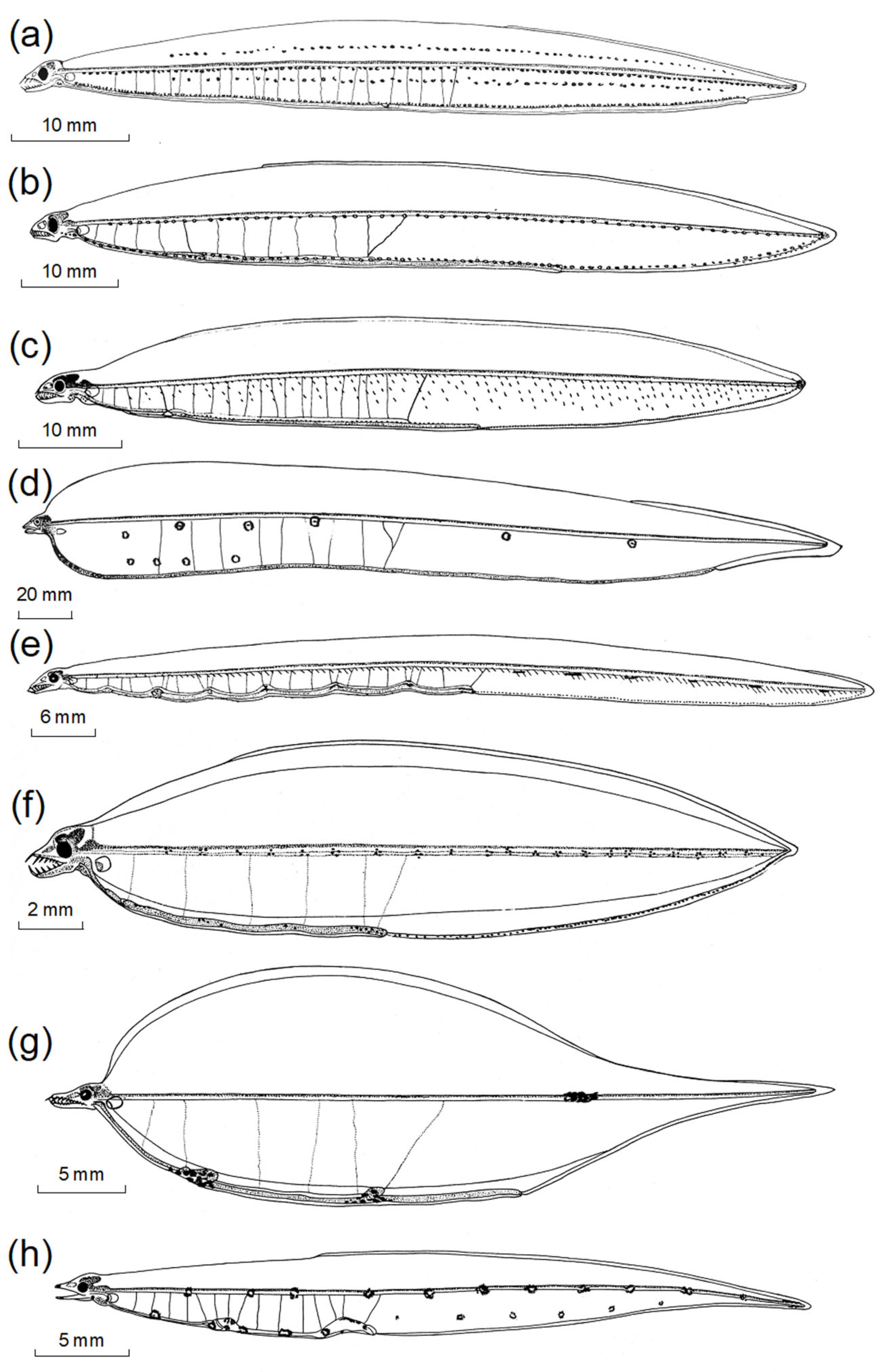
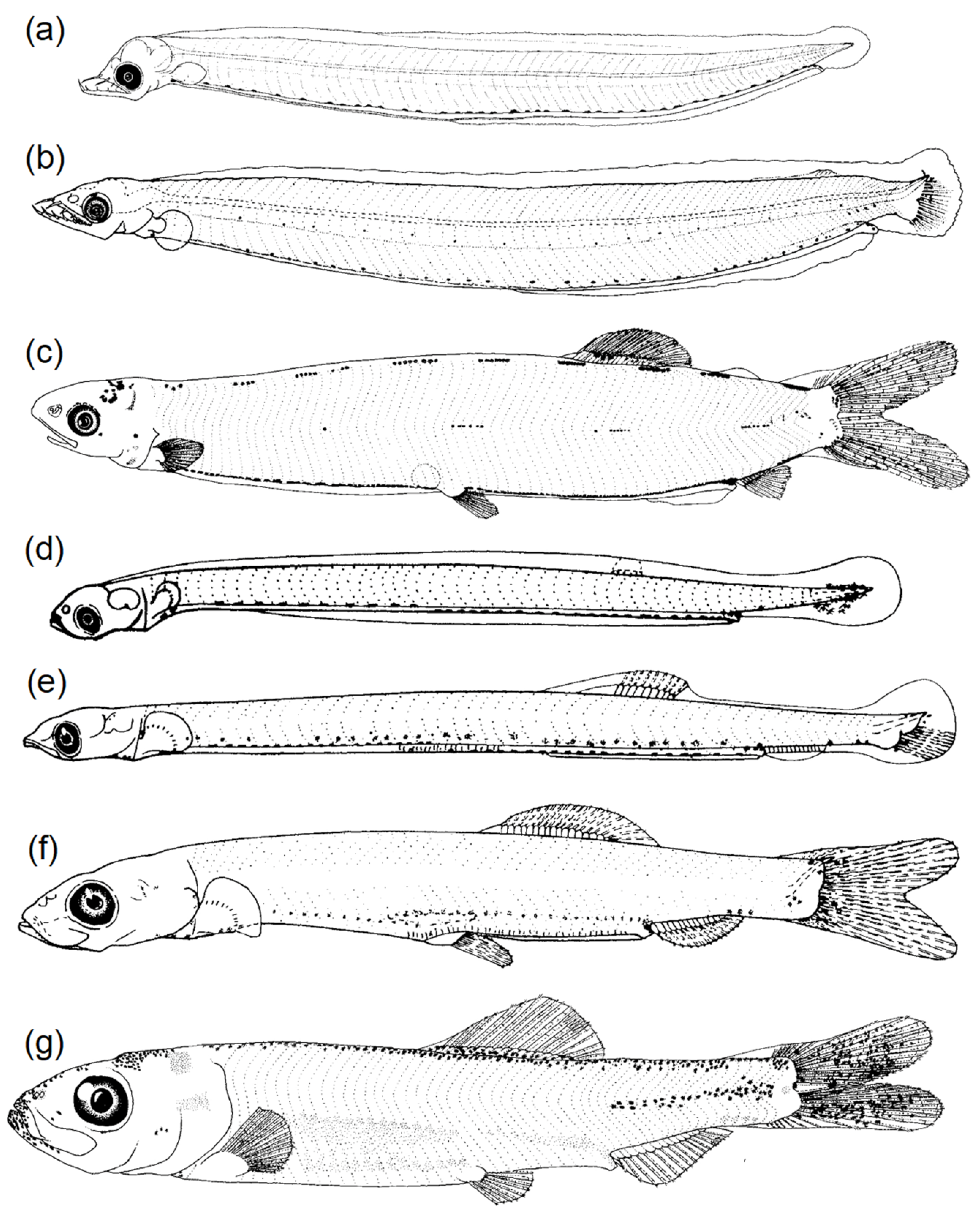
3.2.1. Body Shape and Morphological Structures

Although it might be easy to say that there are more differences between leptocephali and other fish larvae than there are similarities, some general patterns are present in both types of larvae. One obvious similar pattern between them is that they both predominantly hatch out into elongate body-shaped larvae (Figure 5, but with many exceptions for other fish larvae), which can then develop into a wide variety of body shapes. One pattern visible in Figure 2 is that some types of leptocephali such as Conger (1), some nettastomatids (17), and Nemichthys (21) retain a highly elongate body throughout the larval stage. This is also true for larvae of all sizes of the elopomorph families Elopidae (Figure 6b), Albulidae (Figure 6a,c; Charter and Moser [68]), and Notacanthidae [71,74]. For fish larvae, the consistently elongate larvae of the Clupeiformes, such as the Pacific herring (Clupea pallasii) (Figure 6d–f), seem superficially similar to those of the albuliform and elopiform leptocephali (e.g., Moser [37]). All leptocephali of the congrid subfamily Bathymyrinae, which grow larger (~150–300 mm) than most taxa of eel larvae, have generally elongate bodies (Figure 2(8)), and are more elongate for non-exterilium (external gut) types (Figure S2d), like the larvae of garden eels of the congrid subfamily Heterocongrinae (Figure 2(3), Figure 4b,c and Figure S2a) and some Congrinae (Figure 2, Figure 4a and Figure S2b). See Leis and Carson-Ewart [39] for a categorization of which families of coastal fish larvae have elongate bodies in relation to the type of gut structures (length; straight or coiled) or those that have deeper bodies.
Figure 5. Drawings of small western North Atlantic Congridae leptocephali ((a,b), modified from [14]) and representatives of 9 orders of fish larvae from the California Current region ((cl); modified from Moser [37]) showing various patterns of pigmentation, mostly along the margin areas of the body of (a) 15 mm Rhychoconger flavus, (b) 19 mm Pseudophichthys splendens, (c) 6.1 mm Pacific sardine Sardinops sagax (Clupeidae; Clupeiformes), (d) 9.8 mm Red brotula Brosmophycis marginata (Bythitidae; Ophidiiformes), (e) 6.1 mm California lizardfish Synodus lucioceps (Synodontidae; Aulopiformes), (f) 4.7 mm Sheephead Semicossyphus pulcher (Labridae; Labriformes), (g) 9.0 mm Pacific argentine Argentina sialis (Argentinidae; Argentiniformes), (h) 5.8 mm Benttooth bristlemouth Cyclothone acclinidens (Gonostomatidae; Stomiiformes), (i) 5.3 mm Northern lampfish Stenobrachius leucopsarus (Myctophidae; Myctophiformes), (j) 4.8 mm Spotted cusk eel Chilara taylori (Ophidiidae; Ophidiiformes), (k) 8.2 mm Rockweed gunnel Xererpes fucorum (Pholidae; Scorpaeniformes), and (l) 7.7 mm Speckled sanddab Citharichthys stigmaeus (Paralichthyidae; Pleuronectiformes).
Fishes 08 00548 g005
Other than the simple characteristic of a long/thin body, like many other fish larvae, leptocephali also have a wide variety of other body shape variations that include very deep bodies. Perhaps the deepest bodies are seen in the unusual larvae (Figure 2(4)) that grow to 300+ mm or larger, which were historically referred to as Thalassenchelys [75]. Those larvae were morphologically most similar (with some differences) to those of the Chlopsidae, but genetic sequences matched them with poorly known congrid eels of the genus Congriscus [76]. The saccopharyngiform larvae of the Cyematidae and Eurypharyngidae also have short and deep body forms (Figure 2(25,26)). The larvae of the Monognathidae that were thought to probably be cyematids (formerly called Leptocephalus holti [77,78]) have more intermediately deep bodies. In anguilliforms, there can be considerable variation in body shape within families and even seemingly at the genus level, such as in the congrid Gnathophis-types of leptocephali (Figure 2(2,5,6)); in the Muraenidae, there are both long- or deep-bodied species (Figure 2(12,13)). Nettastomatids also have three different types of body shapes (Figure 2(15–17), Figure 4g,h and Figure S2f).
It seems clear, however, that the apparent diversity in the body shapes and features of non-elopomorph fish larvae (Figure 5, Figure 6, Figure 7 and Figure 8) is considerably greater than is seen in leptocephali, perhaps for the simple reason that there are so many different types of marine fishes with quite different body shapes and ecological niches (e.g., [6,11,37,38,39,51]). Part of that impression is due to the fundamental differences between the two types of larvae. Leptocephali generally have simple guts with some swellings or loops as seen in many figures here; this is similar to some small elongate fish larvae, but other fish larvae develop more compact gut areas near the head, particularly in their later stages. Many larger fish larvae also develop larger pectoral and other fins, which do not form in any types of leptocephali. The long extensions of filaments and palps from fins (ornamental rays) or other locations or externally extending guts do not occur in leptocephali other than one recorded specimen of a notacanthid (Figure 7a), one recorded specimen of a Synaphobranchidae species (Ilyophinae) with a short rostral filament that had two black palps attached (Figure 7b), and the exterilum guts of one group of the Bathymyrinae (Figure 2(8), Figure 8a and Figure S3a). Other species types have rostral filaments (Figure 2(20)) that are quite long, branched, or have swellings/pigmented areas (Figure 9d,h and Figure 10a; [9,24,25,79]). At least one species has a very thick rostral filament compared to the size of its head (Figure 9d,h).
Figure 6. Drawings of elopomorph leptocephali and clupeid larvae showing: (a,c) 16.9 mm and 34.2 mm (transforming) bonefish Albula sp. (Albulidae), (b) 16.9 mm Elops affinis (Elopidae), and Pacific herring Clupea pallasii (Clupeidae) at sizes of (d) 10.4 mm, (e) 19.0 mm, (f) 23.8 mm, and (g) 29.5 mm (transforming). From Moser [37].
Fishes 08 00548 g006
The two types of fish larvae share similar types of eye structures, with most species just having large round eyes. However, some species have vertically elongated eyes with visibly spherical lenses. This occurs in some myctophid and stomiid larvae (Figure 8d–f), all synaphobranchid species (Figure 2(18–20), Figure 9g,h and Figure 10a,b,d) and the most frequently caught type of notacanthid larvae (Figure 10e). Figure 9g shows the large lens of an ilyophine leptocephalus very clearly. A few species of fish larvae, such as Idiacanthus (Idiacanthidae) and the Myctophum aurolaternatum (Myctophidae) shown in Figure 8b, have eyes on stalks, which do not occur in leptocephali.
Although there are several general similarities among the taxa of the two types of fish larvae that include the diverse pigmentation patterns examined in the next section, there is one very clear difference, which is the presence of bony structures in fish larvae and an almost total lack of ossification in leptocephali. The most obvious aspect of ossification is the formation of a wide variety of spines (Figure 7e,f) that occur on the heads and fins of some fish larvae, such as the Scorpaeniformes and Perciformes (see Table 1 of Leis and Carson-Ewart [39] for a list of orders that include some spination on the head or fins). The leptocephali of the genus Anguilla (e.g., A. japonica) do not have any ossification outside the jaw region until approaching metamorphosis [80], and red blood cells also do not appear until metamorphosis [81], so the constraints of extreme transparency in leptocephali have resulted in some unique features compared to other marine fish larvae.
Figure 7. Drawings of leptocephali and fish larvae (modified from Moser [37]) with various types of fin rays, other appendages, or spines showing: (a) a 314 mm notacanthid leptocephalus (Notacanthiformes) with a long caudal filament with black palps (from Moser and Charter [71]), (b) the head region of an unknown species of Synaphobranchidae leptocephalus (11 mm TL) with 2 black palps on a short rostral cartilage (redrawn from Miller and Tsukamoto [24]), (c) a 10.0 mm Gulf sanddab Citharichthys fragilis (Paralichthyidae; Pleuronectiformes), (d) a 7.4 mm King-of-the-salmon Trachipterus altivelis (Trachipteridae) that was dissected from an egg, (e) a 5.8 mm Tinsel squirrelfish Sargocentron suborbitalis (Holocentridae; Holocentriformes) with extensive mid-body pigmentation and large rostral, supraoccipital, and preopercular spines, (f) an 8 mm Rosy scorpionfish Pontinus sp. (Scorpaenidae; Scorpaeniformes) with head spines and large pectoral fins; and (g) 17.6 mm Dwarf lanternfish Loweina rara (Myctophidae; Myctophiformes).
Fishes 08 00548 g007

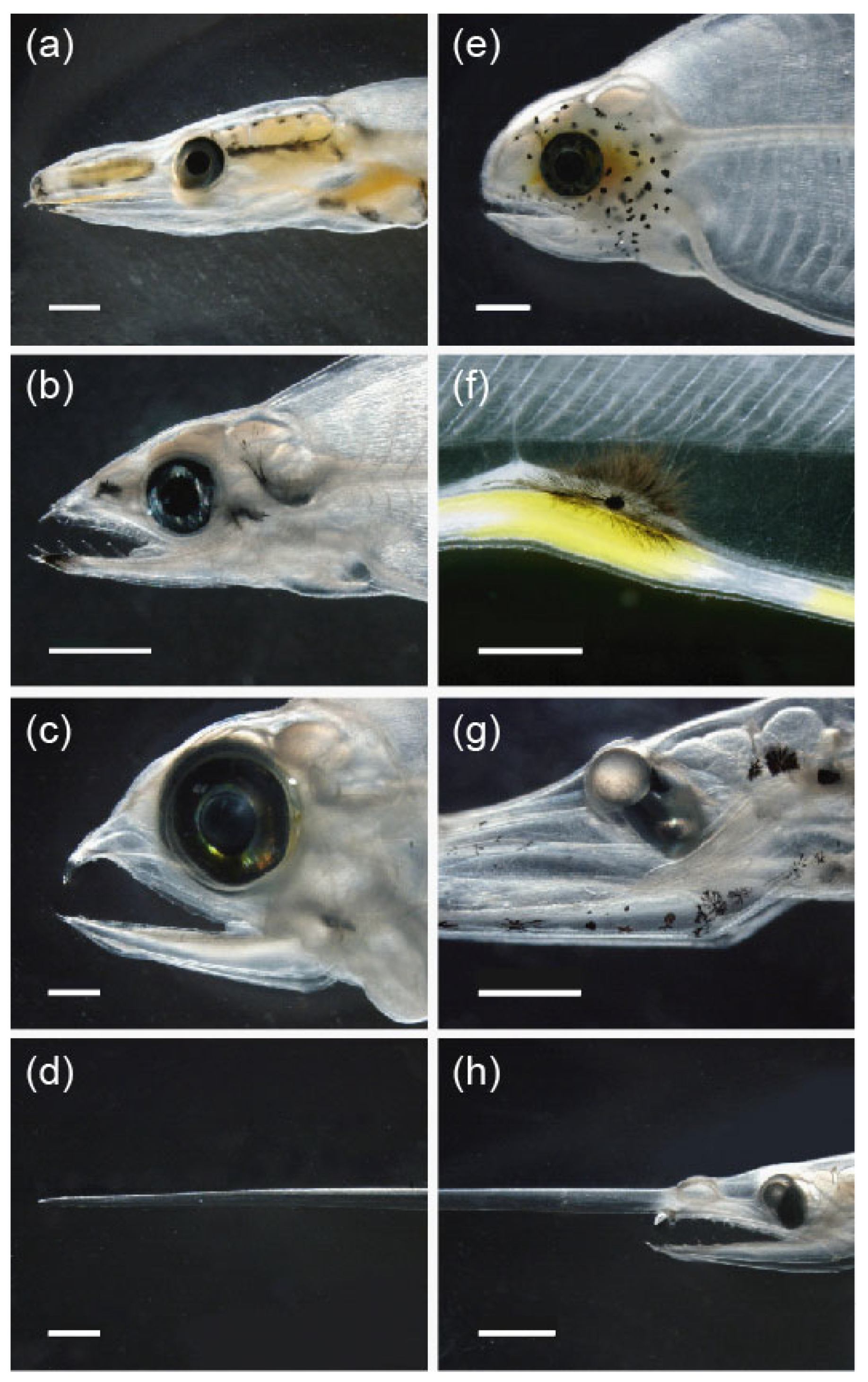
3.2.2. Diversity of Pigmentation

The pigment patterns of all kinds of fish larvae and leptocephali are their distinctive features in addition to the interesting variations in body structures. Moser [1] provided some insight into the pigment diversity of fish larvae, but it is also easy to visualize a larger perspective of the diversity in fish larvae and their diversity in pigmentation patterns by looking at the introductory sections of Leis and Carson-Ewart [39] and Fahay [14], for example, which show examples of each family. Looking through the entirety of those types of documents, including the easily available Atlas No. 33 [37], it is clear that various types of pigmentation occur in almost every part of the body of fish larvae (e.g., Figure 5, Figure 6, Figure 7 and Figure 8). It can occur as single melanophores or pigment patches evenly distributed in rows, such as on the ventral body margin/along the gut, in less regular patches, or in the form of many small spots mostly covering parts of the head or other areas.
Figure 8. Drawings of a leptocephalus and fish larvae (modified from Moser [37]) with various types of extended intestines (ac) or telescopic-type eyes (df) showing: (a) 101 mm congrid Ariosoma anale leptocephalus with an exterilium gut and 3 rows of myoseptal pigment on the side of the body (modified from Fahay [14]), (b) 25.8 mm Golden lanternfish Myctophum aurolaternatum (Myctophidae; Myctophiformes), (c) 34.7 mm Shining loosejaw Aristostomias scintillans (Stomiidae; Stomiiformes), (d) 5.2 mm Evermann’s lanternfish Symbolophorus evermanni (Myctophidae), (e) 15.2 mm Snaggletooth Neonesthes capensis (Stomiidae), and (f) 7.4 mm Slender lanternfish Hygophum reinhardtii (Myctophidae).
Fishes 08 00548 g008
Only a few examples of the variety of pigmentation of fish larvae are presented here (with an emphasis on the types of pigment that seem similar to those in some leptocephali), although most leptocephali largely lack a widespread, thick pigment that would prevent transparency. The general diversity in pigmentation of leptocephali in the WNA is clearly documented by Smith [12] (e.g., Figure S2) and in more extensive detail in each family chapter in Böhlke [13], such as Smith [82]. Blache [11] (reexamined by Miller and Robinet [54]; Figure 4 and Figure S3) presented many highly detailed illustrations of the species of leptocephali from the Gulf of Guinea of West Africa in the eastern South Atlantic. Tabeta and Mochioka [83] and Mochioka and Tabeta [84] documented the pigmentation of leptocephali of each family collected near Japan; in particular, they showed the detailed pigmentation patterns of the distinct species types of the Chlopsidae.
Figure 9. Photographs of freshly collected leptocephali in the western Pacific showing (a) internal yellow pigment in the head region (esophagus, brain, and nasal) of a 112.3 mm Facciolella (Nettastomatidae) leptocephalus, (b,f) internal head pigment in a 58.3 mm Moringua (Moringuidae) and yellow pigment and a large stellate melanophore on the gut loop near the end of the intestine of a 59.6 mm Moringua, (c) a 124 mm Congriscus (Congridae; formerly Thalassenchelys), (d,h) the unusually thick rostral filament (16.6 mm; middle part not shown) and head region of a 116.8 mm Ilyophinae (Synaphobranchidae), (e) a 77.5 mm Muraeninae with many head spots and internal yellow pigment (Muraenidae), and (g) the large eye lens and various shapes of melanophores of a 136.6 mm Ilyophinae. Lengths are TL. Scale bars are 1 mm except in (h) (2 mm; note different scales in (d,h)).
Fishes 08 00548 g009
The complicated but limited range of pigmentation patterns of Indo-Pacific Muraenidae leptocephali was examined more clearly for the first time in Mochioka and Tabeta [84]. Muraenidae leptocephali typically have no lateral pigment, but they generally have small head (Figure 9e and Figure S1f), gut, and body margin pigment spots in various species. In the Indo-Pacific, however, most types of leptocephali have not been matched with their adult species (see Miller and Tsukamoto [24,85]), in part because of the much higher number of eel species present there, which was shown by a recent analysis of catches of leptocephali in different regions [73].
Figure 10. Photographs of freshly caught leptocephali of Synaphobranchidae (ad) in which all species have the telescopic-type eyes with large spherical lenses shown in (d) (Synaphobranchinae; 57 mm), and some Ilyophinae have rostral cartilages ((a), 31 mm TL) or large hourglass-shaped melanophores laterally or dendritic ones on their gut loops ((b,c); 61.9 mm). The Tiluropsis type of notacanthid leptocephali ((e,f), 236 mm) has a less distinct type of telescopic type eye and caudal filaments ((f), 12 mm filament) that may frequently break off but in one unique case were preserved for one species (Figure 7a), although there is no evidence that all species have such ornate filaments. Lengths are TL. Scale bars are 2 mm except in (d) (0.5 mm).
Fishes 08 00548 g010
The line drawings shown here illustrate many of the types of pigmentation of leptocephali, and Miller and Tsukamoto [24] showed photographs of the general pigment types, but whole-body photographs such as those in Figure 2 do not show pigmentation as clearly as in line drawings such as Figure 4, Figure 11, Figures S2 and S3. Leptocephali do not have such a diverse variety in arrangements of pigment as the many types of fish larvae, but pigment frequently occurs on the gut region of many families, in one row (or up to three rows) of pigment spots on the midline (some Congrinae and Heteroconger in Figure 4a,b and Figure S2a,b and in some Chlopsidae in Figure 4f), or in large stellate melanophores such as those in Figure S3c,d. Bathymryinae and some ophichthid leptocephali have myoseptal pigment in the form of short vertical rows of small spots on the borders of the myomeres (muscle segments) (Figure 4e and Figure S2d). Heterocongrinae leptocephali of Gorgasia also have various patterns of myoseptal pigment (Figure 4c) below the midline (see Castle [86] and Smith [87]). Myoseptal pigment is also present in the larvae of the Melanostomiidae in the form of a single row in the middle or lower parts of the lateral body surface or on all the myomeres in various species [88]. Ilyophinae leptocephali can have highly complex melanophores that have hourglass-shaped, stellate, or dendritic structures (Figure 9g and Figure 10a–c)
Figure 11. Drawings of the head (left) and end of gut/last vertical blood vessel (right) regions of leptocephali of (a,b) 99.5 mm and 82.2 mm Ophichthidae from Charter [89], (c) an 88.7 mm nemichthyid, Avocettina bowersii (Charter [90]), and (d) a 31.8 mm nettastomatid, the Dogface witch eel Facciolella gilbertii (Charter [91]). Both (c,d) have small melanophores on parts of the dorsal and ventral body margins, and the ophichthids (a,b) have very low “gut loops” and small melanophore pigmentation that does not appear to be expandable chromatophores like those on top of the very high gut loops shown in Figure S4f,i.
Fishes 08 00548 g011
There are several obviously similar pigment patterns between the two types of larvae. One is the occurrence of evenly spaced spots along the gut or dorsal and ventral body margins, and these types of spots can also form on the midline (Figure 5 and Figure 6), although in some mesopelagic fish larvae these apparent spots may be early photophores (e.g., Moser and Ahlstrom [63]). Larger pigment patches composed of many small melanophores are another shared feature. A few species of leptocephali have pigment spots evenly distributed over the whole body, such as in the chlopsid genus Kaupichthys (Figures S2e and S4b,c), a few ophichthids, and the garden eel species Heteroconger hassi [86].
Regardless of the similarities and differences between the two types of larvae, which are difficult to comprehensively compare in a single paper, it is clear that pigment patterns appear in most types of fish larvae and leptocephali. Curiously for eels, the leptocephali of Anguilla are the only family that has no pigmentation except for some tail pigment in preleptocephali, which later disappears. Only rarely do other fish larvae lack pigment, with the Viperfishes (Chauliodus species) after yolk-sac absorption or the Stareye lightfish (Pollichthys mauli) as examples [92,93].
Various types of examples of the pigmentation of both types of larvae are shown here as if only melanophores are present (black color). However, Baldwin [5] has shown that some freshly caught fish larvae have chromatophores that include other colors that are lost during formalin fixation [4]. Some leptocephali also have a yellow pigment on some body structures [26], as also noted by Baldwin [5]. After observing many freshly caught leptocephali, it is clear that only a few taxa have other colors such as yellow. Some ophichthids (Neenchelys), nettastomatids, and Moringuidae have yellow in parts of their esophagus or intestines, and a few muraenids have yellow in the head region (e.g., Figure 9a,e,f), and some species such as certain Serrivomeridae have yellow on the top of their eyes [25]. But one rare species only reported once in the literature with normal looking black spots on the gut, presumably after being fixed in formalin, was found to have bright orange spots on the gut and to be the larvae of Neocyema, which was considered as a new family of saccopharyngiform fishes (Neocyematidae; Poulsen, et al. [78]).

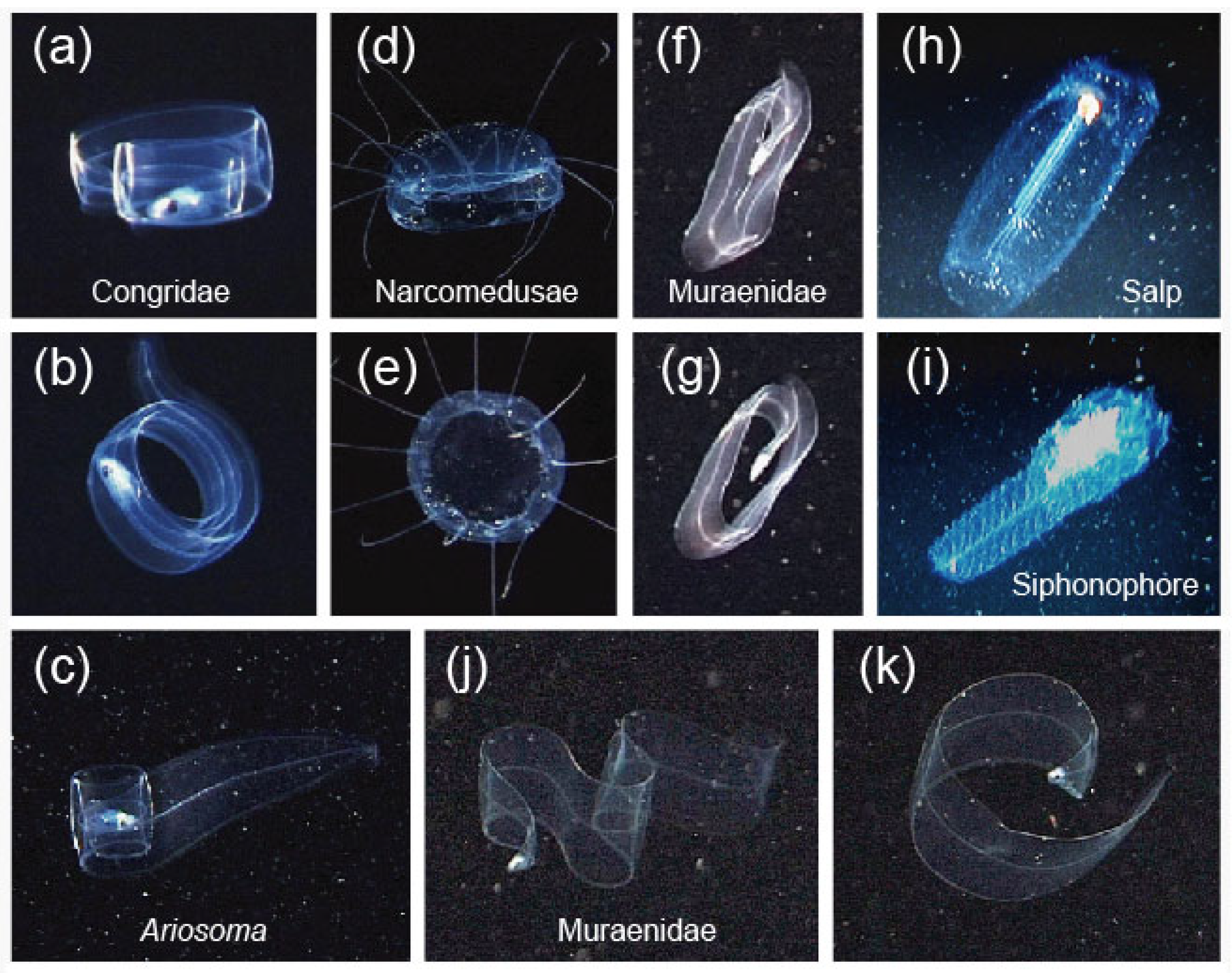
3.3. Mimicry to Reduce Predation

Just as spines that occur in fish larvae are thought to reduce attempts at predation by causing problems or discomfort during ingestion by predators, several morphological and behavioral features of fish larvae and leptocephali seem to be shaped to reduce predation by inhibiting predators from attacking them. Mimicry based on predators avoiding a dangerous or noxious animals (models) is termed Batesian mimicry [41,94]. Mimicry has been documented in a variety of marine fishes. Clear examples of Batesian mimicry are the species of black-and-white-striped ophichthid eels that mimic the color patterns of highly poisonous sea snakes [95]. Some fish larvae appear to have evolved structures that mimic abundant GZ species [1,41] because most fishes do not eat GZ (e.g., large pelagic predators in the Arabian Sea [47]) and may actively avoid consuming them [96] even if some species are specialized to eat them [45,46]. Gelatinous zooplankton have a low nutritional value [41,96,97], and some fish were found to reject fresh tissues of cnidarians and ctenophores, probably due to the presence of active nematocysts [96].
As was prominently featured by Moser [1], fish larvae across a range of taxa have fin, gut, and other extensions from the body that can include pigmented palps (Figure 7 and Figure 8) that seem likely to be examples of mimicry of GZ. A variety of ideas have been considered for what the function of these structures of different origins (dorsal/anal/pelvic fin rays, caudal filaments, etc.) might be [1,4], and they may have multiple functions in addition to Batesian mimicry that include sensory detection or buoyancy, as discussed by Govoni et al. [98] and Greer et al. [41].
Many of the long fin-ray filaments with palps, such as those seen in some species of pearlfish of the Carapidae (studied by Govoni et al. [98]), almost perfectly resemble the tentacle structures of some siphonophores or ctenophores. There is a wide diversity in highly complex body and tentacle structures of siphonophores that include long arrays of retractable tentacles and prey capture systems called tentilla [42], and photographs are now available of the siphonophores, other GZ, and the fish larvae that have structures that appear to mimic parts of their tentacle arrays. A very colorful carapid species shown in Figure 12e has a relatively short vexillum (the extended dorsal fin ray with palps; [98,99]) compared to another carapid that has a longer vexillum and also a long caudal filament (Figure 12g). Other carapids have long fin extensions with fewer or less regularly spaced palps [4], and Sakaue [3] showed a carapid with a vexillum with bright pink and yellow palps.
Figure 12. Photographs of fish larvae with external body extensions in comparison to siphonophore tentacles showing: (a) a cusk eel (Lampogrammus shcherbachevi) larva (Ophidiidae), (b) the main body and tentacle array of a Forskalia tholoides siphonophore, (c) an Agalma okenii siphonophore (Agalmatidae) and its tentacles with orange-colored gastrozooids, (d) a siphonophore of Naomia bijuga photographed on the West Coast off California by Kevin Lee, (e) large palps on a dorsal extension in an orange-colored pearlfish larva of the Carapidae photographed in the Philippines by Ram Yoro, (f) an IKMT-collected species of Liopropoma (Serranidae, Epinphelinae; also see Figure 46 of Baldwin [5]) with long first and second dorsal fin ray extensions before preservation, (g) a pearlfish larva (Carapidae) with a dorsal filament and palps and a caudal filament, (h) Bargmonnia elongata (modified from Mackie et al. [100]), and (i) Naomia bijuga photographed during the day in Welcome Bay of Pender Island of the Gulf Islands of British Columbia in the Salish Sea by Karolle Wall. Photographs in (ac,g) were taken at Roatan Honduras by Mickey Charteris.
Fishes 08 00548 g012
A search of images in published papers or for online images suggests that examples of likely models for the distinct palps on the dorsal vexillum fin-ray filaments of carapids are the evenly spaced gastrozooid structures (from which smaller tentacles and tentilla extend out) of the common physonect siphonophores, such as Nanomia bijuga (Figure 12d,i; [101,102,103]). Other siphonophores have many tentacles with gastrozoods that look like the carapid palps, such as an Agalma okenii (Figure 12c). Interestingly, some types of ctenophores, such as a Pukia falcata shown by Gershwin et al. [104], have evenly spaced palp-like structures with retracted tentilla that also look like the structures seen in carapids and Nanomia siphonophores, and various taxa of ctenophores have long tentacles (see Gibbons et al. [105]). The widely present calycophoran siphonophores also have many gastrozoids on a single tentacle that occur at various spacings [42], and these small types of GZ (1–3 cm) can often be quite abundant (e.g., Luo et al. [106], Lüskow et al. [107]).
Other fish larvae, including the notacanth leptocephalus with a long caudal filament shown in Figure 7a, have various extensions with pigments palps (Figure 7), as discussed by Moser [1] and others. These occur in unrelated families such as the Myctophidae, Trachipteridae, Lophotidae, Paralichthyidae, and Ophidiidae (see citations above). There may also be color factors involved because some siphonophores have the same colors [42] as the fish larvae (e.g., Baldwin [5]) that appear to be using mimicry, such as the carapid shown in Figure 12e. Color may also be important in some whalefish larvae (Cetomimidae) that can have colored gelatinous-appearing bodies [5] and remarkably long caudal filament tail streamers that can extend far behind the fishes [108].
Perhaps the most undeniably clear example of fish larvae mimicking siphonophores are some species of cusk eel larvae of the genus Lamprogrammus (Ophidiidae). A larva that appears to be L. shcherbachevi (Figure 12a), which is known from the Atlantic, Pacific, and Indian oceans [109,110,111], has a “free trailing intestinal loop” as described by Moser [1] for a similar species of Lamprogrammus that he illustrated (see Mundy and Hilton [7]). When living, that loop can be seen to have many palp extensions on the ventral margin, and it also has a filament extension with many small pigmented palps that look almost identical to the siphonophore gastrozooids shown in Figure 12c, but even the overall body shape with the exterilium gut and filaments looks more like a siphonophore than a fish larva. It also has a fan-shaped array of extensions above the head that appear jellyfish-like (Figure 12a), perhaps making the overall appearance of the Lamprogrammus larva function like a “supernormal stimulus” (see Haddock and Dunn [112]) of a dangerous type of stinging-cell gelatinous animal. Other species of the Ophidiiformes have large intestinal loops that are apparently filled with semitransparent areas of expanded liver and do not have any palps to mimic siphonophore tentacles [4,5]. However, those ophidiiform larvae with two separated body regions might generally look like various species of lobate ctenophores that have been photographed [104,105].
The large palps and more ornate structures of the serranid species of Liopropoma (Figure 12f) of the Epinephelinae and other ornate structures might suggest a different type of Batesian mimicry, however. Kendall et al. [113] showed a remarkable example of dorsal spines of an 11 mm Liopropoma larva with complex ornate structures that might be mimicking jellyfish or siphonophore tentacles. Other species of Liopropoma just have large colored palps [4,5] such as those seen in the net-caught larva shown in Figure 12f. Interestingly, Keene and Tighe [114] showed a remarkably similar ornate structure to that of the Liopropoma larva of Kendall et al. [113] except that it formed from pelvic fin rays in a 15.3 mm stephanoberyciform (Gibberichthys pumilus), which represents a remarkable example of convergent evolution to produce similar ornate structures.
Compared to the amazing diversity in structures seen in other fish larvae and the possible list of types of species they might be mimicking, leptocephali have relatively few types of external structures that may function to mimic the tentacles of GZ. One type of structure that is seen in fish larvae (e.g., some myctophids and stomiids; Figure 8b,c) that also occurs in one group of leptocephali is a simple tubular exterilium gut extending outside the body. Leptocephali of the congrid subfamily Bathymyrinae are separated into two types that have slightly different body shapes [115,116,117], but the deeper-bodied forms all have exterilium guts even at sizes of 5.4 mm just after hatching (Figure 2(8), Figure 8a and Figure S3a). These extend outside the body and may have resulted from Batesian mimicry to appear like a tentacle (although they may also have enhanced digestion functions). Whether the simple straight rostral filaments of the Ilyophinae (e.g., Figure S2g) are also related to mimicry (like the one example of having two palps (Figure 7b) or the one illustration of a multi-branched filament [9]) remains to be evaluated. It seems possible that these rostral extensions might mimic the individual tentacles of some ctenophores or jellyfish. More extensive large net sampling in deeper layers might collect more of those larvae, as indicated by collections in the Gulf of Mexico [118], which would help to understand the diversity in and possible function of the rostral filaments in the Ilyophinae.
The mimicry of GZ by leptocephali and possibly other fish larvae also has a behavioral component, however, which includes shape-changing in response to potential threats or using a specific body orientation. The behavioral tactic of shape changing has been reported in various type of marine animals and is thought to be a form of Batesian mimicry to reduce predation by appearing similar to round GZ [40]. Other than a brief mention and drawing of a curled captive metamorphosing leptocephalus shown by Dean [119], the first modern report of shape-change behavior in eel larvae may have been when a diver-caught chlopsid leptocephalus was video-recorded curling its body up into a round coil in a small aquarium onboard a research ship in the Atlantic more than 20 years ago [26]. The extent of the use of this behavior then became clear when scientific divers J. Finn and M. Norman recorded a high percentage of the leptocephali that were observed at Osprey Reef (western Coral Sea near northeastern Australia and the Great Barrier Reef) curling into rounded or flat shapes or changing their swimming style [28]. A possible Conger leptocephalus (Figure 13a,b) was video-recorded to make a perfectly round coil shape [28], and it tried to continuously maintain that coiled shape for about 30 seconds. (https://www.youtube.com/watch?v=ok8MW4pdPXI; accessed on 8 October 2023). A chlopsid leptocephalus formed a similarly rounded shape (see same video link), two muraenids formed more flattened coils (Figure 13f,g), and an Ariosoma formed a coil with the front part of the body with the tail extending out (Figure 13c). A large muraenid formed a partial coil and then swam slowly with its body in a highly curved/convoluted swimming motion (Figure 13j,k). That study showed the shape-change leptocephalus images in comparison with jellyfish, siphonophore, salp, and ctenophore body shapes that were also filmed at Osprey Reef (Figure 13; Miller et al. [28]).
Blackwater divers have subsequently recorded multiple examples of tightly coiled leptocephali at Palau (Sakaue [3]; also see Figure 4a of Miller and Tsukamoto [25]) and the Philippines (R. Yoro; Figure 3b). The coiling behavior of leptocephali was also recorded by an ichthyoplankton imaging system [41]. Robison [40] described other examples of shape-change behavior in marine animals, such as eelpouts coiling their bodies (presumably to mimic GZ to avoid predation), and Sakaue [3] showed an example of a similarly coil-shaped small pelagic bluestriped fangblenny, Plagiotremus rhinorhynchos (Blenniidae), which is a species that mimics cleaner fish as adults, behaving the same way as the eelpout shown by Robison [40]. Most remarkable perhaps is that the elongated ctenophore, the Venus girdle (Cestum veneris), which basically looks like a leptocephalus with no head when it swims while sometimes using an anguilliform swimming style, also has a curling behavior to form partial coils (see Gibbons et al. [105]; Figure 6A), and a blackwater diver video shows that they can curl up into a very tight coil like those seen in some leptocephali (https://www.youtube.com/watch?v=IEKTbX20byM″Cestum veners|Venus girdle—YouTube; accessed on 8 October 2023).
The gastrozooids of siphonophores can appear as dense objects when extended or retracted (Figure 12; [42,101,102,103]), so expandable chromatophores of some leptocephali [27] may also be mimicking clusters of nematocytes, as illustrated in Miller and Tsukamoto [25]. Blackwater divers have documented a previously unknown aspect of the pigmentation of some Ophichthidae leptocephali, which is that some of their pigment patches on their gut loops are actually expandable chromatophores (Figure 3c) that appear to have a greenish color when they reflect bright light from diver lights [27]. This was first realized when a video recording by M.J. D’Avella clearly showed an ophichthid leptocephalus almost seeming to be performing a threat display by swimming in an aggressive posture while paired green chromatophores on top of its gut loops frequently expanded and contracted (https://australian.museum/learn/animals/fishes/ophichthid-leptocephalus-/; accessed on 8 October 2023), showing that they were clearly under the control of the nervous system [27]. That video was recorded off Kona Hawaii, but still photographs of a similar ophichthid leptocephalus were taken by R. Yoro in the Philippines more recently (Figure 3c).
Figure 13. Photographs of various types of shape-change behaviors of leptocephali and several types of GZ at Osprey Reef offshore of the Great Barrier Reef in the Coral Sea near Australia showing: (a,b) a curled Congridae (possibly Conger) leptocephalus, (c) an Ariosoma leptocephalus curling only the front part of its body, (d,e) a jellyfish of the order Narcomedusae, (f,g) a Muraenidae leptocephalus making elongate flattened coils, (h) a salp (order Salpa), (i) Forskalia (order Siphonophora) with its tentacles all retracted, and (j) a deep-bodied Muraenidae using highly curved-body swimming and also forming a partial curl (k). Photographs were taken by Julian Finn and Mark Norman and were modified from Miller et al. [28].
Fishes 08 00548 g013
Smaller patches of pigment on the guts of some ophichthids may not be expandable (Figure 11a,b and Figure S5) compared to the larger patches on other species (Figure S4f,i), but that is not yet studied. However, when exposed to bright light at night by divers, these chromatophores have the same greenish color even if they are not expanded, as shown by M. Charteris at Roatan, Honduras (Figure 3d,e). In fact, ophichthid leptocephali are the most diverse according to the number of eel species (see Table 1 of Miller et al. [56]) and due to having a wide range of gut morphologies and pigmentation patterns that includes the three subfamily level groups of the Ophichthinae, Myrophinae [120], and the Neenchelys type [121], which are distinguished by the number and structures of the gut loops/liver swellings on the anterior part of the gut. If leptocephali such as ophichthids with gut swellings also perform curling behaviors, the swellings might mimic the clusters of retracted tentacles of jellyfish and siphonophores as the expandable chromatophores might do.
The diversity in the morphology in ophichthids and other taxa of leptocephali indicates that much remains to be learned about the structure and function of their pigmentation and chromatophores because the era of gaining knowledge from underwater photography is relatively new. Historically, fish larvae were typically collected by plankton nets, and fragile body parts such as long filaments became damaged or broken, or large gelatinous body-margin or gut areas contracted after formalin fixation [4]. With more recent methods, some have been collected intact (e.g., Figure 7), and divers have photographed many others ([3,4]; see online imagery). Nonaka et al. [4] showed that the structures of the sometimes incredibly ornate sets of filaments, palps, or gelatinous areas do not always remain intact after formalin fixation, which explains how the full details of these structures were not well understood until recently when divers photographed them and began interacting with ichthyologists.

4. Discussion

4.1. Reasons for Similarities and Differences between Fish Larvae and Leptocephali

Leptocephali and other fish larvae live in the surface layer of the ocean, so they experience similar or at least overlapping environmental conditions. Leptocephali are mostly found in the upper 100 m at night in offshore areas, and at least some larger species vertically migrate to deeper depths during the day within approximately the upper 250–300 m [25,26,122,123,124,125], but sometimes they may be most abundant in approximately the upper 10 m layer [126]. The depth distributions of other types of fish larvae are mostly in the upper 150 m (depending on the species and hydrographic conditions), and they have been studied more than for leptocephali (e.g., Moser and Pommeranz [127] and Rodriguez et al. [128]). Leptocephali and all types of fish larvae have large eyes that are likely adapted for visual feeding or predator avoidance in low light levels down to about 200 m deep [129,130,131].
These shared environmental conditions seem to have resulted in both types of larvae having some similar features. Fish larvae and leptocephali both have a variety of body shapes and diversity in pigmentation patterns, and some species of both types have telescopic eyes or even long body appendages. This diversity in morphology fascinated HGM and was nicely reviewed for fish larvae by Moser [1] and in many publications that followed. The reasons for such diversity are obviously complex and related to numerous biological and ecological factors determined by the ocean environments and biological communities where the larvae have lived during their evolutionary history. The factors also would include developmental constraints related to body shapes of the growing larvae that need to efficiently transition into the juvenile and adult stages after maximizing survival as larvae. For some species, ornate structures form very early in development and in some cases possibly before hatching, such as in the bathymyrin leptocephalus, which has an exterilium gut present before yolk absorption or feeding (Figure S3a). More clearly, a 7.4 mm King-of-the-salmon had dorsal and pelvic fin ray extensions that were already longer than its body (Figure 7d) that formed while still an embryo inside its egg [132]. This underscores the importance of avoiding predation even at the earliest stages of both types of larvae. The wide variety of structures that have evolved to visually deter predation often last until the juvenile stage begins or disappear even sooner.
For leptocephali, however, they undergo an extreme form of metamorphosis at the end of their larval stage, during which they change from highly transparent, laterally compressed larvae to totally different eel-like (anguilliforms) (Figure 1) or fish-like (elopomorphs) bodies. Other fish larvae also clearly have many distinct structures that are present in the larvae but disappear before the juvenile stage.
The biological and physiological factors that may influence the morphology of marine teleost larvae are outside of the scope of this paper, but the presence of morphological and pigmentation features that may have been shaped by natural selection to help to reduce predation appears to be a common feature between both types of larvae. A typical pattern in many leptocephali are rows of small spots or larger pigment patches along the body margins, and many types of fish larvae also have these. Moser [1] pointed out that these serially arranged melanophores on the gut might help conceal the larvae by masking their gut contents. Another possibility is that spots along the body margins in many species of leptocephali and fish larvae might help to break up the body outline making it harder for predators to see them due to the alternation of light and dark edges.
Another shared pattern among some elongate species of both types of larvae are the long lines of pigment spots (or expanded melanophores in leptocephali) on the midline of the bodies, which might be related to what occurs when the larvae are swimming. During swimming, the body undulations cause the spots on the midline to move rapidly in many different directions, causing a chaotic image referred to as “flicker fusion” [1,133]. This might make it difficult to see a larva swimming laterally in front of a predator. The long linear shape of some leptocephali and their anguilliform swimming style might make the flicker fusion effect even more pronounced. Even one large spot on the body, such as that in Nettenchelys (Figure S2f) and Nettastoma (Figure 4g), might have that kind of effect. Having more lateral expanded stellate melanophores, such as in Moringua edwardsi (Figure 2(14)), Xenomystax congroides (Figure 4d), and Hoplunnis punctata (Figure 4h) might create a very complex flicker fusion effect, as would the various pigment patterns of the small fish larvae in Figure 5.
For leptocephali, which grow to large sizes, there may also be different predation pressures on the small and large individuals that have resulted in developmental changes in pigment patterns. For example, small Ariosoma leptocephali have relatively large spots on both the dorsal and ventral margins of the body [134], but those spots disappear in the larger larvae (Figure S2d), suggesting they may be more important in the small larvae of that genus than for larger ones. It also seems that the larger types of individual melanophores on the lateral body surface may frequently be larger relative to the body size in the smaller leptocephali. This seems true for nettastomatids (Figure 4h and Figure S3d), nemichthyids [134], Ilyophinae [135], and at least a few ophichthids [120]. That suggests that there is a greater importance of pigmentation during the early larval period, possibly because the range of predators is reduced as the leptocephali become larger. For example, small leptocephali, which might experience some predation in the open ocean by the highly abundant myctophids [136,137], would eventually grow and become larger than many of those predators, thus reducing the predation pressure by some of the most abundant fishes in the oceanic communities.
Both types of fish larvae have also evolved body extensions as described above. However, there are three taxa of leptocephali that have body extensions that seem to facilitate mimicry (notacanthids with caudal filaments, Ilyophinae of the Synaphobranchidae with rostral filaments, and bathymyrins with exterilium guts), and the diversity in taxonomic groups and types of body extensions is much greater in other teleost fish larvae, as shown in part here. One possible reason for this is that the body shapes of leptocephali are constrained by needing to support the GAG pouch that supports the overall body structure [10,18,26]. The bodies of all leptocephali have rounded body edges and very reduced fins [12,13]. Many fish larvae with long fin or body extensions are more heavily ossified than leptocephali and likely gain more of their propulsion from the caudal fin with fewer body undulations. In general, the soft fragile bodies of leptocephali have no structural support for long fin extensions.
What is also suggested is that the leptocephalus body form has been shaped with extreme transparency as a major feature that allows them to grow to large sizes. The result of this is a larval form that is very different from many of the wide array of fish larvae, including those with defensive spines, and ossification occurs much earlier in fish larvae than in leptocephali, which only begin ossification (aside from the jaws) after metamorphosis.

4.2. Predation on Fish Larvae as a Selective Force

As mentioned by Lasker [138] in the same 1981 book that contained the Moser [1] chapter, one of the driving forces to extensively study fish larvae through net sampling in the ocean was the hypothesis of Hjort [139] that the fluctuations in the year-class strengths of the great fisheries species of Europe might be determined by the larval survival of each cohort from a spawning season based on their feeding success. Lasker [138] discussed this issue, but not much was known about the effects of predation at that time [32]. Many aspects of the ideas proposed by Hjort [139] and their importance to fisheries science up to now were examined in a previously published special issue of papers [140] and will not be re-examined in detail here.
It is presently unclear how important predation is to the survival of leptocephali compared to other sources of mortality. Leptocephali are extremely rare in predator fish gut contents, but they also seem to be easily digestible [141]. However, in addition to reducing predation, after some growth, leptocephali may be more resistant to starvation than other fish larvae, because their bodies contain ample energy (GAGs) to enable survival during low food periods. It has been hypothesized, however, that the amount and quality of marine snow available to the first-feeding larvae could determine the survival rate from each spawning event [142,143]. This is the “critical period” concept in fisheries science (e.g., Robert et al. [144]) that proposes that the first-feeding larvae are the most vulnerable to starvation and that many may die from starvation or predation at that time.
As described above, many common aspects of teleost fish larvae and leptocephali appear to be related to reducing predation to increase larval survival. These features include pigmentation to reduce visibility, body structures that appear to mimic GZ, and the shape-change behavior of leptocephali. Some elaborate spines in fish larvae may increase the gape limitation for predators but may also serve to mimic heavily spinous and low-nutritional-value crustaceans [145]. The ocean is full of these types of diverse predator–prey interactions that are similar to those seen for fish larvae. For example, some small, slowly swimming siphonophores may have tentacles that seem to mimic copepods to attract and capture small fish larvae [145]. A squid species appears to have evolved a juvenile stage to mimic the Naomia siphonophores mentioned above by developing a long streamer [146]. Cnidarians such as hydromedusa may use fluorescent structures to attract fish larvae prey [112]. In addition, some siphonophores are even able to capture and consume leptocephali [147], and the highly abundant, small calycophoran siphonophores [107] frequently contribute detrital materials to the marine snow that leptocephali consume [30,148,149,150], so the evolution of siphonophores and fish larvae appears to be intertwined.
Other than the use of spines, leptocephali seem to have evolved similar predation-reduction strategies such as having pigmentation to reduce visibility to predators or to make it hard for them to be clearly seen through the flicker fusion effect. Many fish larvae may be relatively transparent at small sizes, but leptocephali have transparency as one of their primary adaptations to avoid predation. Reducing visibility to predators through transparency occurs in a wide range of marine animals [151,152,153,154], but for leptocephali, which reach maximum sizes usually of about 60–300 mm, it appears especially important. The extreme transparency is achieved by the unusual body structure of the internal gelatinous pouch that contains clear energy-storage materials and a high percentage of water [155].
Although it is easy to suggest that unique larval features of leptocephali and other teleosts are attributable to being adaptations to reduce predation, the metabolic costs of producing and reabsorbing them may have prohibited their evolution in many taxa. Simpler features such as pigmentation may be enough to reduce predation mortality in many species. Similarly, for leptocephali that use curling behavior, there may be tradeoffs between having protection from predators that avoid GZ and their mimics (coiled leptocephali) and having vulnerability to predators that consume GZ, such as some tunas, lancetfish (Alepisauridae), or ocean sunfish [141,156].

4.3. Future Research on Fish Larvae

H. Geoffrey Moser was part of a generation of scientists who participated in an era of scientific advancements of discovering the amazing morphological diversity of fish larvae and which larvae belonged to which species of fishes around the world. Since their work, DNA identification methodologies have rapidly increased (e.g., Richardson et al. [157] and Hubert et al. [158,159]) and have offered some alternatives for at least partial identifications of some types of fish larvae, including leptocephali [76,160,161,162]. However, not enough species have been sequenced yet [4] for genetic identifications to replace the detailed knowledge passed on by the individuals who established a body of literature to teach future generations of scientists how to distinguish the larvae of the many different species of teleost fishes via their morphology. In fact, teaching larval fish identification was one of the important components of the career of HGM, as outlined by Mundy and Hilton [7].
Great progress was made to work out the species identities of the leptocephali in the WNA [12,13,120], but the much greater diversity of marine eels in the Indo-Pacific has prevented major progress from being made on matching most leptocephali with individual species [24,85]. Part of this is due to the need to have specimens of intermediary transitionary forms during late stages of metamorphosis that have some characteristics of both the leptocephali and the juveniles that can provide essential clues to match the larvae to their adult species. Collections of these late stages may be lacking partly because even though much sampling has been conducted for leptocephali in the Indo-Pacific [25], most of those collections were made over deep water in search of anguillid spawning areas, where metamorphosing leptocephali are rare. Another factor is that even though many leptocephali are now available for DNA sequencing from the Indo-Pacific surveys, there are probably many adult marine eel species that have not been genetically sequenced to enable matching with leptocephali. This represents a major new research frontier for future research, such as that of Ma et al. [160,161,162], Kurogi et al. [76], and Poulsen et al. [78].
Even more difficult than obtaining specimens of marine eel species that are often hard to find or dangerous to handle for DNA matching is learning about the behavioral predator–prey interactions of leptocephali and other fish larvae with their potential predators. Divers with cameras and lights represent highly unnatural examples of a predatory threat, so how to avoid that bias to learn more about how the morphology and behavior of these larvae function to reduce predation seems uncertain. However, underwater observations by blackwater divers at night [3,4] or even during the day [29] still seem like useful approaches if combined with scientific documentation. Greer et al. [41] also outlined some research directions for studying mimicry in fish larvae and how they respond behaviorally to cues in their environment (e.g., Leis [163]).
Therefore, perhaps there are three useful approaches to future research on all types of fish larvae: (1) training of young scientists to morphologically identify fish larvae to facilitate research in less studied parts of the world (HGM conducted many training courses, and they are ongoing by others), such as proposed by Syahailatua et al. [164] for Indonesia, where research efforts have increased in recent years; (2) increasing databases of genetic sequences of all types of fish species worldwide for regional comparisons with larvae; and (3) increasing efforts to create ways for recreational divers to interact with scientists to document the behavior and morphology of fish larvae in their natural environments and to obtain live specimens for laboratory behavioral experiments and preserved specimens.
The latter approach is related to the “citizen scientist” concept [27,165,166,167], and the wide range of images already online are an existing resource as well. Nonaka et al. [4] discussed some of these goals, and their paper showed examples of some types of things that can be learned through collaboration with citizen scientist divers. Gibbons et al. [105] also considered the value of divers photographing ctenophores in the ocean. Standardized blackwater diver survey methods can be developed to learn more about the communities where fish larvae live and to obtain photographs or specimens [168], so much remains to be learned about the behavioral ecology of teleost fish larvae and the communities in which they live.

5. Conclusions

The last century saw great advances in the understanding of the morphology, taxonomy and ecology of fish larvae, and H. Geoffrey Moser was an important part of that effort, which has extended into the 21st century. The time series of fish larvae collections created by the CalCOFI program was a major facilitator of linking fisheries investigations with larval fish research. Moser’s [1] 1981 book chapter helped to inspire the present author to further explore the common principles between leptocephali and all other fish larvae by attempting to identify some of their shared attributes. There are clearly similarities that likely have resulted from the heavy selection pressure on the survival of the early life history stages that strongly influence the population dynamics of marine fish species, but there are also some very large differences between the leptocephalus larval form and the amazing diversity in other fish larvae. Natural selection appears to have led leptocephali to be highly transparent, and their transparency in combination with shape-change behaviors and morphological features mimic the similarly transparent gelatinous zooplankton groups. Some other fish larvae also seem to use Batesian mimicry of gelatinous zooplankton to reduce predation, but many fish larvae have also evolved defenses provided by ossified spines, which are not seen in leptocephali. Each taxonomic group of fish larvae or leptocephali have traveled along their own separate evolutionary pathways, and it is fascinating to see the remarkable results of how natural selection has created such truly amazing diversity. H. Geoffrey Moser understood this wonderful diversity as much as anyone, and his accomplishments to document that will not be forgotten.

Supplementary Materials

The following supporting information can be downloaded at: https://www.mdpi.com/article/10.3390/fishes8110548/s1. Supplementary Figures S1–S5 showing the head regions, body shapes and pigmentation patterns of leptocephali.

Funding

There is no external funding associated with this review paper.

Acknowledgments

Considering the broad scale of the content of this paper, the efforts of many people, such as larval fish experts and ship crews during research surveys over many years, contributed to making it possible. Special thanks to the divers and underwater photographers who provided new images: R. Yoro, M. Charteris, K. Lee, and K. Wall. B. Mundy facilitated improvements to many aspects of the review. The support of K. Tsukamoto, M. Kuroki, and A. Takasuka of the University of Tokyo to help make this paper possible is appreciated.

Conflicts of Interest

The author declares no conflict of interest.

References

  1. Moser, H.G. Morphological and functional aspects of marine fish larvae. In Marine Fish Larvae: Morphology, Ecology, and Relation to Fisheries; Lasker, R., Ed.; Washington Sea Grant Program: Seattle, WA, USA, 1981; pp. 89–131. [Google Scholar]
  2. Lasker, R. (Ed.) Marine Fish Larvae: Morphology, Ecology, and Relation to Fisheries; Washington Sea Grant Program: Seattle, WA, USA, 1981. Available online: https://swfsc-publications.fisheries.noaa.gov/publications/CR/1981/1981XD.pdf (accessed on 6 November 2023).
  3. Sakaue, J. The Wonder World of Juvenile Fish; Aqualife Publishing: Yokohama Japan, 2016. (In Japanese) [Google Scholar]
  4. Nonaka, A.; Milisen, J.W.; Mundy, B.C.; Johnson, G.D. Blackwater diving: An exciting window into the planktonic arena and its potential to enhance the quality of larval fish collections. Ichthyol. Herpetol. 2021, 109, 138–156. [Google Scholar] [CrossRef]
  5. Baldwin, C.C. The phylogenetic significance of colour patterns in marine teleost larvae. Zool. J. Linn. Soc. 2013, 168, 496–563. [Google Scholar] [CrossRef] [PubMed]
  6. Moser, H.G.; Richards, W.J.; Cohen, D.M.; Fahay, M.P.; Kendall, A.W.; Richardson, S.L. (Eds.) Ontogeny and Systematics of Fishes; Special Publication No. 1; American Society of Ichthyologists and Herpetologists: Lawrence, KS, USA, 1984; Available online: https://www.biodiversitylibrary.org/item/23343#page/13/mode/1up (accessed on 6 November 2023).
  7. Mundy, B.C.; Hilton, E.J. H Geoffrey Moser. Larval fishes: Taxonomy, distribution, and fisheries biology. Ichthyol. Herpetol. 2022, 110, 162–173. [Google Scholar] [CrossRef]
  8. Richards, W.J. Elopiformes: Development. In Ontogeny and Systematics of Fishes; Moser, H.G., Richards, W.J., Eds.; Spec. Publ. No. 1.; American Society of Ichthyologists and Herpetologists; Allen Press: Lawrence, KS, USA, 1984; pp. 60–62. [Google Scholar]
  9. Castle, P.H.J. Notacanthiformes and Anguilliformes: Development. In Ontogeny and Systematics of Fishes; Moser, H.G., Richards, W.J., Eds.; Spec. Publ. No. 1.; Amer. Soc. Ichthyology and Herpetology; Allen Press: Lawrence, KS, USA, 1984; pp. 62–93. [Google Scholar]
  10. Smith, D.G. Elopiformes, Notacanthiformes and Anguilliformes: Relationships. In Ontogeny and Systematics of Fishes; Moser, H.G., Richards, W.J., Eds.; Spec. Publ. No. 1.; Amer. Soc. Ichthyology and Herpetology; Allen Press: Lawrence, KS, USA, 1984; pp. 94–108. [Google Scholar]
  11. Blache, J. Leptocephales des poissons Anguilliformes dans la zone sud du Golfe de Guinée. Faune Trop. 1977, 10, 1–381. [Google Scholar]
  12. Smith, D.G. Guide to the Leptocephali (Elopiformes, Anguilliformes, and Notacanthiformes); NOAA Technical Report NMFS Circular 424; US Department of Commerce: Washington, DC, USA, 1979; p. 39. [Google Scholar]
  13. Böhlke, E.B. (Ed.) Leptocephali. In Fishes of the Western North Atlantic; Yale University Press: New Haven, CT, USA, 1989; Volume 2, pp. 657–1055, Part 9, Sears Found. Mar. Res. [Google Scholar]
  14. Fahay, M.P. Early Stages of Fishes in the Western North Atlantic Ocean (Davis Strait, Southern Greenland and Flemish Cap to Cape Hatteras); Northwest Atlantic Fisheries Organization (NAFO): Dartmouth, NS, USA, 2007. [Google Scholar]
  15. Schmidt, J. Remarks on the metamorphosis and distribution of the larvae of the eel (Anguilla vulgaris, Turt.). Med. Komm. Hamunders. Ser. Fisk. 1909, 3, 1–17. [Google Scholar]
  16. Schmidt, J. On the early larval stages of the fresh-water eels (Anguilla) and some other North Atlantic muraenoids. Medd. Komm. Havunders. Ser. Fisk. 1916, 4, pls-1. [Google Scholar]
  17. Schmidt, J. The breeding places of the eel. Phil. Trans. Roy. Soc. Lond. 1922, 211, 179–208. [Google Scholar]
  18. Smith, D.G. Introduction to leptocephali. In Fishes of Western North Atlantic; Böhlke, E.B., Ed.; Yale University Press: New Haven, CT, USA, 1989; Volume 2, pp. 657–668, Part 9, Leptocephali, Sears Found. Mar. Res. [Google Scholar]
  19. Kleckner, R.C.; McCleave, J.D. The northern limit of spawning by Atlantic eels (Anguilla spp.) in the Sargasso Sea in relation to thermal fronts and surface water masses. J. Mar. Res. 1988, 46, 647–667. [Google Scholar] [CrossRef]
  20. Miller, M.J.; Bonhommeau, S.; Munk, P.; Castonguay, M.; Hanel, R.; McCleave, J.D. A century of research on the larval distributions of the Atlantic eels: A re-examination of the data. Biol. Rev. 2015, 90, 1035–1064. [Google Scholar] [CrossRef]
  21. Miller, M.J.; McCleave, J.D. Species assemblages of leptocephali in the Subtropical Convergence Zone of the Sargasso Sea. J. Mar. Res. 1994, 52, 743–772. [Google Scholar] [CrossRef]
  22. Miller, M.J.; McCleave, J.D. Species assemblages of leptocephali in the southwestern Sargasso Sea. Mar. Ecol. Prog. Ser. 2007, 344, 197–212. [Google Scholar]
  23. Böhlke, E.B. (Ed.) Orders Anguilliformes and Saccopharyngiformes. In Fishes of Western North Atlantic; Yale University Press: New Haven, CT, USA, 1989; Volume 1, p. 655, Part 9, Sears Found. Mar. Res. [Google Scholar]
  24. Miller, M.J.; Tsukamoto, K. An Introduction to Leptocephali: Biology and Identification; Ocean Research Institute, The University of Tokyo: Tokyo, Japan, 2004; p. 96. [Google Scholar]
  25. Miller, M.J.; Tsukamoto, K. The behavioral ecology and distribution of leptocephali: Marine fish larvae with unforeseen abilities. Mar. Biol. 2020, 167, 1–21. [Google Scholar] [CrossRef]
  26. Miller, M. Ecology of Anguilliform Leptocephali: Remarkable Transparent Fish Larvae of the Ocean Surface Layer. Aqua-BioScience Monogr. 2009, 2, 1–94. [Google Scholar] [CrossRef]
  27. Miller, M.J.; D’Avella, M.J.; Tsukamoto, K. Enlarged chromatophores in an actively swimming ophichthid leptocephalus observed over deep-water off Kona, Hawaii. Zool. Stud. 2010, 49, 24. [Google Scholar]
  28. Miller, M.J.; Norman, M.D.; Tsukamoto, K.; Finn, J.K. Evidence of mimicry of gelatinous zooplankton by anguilliform leptocephali for predator avoidance. Mar. Freshw. Behav. Physiol. 2013, 45, 375–384. [Google Scholar] [CrossRef]
  29. Miller, M.J.; Rutgers, R.; Haythorne, B.; Yavuzdogan, T.; Obata, S.; Wu, T.; Rutgers, H.; Powell, J.; Tsukamoto, K. Observations of large muraenid leptocephali in coastal Indonesia: Locations of sightings and behavior of the larvae. Mar. Biodiv. Rec. 2013, 6, e82. [Google Scholar] [CrossRef]
  30. Miller, M.J.; Hanel, R.; Feunteun, E.; Tsukamoto, K. The food source of Sargasso Sea leptocephali. Mar. Biol. 2020, 167, 57. [Google Scholar] [CrossRef]
  31. Tsukamoto, K.; Miller, M.J. The mysterious feeding ecology of leptocephali: A unique strategy of consuming marine snow materials. Fish. Sci. 2020, 87, 11–29. [Google Scholar] [CrossRef]
  32. Hunter, J.R. Feeding ecology and predation of marine fish larvae. In Marine Fish Larvae: Morphology, Ecology and Relation to Fisheries; Lasker, R., Ed.; Washington Sea Grant Program: Seattle, DC, USA, 1981; pp. 33–77. [Google Scholar]
  33. Sumida, B.Y.; Moser, H.G. Food and feeding of bocaccio (Sebastes paucispinis) and comparison with Pacific hake (Merluccius productus) larvae in the California Current. Calif. Coop. Oceanic Fish. Investig. Rep. 1984, 25, 112–118. [Google Scholar]
  34. Llopez, J.K.; Cowan, R.K. Variability in the trophic role of coral reef fish larvae in the oceanic plankton. Mar. Ecol Prog. Ser. 2009, 381, 259–272. [Google Scholar] [CrossRef]
  35. Alldredge, A.L.; Silver, M.W. Characteristics, dynamics and significance of marine snow. Progr. Oceanogr. 1988, 20, 41–82. [Google Scholar] [CrossRef]
  36. Quigg, A.; Santschi, P.H.; Burd, A.; Chin, W.-C.; Kamalanathan, M.; Xu, C.; Ziervogel, K. From nano-gels to marine snow: A synthesis of gel formation processes and modeling efforts involved with particle flux in the ocean. Gels 2021, 7, 114. [Google Scholar] [CrossRef]
  37. Moser, H.G. (Ed.) The Early Stages of Fishes in the California Current Region; CalCOFI Atlas 33; Allen Press: Lawrence, KS, USA, 1996; Available online: https://calcofi.org/downloads/publications/atlases/CalCOFI_Atlas_33.pdf (accessed on 6 November 2023).
  38. Richards, W.J. Early Stages of Atlantic Fishes: An Identification Guide for the Western Central North Atlantic; Taylor & Francis: Boca Raton, FL, USA, 2006. [Google Scholar]
  39. Leis, J.M.; Carson-Ewart, B.M. (Eds.) The Larvae of Indo-Pacific Coastal Fishes: An Identification Guide to Marine Fish Larvae; Brill: Leiden, The Netherlands, 2000; 850p. [Google Scholar]
  40. Robison, B.H. Shape change behavior by mesopelagic animals. Mar. Freshw. Behav. Physiol. 1999, 32, 17–25. [Google Scholar] [CrossRef]
  41. Greer, A.; Woodson, C.; Guigand, C.; Cowen, R. Larval fishes utilize Batesian mimicry as a survival strategy in the plankton. Mar. Ecol. Prog. Ser. 2016, 551, 1–12. [Google Scholar] [CrossRef]
  42. Mapstone, G.M. Global diversity and review of Siphonophorae (Cnidaria: Hydrozoa). PLoS ONE 2014, 9, e87737. [Google Scholar] [CrossRef]
  43. Hetherington, E.D.; Damian-Serrano, A.; Haddock, S.H.D.; Dunn, C.W.; Choy, C.A. Integrating siphonophores into marine food-web ecology. Limnol. Oceanogr. Lett. 2022, 7, 81–95. [Google Scholar] [CrossRef]
  44. Bouillon; Gravili, C.; Pagès, F.; Gili, J.-M.; Boero, F. An Introduction to Hydrozoa; Mémoires du Muséum National d’Histoire Naturelle: Paris, France, 2006; p. 194. [Google Scholar]
  45. Purcell, J.E.; Arai, M.N. Interactions of pelagic cnidarians and ctenophores with fish: A review. Hydrobiologia 2001, 451, 27–44. [Google Scholar] [CrossRef]
  46. Arai, M.N. Predation on pelagic coelenterates: A review. J. Mar. Biol. Assoc. U.K. 2005, 85, 523–536. [Google Scholar] [CrossRef]
  47. Varghese, S.P.; Somvanshi, V.S.; Dalvi, R.S. Diet composition, feeding niche partitioning and trophic organisation of large pelagic predatory fishes in the eastern Arabian Sea. Hydrobiologia 2014, 736, 99–114. [Google Scholar] [CrossRef]
  48. Matarese, A.C.; Kendall, A.W., Jr.; Blood, D.M.; Vinter, B.M. Laboratory Guide to Early Life History Stages of Northeast Pacific Fishes; US Department of Commerce: Washington, DC, USA, 1989; NOAA Technical Rep. NMFS 80. [Google Scholar]
  49. Konishi, Y.; Chayakul, R.; Chamchang, C.; Duangdee, T. Early Stages of Marine Fishes in the Southeast Asian Region; Southeast Asian Fisheries Development Center: Samutprakan, Thailand, 2012. [Google Scholar]
  50. Okiyama, M. (Ed.) An Atlas of the Early Stage Fishes in Japan; Tokai University Press: Tokyo, Japan, 1988. (In Japanese) [Google Scholar]
  51. Okiyama, M. (Ed.) An Atlas of Early Stage Fishes in Japan, 2nd ed.; Tokai University Press: Tokyo, Japan, 2014. (In Japanese) [Google Scholar]
  52. Leis, J.M. Taxonomy and systematics of larval Indo-Pacific fishes: A review of progress since 1981. Ichthyol. Res. 2015, 62, 9–28. [Google Scholar] [CrossRef]
  53. Sköld, H.N.; Aspengren, S.; Cheney, K.L.; Wallin, M. Fish Chromatophores—From molecular motors to animal behavior. Internat. Rev. Cell Molec. Biol. 2016, 321, 171–219. [Google Scholar]
  54. Miller, M.J.; Robinet, T. Life history and morphology of eel larvae in the Gulf of Guinea of western Africa: Revisiting Jacques Blache’s research (1960–1977) 40 years later. Rev. Fish Biol. Fish. 2018, 28, 355–379. [Google Scholar] [CrossRef]
  55. Aoyama, J.; Watanabe, S.; Miller, M.J.; Mochioka, N.; Otake, T.; Yoshinaga, T.; Tsukamoto, K. Spawning sites of the Japanese eel in relation to oceanographic structure and the West Mariana Ridge. PLoS ONE 2014, 9, e88759. [Google Scholar] [CrossRef] [PubMed]
  56. Miller, M.J.; Shimizu, M.; Aoyama, J.; Watanabe, S.; Kuroki, M.; Feunteun, E.; Higuchi, T.; Takeuchi, A.; Han, Y.-S.; Sasal, P.; et al. Distribution and abundance of leptocephali in the western South Pacific region during two large-scale sampling surveys. Prog. Oceanogr. 2022, 206, 102853. [Google Scholar] [CrossRef]
  57. Tsukamoto, K.; Chow, S.; Otake, T.; Kurogi, H.; Mochioka, N.; Miller, M.J.; Aoyama, J.; Kimura, S.; Watanabe, S.; Yoshinaga, T.; et al. Oceanic spawning ecology of freshwater eels in the western North Pacific. Nat. Commun. 2011, 2, 179. [Google Scholar] [CrossRef] [PubMed]
  58. Miller, M.J.; Feunteun, E.; Aoyama, J.; Watanabe, S.; Kuroki, M.; Lecomte-Finiger, R.; Minegishi, Y.; Robinet, T.; Réveillac, E.; Gagnaire, P.-A.; et al. Biodiversity and distribution of leptocephali west of the Mascarene Plateau in the southwestern Indian Ocean. Prog. Oceanogr. 2015, 137, 84–102. [Google Scholar] [CrossRef]
  59. Miller, M.J.; Wouthuyzen, S.; Feunteun, E.; Aoyama, J.; Watanabe, S.; Syahailatua, A.; Kuroki, M.; Robinet, T.; Hagihara, S.; Otake, T.; et al. Contrasting biodiversity of eel larvae across the central Indian Ocean subtropical gyre. Deep Sea Res. Part II Top. Stud. Oceanogr. 2019a, 161, 120–131. [Google Scholar] [CrossRef]
  60. Miller, M.J.; Wouthuyzen, S.; Sugeha, H.Y.; Kuroki, M.; Tawa, A.; Watanabe, S.; Syahailatua, A.; Suharti, S.; Tantu, F.Y.; Otake, T.; et al. High biodiversity of leptocephali in Tomini Bay Indonesia in the center of the Coral Triangle. Reg. Stud. Mar. Sci. 2016, 8, 99–113. [Google Scholar] [CrossRef]
  61. Miller, M.J.; Marohn, L.; Wysujack, K.; Freese, M.; Pohlmann, J.-D.; Westerberg, H.; Tsukamoto, K.; Hanel, R. Morphology and gut contents of anguillid and marine eel larvae in the Sargasso Sea. Zool. Anz. A J. Comp. Zool. 2019, 279, 138–151. [Google Scholar] [CrossRef]
  62. Pfeiler, E. Developmental physiology of elopomorph leptocephali. Comp. Biochem. Physiol. Part A Mol. Integr. Physiol. 1999, 123, 113–128. [Google Scholar] [CrossRef]
  63. Rass, T.S.; Moser, H.G.; Ahlstrom, E.H. Development of Lanternfishes (Family Myctophidae) in the California Current. Pt I. Species with Narrow-Eyed Larvae. Bull. Mus. Nat. Hist. Sci. 1970, 1970, 792–794. [Google Scholar] [CrossRef]
  64. Moser, H.G.; Ahlstrom, E.H. Myctophidae: Lanternfishes. In The Early Stages of Fishes in the California Current Region; CalCOFI Atlas 33; Moser, H.G., Ed.; Allen Press: Lawrence, KS, USA, 1996; pp. 387–475. [Google Scholar]
  65. Olivar, M.P.; Moser, H.G.; Beckley, L.E. Lanternfish larvae from the Agulhas current (SW Indian Ocean). Sci. Mar. 1999, 63, 101–120. [Google Scholar] [CrossRef]
  66. Moser, H.G.; Watson, W. Preliminary Guide to the Identification of the Early Life History Stages of Myctophiform Fishes of the Central Western Atlantic; NOAA Technical Memorandum, NOAA-TM-NMFS-SEFSC-453; US Department of Commerce: Washington, DC, USA, 2001. [Google Scholar]
  67. Sassa, C.; Moser, H.G.; Kawaguchi, K. Horizontal and vertical distribution patterns of larval myctophid fishes in the Kuroshio Current region. Fish. Oceanogr. 2002, 11, 1–10. [Google Scholar] [CrossRef]
  68. Charter, S.R.; Moser, H.G. Elopidae: Tenpounders. In The Early Stages of Fishes in the California Current Region; Moser, H.G., Ed.; Allen Press: Lawrence, KS, USA, 1996; pp. 75–77, CalCOFI Atlas 33. [Google Scholar]
  69. Charter, S.R.; Moser, H.G. Albulidae: Bonefishes. In The Early Stages of Fishes in the California Current Region; Moser, H.G., Ed.; Allen Press: Lawrence, KS, USA, 1996; pp. 79–81, CalCOFI Atlas 33. [Google Scholar]
  70. Charter, S.R.; Moser, H.G. Muraenidae: Morays. In The Early Stages of Fishes in the California Current Region; Moser, H.G., Ed.; Allen Press: Lawrence, KS, USA, 1996; pp. 88–91, CalCOFI Atlas 33. [Google Scholar]
  71. Moser, H.G.; Charter, S.R. Notacanthidae: Spiny eels. In The Early Stages of Fishes in the California Current Region; Moser, H.G., Ed.; Allen Press: Lawrence, KS, USA, 1996; pp. 82–85, CalCOFI Atlas 33. [Google Scholar]
  72. Miller, M.J.; Tsukamoto, K. The ecology of oceanic dispersal and survival of anguillid leptocephali. Can. J. Fish. Aquat. Sci. 2017, 74, 958–971. [Google Scholar] [CrossRef]
  73. Miller, M.J.; Wouthuyzen, S.; Aoyama, J.; Sugeha, H.Y.; Watanabe, S.; Kuroki, M.; Syahailatua, A.; Suharti, S.; Hagihara, S.; Tantu, F.Y.; et al. Will the high biodiversity of eels in the Coral Triangle be affected by climate change? In Proceedings of the IOP Conference Series: Earth and Environmental Science, Jakarta Selatan, Indonesia, 20 November 2020; Volume 789, p. 12011. [Google Scholar] [CrossRef]
  74. Smith, D.G. Notacanthiform leptocephali in the Western North Atlantic. Copeia 1970, 1970, 1–9. [Google Scholar] [CrossRef]
  75. Castle, P.H.J.; Raju, N.S. Some rare leptocephali from the Atlantic and Indo-Pacific Oceans. Dana Rep. 1975, 85, 25. [Google Scholar]
  76. Kurogi, H.; Chow, S.; Yanagimoto, T.; Konishi, K.; Nakamichi, R.; Sakai, K.; Ohkawa, T.; Saruwatari, T.; Takahashi, M.; Ueno, Y.; et al. Adult form of a giant anguilliform leptocephalus Thalassenchelys coheni Castle and Raju 1975 is Congriscus megastomus (Günther 1877). Ichthyol. Res. 2016, 63, 239–246. [Google Scholar] [CrossRef]
  77. Smith, D.G.; Miller, M.J. Cyematid larvae of the Leptocephalus holti group in the Atlantic and Pacific oceans (Pisces: Saccopharyngiformes). Breviora 1996, 503, 1–12. [Google Scholar]
  78. Poulsen, J.Y.; Miller, M.J.; Sado, T.; Hanel, R.; Tsukamoto, K.; Miya, M. Resolving deep-sea pelagic saccopharyngiform eel mysteries: Identification of Neocyema and Monognathidae leptocephali and indication of a new fish family “Neocyematidae” based on larvae, adults and mitogenomic gene orders. PLoS ONE 2018, 13, e0199982. [Google Scholar] [CrossRef] [PubMed]
  79. Smith, D.G. Dysommid eel larvae in the western North Atlantic. Copeia 1974, 1974, 671. [Google Scholar] [CrossRef]
  80. Masuda, Y.; Shima, Y.; Tamaru, O.; Takahashi, Y.; Ohmura, Y.; Iwasaki, T.; Kamoshida, M.; Arimoto, M.; Yamano, K.; Yatabe, T. Japanese eel jaw and vertebra ossification occurring respectively during the larval stage and metamorphosis. Fish. Sci. 2019, 85, 1045–1054. [Google Scholar] [CrossRef]
  81. Hulet, W.C.; Robins, C.R. The evolutionary significance of the leptocephalus larva. In Fishes of the Western North Atlantic; Böhlke, E.B., Ed.; Yale University Press: New Haven, CT, USA, 1989; Volume 2, pp. 669–677, Part 9, Sears Found. Mar. Res. [Google Scholar]
  82. Smith, D.G. Congridae: Leptocephali. In Fishes of Western North Atlantic; Böhlke, E.B., Ed.; Yale University Press: New Haven, CT, USA, 1989; Volume 2, pp. 723–763, Leptocephali, Sears Found. Mar. Res. [Google Scholar]
  83. Tabeta, O.; Mochioka, N. Leptocephali. In An Atlas of the Early Stages Fishes in Japan; Okiyama, M., Ed.; Tokai University Press: Tokyo, Japan, 1988; pp. 15–64. (In Japanese) [Google Scholar]
  84. Mochioka, N.; Tabeta, O. Leptocephali. In An Atlas of the Early Stage Fishes in Japan, 2nd ed.; Okiyama, M., Ed.; Tokai University Press: Tokyo, Japan, 2014; pp. 2–89. (In Japanese) [Google Scholar]
  85. Miller, M.J.; Tsukamoto, K. Studies on eels and leptocephali in Southeast Asia: A new research frontier. Coastal Mar. Sci. 2006, 30, 283–292. [Google Scholar]
  86. Castle, P.H.J. Garden eel leptocephali: Characters, generic identification, distribution, and relationships. Bull. Mar. Sci. 1997, 60, 6–22. [Google Scholar]
  87. Smith, D.G. Larvae of the garden eel genus Gorgasia (Congridae, Heterocongrinae) from the western Caribbean Sea. Bull. Mar. Sci. 2002, 70, 831–836. [Google Scholar]
  88. Kawaguchi, K.; Moser, H.G. Stomiatoidea: Development. In Ontogeny and Systematics of Fishes; Moser, H.G., Richards, W.J., Cohen, D.M., Fahay, M.P., Kendall, A.W., Jr., Richardson, S.L., Eds.; American Society of Ichthyologists and Herpetologists: Lawrence, KS, USA, 1984; pp. 169–181, Spec. Publ. 1. [Google Scholar]
  89. Charter, S.R. Ophichthidae: Snake eels and worm eels. In The Early Stages of Fishes in the California Current Region; Moser, H.G., Ed.; Allen Press: Lawrence, KS, USA, 1996; pp. 93–99, CalCOFI Atlas 33. [Google Scholar]
  90. Charter, S.R. Nemichthyidae: Snipe Eels. In The Early Stages of Fishes in the California Current Region; Moser, H.G., Ed.; Allen Press: Lawrence, KS, USA, 1966; pp. 122–129, CalCOFI Atlas 33. [Google Scholar]
  91. Charter, S.R. Nettastomatidae: Duckbill eels. In The Early Stages of Fishes in the California Current Region; Moser, H.G., Ed.; Allen Press: Lawrence, KS, USA, 1966; pp. 135–139, CalCOFI Atlas 33. [Google Scholar]
  92. Ozawa, T. Early life history of the gonostomatid fish, Pollichthys mauli, in the oceanic region off southern Japan. Japan. J. Ichthyol. 1976, 23, 43–54. [Google Scholar]
  93. Moser, H.G. Chauliodontidae: Viperfishes. In The Early Stages of Fishes in the California Current Region; Moser, H.G., Ed.; Allen Press: Lawrence, KS, USA, 1996; pp. 297–299, CalCOFI Atlas 33. [Google Scholar]
  94. Ruxton, G.D.; Sherratt, T.N.; Speed, M.P. Avoiding Attack: The Evolutionary Ecology of Crypsis, Mimicry and Aposematism; Oxford University Press: New York, NY, USA, 2004. [Google Scholar]
  95. Randall, J.E. A review of mimicry in marine fishes. Zool. Stud. 2005, 44, 299–328. [Google Scholar]
  96. Bullard, S.G.; Hay, M.E. Palatability of marine macro-holoplankton: Nematocysts, nutritional quality, and chemistry as defenses against consumers. Limnol. Oceanogr. 2002, 47, 1456–1467. [Google Scholar] [CrossRef]
  97. Kiørboe, T. Zooplankton body composition. Limnol. Oceanogr. 2013, 58, 1843–1850. [Google Scholar] [CrossRef]
  98. Govoni, J.J.; Olney, J.E.; Markle, D.F.; Curtsinger, W.R. Observations on structure and evaluation of possible functions of the vexillum in larval Carapidae (Ophidiiformes). Bull. Mar. Sci. 1984, 34, 60–70. [Google Scholar]
  99. Olney, J.E.; Markle, D.F. Description and occurrence of vexillifer larvae of Echiodon (Pisces, Carapidae) in the western North-Atlantic and notes on other carapid vexillifers. Bull. Mar. Sci. 1979, 29, 365–379. [Google Scholar]
  100. Mackie, G.O.; Pugh, P.R.; Purcell, J.E. Siphonophore biology. Adv. Mar. Biol. 1988, 24, 97–262. [Google Scholar]
  101. Siebert, S.; Robinson, M.D.; Tintori, S.C.; Goetz, F.; Helm, R.R.; Smith, S.A.; Shaner, N.; Haddock, S.H.D.; Dunn, C.W. Differential gene expression in the siphonophore Nanomia bijuga (Cnidaria) assessed with multiple next generation sequencing workflows. PLoS ONE 2011, 6, e22953. [Google Scholar] [CrossRef] [PubMed]
  102. Munro, C.; Siebert, S.; Zapata, F.; Howison, M.; Damian-Serrano, A.; Church, S.H.; Goetz, F.E.; Pugh, P.R.; Haddock, S.H.; Dunn, C.W. Improved phylogenetic resolution within Siphonophora (Cnidaria) with implications for trait evolution. Mol. Phylogenetics Evol. 2018, 127, 823–833. [Google Scholar] [CrossRef] [PubMed]
  103. Damian-Serrano, A.; Hetherington, E.D.; Choy, C.A.; Haddock, S.H.D.; Lapides, A.; Dunn, C.W. Characterizing the secret diets of siphonophores (Cnidaria: Hydrozoa) using DNA metabarcoding. PLoS ONE 2022, 17, e0267761. [Google Scholar] [CrossRef]
  104. Gershwin; Lewis, M.; Gowlett-Holmes, K.; Kloser, R. The Ctenophores. In Pelagic Invertebrates of South-Eastern Australia: A Field Reference Guide; Version 1.1.; CSIRO Marine and Atmospheric Research: Hobart, TAS, Australia, 2014; Available online: https://publications.csiro.au/rpr/download?pid=csiro:EP1312314&dsid=DS2 (accessed on 6 November 2023).
  105. Gibbons, M.J.; Haddock, S.H.; Matsumoto, G.I.; Foster, C. Records of ctenophores from South Africa. PeerJ 2021, 9, e10697. [Google Scholar] [CrossRef] [PubMed]
  106. Luo, J.Y.; Grassian, B.; Tang, D.; Irisson, J.-O.; Greer, A.T.; Guigand, C.M.; McClatchie, S.; Cowen, R.K. Environmental drivers of the fine-scale distribution of a gelatinous zooplankton community across a mesoscale front. Mar. Ecol. Prog. Ser. 2014, 510, 129–149. [Google Scholar] [CrossRef]
  107. Lüskow, F.; Neitzel, P.; Miller, M.J.; Marohn, L.; Wysujack, K.; Freese, M.; Pohlmann, J.-D.; Hanel, R. Distribution and abundance of net-captured calycophoran siphonophores and other gelatinous zooplankton in the Sargasso Sea European eel spawning area. Mar. Biodivers. 2019, 49, 2333–2349. [Google Scholar] [CrossRef]
  108. Johnson, G.D.; Paxton, J.R.; Sutton, T.T.; Satoh, T.P.; Sado, T.; Nishida, M.; Miya, M. Deep-sea mystery solved: Astonishing larval transformations and extreme sexual dimorphism unite three fish families. Biol. Lett. 2009, 5, 235–239. [Google Scholar] [CrossRef]
  109. Fraser, T.H.; Smith, M.M. An exterilium larval fish from South Africa with comments on its classification. Copeia 1974, 1974, 886. [Google Scholar] [CrossRef]
  110. Fahay, M.P.; Nielsen, J.G. Ontogenetic evidence supporting a relationship between Brotulotaenia and Lamprogrammus (Ophidiiformes: Ophidiidae) based on the morphology of exterilium and rubaniform larvae. Ichthyol. Res. 2003, 50, 209–220. [Google Scholar] [CrossRef]
  111. Fukui, A.; Kuroda, H. Larvae of Lamprogrammus shcherbachevi (Ophidiiformes: Ophidiidae) from the western North Pacific Ocean. Ichthyol. Res. 2007, 54, 74–80. [Google Scholar] [CrossRef]
  112. Haddock, S.H.D.; Dunn, C.W. Fluorescent proteins function as a prey attractant: Experimental evidence from the hydromedusa Olindias formosus and other marine organisms. Biol. Open 2015, 4, 1094–1104. [Google Scholar] [CrossRef]
  113. Kendall, A.W., Jr.; Ahlstrom, E.H.; Moser, H.G. Early life history stages of fishes and their characters. In Ontogeny and Systematics of Fishes; Moser, H.G., Richards, W.J., Cohen, D.M., Fahay, M.P., Kendall, A.W., Jr., Richardson, S.L., Eds.; American Society of Ichthyologists and Herpetologists: Lawrence, KS, USA, 1984; pp. 11–22, Spec. Publ. 1. [Google Scholar]
  114. Keene, M.J.; Tighe, K.A. Beryciformes: Development and relationships. In Ontogeny and Systematics of Fishes; Moser, H.G., Richards, W.J., Cohen, D.M., Fahay, M.P., Kendall, A.W., Jr., Richardson, S.L., Eds.; American Society of Ichthyologists and Herpetologists: Lawrence, KS, USA, 1984; pp. 292–383, Spec. Publ. 1. [Google Scholar]
  115. Mochioka, N.; Kakuda, S.; Tabeta, O. Congrid leptocephali in the western North and Middle Pacific I: Exterilium Ariosoma-type larvae. J. Fac. Appl. Biol. Sci. Hirosh. Univ. 1982, 21, 35–66. [Google Scholar]
  116. Mochioka, N.; Tabeta, O.; Kakuda, S.; Tsukahara, H. Congrid leptocephali in the western north and middle Pacific II. Non-exterilium Ariosoma type larvae. Bull. Mar. Sci. 1991, 48, 606–622. [Google Scholar]
  117. Miller, M.J.; Yamaguchi, M.; Wouthuyzen, S.; Aoyama, J.; Suharti, S.; Ma, T.; Yoshinaga, T.; Minegishi, Y.; Kawakami, T.; Tsukamoto, K. Ariosoma-type Leptocephali (Congridae: Bathymyrinae) in the Mentawai Islands region off western Sumatra, Indonesia. Zool. Stud. 2013, 52, 26. [Google Scholar] [CrossRef]
  118. Moore, J.A.; Fenolio, D.B.; Cook, A.B.; Sutton, T.T. Hiding in plain sight: Elopomorph larvae are important contributors to fish biodiversity in a low-latitude oceanic ecosystem. Front. Mar. Sci. 2020, 7, 169. [Google Scholar] [CrossRef]
  119. Dean, B. Changes in the behavior of the eel during transformation. Ann. N.Y. Acad. Sci. 1912, 22, 321–326. [Google Scholar] [CrossRef]
  120. Leiby, M.M. Family Ophichthidae: Leptocephali. In Fishes of Western North Atlantic; Böhlke, E.B., Ed.; Yale University Press: New Haven, CT, USA, 1989; pp. 764–897, Part 9, Vol. 2 Leptocephali, S Sears Found. Mar. Res. [Google Scholar]
  121. Castle, P.H.J. Larvae of the ophichthid genus Neenchelys in the Indo-Pacific. Pac. Sci. 1980, 34, 165–171. [Google Scholar]
  122. Castonguay, M.; McCleave, J.D. Vertical distributions, diel and ontogenetic vertical migrations and net avoidance of leptocephali of Anguilla and other common species in the Sargasso Sea. J. Plankt. Res. 1987, 9, 195–214. [Google Scholar] [CrossRef]
  123. Miller, M.J. Nighttime vertical distribution and regional species composition of eel larvae in the western Sargasso Sea. Reg. Stud. Mar. Sci. 2015, 1, 34–46. [Google Scholar] [CrossRef]
  124. Onda, H.; Miller, M.J.; Takeshige, A.; Miyake, Y.; Kuroki, M.; Aoyama, J.; Kimura, S. Vertical distribution and assemblage structure of leptocephali in the North Equatorial Current region of the western Pacific. Mar. Ecol. Prog. Ser. 2017, 575, 119–136. [Google Scholar] [CrossRef]
  125. Quattrini, A.M.; McClain-Counts, J.; Artabane, S.J.; Roa-Varon, A.; McIver, T.C.; Rhode, M.; Ross, S.W. Assemblage structure, vertical distributions, and stable isotopic compositions of anguilliform leptocephali in the Gulf of Mexico. J. Fish Biol. 2019, 94, 621–647. [Google Scholar] [CrossRef] [PubMed]
  126. Miller, M.J.; Tsukamoto, K.; Leis, J.M. Shallow larval depth-distribution and life history characteristics of the tropical congrid eel Ariosoma scheelei in the Northwest Coral Sea. Reg. Stud. Mar. Sci. 2021, 42, 101610. [Google Scholar] [CrossRef]
  127. Moser, H.G.; Pommeranz, T. Vertical distribution of eggs and larvae of northern anchovy, Engraulis mordax, and of the larvae of associated fishes at two sites in the Southern California Bight. Fish. Bull. 1999, 97, 920–943. [Google Scholar]
  128. Rodriguez, J.; Cabrero, A.; Gago, J.; Guevara-Fletcher, C.; Herrero, M.; de Rojas, A.H.; Garcia, A.; Laiz-Carrion, R.; Vergara, A.; Alvarez, P.; et al. Vertical distribution and migration of fish larvae in the NW Iberian upwelling system during the winter mixing period: Implications for cross-shelf distribution. Fish. Oceanogr. 2015, 24, 274–290. [Google Scholar] [CrossRef]
  129. Job, S.D.; Bellwood, D.R. Light sensitivity in larval fishes: Implications for vertical zonation in the pelagic zone. Limnol. Oceanogr. 2000, 45, 362–371. [Google Scholar] [CrossRef]
  130. Taylor, S.M.; Loew, E.R.; Grace, M.S. A rod-dominated visual system in leptocephalus larvae of elopomorph fishes (Elopomorpha: Teleostei). Environ. Biol. Fishes 2011, 92, 513–523. [Google Scholar] [CrossRef]
  131. Taylor, S.M.; Loew, E.R.; Grace, M.S. Ontogenic retinal changes in three ecologically distinct elopomorph fishes (Elopomorpha:Teleostei) correlate with light environment and behavior. Vis. Neurosci. 2015, 32, E005. [Google Scholar] [CrossRef]
  132. Charter, S.R.; Moser, H.G. Trachipteridae: Ribbonfishes. In The Early Stages of Fishes in the California Current Region; Moser, H.G., Ed.; Allen Press: Lawrence, KS, USA, 1996; pp. 669–677, CalCOFI Atlas 33. [Google Scholar]
  133. Umeton, D.; Read, J.C.A.; Rowe, C. Unravelling the illusion of flicker fusion. Biol. Lett. 2017, 13, 20160831. [Google Scholar] [CrossRef]
  134. Smith, D.G. Nemichthyidae: Leptocephali. In Fishes of Western North Atlantic; Böhlke, E.B., Ed.; Yale University Press: New Haven, CT, USA, 1989; Volume 2, pp. 925–932, Part 9, Leptocephali, Sears Found. Mar. Res. [Google Scholar]
  135. Smith, D.G. Synaphobranchide: Leptocephali. In Fishes of Western North Atlantic; Böhlke, E.B., Ed.; Yale University Press: New Haven, CT, USA, 1989; Volume 2, pp. 682–698, Part 9, Leptocephali, Sears Found. Mar. Res. [Google Scholar]
  136. Appelbaum, S. Studies on food organisms of pelagic fishes as revealed by the 1979 North Atlantic Eel Expedition. Helgol. Mar. Res. 1982, 35, 357–367. [Google Scholar] [CrossRef]
  137. Jensen, M.R.; Knudsen, S.W.; Munk, P.; Thomsen, P.F.; Møller, P.R. Tracing European eel in the diet of mesopelagic fishes from the Sargasso Sea using DNA from fish stomachs. Mar. Biol. 2018, 165, 130. [Google Scholar] [CrossRef]
  138. Lasker, R. The role of a stable ocean in larval fish survival and subsequent recruitment. In Marine Fish Larvae: Morphology, Ecology, and Relation to Fisheries; Lasker, R., Ed.; Washington Sea Grant Program: Seattle, WA, USA, 1981; pp. 80–87. [Google Scholar]
  139. Hjort, J. Fluctuations in the great fisheries of Northern Europe. Rapp. Internat. Conseil Pour L’Explor. Mer. 1914, 20, 1–228. [Google Scholar]
  140. Browman, H.I. Commemorating 100 years since Hjort’s 1914 treatise on fluctuations in the great fisheries of northern Europe: Where we have been, where we are, and where we are going. ICES J. Mar. Sci. 2014, 71, 1989–1992. [Google Scholar] [CrossRef]
  141. Miller, M.J.; Dubosc, J.; Vourey, E.; Tsukamoto, K.; Allain, V. Low occurrence rates of ubiquitously present leptocephalus larvae in the stomach contents of predatory fish. ICES J. Mar. Sci. 2015, 72, 1359–1369. [Google Scholar] [CrossRef]
  142. Miller, M.J.; Feunteun, E.; Tsukamoto, K. Did a “perfect storm” of oceanic changes and continental anthropogenic impacts cause Northern Hemisphere anguillid recruitment reductions? ICES J. Mar. Sci. 2016, 73, 43–56. [Google Scholar] [CrossRef]
  143. Chang, Y.-L.K.; Miller, M.J. Possible influence of typhoons, volcanic ash, and ocean productivity on early larval survival and Japanese eel annual cohort recruitment. Deep Sea Res. Part I: Oceanogr. Res. Pap. 2022, 192, 103940. [Google Scholar] [CrossRef]
  144. Robert, H.M.; Murphy, H.M.; Jenkins, G.P.; Fortier, L. Poor taxonomical knowledge of larval fish prey preference is impeding our ability to assess the existence of a “critical period” driving year-class strength. ICES J. Mar. Sci. 2014, 71, 2042–2052. [Google Scholar] [CrossRef]
  145. Purcell, J.E. Influence of siphonophore behavior upon their natural diets: Evidence for aggressive mimicry. Science 1980, 209, 1045–1047. [Google Scholar] [CrossRef]
  146. Burford, B.P.; Robison, B.H.; Sherlock, R.E. Behaviour and mimicry in the juvenile and subadult life stages of the mesopelagic squid Chiroteuthis calyx. J. Mar. Biol. Assoc. United Kingd. 2014, 95, 1221–1235. [Google Scholar] [CrossRef]
  147. Pagès, F.; Madin, L.P. Siphonophores eat fish larger than their stomachs. Deep Sea Res. II 2010, 57, 2248–2250. [Google Scholar] [CrossRef]
  148. Ayala, D.J.; Munk, P.; Lundgreen, R.B.C.; Traving, S.J.; Jaspers, C.; Jørgensen, T.S.; Hansen, L.H.; Riemann, L. Gelatinous plankton is important in the diet of European eel (Anguilla anguilla) larvae in the Sargasso Sea. Sci. Rep. 2018, 8, 6156. [Google Scholar] [CrossRef] [PubMed]
  149. Chow, S.; Inaba, N.; Nagai, S.; Kurogi, H.; Nakamura, Y.; Yanagimoto, T.; Tanaka, H.; Hasegawa, D.; Asakura, T.; Kikuchi, J.; et al. Molecular diet analysis of Anguilliformes leptocephalus larvae collected in the western North Pacific. PLoS ONE 2019, 14, e0225610. [Google Scholar] [CrossRef]
  150. Watanabe, T.; Nagai, S.; Kawakami, Y.; Asakura, T.; Kikuchi, J.; Inaba, N.; Taniuchi, Y.; Kurogi, H.; Chow, S.; Tomoda, T.; et al. 18Sr RNA gene sequences of leptocephalus gut contents, particulate organic matter, and biological oceanographic conditions in the western North Pacific. Sci Rep. 2021, 11, 5488. [Google Scholar] [CrossRef]
  151. Johnsen, S.; Widder, E.A. Transparency and visibility of gelatinous zooplankton from the Northwestern Atlantic and Gulf of Mexico. Biol. Bull. 1998, 195, 337–348. [Google Scholar] [CrossRef] [PubMed]
  152. Johnsen, S. Hidden in plain sight: The ecology and physiology of organismal transparency. Biol. Bull. 2001, 201, 301–318. [Google Scholar] [CrossRef]
  153. Johnsen, S. Lifting the cloak of invisibility: The effects of changing optical conditions on pelagic crypsis. Integr. Comp. Biol. 2003, 43, 580–590. [Google Scholar] [CrossRef]
  154. Johnsen, S. Hide and seek in the open sea: Pelagic camouflage and visual countermeasures. Annu. Rev. Mar. Sci. 2014, 6, 369–392. [Google Scholar] [CrossRef]
  155. Donnelly, J.; Torres, J.J.; Crabtree, R.E. Proximate composition and nucleic acid content of premetamorphic leptocephalus larvae of the congrid eel Ariosoma balearicum. Mar. Biol. 1995, 123, 851–858. [Google Scholar] [CrossRef]
  156. Matsui, I.; Takai, T.; Kataoka, A. Leptocephalae of the eel Anguilla obscura found in the stomachs of skipjack tuna Katsuwonus pelamis caught near New Guinea. J. Shimonoseki Univ. Fish. 1970, 19, 25–28. [Google Scholar]
  157. Richardson, D.E.; Vanwye, J.D.; Exum, A.M.; Cowen, R.K.; Crawford, D.L. High-throughput species identification: From DNA isolation to bioinformatics. Mol. Ecol. Notes 2007, 7, 199–207. [Google Scholar] [CrossRef]
  158. Hubert, N.; Delrieu-Trottin, E.; Irisson, J.-O.; Meyer, C.; Planes, S. Identifying coral reef fish larvae through DNA barcoding: A test case with the families Acanthuridae and Holocentridae. Mol. Phylogenetics Evol. 2010, 55, 1195–1203. [Google Scholar] [CrossRef] [PubMed]
  159. Hubert, N.; Espiau, B.; Meyer, C.; Planes, S. Identifying the ichthyoplankton of a coral reef using DNA barcodes. Mol. Ecol. Resour. 2014, 15, 57–67. [Google Scholar] [CrossRef] [PubMed]
  160. Ma, T.; Miller, M.J.; Aoyama, J.; Tsukamoto, K. Genetic identification of Conger myriaster leptocephali in the East China Sea region. Fish. Sci. 2007, 73, 989–994. [Google Scholar] [CrossRef]
  161. Ma, T.; Miller, M.J.; Aoyama, J.; Minagawa, G.; Inoue, J.G.; Watanabe, S.; Tsukamoto, K. Genetic identification of two species of Ariosoma leptocephali. Coastal Mar. Sci. 2008, 32, 48–53. [Google Scholar]
  162. Ma, T.; Aoyama, J.; Miller, M.J.; Minegishi, Y.; Inoue, J.G.; Tsukamoto, K. Genetic differentiation in the genus Uroconger in the Indo-Pacific region. Aquat. Biol. 2008, 2, 29–35. [Google Scholar] [CrossRef]
  163. Leis, J.M. Ontogeny of behaviour in larvae of marine demersal fishes. Ichthyol. Res. 2010, 57, 325–342. [Google Scholar] [CrossRef]
  164. Syahailatua, A.; Taufik, M.; Wagiyo, K.; Sugeha, H.Y.; Simanjuntak, C.P.H.; Wouthuyzen, S.; Miller, M.J.; Aoyama, J. A century of ichthyoplankton research in Indonesian waters: Lessons from the past, challenges for the future. Rev. Fish Biol. Fish. 2003, 57, 325–342. [Google Scholar] [CrossRef]
  165. Langenkämper, D.; Simon-Lledó, E.; Hosking, B.; Jones, D.O.B.; Nattkemper, T.W. On the impact of Citizen Science-derived data quality on deep learning based classification in marine images. PLoS ONE 2019, 14, e0218086. [Google Scholar] [CrossRef]
  166. Newcomer, K.; Tracy, B.M.; Chang, A.L.; Ruiz, G.M. Evaluating performance of photographs for marine citizen science applications. Front. Mar. Sci. 2019, 6, e0218086. [Google Scholar] [CrossRef]
  167. Lüskow, F.; Johnson, S.; Johnson, J.; Pakhomov, E.A. Gelatinous zooplankton of the Marshall Islands, Central Tropical Pacific: An inventory. Mar. Biodivers. 2021, 51, 336. [Google Scholar] [CrossRef]
  168. Milisen, J.W.; Matye, S.A.; Kobayashi, D.R. Nocturnal visual census of pelagic fauna using scuba near Kona, Hawai‘i. Pac. Sci. 2018, 72, 399–410. [Google Scholar] [CrossRef]
Disclaimer/Publisher’s Note: The statements, opinions and data contained in all publications are solely those of the individual author(s) and contributor(s) and not of MDPI and/or the editor(s). MDPI and/or the editor(s) disclaim responsibility for any injury to people or property resulting from any ideas, methods, instructions or products referred to in the content.

