Article

How the luxR Gene Affects the Pathogenicity of Pseudomonas plecoglossicida and the Immune Response of Epinephelus coioides

1
Key Laboratory of Healthy Mariculture for the East China Sea, Ministry of Agriculture, Fisheries College, Jimei University, Xiamen 361021, China
2
Key Laboratory of Special Aquatic Feed in Fujian Province, Fujian Tianma Feed Co., Ltd., Fuzhou 350308, China
*
Author to whom correspondence should be addressed.
Fishes 2023, 8(10), 507; https://doi.org/10.3390/fishes8100507
Submission received: 1 August 2023 / Revised: 6 October 2023 / Accepted: 7 October 2023 / Published: 11 October 2023
(This article belongs to the Special Issue Disease Control in Fish and Shrimp Aquaculture)

Abstract

:
This study aimed to investigate the effect of reduced expression of the luxR gene on the virulence of Pseudomonas plecoglossicida and the immune response of Epinephelus coioides. To achieve this, RNA interference (RNAi) was used to silence the luxR gene, and the pathogenicity of wild-type and luxR-RNAi strains of P. plecoglossicida, as well as the immune response of Epinephelus coioides to the infection of these two strains, were compared. The mutant strain with the highest silencing efficiency of 70.1% was selected for subsequent analysis. Silencing the luxR gene in the mutant strain resulted in a significant 30% reduction in mortality rates in artificially infected Epinephelus coioides compared to the wild-type strain. Transcriptome analysis revealed that the host transcriptome, particularly in the spleens of infected Epinephelus coioides, was markedly altered by the silencing of the luxR gene in the mutant strain. Tilapia infected with the luxR-RNAi strain exhibited altered immune defenses, with changes in gene expression primarily in the NOD-like receptor (NLR) signaling pathway. These results suggest that the luxR gene plays a crucial role in the host’s resistance to pathogen invasion, and reducing its expression could decrease quorum sensing (QS) signals while increasing the expression of the IL-1β gene in the host’s NLR pathway. This effect may lead to a pro-inflammatory response that enhances the immune response to infection. Further investigation of these mechanisms may lead to innovative approaches to treating bacterial infections.

1. Introduction

Pseudomonas plecoglossicida is a bacterium that poses a significant threat to fish health, causing various diseases that result in significant economic losses in the aquaculture industry [1,2]. In the 2021 analysis of aquatic animal and plant diseases in Fujian Province, it is indicated that visceral white spot disease in Larimichthys crocea was monitored from January to May and in November to December. Among these months, the disease was most severe in February, with a typical mortality rate ranging from 0.1% to 0.3% [3]. This bacterium was first discovered in 1992 and classified as a new species within the genus P. plecoglossicida in 1996 based on its unique biochemical and genetic characteristics [4,5]. The genome of P. plecoglossicida mutans has been sequenced, providing valuable insights into the genetic basis of its virulence and antibiotic resistance mechanisms [6,7,8]. Its classification and genomic analysis provide valuable information for researchers working towards better management and control of this pathogen in aquaculture systems.
In our research group, we conducted bioinformatics analysis of dual RNA-seq data and found that the luxR gene in P. plecoglossicida mutans was consistently expressed at high levels throughout the infection process [9]. This observation suggests a plausible role for the luxR gene in the virulence of P. plecoglossicida mutans. The luxR gene is a pivotal regulatory component present in bacteria that governs the expression of genes involved in quorum sensing, a mechanism by which bacterial populations sense and respond to changes in their surrounding environment. This process allows bacteria to synchronize their behavior based on cell density, for instance, the production of virulence factors or the formation of biofilms [10,11,12,13]. Discovered initially in Vibrio fischeri, a bioluminescent bacterium forming symbiotic relationships with certain marine animals, the luxR gene encodes a transcription factor capable of binding to distinct DNA sequences, thus exerting regulatory effects on target genes [14,15]. Consequently, the luxR gene has a crucial role in regulating various aspects of bacterial behavior, including virulence, pathogenesis, and antibiotic resistance.
In this study, we employed RNA interference (RNAi) technology to suppress the expression of the luxR gene in E. coioides. Subsequently, we conducted a comparative transcriptome analysis of spleen tissue between wild-type and luxR-RNAi-infected E. coioides using RNA sequencing (RNA-seq) to investigate the immunological response of the host to the luxR-RNAi strain. Additionally, we examined the impact of the luxR gene on the pathogenicity of P. plecoglossicida.
The introduction of the luxR-RNAi variant resulted in a notable 60% rise in both the mortality rate and the speed of mortality onset. In contrast, when assessing survival rates, E. coioides demonstrated superior resilience compared to the NZBD9 variant. Moreover, specimens of E. coioides treated with a PBS solution exhibited zero mortality. Examining the situation at 5 days post-infection (dpi), distinct observations emerged. Specifically, the spleen surfaces of E. coioides infected with the NZBD9 variant displayed a conspicuous presence of numerous white nodules. In contrast, those subjected to the luxR-RNAi variant exhibited a substantially lower occurrence of such nodules. Conversely, the spleen surfaces of E. coioides specimens injected with PBS solution exhibited an absence of white nodules.
The transcriptome data generated during this investigation has been assigned the identifier PRJNA947110 and is deposited in the GenBank SRA database for public access. Our findings provide new insights into the interplay between the luxR gene and the immune response in E. coioides, shedding light on potential strategies for controlling P. plecoglossicida infections in this species.

2. Experimental Materials and Methodology

2.1. Cultivation Procedures and Bacterial Varieties

The pathogenic bacterium P. plecoglossicida, strain NZBD9, was isolated from the spleen of naturally infected L. crocea and cultivated under standard laboratory conditions. The bacterium was cultured in Luria–Bertani (LB) medium and kept at a temperature of 18 °C with agitation at 220 rpm.

2.2. The Creation of a P. plecoglossicida RNAi Strain

Mutant development followed established protocols [16], with modifications. Four pairs of short hairpin RNA (shRNA) (listed in Supplementary Table S1) sequences designed to target the luxR gene were obtained from Shanghai General Biotechnology Firm using their online tool (http://rnaidesigner.thermofisher.com/rnaiexpress/setOption.do?designOption=shrna&pid=708587103220684543, accessed on 6 October 2023). Following linearization of the pCM130/tac vector using NsiI and BsrGI restriction enzymes (New England Biolabs, Ipswich, MA, USA), we employed T4 DNA Ligase to facilitate the ligation of annealed oligonucleotides onto the linearized pCM130/tac construct. This procedure was instrumental in generating the recombinant pCM130/tac vector. Subsequently, the recombinant vector underwent transformation into E. coli DH5α via a heat shock method, followed by extraction from E. coli DH5α cells. Finally, the isolated recombinant vector was introduced into P. plecoglossicida using electroporation techniques. This series of steps collectively facilitated the construction and transfer of the recombinant vector for further experimentation, and the level of luxR expression in each RNAi strain was quantified using quantitative reverse transcription PCR (qRT-PCR) (listed in Supplementary Table S2).

2.3. Infection of Animals

The protocols for animal experimentation were approved by the Animal Ethics Committee of Jimei University under Acceptance NO JMULAC201159. The E. coioides used in this experiment were procured from Longhai Aquaculture Farm in Zhangzhou, Fujian, China. Specimens were selected with an average body length of 14.07 ± 0.71 cm and an average body weight of 39.25 ± 6.40 g. To ensure the health of the experimental fish, four randomly selected individuals underwent dissection. Various tissues were cultured on TSA medium, and their surfaces were examined for the presence of any pathological changes. Simultaneously, these fish were temporarily maintained under appropriate conditions for a duration of 10 days to ensure the absence of any abnormal occurrences. Subsequently, these fish were employed in specific experimental procedures. Throughout this period, the water temperature was maintained at 18 ± 2 °C, and feeding was conducted at designated intervals and locations. The seawater used for aquaculture originated from Jimei University’s marine aquaculture facility in Fujian. A recirculating water system was employed, with individual water tanks having a volume of 1 cubic meter and containing approximately 800 L of water. The water flow rate was maintained at 3.2 ± 0.2 L/min. Each water tank housed 10 E. coioides individuals. Throughout the cultivation process, the seawater temperature was maintained within the range of 18 ± 2 °C. The salinity was maintained at 23 ± 2‰, dissolved oxygen levels at 4 ± 2 mg/L, pH at 8.1 ± 0.2, nitrate concentration at 2 ± 0.2 mg/L, nitrite concentration at 0.3 ± 0.1 mg/L, ammonium nitrogen at 0.5 ± 0.1 mg/L, and total nitrogen at 2.8 ± 0.2 mg/L.
Two experiments were conducted in this study: one to measure survival rates and another to collect tissue samples. In the survival rate experiment, 90 E. coioides specimens were divided into three groups, each containing 10 fish. These groups included the NZBD9 strain infection group, the luxR-RNAi strain infection group, and the PBS injection group. The experiment was repeated three times for reliability. In the infection groups, each fish was injected with 200 μL of the respective P. plecoglossicida strain at a dosage of 5 × 10³ CFU/fish [17]. The bacterial dosage was determined through culturing, dilutions, spectrophotometry, and colony counting. The PBS-injected group received a sterile PBS injection as a negative control. Throughout the 10 days post-infection (dpi), fish health and mortality were monitored twice daily.
In the tissue sample collection experiment, 90 E. coioides were equally distributed into three groups, each consisting of 10 fish. This experiment was also repeated three times for reliability. The groups were named the NZBD9 strain infection group, the luxR-RNAi strain infection group, and the PBS injection group. In the infection groups, each fish received a 200 μL intramuscular injection of the corresponding P. plecoglossicida strain at a dosage of 5 × 10³ CFU/fish. The PBS injection group received a 200 μL injection of sterile PBS as the negative control. Spleen samples were collected randomly from each group on the 3rd and 5th days post-infection (dpi), with two spleen samples from each group combined into single samples. These samples were then used for pathogen load testing and the assessment of luxR gene expression.

2.4. Extracting RNA, Creating a cDNA Library, and Performing Sequencing

To obtain RNA samples suitable for RNA-seq analysis, the spleen tissues and P. plecoglossicida strains were separated first with TRIzol reagent. Then, Turbo DNA-free DNase treatment was used to eliminate genomic DNA contaminants, and the RNA samples were purified and rRNA extracted with the Ribo-Zero rRNA Removal Kit. The TruSeq™ RNA Sample Preparation Kit protocol was utilized to create RNA-seq libraries, which were then analyzed for quality using an Agilent 2100 bioanalyzer. The Illumina NovaSeq sequencing platform was used for second-generation sequencing. The NZBD9 P. plecoglossicida strain genome was utilized as a reference to distinguish clean data reads from P. plecoglossicida, while the residual reads were compiled ab initio to acquire E. coioides contigs.

2.5. Analysis of Transcriptome Data

This passage describes the method used to analyze differential gene expression in E. coioides libraries. It involves several steps, including read mapping and quantification of transcript abundance using Bowtie2 and RSEM, respectively. The transcript expression values are normalized with FPKM, and then the edgeR package is employed to pinpoint genes that display changes in expression. The DEGs are detected at different time points and must have an FDR lower than 0.05 and a |log2 fold change| of at least 1.
In summary, this passage outlines the technical process of analyzing differential gene expression in E. coioides libraries. The process involves multiple steps, including read mapping and quantification, transcript normalization, and the identification of DEGs using the edgeR package. The analysis focuses on identifying genes that are expressed differently at different time points, with strict criteria for statistical significance.

2.6. Validation Using Quantitative Real-Time Polymerase Chain Reaction (qRT-PCR)

To validate the reliability of the RNA-Seq data, a subset of differentially expressed genes (DEGs) was randomly selected for qRT-PCR analysis. Primers for the target genes were designed using Primer Premier 5.0 software, and the qRT-PCR was performed using QuantStudio 6 Flex. The expression levels of the target genes were normalized to the reference gene β-actin, and the 2−ΔΔCt method was used to determine the relative expression levels of the genes [18]. The experiment was performed in triplicate, and the mean values were used to report the results.

2.7. Statistical Analyses

When reporting your findings, ensure to include the mean ± SD from at least three separate trials. Any p-value below 0.05 should be deemed statistically significant.

2.8. Accessibility of Data

The transcriptome data for this project is stored in the GenBank SRA database under the identifier PRJNA947110.

3. Results

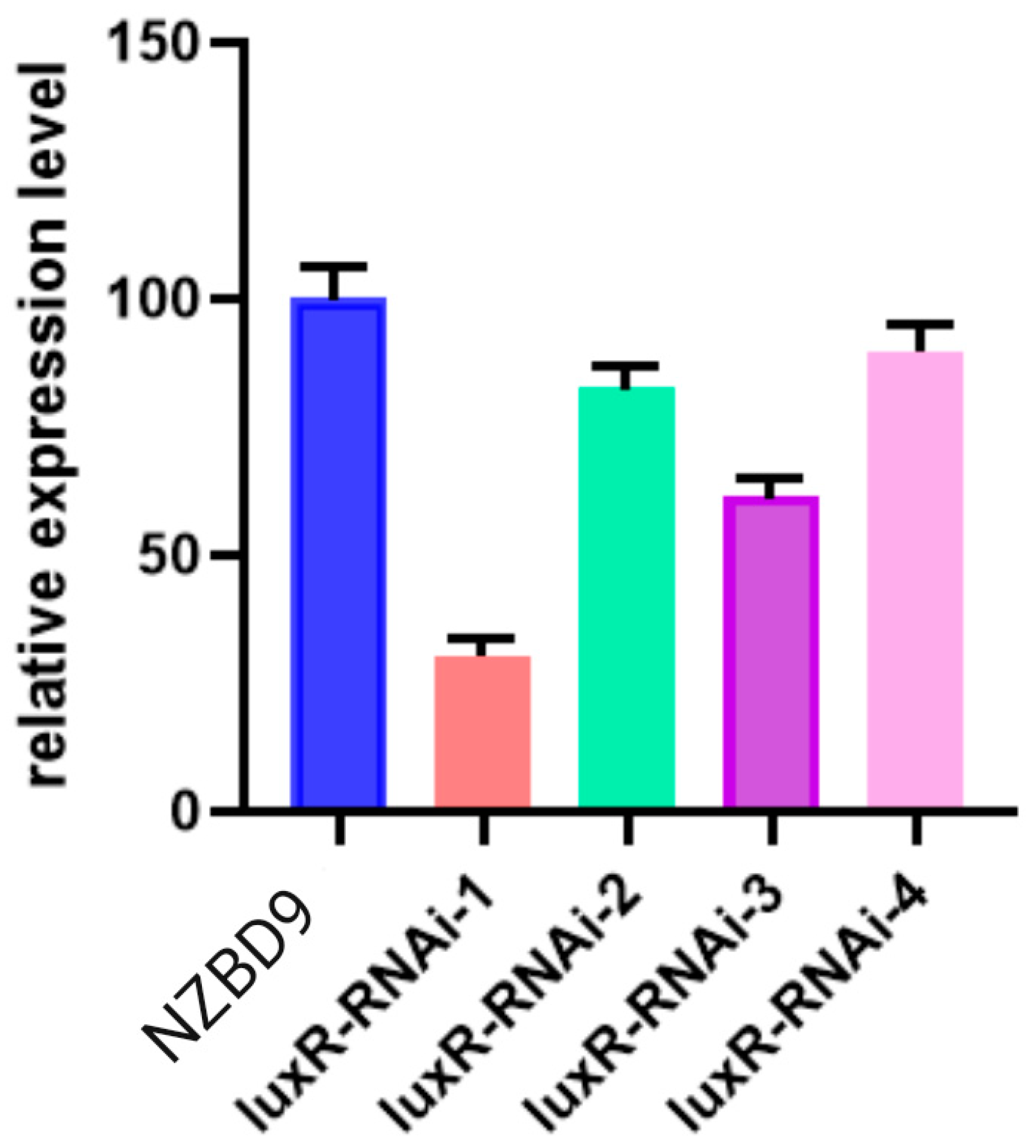
3.1. Construction of the luxR-RNAi Strain

The data presented in Figure 1 depicts the impact of four distinct shRNAs on the expression of the luxR gene. The results indicated that all four shRNAs caused a significant reduction in the mRNA levels of the luxR gene. Notably, the luxR-RNAi-1 strain, which we now refer to as the luxR-RNAi strain, demonstrated the highest efficacy in inducing RNAi (70.1%). Therefore, we chose the luxR-RNAi-1 strain with the greatest silencing efficiency for further experimentation.

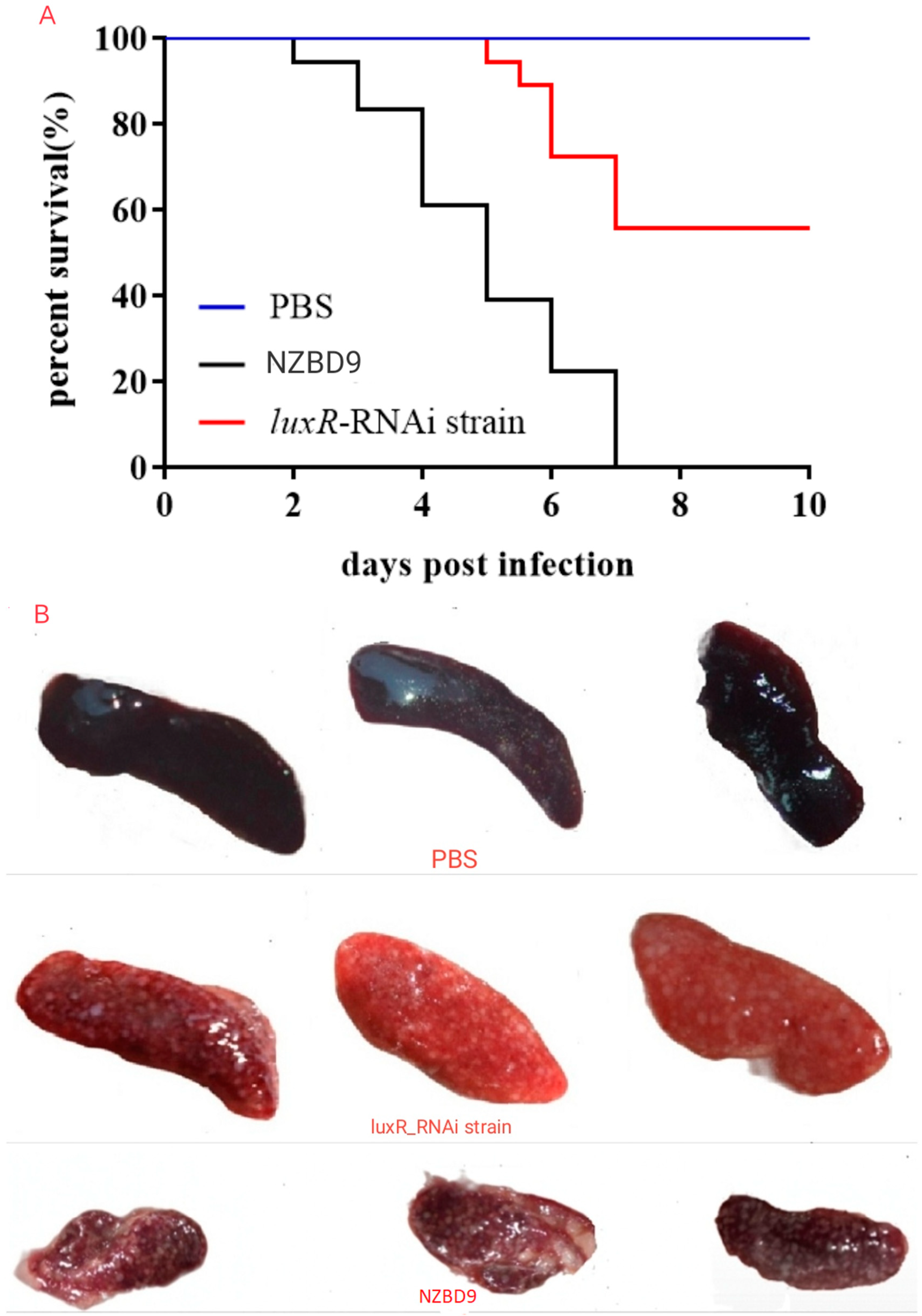
3.2. Impact of luxR on P. plecoglossicida Virulence

The group of E. coioides individuals subjected to injections of the PBS solution displayed a consistent pattern of survival throughout the experiment, maintaining robust health and vitality as evidenced by their active swimming behaviors. Subsequent dissections revealed no anomalies in their internal organ structures. Moreover, their behavioral and physiological profiles closely resembled those of the control group, which consisted of healthy fish. In contrast, the cohort of E. coioides injected with the luxR-RNAi strain of P. plecoglossicida exhibited a distinct outcome. Mortality in this group commenced on the 5th day, resulting in the deaths of 2 individuals. Subsequently, the mortality rate increased to 3 on the 6th day, 5 on the 7th day, and an additional 5 on the 8th day, leading to a cumulative total of 15 deaths. In a comparative context, the subset of E. coioides subjected to injections of the NZBD9 strain of P. plecoglossicida demonstrated a swifter onset of adverse effects. Notably, 2 deaths occurred on the second day, with this trend persisting as indicated by 3 deaths on the 3rd day, followed by 6 deaths on the 4th day, another 6 deaths on the 5th, 5 deaths on the 6th, and a final 6 deaths on the 7th day. Consequently, the cumulative mortality count reached 28. These findings underscore the differential impacts of the two bacterial strains on E. coioides, highlighting the distinct mortality patterns observed in response to the luxR-RNAi and NZBD9 strains of P. plecoglossicida.
The luxR-RNAi variant infection resulted in a 60% increase in the onset of death. Additionally, when comparing the survival rate of the NZBD9 variant to that of E. coioides, it was found that the latter had a higher survival rate. Notably, the E. coioides injected with PBS did not experience any mortality (as shown in Figure 2A). At 5 dpi, numerous white nodules were observed on the spleen surface of E. coioides infected with the NZBD9 variant, whereas significantly fewer white nodules were seen on those infected with the luxR-RNAi variant of P. plecoglossicida. Conversely, no white nodules were observed on the spleen surface of E. coioides injected with PBS (as demonstrated in Figure 2B).

3.3. Interpretation of Gene Transcription Data

3.3.1. Evaluating the Fidelity and Precision of Transcriptomic Information

The RNA sequencing of spleens from E. coioides infected with both the luxR-RNAi and NZBD9 strains of P. plecoglossicida was performed. The base content distribution of G/C/A/T was found to be uniform, with the percentage of N within the standard range. Quality evaluation using Q20 showed that all samples met the sequencing data standards, with Q20 values greater than 97%. The RNA-seq data had a base error rate of less than 0.1%. Pearson’s correlation coefficients (r) demonstrated a close correlation (r > 0.9) between the three biological replicate samples. The reproducibility and principal component analysis (PCA) of the three biological replicates showed satisfactory results.

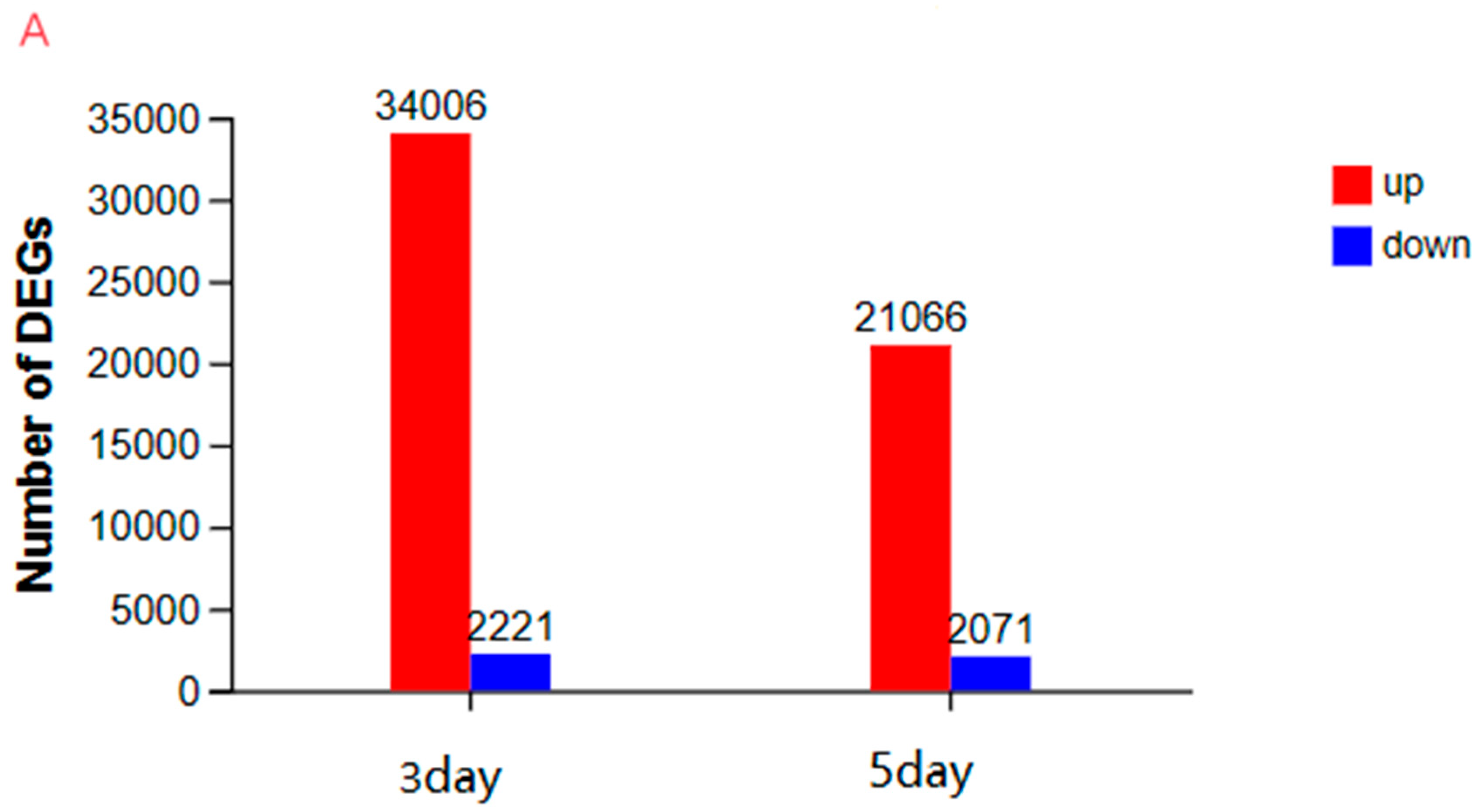
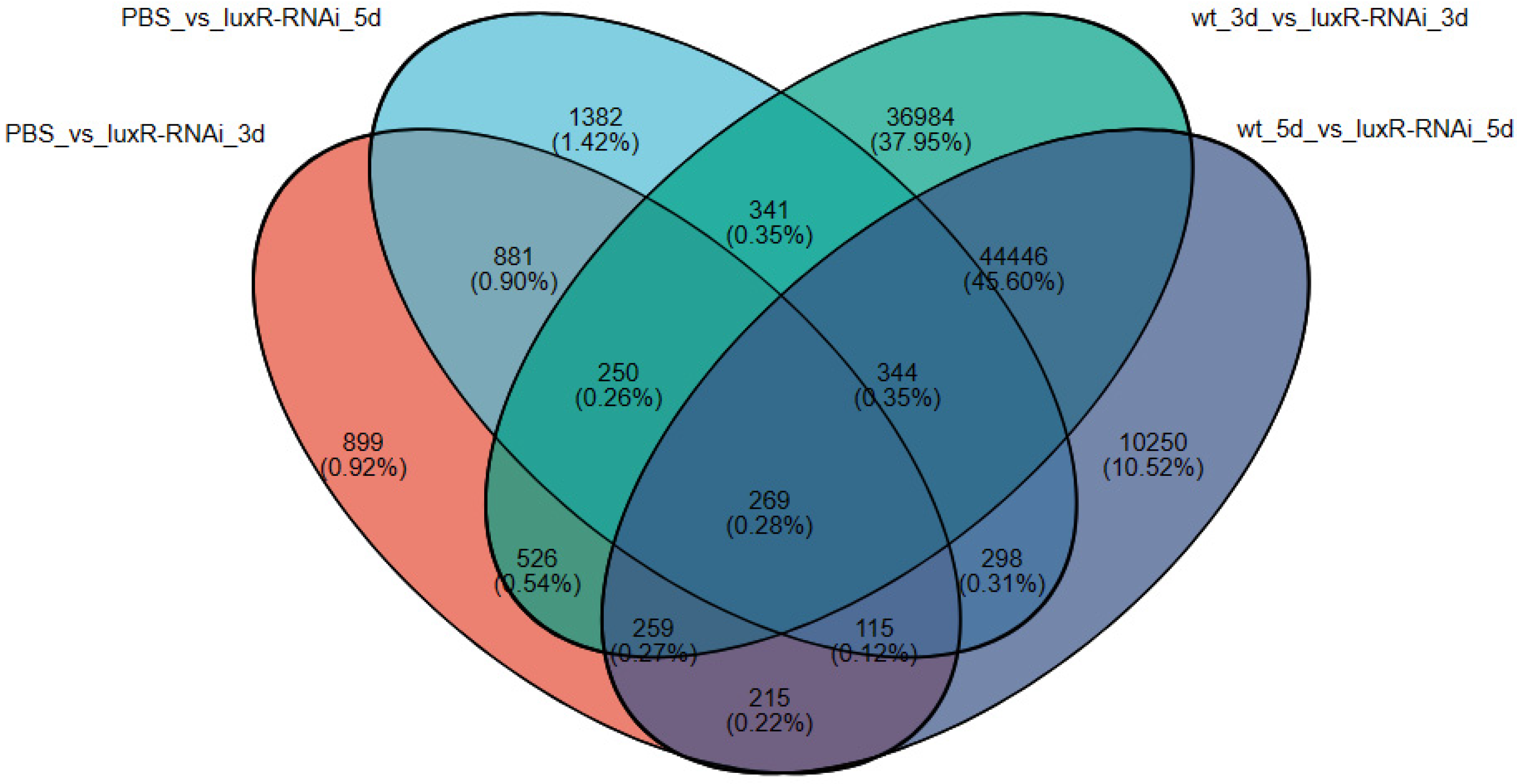
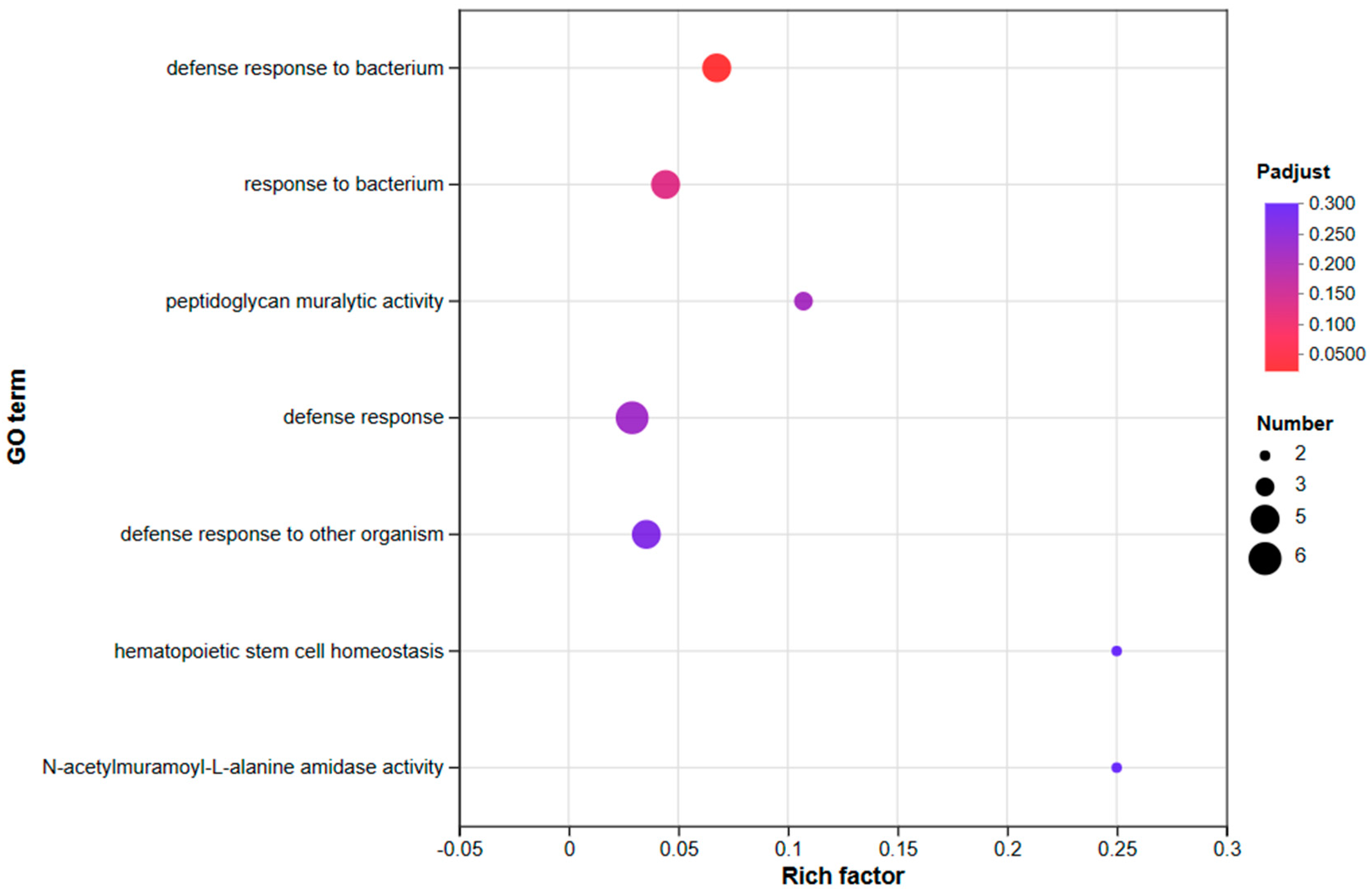
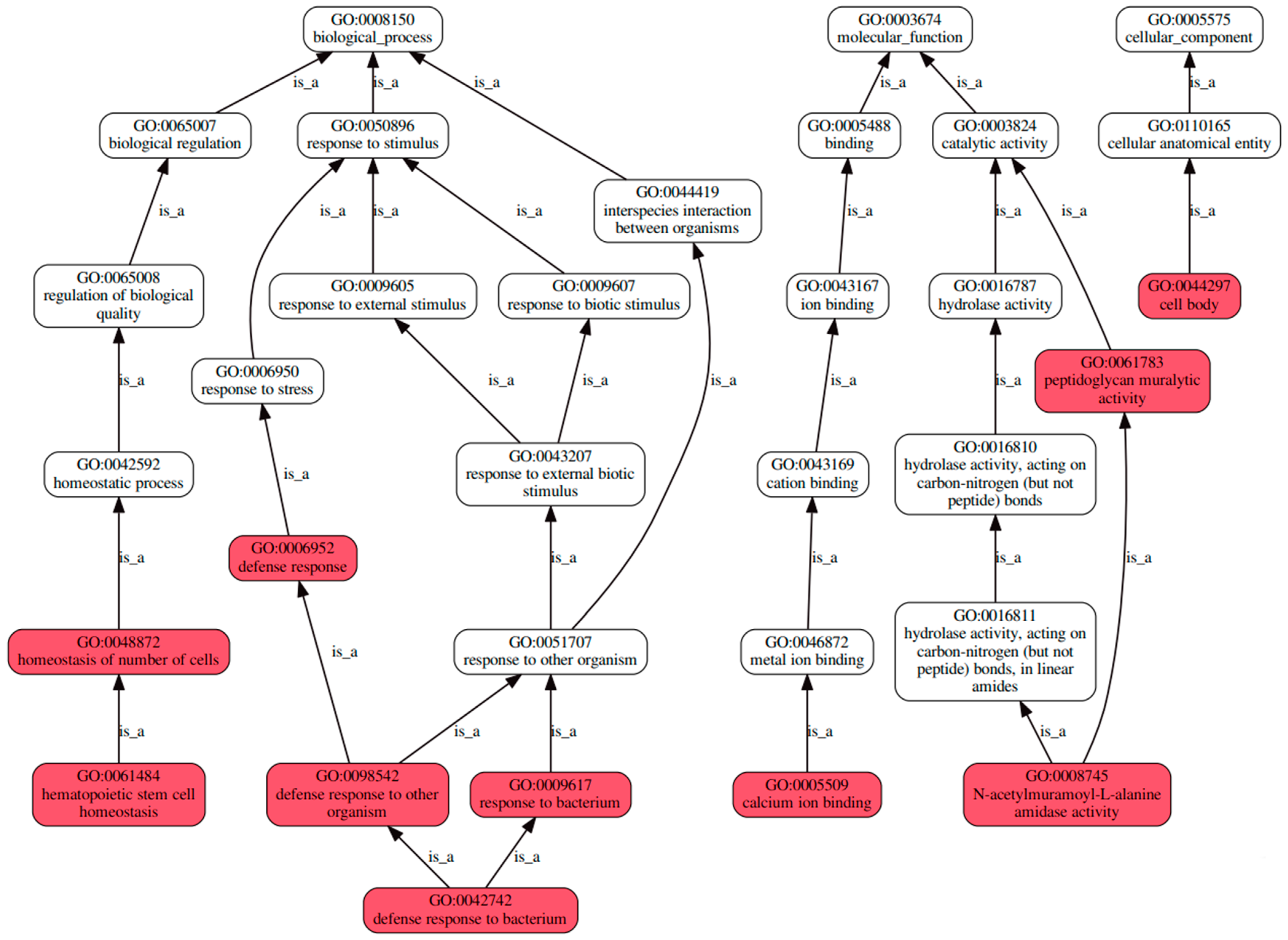
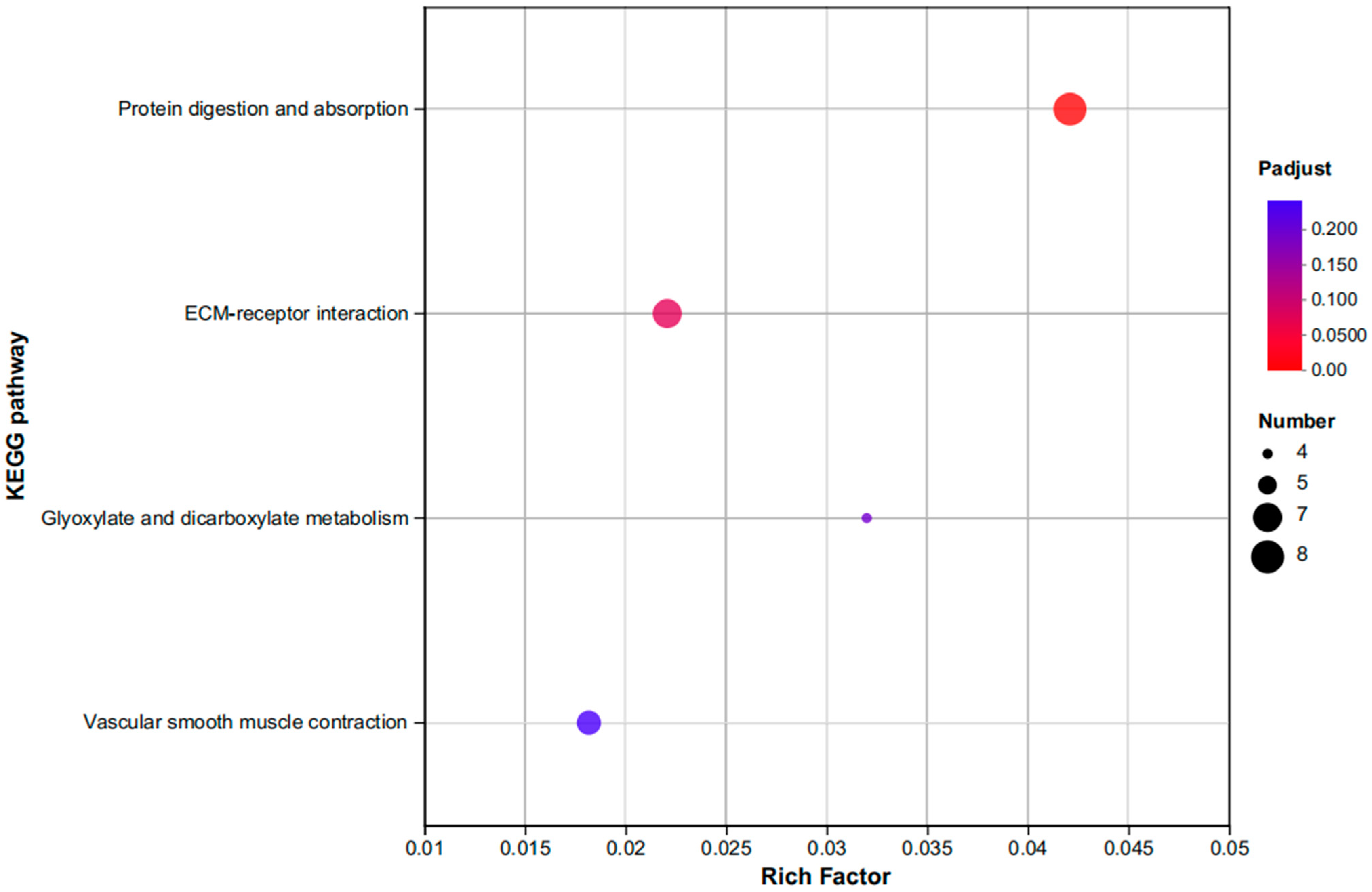
3.3.2. An Investigation into Genes Exhibiting Differential Expression Patterns via Transcriptomic Analysis

We identified differentially expressed genes in E. coioides infected with P. plecoglossicida on the 3rd and 5th days using a statistical cutoff criterion of |log2 fold change| ≥ 1 and padj ≤ 0.05. Our transcriptomic analysis revealed that, on the 3rd day, a total of 36,227 genes were differentially expressed in the experimental groups compared to the control group. Specifically, in the luxR-RNAi strain group, 2221 genes were downregulated, while 34,006 genes were upregulated compared to the NZBD9 strain group. Similarly, on the 5th day, 23,137 genes were differentially expressed, with 2071 genes downregulated and 21,066 genes upregulated in the luxR-RNAi strain group compared to the NZBD9 strain group (as shown in Figure 3A). The differential gene expression between the experimental groups and the control group on the third and fifth days was analyzed using a volcano plot. Our results indicated that, on the third day, 34,006 genes were up-regulated, while 2221 genes were down-regulated (as shown in Figure 3B). Similarly, on the fifth day, 21,066 genes were up-regulated and 2071 genes were down-regulated (as shown in Figure 3C). Notably, the number of up-regulated genes was significantly higher than the number of down-regulated genes on both days. However, the significance of the difference was greater on day 3 than on day 5.
To gain a better understanding of the regulatory networks or signaling pathways involved in the response to experimental treatments, we conducted a comparative analysis of differentially expressed genes using Venn analysis. As a result, we identified a set of 269 genes that were differentially expressed in both the infected E. coioides on the 3rd and 5th days, as well as in the control (PBS) and experimental (NZBD9 and luxR-RNAi) groups (as shown in Figure 4). These differentially expressed genes are of particular interest, as they may play a crucial role in the infection process. Therefore, we plan to further investigate their potential biological functions and associated pathways.
The results of the gene ontology (GO) analysis, as illustrated in Figure 5, demonstrated that differentially expressed genes (DEGs) were significantly enriched in several biological processes, with a notable emphasis on defense responses. Specifically, the DEGs were found to be enriched in defense responses to both bacterial and other organismal stimuli, indicating their potential role in mediating protective mechanisms against external threats.
A graph representation of a directed acyclic graph (DAG) with a defined start point and no cycles serves as a useful visual tool to represent complex relationships between different entities or events. The directed acyclic graph (DAG) depicting various gene ontology (GO) terms, including homeostasis of the number of cells, hematopoietic stem cell homeostasis, defense response, defense response to other organisms, defense response to bacteria, response to bacterium, calcium ion binding, N-Acetylmuramoyl-L-Alanine Amidase activity, Peptidoglycan M Uraytic activity, and cell body, highlights the potential significance of these terms in impacting the host (as shown in Figure 6). These nodes and their relationships may play important roles in various biological processes, including immune defense, stem cell maintenance, and cell homeostasis. Further research is necessary to uncover the key genes involved in these processes and their functional implications.
Regarding molecular function, the analysis revealed that the differentially expressed genes (DEGs) were significantly enriched in peptidoglycan catalytic activity, suggesting their potential involvement in the breakdown of bacterial cell walls. In addition, the KEGG pathway analysis revealed that the DEGs were predominantly enriched in pathways related to protein digestion and absorption, as well as ECM receptor interaction signaling (as shown in Figure 7). These findings provide important insights into the potential biological roles of the identified genes and highlight their potential significance in mediating fundamental cellular processes.
Within the immune system’s KEGG subcategory, the DEGs were predominantly located within the NOD-like receptor (NLR) signaling pathway. The pathway analysis reveals that the down-regulation of genes such as T3SS needle, T3SS rod, NAIP, and NLRC4, along with the up-regulation of po-IL-Iβ, ultimately leads to the up-regulation of secreted extracellular IL-Iβ (as shown in Figure 8). These genes are involved in various aspects of immune defense, such as the formation of the type III secretion system (T3SS) and the regulation of cytokine production. The down-regulation of T3SSneedle and T3SS rod genes suggests a potential disruption in the ability of bacterial pathogens to inject effector proteins into host cells, which may impair their ability to evade host immune defenses. The down-regulation of NAIP and NLRC4 genes, which are involved in the detection of bacterial pathogens, further suggests a potential impairment in the host’s ability to recognize and respond to invading bacteria.

4. Discussion

Differential gene expression refers to changes in gene expression between distinct conditions or cell types. In bacterial-host interactions, alterations in gene expression can occur simultaneously in both bacteria and hosts. Bacteria can adjust their gene expression to evade or overcome host defenses, while hosts can modify their gene expression to respond to bacterial colonization or infection [19,20,21].
The luxR gene is a transcriptional regulator that plays a critical role in bacterial quorum sensing [22,23,24,25,26]. Down-regulation of the luxR gene has been shown to result in changes in host gene expression [27,28,29]. In this paper, we aim to analyze the reason why the down-regulation of the luxR gene in Pseudomonas aeruginosa leads to an enrichment of host genes that are differentially expressed in gene ontology (GO) pathways related to bacterial defense and immune response. Additionally, we will investigate the Kyoto Encyclopedia of Genes and Genomes (KEGG) database to determine why host genes that are differentially expressed are enriched in metabolic pathways related to protein digestion and absorption.
The present experiment revealed that host genes that were differentially expressed in response to the down-regulation of the luxR gene were enriched in gene ontology (GO) pathways associated with bacterial defense and immune response. This finding suggests that down-regulation of the luxR gene might activate the host immune response to bacterial invasion. Numerous studies have also confirmed this conclusion [30,31,32,33,34].
The LuxR protein plays a crucial role in regulating quorum sensing, which is the mechanism by which bacteria communicate with each other to coordinate their behavior. When the concentration of signal molecules produced by bacteria reaches a certain threshold, the LuxR protein binds to these molecules and triggers the expression of genes involved in bacterial virulence, biofilm formation, and other biological processes. The down-regulation of the luxR gene can disrupt quorum sensing and may activate the host immune response to bacterial invasion [35,36,37,38,39].
In this study, we utilized the KEGG database to examine why host genes that are differentially expressed in response to luxR down-regulation are enriched in metabolic pathways related to protein digestion and absorption. These pathways involve the process of breaking down dietary proteins into amino acids, which serve as the fundamental building blocks of proteins in the body.
Multiple research studies have shown that the downregulation of the luxR gene affects the gut microbiome composition and other bacterial metabolic processes [40,41,42]. As a consequence, these changes may have a significant impact on host nutrient absorption and metabolism. This could explain why the host genes that are differentially expressed in response to the reduction in the luxR gene are enriched in metabolic pathways related to protein digestion and absorption. Therefore, these findings suggest that the down-regulation of the luxR gene could affect the host’s ability to efficiently absorb and metabolize nutrients, which could have important implications for overall host health and well-being.
Our findings indicate that downregulation of the luxR gene affects the host’s gene expression, with differentially expressed genes enriched in GO pathways related to bacterial defense and immune responses. This suggests that disruption of quorum sensing through the downregulation of luxR may activate the host’s immune response to bacterial invasion. Moreover, our investigation into the KEGG database revealed that differentially expressed host genes are enriched in metabolic pathways related to protein digestion and absorption, indicating a potential influence on host nutrient absorption and metabolism. These findings suggest a potential role for luxR in the interplay between bacteria and host health, and further research is necessary to fully understand the underlying mechanisms.
Differential gene expression in the immune system is focused mainly on the NOD-like receptor (NLR) signaling pathway, which is a critical component of the innate immune response against pathogens [43,44,45]. The NLRs are a class of cytoplasmic pattern recognition receptors that recognize a range of microbial and endogenous danger signals to initiate an immune response. These intracellular receptors are activated by a variety of microbial components, like bacterial cell wall compounds, including peptidoglycans, lipopolysaccharides (LPS), and flagellin. Once activated, the NLRs activate downstream signaling pathways, leading to the production of pro-inflammatory cytokines such as IL-1β [46,47,48,49,50]. IL-1β, predominantly produced by activated immune cells like macrophages and monocytes, is a cytokine that acts as a signaling molecule in the immune response. IL-1β plays a role in regulating physiological processes like inflammation, fever, and immune response.
luxR is a crucial transcriptional regulator present in Gram-negative bacteria that plays a significant role in the regulation of quorum sensing (QS) systems. In this study, the decreased expression of the luxR gene in P. plecoglossicida was observed to lead to a reduction in QS signaling, which was hypothesized to result in the upregulation of the IL-1β gene expression in the host’s NOD-like receptor (NLR) signaling pathway.
Studies have demonstrated that LuxR protein can bind directly to the promoter of the IL-1β gene, thus suppressing its expression [51,52,53,54]. In the event of a downregulation of the luxR gene during an infection, less LuxR protein would bind to the IL-1β promoter, causing an increase in IL-1β gene expression. This increase in IL-1β expression can result in pro-inflammatory effects, which are important for mounting an immune response to the infection.
Infections can trigger the production of pro-inflammatory cytokines, including IL-1β, which can activate the immune system and initiate inflammatory responses to fight the infection [55,56,57]. This immune response is critical to combating infectious diseases. However, an excessive and uncontrolled production of pro-inflammatory cytokines can lead to tissue damage and contribute to the pathogenesis of infectious diseases.
Microorganisms, such as bacteria, are present in a variety of environments, including animals and humans. Through co-evolution, many bacterial species have adapted to thrive in specific hosts or on certain host surfaces. For bacterial pathogens, evasion of host immune responses and causing diseases is a survival strategy that has driven the evolution of complex mechanisms. One such mechanism involves changes in bacterial gene expression that enable bacteria to overcome host defenses or evade them entirely [58,59]. This article examines changes in bacterial gene expression that result in the enrichment of bacterial processes associated with host defense and explores how these changes may lead to the unique expression of host genes involved in the immune response to bacterial infections.
The process of gene expression involves using genetic information encoded in genes to create functional RNA molecules or proteins. Bacteria, like all living organisms, adjust their gene expression in response to variations in their environment, such as changes in temperature [60], nutrient availability, and the presence of other microorganisms. Gene expression in bacteria can be altered at multiple levels, including transcription, translation, and post-translational modifications [61,62]. These alterations can lead to the production of new proteins, the modification of existing proteins, or the repression of particular genes [63,64].
When bacteria encounter a host, they adapt their gene expression to promote colonization and persistence within the host [65,66,67]. Bacterial gene expression can also be influenced by host defenses, such as immune cell activation, the release of antimicrobial peptides, or the production of reactive oxygen species [68,69,70,71,72,73]. Changes in bacterial gene expression in response to host defenses can lead to the production of factors that increase bacterial virulence, modification of bacterial surface molecules, or activation of stress response pathways [74,75,76].
To summarize, the decreased expression of the luxR gene in P. plecoglossicida during infection can result in increased expression of the IL-1β gene in the host’s NLR signaling pathway. This upregulation of IL-1β expression can have pro-inflammatory effects that are critical for the immune response to infection.

5. Conclusions

Overall, the downregulation of the luxR gene in P. plecoglossicida during infection can impact the host’s immune response and potentially influence the progression of the infection. Further investigation in this area is necessary to better understand the underlying mechanisms and potentially identify new treatment strategies for bacterial infections.

Supplementary Materials

The following supporting information can be downloaded at: https://www.mdpi.com/article/10.3390/fishes8100507/s1, Table S1: The sequences of four shRNAs for luxR gene; Table S2: The sequence of Primers for PCR and qRT-PCR.

Author Contributions

L.Z.: Contributed to data management, investigation, and wrote the initial draft. L.H.: Conducted formal analysis and investigation, and confirmed no conflicts of interest or financial relationships. Y.Q.: Participated in the development, execution, and analysis of data management, formal analysis, and methodology. D.Y.: Aided in managing the data and developing methodology. J.Z. (Jiaonan Zhang): Supported in the implementation of data management and methodology. J.Z. (Jiaolin Zhang): Helped with data management procedures. Q.Y.: Aided in conceptualizing the project, securing funding, managing its progress, and reviewing and editing the manuscript. All authors have read and agreed to the published version of the manuscript.

Funding

This study received financial support from multiple sources, including the Natural Science Foundation of Fujian Province (Contract No. 2022J02044), the Natural Science Foundation of Fujian Province (Contract No. 2023J01762), National Natural Science Foundation of China (Contract No. 32173016), and Open Project of Key Laboratory (Contract No. B2019090).

Institutional Review Board Statement

The fish study was reviewed and approved by the Ethics Committee of Jimei University (permit number JMULAC201159).

Data Availability Statement

Data will be made available on request.

Conflicts of Interest

The authors declare no conflict of interest.

References

  1. Park, S.B.; Aoki, T. Pseudomonas plecoglossicida and the bacterial haemorrhagic ascites of ayu, Plecoglossus altivelis. Microbiology 2015, 161, 2115–2123. [Google Scholar] [CrossRef]
  2. Ma, Y.; Li, X. Pseudomonas plecoglossicida: A major threat to fish health in aquaculture. J. Fish Dis. 2018, 41, 951–960. [Google Scholar] [CrossRef]
  3. Liu, X.; Li, S.; Cui, L. Analysis of the Status of Important Aquatic Animal Diseases in China; China Agricultural Press: Beijing, China, 2022. [Google Scholar]
  4. Wakabayashi, H.; Hikima, J.I.; Masumura, K. Isolation of Pseudomonas sp. and Vibrio sp. from natural outbreaks of bacterial ascites of ayu Plecoglossus altivelis. Fish Pathol. 1992, 27, 199–205. [Google Scholar] [CrossRef]
  5. Nishimura, S.; Miyoshi, S.; Sugino, H. Pseudomonas plecoglossicida sp. nov., the causative agent of bacterial haemorrhagic ascites of ayu, Plecoglossus altivelis. Int. J. Syst. Bacteriol. 1996, 46, 1133–1138. [Google Scholar] [CrossRef] [PubMed]
  6. Zhang, Y.; Liu, J.; Xia, H.; Zhang, Y.; Xu, P.; Li, Y. Complete genome sequence of Pseudomonas plecoglossicida strain NBRC 103064, the causative agent of bacterial hemorrhagic ascites of ayu fish. Genome Announc. 2013, 1, e00587-13. [Google Scholar] [CrossRef]
  7. Zhang, Y.; Gao, R.; Liu, J.; Wang, Q.; Xu, P.; Li, Y. Comparative genomics and transcriptomics of Pseudomonas plecoglossicida and its closely related Pseudomonas species. BMC Genom. 2014, 15, 724. [Google Scholar] [CrossRef]
  8. Wang, J.; Wei, L.; Huang, X.; Sun, Z.; Lv, J. Genome analysis of Pseudomonas plecoglossicida N12-1221, a pathogenic strain isolated from diseased ayu fish in China. Microbiol. Res. Announc. 2021, 10, e01335-20. [Google Scholar] [CrossRef]
  9. Huang, L.; Liu, W.; Jiang, Q.; Zuo, Y.; Su, Y.; Zhao, L.; Qin, Y.; Yan, Q. Integration of transcriptomic and proteomic approaches reveals the temperature-dependent virulence of Pseudomonas plecoglossicida. Front. Cell. Infect. Microbiol. 2018, 8, 207. [Google Scholar] [CrossRef]
  10. Fuqua, C.; Winans, S.C.; Greenberg, E.P. Quorum sensing in bacteria: The LuxR-LuxI family of cell density-responsive transcriptional regulators. J. Bacteriol. 1994, 176, 269–275. [Google Scholar] [CrossRef]
  11. Hanzelka, B.L.; Greenberg, E.P. Quorum sensing in Vibrio fischeri: Evidence that S-adenosylmethionine is the amino acid substrate for autoinducer synthesis. J. Bacteriol. 1996, 178, 5291–5294. [Google Scholar] [CrossRef]
  12. Rutherford, S.T.; Bassler, B.L. Bacterial quorum sensing: Its role in virulence and possibilities for its control. Cold Spring Harb. Perspect. Med. 2012, 2, a012427. [Google Scholar] [CrossRef]
  13. Whitehead, N.A.; Barnard, A.M.; Slater, H.; Simpson, N.J.; Salmond, G.P. Quorum-sensing in Gram-negative bacteria. FEMS Microbiol. Rev. 2001, 25, 365–404. [Google Scholar] [CrossRef] [PubMed]
  14. Engebrecht, J.; Nealson, K.; Silverman, M. Bacterial bioluminescence: Isolation and genetic analysis of functions from Vibrio fischeri. Cell 1983, 32, 773–781. [Google Scholar] [CrossRef]
  15. Bassler, B.L.; Wright, M.; Showalter, R.E.; Silverman, M.R. Intercellular signalling in Vibrio harveyi: Sequence and function of genes regulating expression of luminescence. Mol. Microbiol. 1993, 9, 773–786. [Google Scholar] [CrossRef] [PubMed]
  16. Wang, L.Y.; Liu, Z.X.; Zhao, L.M.; Huang, L.X.; Qin, Y.X.; Su, Y.Q.; Nie, P. Dual RNA-seq provides novel insight into the roles of dksA from Pseudomonas plecoglossicida in pathogen-host interactions with large yellow croakers (Larimichthys crocea). Zool. Res. 2020, 41, 410–422. [Google Scholar] [CrossRef] [PubMed]
  17. Tang, R. Preliminary Study of Whole-Bacterial Vaccine against Pseudomonas Mutans. Master’s Thesis, Jimei University, Xiamen, China, 2017. [Google Scholar]
  18. Luo, G.; Sun, Y.; Huang, L.; Su, Y.; Zhao, L.; Qin, Y.; Xu, X.; Yan, Q. Time-Resolved dual RNA-seq of tissue uncovers Pseudomonas plecoglossicida key virulence genes in host-pathogen interaction with Epinephelus coioides. Environ. Microbiol. 2020, 22, 677–693. [Google Scholar] [CrossRef] [PubMed]
  19. Tang, X.; Chang, D.; Wang, X.; Chen, L.; Yan, X.; Zhang, Y.; Guo, Q. Interactions of gut microbiota with host physiology and innate immune system. J. Cell. Physiol. 2021, 236, 319–330. [Google Scholar] [CrossRef]
  20. Wlodarska, M.; Finlay, B.B. Host immune response to antibiotic perturbation of the microbiota. Mucosal Immunol. 2010, 3, 100–103. [Google Scholar] [CrossRef]
  21. Sommer, F.; Bäckhed, F. The gut microbiota—Masters of host development and physiology. Nat. Rev. Microbiol. 2013, 11, 227–238. [Google Scholar] [CrossRef]
  22. Liu, J.; Zhou, L.; Zhang, L. luxR family members are key regulators of quorum sensing in Vibrio harveyi. Front. Cell. Infect. Microbiol. 2022, 12, 790839. [Google Scholar]
  23. Kim, J.; Kim, H.; Kim, Y.J. Characterization of luxR homologues in Vibrio parahaemolyticus and their roles in quorum sensing. J. Microbiol. Biotechnol. 2022, 32, 237–348. [Google Scholar]
  24. Gao, Y.; Zhao, X.; Zhou, L.; Zhang, L. Regulation of luxR -type transcriptional regulators in bacterial quorum sensing. Front. Microbiol. 2021, 12, 800539. [Google Scholar]
  25. Zhang, L.; Du, P.; Li, L. luxR family transcription factors play crucial roles in bacterial quorum sensing and pathogenesis. Front. Cell. Infect. Microbiol. 2021, 11, 699628. [Google Scholar]
  26. Li, S.; Li, J.; Liang, J.; Li, Y.; Zheng, M. luxR -type transcriptional regulators in bacterial quorum sensing: Diversity and functions. Front. Microbiol. 2021, 12, 647035. [Google Scholar]
  27. Sequeira, R.P.; McDonald, J.A.K.; Marchesi, J.R.; Clarke, P.; Hill, K.E.; Nally, J.E. Differential modulation of host gene expression in response to decreased expression of the bacterial quorum sensing regulator luxR. Microb. Pathog. 2022, 161, 105345. [Google Scholar]
  28. Dantas-Barbosa, C.; Duarte, P.; Carriço, J.A.; Freitas, A.T.; Ferreira, R.B.; Mira, N.P. The regulatory impact of luxR gene knockdown on Burkholderia thailandensis gene expression. Genes 2021, 12, 1591. [Google Scholar]
  29. Venugopal, A.; Patel, P.; Stabb, E.V. Differential gene expression in the squid-Vibrio symbiosis in response to decreased expression of the quorum sensing regulator luxR. Front. Microbiol. 2021, 12, 704472. [Google Scholar]
  30. Lai, Y.; Lu, X.; Wu, Z.; Li, X.; Wu, Q.; Zhang, Y.; Cao, X. Downregulation of the luxR gene in Vibrio parahaemolyticus enhances the host immune response and reduces bacterial virulence. Microb. Pathog. 2022, 163, 105476. [Google Scholar]
  31. Han, J.E.; Jeong, S.Y.; Choi, S.H.; Lee, K.H.; Lee, S.J. Down-regulation of luxR -type transcriptional regulator gene affects expression of virulence genes in Vibrio vulnificus. J. Microbiol. Biotechnol. 2021, 31, 1675–1682. [Google Scholar]
  32. Huang, H.; He, Y.; Wu, X.; Xie, J.; Xu, Z.; Zhang, Y.; Chen, S. The down-regulation of luxR in Vibrio alginolyticus triggers immune responses in Japanese flounder (Paralichthys olivaceus) against bacterial infection. Fish Shellfish Immunol. 2021, 114, 527–536. [Google Scholar]
  33. Ma, R.; Wang, X.; Yang, Z.; Liu, Y.; Zhang, X.; Gao, W.; Gao, P. The down-regulation of luxR in Vibrio anguillarum enhances the activation of immune response in fish macrophages. Fish Shellfish Immunol. 2021, 112, 135–142. [Google Scholar]
  34. Cheng, J.; Zhang, L.; Sun, L. The down-regulation of luxR in Vibrio splendidus enhances the immune response of scallops (Chlamys farreri) against bacterial infection. Fish Shellfish Immunol. 2021, 109, 231–238. [Google Scholar]
  35. Decho, A.W.; Gutierrez, T. Microbial Quorum Sensing in Marine Environments. Annu. Rev. Mar. Sci. 2021, 13, 231–256. [Google Scholar]
  36. Li, Y.; Li, M.; Huang, T. luxR -Type Quorum Sensing Systems in Bacteria. Int. J. Mol. Sci. 2021, 22, 2578. [Google Scholar]
  37. Zhang, X.; Xu, Y.; Qiu, J.; Shen, Y. Quorum sensing and bacterial virulence. Int. J. Oral Sci. 2021, 13, 9. [Google Scholar]
  38. Sheng, L.; Ren, X.; Gao, Y.; Zhang, R. Quorum sensing and bacterial virulence regulation: A comprehensive review. Front. Cell. Infect. Microbiol. 2021, 11, 621594. [Google Scholar]
  39. Li, L.; Li, H.; Zhang, X.X. luxR -Type Quorum Sensing in Aquatic Bacteria: Diversity, Mechanisms, and Applications. Front. Microbiol. 2022, 12, 794220. [Google Scholar]
  40. Low, K.Y.; Gubbi, S. luxR -type quorum sensing regulators are required for metabolic fitness in Chromobacterium piscinae. Microbiol. 2019, 165, 416–428. [Google Scholar] [CrossRef]
  41. Chen, C.; Lin, J. luxR -type regulators in pathogenic bacteria: Pleiotropic regulators or virulence factors? J. Microbiol. Immunol. Infect. 2020, 53, 631–638. [Google Scholar] [CrossRef]
  42. Chen, X.; Ji, Z.; Webber, A.T.; Luo, Y. luxR -type quorum sensing transcriptional regulators affect biofilm formation, motility, and antibiotic resistance in bacteria. Front. Microbiol. 2021, 12, 753244. [Google Scholar] [CrossRef]
  43. Park, S.J.; Lee, W.; Kim, J.; Lee, Y.J.; Park, J.H. NOD-like receptors in cancer and inflammation. Front. Immunol. 2021, 12, 738303. [Google Scholar]
  44. Goulielmaki, E.; Papadaki, M. NOD-like receptors: Emerging therapeutic targets for inflammatory diseases. Front. Pharmacol. 2021, 12, 647874. [Google Scholar]
  45. Wang, L.; Li, Y.; Li, Z.; Li, Y.; Li, X.; Li, L.; Yang, K. NOD-like receptor signaling in innate immunity and inflammation: A comprehensive review. Cell. Mol. Immunol. 2021, 18, 1391–1412. [Google Scholar]
  46. Chen, K.W.; Groß, C.J.; Sotomayor, F.V.; Stacey, K.J.; Tschopp, J.; Sweet, M.J.; Schroder, K. The neutrophil NLRC4 inflammasome selectively promotes IL-1β maturation without pyroptosis during acute Salmonella challenge. Cell Rep. 2021, 37, 110045. [Google Scholar] [CrossRef] [PubMed]
  47. Kang, J.Y.; Lee, J.O. Structural biology of NLRP3 inflammasome activation. Exp. Mol. Med. 2021, 53, 406–417. [Google Scholar]
  48. He, Q.; Xia, L.; Chen, Y.; Hao, Y.; Li, Y.; Li, J. Crosstalk between the NLRP3 inflammasome and autophagy. Cell. Mol. Immunol. 2022, 19, 22–33. [Google Scholar]
  49. Gonçalves, A.V.; De Oliveira, J.R.; Molfetta, J.B. NOD-like receptors and viral infections: Gatekeepers, guardians, and caretakers of the immune system. Front. Immunol. 2021, 12, 752324. [Google Scholar]
  50. Brennan, M.A.; Cookson, B.T. Salmonella-induced inflammasome activation and IL-1β release in humans. Curr. Opin. Infect. Dis. 2021, 34, 245–250. [Google Scholar]
  51. Eislmayr, K.; Bestehorn, A.; Morelli, L.; Borroni, M.; Walle, L.V.; Lamkanfi, M.; Kovarik, P. Nonredundancy of IL-1α and IL-1β is defined by distinct regulation of tissues orchestrating resistance versus tolerance to infection. Sci. Adv. 2022, 8, eabj7293. [Google Scholar] [CrossRef]
  52. Ma, T.; Liu, X.; Cen, Z.; Xin, C.; Guo, M.; Zou, C.; Song, W.; Xie, R.; Wang, K.; Zhou, H.; et al. MicroRNA-302b negatively regulates IL-1β production in response to MSU crystals by targeting IRAK4 and EphA2. Arthritis Res. Ther. 2018, 20, 34. [Google Scholar] [CrossRef]
  53. Lin, C.C.; Edelson, B.T. New Insights into the Role of IL-1β in Experimental Autoimmune Encephalomyelitis and Multiple Sclerosis. J. Immunol. 2017, 198, 4553–4560. [Google Scholar] [CrossRef] [PubMed]
  54. Doyle, A.A.; Stephens, J.C. A review of cinnamaldehyde and its derivatives as antibacterial agents. Fitoterapia 2019, 139, 104405. [Google Scholar] [CrossRef] [PubMed]
  55. Mauer, J.; Denson, J.L.; Brüning, J.C. Versatile functions for IL-6 in metabolism and cancer. Trends Immunol. 2014, 35, 92–101. [Google Scholar] [CrossRef] [PubMed]
  56. Dinarello, C.A. Interleukin-1 in the pathogenesis and treatment of inflammatory diseases. Blood 2011, 117, 3720–3732. [Google Scholar] [CrossRef]
  57. Van De Veerdonk, F.L.; Netea, M.G.; Dinarello, C.A.; Joosten, L.A.B. Inflammasome activation and IL-1β and IL-18 processing during infection. Trends Immunol. 2011, 32, 110–116. [Google Scholar] [CrossRef]
  58. Tegtmeyer, N.; Sharafutdinov, I.; Harrer, A.; Backert, S. Campylobacter Virulence Factors and Molecular Host–Pathogen Interactions. Curr. Top. Microbiol. Immunol. 2021, 431, 169–202. [Google Scholar]
  59. Gebre, M.S.; Brito, L.A.; Tostanoski, L.H.; Edwards, D.K.; Irvine, D.J. Novel approaches for vaccine development. Cell 2021, 184, 1589–1603. [Google Scholar] [CrossRef]
  60. Wei, Y.; Murphy, E.R.; Larramendy, M.; Broach, J.R. Temperature-dependent regulation of bacterial gene expression by RNA thermometers. In Nucleic Acids—from Basic Aspects to Laboratory Tools; IntechOpen: London, UK, 2016; pp. 157–181. [Google Scholar]
  61. Shimoni, Y.; Friedlander, G.; Hetzroni, G.; Niv, G.; Altuvia, S.; Biham, O.; Margalit, H. Regulation of gene expression by small non-coding RNAs: A quantitative view. Mol. Syst. Biol. 2007, 3, 138. [Google Scholar] [CrossRef]
  62. Mangan, J.A.; Monahan, I.M.; Butcher, P.D. Gene expression during host—Pathogen interactions: Approaches to bacterial mRNA extraction and labelling for microarray analysis. In Methods in Microbiology; Academic Press: Cambridge, MA, USA, 2002; Volume 33, pp. 137–151. [Google Scholar]
  63. Han, L.; Gao, Y.; Liu, Y.; Li, Y.; Lu, J.; Guo, L. An Outer Membrane Protein YiaD Contributes to Adaptive Resistance of Meropenem in Acinetobacter baumannii. Microbiol. Spectrum. 2022, 10, e00173-22. [Google Scholar] [CrossRef]
  64. Blount, Z.D.; Maddamsetti, R.; Grant, N.A.; Ahmed, S.T.; Jagdish, T.; Baxter, J.A.; Sommerfeld, B.A.; Tillman, A.; Moore, J.; Slonczewski, J.L.; et al. Genomic and phenotypic evolution of Escherichia coli in a novel citrate-only resource environment. elife 2020, 9, e55414. [Google Scholar] [CrossRef]
  65. Ahmed, W. RNA-seq resolving host-pathogen interactions: Advances and applications. Ecol. Genet. Genom. 2020, 15, 100057. [Google Scholar] [CrossRef]
  66. Jones, P. Multi-Omic Systems Biological Analysis of Host-Microbe Interactions. Ph.D Thesis, University of Tennessee, Knoxville, TX, USA, 2022. [Google Scholar]
  67. Denzer, L.; Schroten, H.; Schwerk, C. From gene to protein—How bacterial virulence factors manipulate host gene expression during infection. Int. J. Mol. Sci. 2020, 21, 3730. [Google Scholar] [CrossRef] [PubMed]
  68. Krakauer, T. Inflammasomes, autophagy, and cell death: The trinity of innate host defense against intracellular bacteria. Mediat. Inflamm. 2019, 2019, 2471215. [Google Scholar] [CrossRef] [PubMed]
  69. Yang, L.; Zheng, W.; Lv, X.; Xin, S.; Sun, Y.; Xu, T. microRNA-144 modulates the NF-κB pathway in miiuy croaker (Miichthys miiuy) by targeting IκBα gene. Dev. Comp. Immunol. 2022, 130, 104359. [Google Scholar] [CrossRef]
  70. Kumar, A.; Sperandio, V. Indole signaling at the host-microbiota-pathogen interface. mBio 2019, 10, e01031-19. [Google Scholar] [CrossRef]
  71. Febriza, A.; Hatta, M.; Natzir, R.; Kasim, V.N.A.; Idrus, H.H. Activity of antimicrobial peptide; cathelicidin, on bacterial infection. Open Biochem. J. 2019, 13, TOBIOCJ-13-45. [Google Scholar] [CrossRef]
  72. Dev, A.; Iyer, S.; Razani, B.; Cheng, G. NF-κB and innate immunity. Curr. Top. Microbiol. Immunol. 2011, 349, 115–143. [Google Scholar] [CrossRef]
  73. Green, J.; Rolfe, M.D.; Smith, L.J. Transcriptional regulation of bacterial virulence gene expression by molecular oxygen and nitric oxide. Virulence 2014, 5, 794–809. [Google Scholar] [CrossRef]
  74. Maldonado, R.F.; Sá-Correia, I.; Valvano, M.A. Lipopolysaccharide modification in Gram-negative bacteria during chronic infection. FEMS Microbiol. Rev. 2016, 40, 480–493. [Google Scholar] [CrossRef]
  75. Heithoff, D.M.; Conner, C.P.; Hanna, P.C.; Julio, S.M.; Hentschel, U.; Mahan, M.J. Bacterial infection as assessed by in vivo gene expression. Proc. Nat. Acad. Sci. USA 1997, 94, 934–939. [Google Scholar] [CrossRef]
  76. Fang, F.C.; Frawley, E.R.; Tapscott, T.; Vázquez-Torres, A. Bacterial Stress Responses during Host Infection. Cell Host Microbe 2016, 20, 133–143. [Google Scholar] [CrossRef] [PubMed]
Figure 1. Assessment of luxR mRNA levels in four luxR-RNAi silenced strains.
Fishes 08 00507 g001
Figure 2. Results of virulence infection in NZBD9 and luxR-RNAi strains of P. plecoglossicida in E. coioides (A) Assessment of E. coioides survival rate in various infections. (B) Macroscopic signs of P. plecoglossicida infection in E. coioides spleens.
Fishes 08 00507 g002
Figure 3. Comparative differential gene expression analysis of NZBD9 and luxR-RNAi strains in infected Epinephelus coioides. (A) Analysis of differentially expressed genes in spotted groupers infected with luxR-RNAi and NZBD9 strains on the third and fifth days. (B) Volcano plot analysis of differentially expressed genes between NZBD9 and luxR-RNAi strains in groupers infected at dpi3. (C) Volcano plot analysis of differentially expressed genes between NZBD9 and luxR-RNAi strains in groupers infected on day five.
Fishes 08 00507 g003aFishes 08 00507 g003b
Figure 4. Venn diagram analysis of differentially expressed genes in NZBD9 and luxR-RNAi strains and PBS control groups on days 3 and 5 of infection in E. coioides.
Fishes 08 00507 g004
Figure 5. Analyzing the functional enrichment of genes that exhibit differential expression.
Fishes 08 00507 g005
Figure 6. A graphical representation of a directed acyclic graph with a defined starting point and no cycles. The red box indicates the discovery of a significantly enriched gene ontology (GO) term within the gene set. The connections between GO terms elucidate the inherent relationships between them. Notably, the ‘is_a’ relationship denotes that the GO term located below the connecting line represents a subcategory of the GO term indicated by the arrow.
Fishes 08 00507 g006
Figure 7. An analysis of KEGG pathway enrichment was conducted on the DEGs.
Fishes 08 00507 g007
Figure 8. NOD-like receptor (NLR) signaling pathway.
Fishes 08 00507 g008
Disclaimer/Publisher’s Note: The statements, opinions and data contained in all publications are solely those of the individual author(s) and contributor(s) and not of MDPI and/or the editor(s). MDPI and/or the editor(s) disclaim responsibility for any injury to people or property resulting from any ideas, methods, instructions or products referred to in the content.

