Article

Localization of Acetylcholine, Alpha 7-NAChR and the Antimicrobial Peptide Piscidin 1 in the Macrophages of Fish Gut: Evidence for a Cholinergic System, Diverse Macrophage Populations and Polarization of Immune Responses

1
Department of Veterinary Sciences, University of Messina, 98168 Messina, Italy
2
Department of Chemical, Biological, Pharmaceutical and Environmental Sciences, University of Messina, 98166 Messina, Italy
3
Department of Cell and Tissues, Faculty of Veterinary Medicine, Assiut University, Assiut 71526, Egypt
4
Department of Histology and Anatomy, School of Veterinary Medicine, Badr University in Assiut, Asyut 71526, Egypt
5
Institute of Marine Biological Resources and Biotechnology (IRBIM), National Research Council (CNR), 98164 Messina, Italy
6
Faculty of Biosciences and Aquaculture, Nord University, 8049 Bodø, Norway
7
Department of Anatomy and Embryology, Faculty of Veterinary Medicine, Sohag University, Sohag 82524, Egypt
8
Poison Information Center, Department of Toxicology and Environmental Disease, Faculty of Medicine, Jagellonian University, 30501 Cracow, Poland
9
Department of Comparative Anatomy, Faculty of Biology, Institute of Zoology and Biomedical Research, Jagellonian University, 30387 Cracow, Poland
10
Department of Anatomy and Cell Biology, Poligono de Cazona, Faculty of Medicine, University of Cantabria, 39011 Santander, Spain
*
Authors to whom correspondence should be addressed.
Fishes 2023, 8(1), 43; https://doi.org/10.3390/fishes8010043
Submission received: 13 November 2022 / Revised: 5 January 2023 / Accepted: 6 January 2023 / Published: 8 January 2023
(This article belongs to the Special Issue Neuroimmune Communication in Fishes)

Abstract

:
The recognition and elimination of invading pathogens are vital for host survival. Macrophages play a central role in host protection and cells functionally reminiscent of vertebrate macrophages are present in all multicellular organisms. A pattern responsible for bacterial recognition found on the surface of macrophages is CD14. These cells possess a repertoire of antimicrobial molecules stored in their granules and lysosomes. Polarization states observed in mammalian macrophages termed M1 and M2 also likely exist in fish macrophages. Markers for macrophage subtypes are slowly but definitively emerging in fish species. In the present study cell markers such as CD14, acetylcholine, alpha 7 acetylcholine nicotinic receptor (nAChR) subtype, the inducible nitric oxidase synthase (iNOS), and the antimicrobial peptide piscidin 1 are reported for the first time in the intestinal macrophages of both catfish Heteropneustes fossilis (Bloch, 1794) and the African bonytongue Heterotis niloticus (Cuvier, 1829) along the anterior and the posterior axis and the concentric muscle layers. Many antimicrobial effector responses of vertebrate macrophages including respiratory burst and NO induction are similar across the diverse animal taxa. Antibodies against calbindin coupled with ones to VAChT and tubulin revealed the localization of myenteric and submucosal plexuses, which are made up of enteric neurons, glial cells, and nerves near macrophages. Current studies allow for the elucidation of multiple roles of macrophages in disease models providing an insight into their in vivo function in fish.

1. Introduction

The immune system of all vertebrates is to a larger extent dependent on macrophage-lineage cells, and while the ontogeny and function of diverse macrophage subsets and lineages have been studied in mammals [1,2], they remain to be defined in aquatic vertebrates such as teleost fishes [3]. In fish macrophages with polarized profiles M1 and M2 have been demonstrated. M1 macrophages were activated by microbial infection without any influence of adaptative immune cells. M2 macrophages having anti-inflammatory phenotypes were important for cell growth and division [4].
Macrophages and mast cells are regarded as the key regulators of gut physiological functions. Mast cells are crucial for maintaining gut mucosal homeostasis by contributing to intestinal functions (including host defense, vascular permeability, and peristalsis) [5,6]. Macrophages play a central role in host protection and many of the antimicrobial effector responses of vertebrate phagocytes are similar across diverse animal taxa. These immune cells are a heterogeneous mix of diversified and functionally specialized cell subsets. Neuron-associated macrophages are important for the survival and homeostasis of enteric neurons that control gastrointestinal motility and intestinal secretion. Blood vessel-associated macrophages also ensure blood vessel integrity and regulate blood vessel permeability [7]. In fish macrophages play critical roles in host protection and homeostasis. Various mechanisms determine and regulate functional phenotypes of macrophages, including antimicrobial host defenses (pro-inflammatory, M1 type) and resolution and repair functions (anti-inflammatory/regulatory, M2 type) [8]. The M1/M2 paradigm describes the two major and opposing activities of macrophages.
The intestinal epithelial layer forms a physical barrier between the microbe-rich environment of the lumen and the mucosal immune system, further aiding in preventing infection [9]. Epithelial cells have innate immunity functions and detect potential pathogens through the secretion of cytokines and antimicrobial peptides (AMPs) [10]. Recently, macrophages have been found in the apical membrane of the intestinal epithelial cells as well [11], thus suggesting the complex epithelial-macrophage relationship that is critical for innate immunity within the intestinal tract during infection and maintenance of the host microbiome.
The teleost fish inflammatory M1 macrophage populations have been the best studied and shown to rapidly kill pathogens through phagocytosis [12], and the production of reactive oxygen and nitrogen intermediates [13,14]. During injury and/or infection, resident macrophages detect tissue damage and/or infiltrate pathogens by either extracellular or intracellular recognition of receptors (PRRs) [15]. They have been classified into five groups that are TLRs, CLRs, NLRs, RLRs, and ALR.
CD14 (cluster of differentiation 14) is a receptor of LPS that is a component of the outer membrane of Gram-negative bacteria and the strongest activator of macrophages. It is expressed on the cell membrane and the endoplasmic reticulum of intestinal macrophages [16] and is known to serve as a co-receptor for several Toll-like-Receptors (TLRs) [17]. Circulating monocytes and tissue macrophages are typically CD14+ and express chemokine receptors, adhesion molecules, and TLRs, surface molecules that contribute to the pathogen-associated molecular pattern recognition at sites of infection and/or inflammation [18,19]. Although the fish receptors that can recognize lipopolysaccharide (LPS) are not so clear, a recent study demonstrated the recognition of LPS and activation of NF-kB by cytosolic sensor NOD1 in teleost fish [20]. The transition of monocytes to a tissue-resident phenotype has been described in mucosal macrophages where a population of monocytes sharing CD14 markers is transitioning to a CD14 macrophage phenotype [21].Monocytes move from flowing blood to the tissues after 1–2 days. When monocytes enter the tissue, they become known as macrophages and are responsible for fighting foreign bodies or pathogen and debris by engulfing and inactivating and digesting them in a process known as phagocytosis.
A critical component of the bi-directional gut-brain communication system is the expression of the nAChRs. These ligand-gated ion channels serve myriad roles including mediating synaptic transmission between autonomic pre- and post-ganglionic neurons, modulation of neurotransmitter release from peripheral sensory and enteric neurons, and modulation of cytokine release from immune cells [22]. The nicotinic ACh receptors expression by autonomic ganglion neurons innervating the gut suggests the presence of several subtypes that contain alpha 2, 3, 4, 5, beta 2, 4 subunits [23]. The role of the nAChR alpha 7 subunit in distinct immune cells may differ depending on cell type and function. In macrophages nAChR alpha 7 subunit, besides decreasing the release of inflammatory cytokines, also stimulates the survival and polarization of the anti-inflammatory M2 phenotype [24,25]. The alpha 7 nAChR has been described as an essential regulator of inflammation as this receptor mediates the inhibition of the cytokine synthesis through the cholinergic anti-inflammatory pathway.
The definition of non-neuronal acetylcholine (ACh) was originally proposed by Morris et al. (1996) [26]. Since then, the expressions of components involved in the cholinergic system, including acetylcholinesterase (ChAT) and vesicular ACh transporter (VAChT) have been confirmed in various immune cells including macrophages [27], and epithelial cells [28]. ACh synthesized by immune cells plays a key role in the regulation of immune function by triggering signals that initiate and terminate cytokine production in immune cells.
Distribution and molecular signatures of cholinergic tuft cells have been identified by Schutz et al. (2019) in the human digestive tract [29], and recent studies have demonstrated more mechanisms for tuft cells in detecting invaders and maintaining epithelial homeostasis [30]. Several studies have demonstrated that most epithelial cells release ACh directly from cytoplasm rather than concentrate it into vesicles [31]. ACh regulates epithelial cell proliferation via muscarinic receptors and tuft cell derived ACh participates in maintaining epithelial homeostasis. The recent localization of ACh in mast cells and the pavement cells as well as the interaction of the mast cells with eosinophils and the innervation of the latter by nAChR alpha 2 subunit were correlated with the modulation of the immune function in fish gill [32]. Both muscarinic and nicotinic cholinergic receptors have been identified on mast cells [33,34], but the functional expression of nAChR subunits and implication of cholinergic activation is, however, not known in fishes, and rather limited in mammals [35]. In the latter, the mechanism by which the activation of the nAChR alpha 7 subtype regulates pro-inflammatory responses is the subject of intense research.
An important function of the gastrointestinal tract is to sense and respond to external cues. In addition, the interaction between the nervous and immune systems and their proximity is a dynamic ecosystem beside the microbiome that the gut harbors [36]. The gut microbiota regulates innate and adaptative immune homeostasis [37]. Having co-evolved with microbiota, a key feature of intestinal antigen presenting cells is their ability to protect the body against infection while still maintaining immune tolerance to the normal gut microbiota. Current information is not available on the regulatory mechanisms of ACh and the biology of the cholinergic system in fish gut. In mammals, the mechanism of cholinergic control of epithelial ion transport across the intestine has been investigated [38]. We report in this first contribution on novel findings about the existence of cholinergic system components in the immune cells (macrophages and mast cells) and epithelial mucous cells and the expression of the antimicrobial peptide piscidin 1 in the immune cells of the gut in two teleost species, the African bonytongue and the Indian catfish.

2. Materials and Methods

2.1. Ethical Statement

Handling and care of animals were conducted following the ethical principles indicated by the European Union Directive (63/2010/EU) on the use of animals for scientific purposes.

2.2. Animals and Tissue Preparation

Samples of adult Indian catfish (n = 10), 6.4–7.0 cm in total length, were obtained by a local supplier and kept at our laboratory in aerated aquaria at 25 °C with a 12/12 h photoperiod. Fish were fed twice a day with commercial aquarium fish food consisting of spirulina and minerals (ELOS, the Aquarium Company, Valeggio sul Mincio, Italy). 10 specimens of the African bonytongue, 30 cm in total length, originally collected in the lake Mai-Ndombe (Congo), were acquired from a local dealer. These fishes feed predominantly on aquatic invertebrates and seeds.
Basic principles of fish transport and the main factors, namely fish species, temperature, and oxygen content are evaluated based on an analysis of the pertinent literature.
Both the two fish species were euthanized with MS-222 (3% tricaine). The entire alimentary tract of the two fish was processed for histological and immunohistochemical analysis [39]. The intestines were immersed in 4% paraformaldehyde (PFA) in phosphate-buffered saline (PBS) pH = 7.4, for 6–8 h. After washing in PBS the intestines were dehydrated in graded ethanol, cleared in xylene and embedded in Paraplast (McCormick-Scientific, St Louis, MO, USA), and cut into 5–10 millimicron sections using a rotary microtome (Leica, RM2135, Wetzlar, Nussloch, Germany) [40,41] and collected on gelatin-coated microscope slides.

2.3. Immunofluorescence

After being exposed to 2.5% bovine serum albumin (BSA) for an hour, the serial slices were deparaffinized progressively, rehydrated in PBS and then blocked. In a humid chamber overnight at 4 °C, the sections were incubated with primary antibodies against CD14, VAChT [42], piscidin 1 [43], nAChr-alpha7, iNOS [44,45], Tubulin [46], Calbindin [47]. After that, each section was assessed separately and in double-label tests. Alexa Fluor 594 donkey anti-rabbit IgG TRITC conjugated and Alexa Fluor 488 donkey anti-mouse IgG FITC conjugated secondary antibodies were used. The sections were mounted with Vectashield (Vector Labs, Burlingame, CA, USA) to stop photobleaching, and the cover was taken off after washing [48]. Experiments were carried out without the primary antibodies as a negative control. To confirm the immunopositivity of the primary antibodies, rat intestine tissues were employed as a positive control. The information about antibodies is summarized in Table 1. Other counter-experiments included the preabsorption of the above primary antisera with the respective antigens according to the guidelines of the suppliers leading to the complete elimination of the immunostaining in all the cases.
All primary antibodies were chosen based on previous morphological studies performed on zebrafish (Danio rerio) and other teleost fish species. The characterization, specificity, and reliability of the antibodies used in this study and their application in morphological studies on fish tissues have already been described in detail previously (see [49]).

2.4. Laser Confocal Immunofluorescence

With the help of a Zeiss LSM DUO confocal laser scanning microscope (Carl Zeiss MicroImaging GmbH, Jena, Germany, Europe) with a META module, sections were analyzed, and pictures were taken. Two helium-neon lasers (543 and 633 l) and two argon lasers are included in this microscope (458 and 488 l). A 2048 × 2048 pixel array with an 8-bit resolution was created by digitizing each image. Using helium-neon (543 nm) and argon (458 nm) lasers with scanning speeds of 1 min and 2 s, optical slices of fluorescence samples were created. The images were enhanced with Zen 2011 (LSM 700 Zeiss software, Oberkochen, Germany, Europe) [50,51]. Each photo was taken as soon as possible to prevent photo deterioration. Digital photo cropping was completed in Adobe Photoshop CC (Adobe Systems, San Jose, CA, USA) to produce the figure composite.

2.5. Transmission Electron Microscopy

Small tissue samples were fixed in 3% glutaraldehyde in PBS, postfixed in 1% osmium tetroxide, dehydrated in graded acetone, and embedded in Araldite (Fluka, Buchs, Switzerland), following routine procedures. Semithin sections, for control purposes, were cut with an LKB III ultratome, stained with 1% toluidine blue, and observed with a Zeiss Axioskop 2 plus microscope. For TEM, ultrathin sections cut with a Leica ultracut UCT were stained with uranyl acetate and lead citrate and examined with a Jeol-JEM-1011 working at 80 KV and equipped with a Gatan ORISUS SC 1000 CCD camera.

2.6. Statistical Analysis

For each sample, five sections and 10 fields were evaluated to gather data for statistical analysis. The observation fields were chosen based on how well the cells responded. Using ImageJ software, each field was evaluated [52]. The background was removed, the image was converted to 8 bits, and the cells were identified using a “Threshold” filter and a mask. The “Analyze particles” plug-in was then utilized to count the cells. SigmaPlot version 14.0 (Systat Software, San Jose, CA, USA) was used to count the number of macrophages that were positive for piscidin1, CD14, nAChr-alpha7, and VAChT. The normally distributed data were analyzed using One-way ANOVA, then Student’s t-test. Data are displayed in mean values and standard deviation (SD). Statistical significance was given to the following p values in this order: ** p ≤ 0.01, * p ≤ 0.05.

3. Results

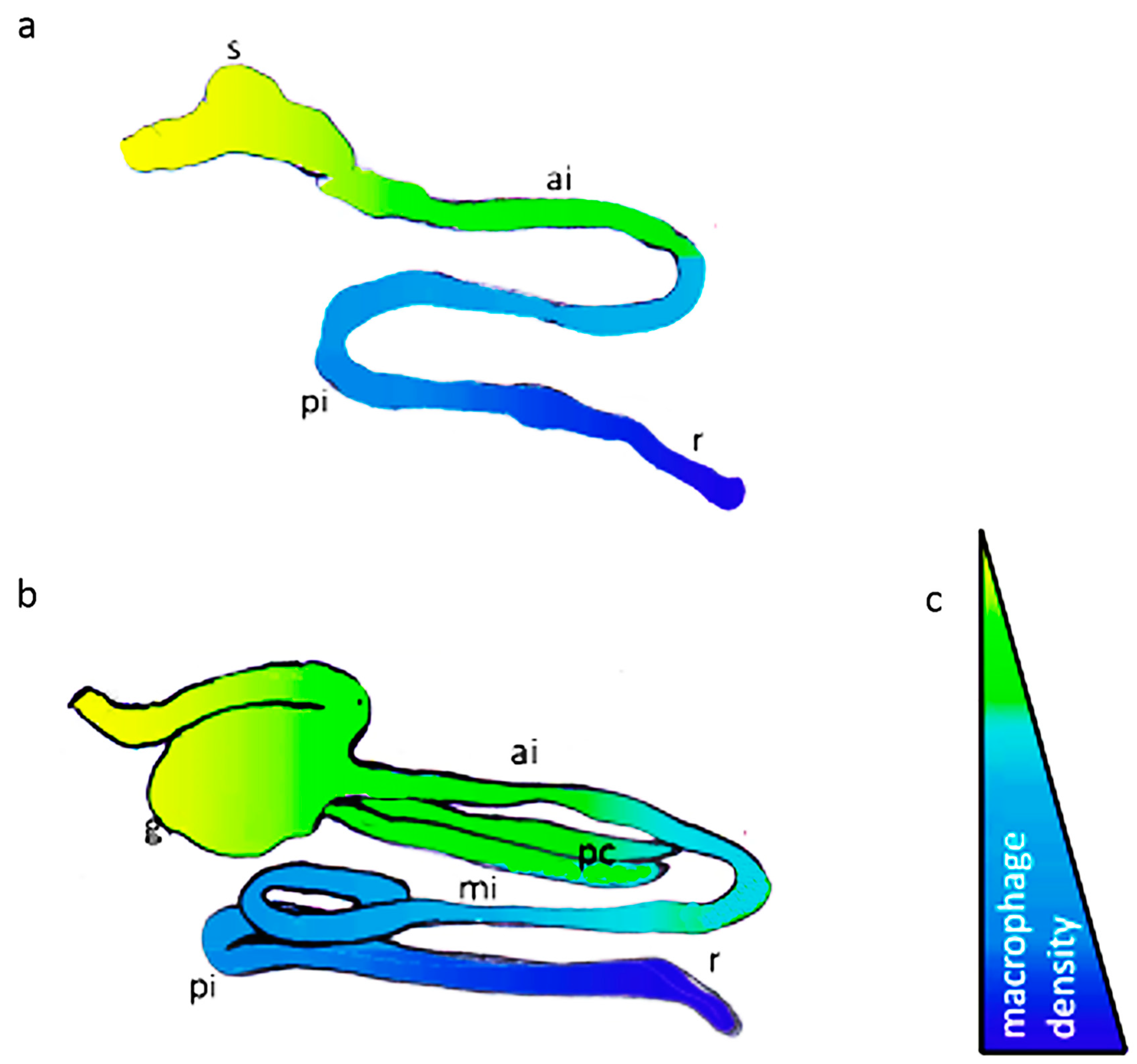
3.1. Intestinal Segmentation and Macrophage Density in the Anterior-Posterior Gut Axis of African Bonytongue and Indian Catfish

The gastrointestinal tract of both African bonytongue and Indian catfish is organized along the anterior-posterior (A-P) axis from the esophagus to the distal intestine. The regionalization of the gut along the A-P axis of the two species, unlike that reported in zebrafish [53], was not previously studied by differences in molecular, cellular, functional, and immunological properties. A morphological and structural study on the intestine of bonytogue is recently reported by Guerrera et al. (2022) [54].
The intestine of Indian catfish is long, coiled, and divided into three parts: proximal, middle, and distal intestine. The mucosa of the proximal intestine had numerous elongated and deep finger-like folds called villi, lined with columnar epithelial cells consisting of absorptive cells and mucous-secreting goblet cells. The stomach of H. fossilis consists of two parts: cardio-fundic and pyloric. Pyloric caeca are observed in the pyloric region of the stomach. The distal intestine showed an increase in mucous-secreting goblet cells.
In the African bonytongue the oropharyngeal cavity connects the stomach by a short esophagus. This latter is adapted to mechanical trituration, and it is divided into a pars glandularis and a thick-walled pars muscularis (the gizzard). The gizzard flows into the anterior intestine and two blind pyloric appendages. The anterior intestine continues with the middle and posterior tracts up into the rectum. According to the histological observations, all regions of the alimentary tract have common structural features, typical of hollow organs, with differences in the mucosa structure that reflects the different functions of the apparatus, from mouth to anus. H. niloticus, a specie of commercial interest, is distinguished by individual morphological characteristics showing a digestive tract similar to that of reptiles and birds [54].
The two studied fish species, such as the zebrafish, have elongated finger-like villi that project into the luminal space and are lined with a single layer of intestinal epithelial cells mediating the environmental interface of the gut tube [53,55]. Similar to mammals, the gut wall contains two muscle layers (circular and longitudinal), which are found surrounding the myenteric plexus containing intrinsic neurons of the enteric nervous system (ENS) and associated nerves and neuronal cell bodies as individuals or small groups.
As for previous comparative studies [53], we used a regionalization schema focused on the morphology and density of macrophages along the various segments of the gut (Figure 1) by statistical analysis and confocal imaging with double immunolabeling procedures. Interestingly, we noted an increasing anterior-to-posterior gradient of macrophages in the gut of the two fish species from a uniform density in the anterior intestine to a higher density in the distal intestine (Figure 1).
African bonytongue and Indian catfish harbor distinct mucosal, submucosal, and muscularis-associated gut macrophages.
Macrophages carry out diverse functions in the intestine that vary to their anatomical localization. We found macrophages that localize in the epithelium and subepithelium, the latter known as the lamina propria. A distinct group of macrophages localizes in the forefront of the mucosal epithelial barrier of the intestinal epithelium, beneath the lamina propria, also around the blood vessels and luminal space, between the circular and longitudinal muscle layers in the tissue region known as the muscularis externa.

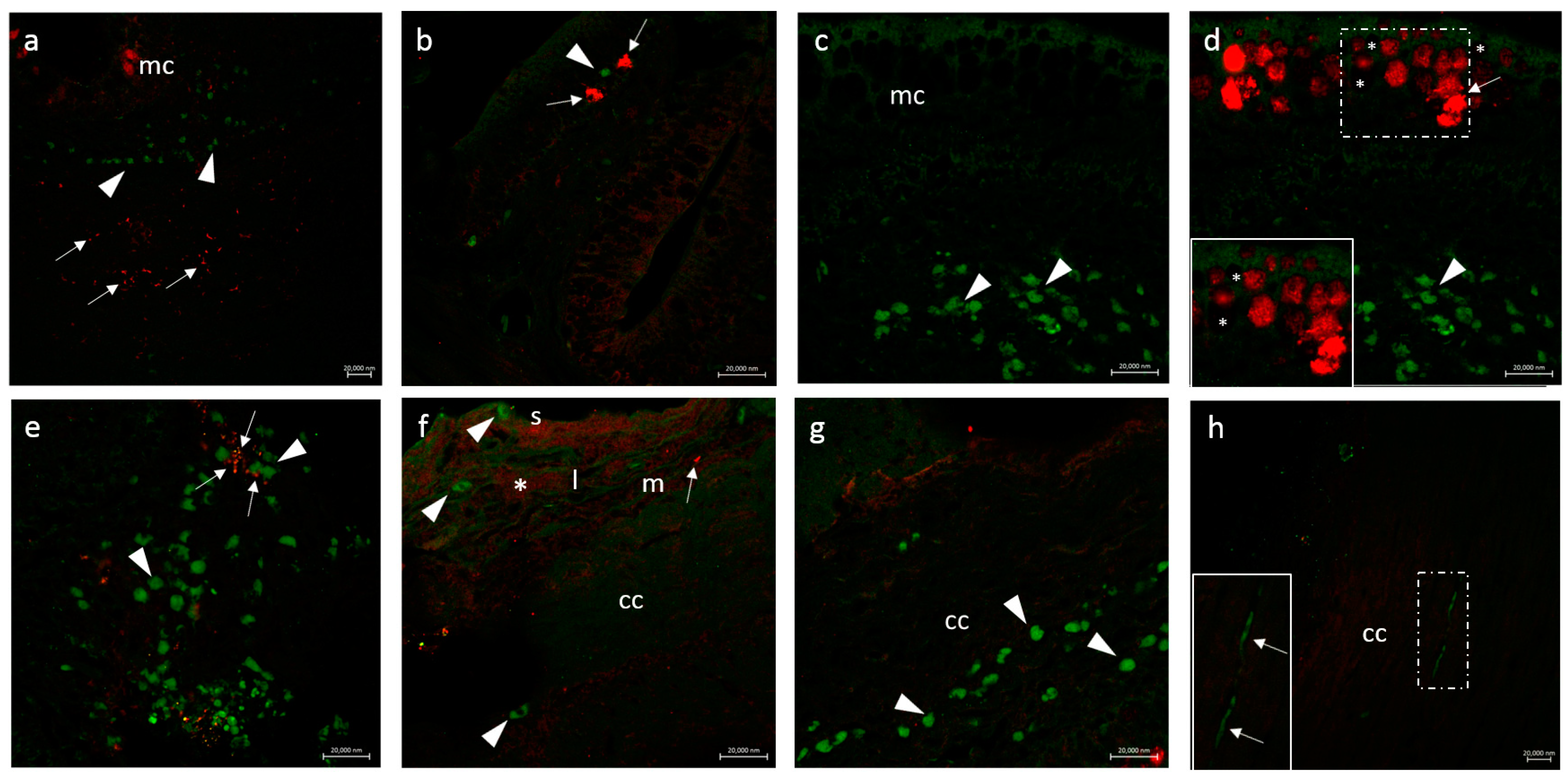
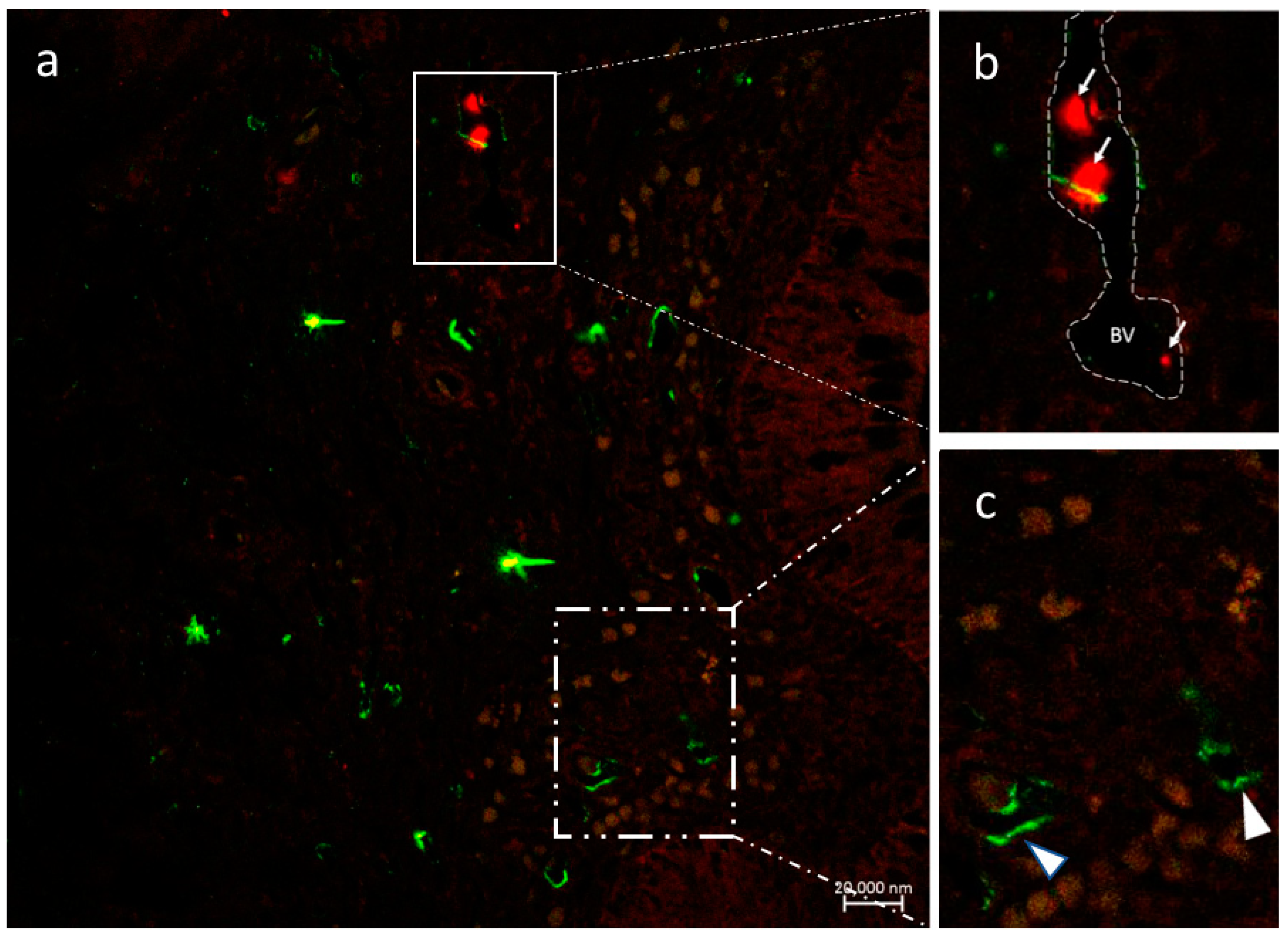
3.2. Double Labeling Methods

3.2.1. CD14 and VAChT

Imaging intestines of the two species studied and using double immunolabeling with antibodies against CD14 and VAChT, enabled observation of distinct cell populations in both the intestinal mucosa/lamina propria). This antibody pair was used to label intestinal macrophage phenotypes.
Macrophages containing VAChT immunoreactivity mainly localize in the intestinal epithelial layers, those immunoreacting to CD14 reside in the subepithelial layers of the intestine (Figure 2a,b). Differences in cellular morphologies were encountered in the macrophages located in the outermost intestinal epithelial layers. Few cells immunostained with VAChT antibody are lined with a single layer of epithelial cells or are seen exposed to the surface layers. Some cells showing a different morphology are found surrounding the mucous cells in the outer cell layers (Figure 2c,d). Interestingly, intestinal mucous cells produce acetylcholine (cholinergic mucous cells) and are labeled by the VAChT antibodies (Figure 2a).
Blood vessel-associated CD14-positive macrophages were observed in subepithelial layers. Macrophages in subepithelial localization are seen often in close association with VAChT-positive nerve fibers (Figure 2e). Macrophages associated with VAChT-positive nerve fibers are seen spanning the longitudinal muscle, the myenteric plexus, and the circular muscle (Figure 2f–h). Individual or grouped macrophages can be found extending their processes that align with the circular muscle. Sometimes more packed macrophages in the circular muscle show an intimate interaction with VAChT-positive nerve fibers.

3.2.2. CD14 and nAChR Alpha 7 Subunit

Macrophages were present in all the intestinal epithelial and subepithelial layers showing distinctive phenotypic markers (CD14 and alpha 7 subunit nAChR). Perivascular macrophages (PVMs) had close contact with blood vessels and lay on the outer (abluminal) surface of blood channels or the luminal surface. Figure 3 shows the presence of CD14 immunoreactive macrophages enwrapping a blood vessel (dashed line). Some macrophages are also found in the lumen showing immunoreactivity for alpha 7 subunit nAChR thus suggesting the presence of two cell phenotypes presumably involved in vascular integrity. Other cells showing quite different morphology are immunostained by the CD14 antibody (Figure 3).

3.2.3. Secretion of the Antimicrobial Peptide Piscidin 1 by Intestinal Macrophages: CD14 and Piscidin 1 Double Immunolabeling

Besides the CD14 phenotype, the macrophages are strongly labeled by the piscidin 1 antibodies. The piscidin 1 macrophages represent an abundant cell type and a distinct macrophage phenotype in the intestinal epithelium (Figure 4). In addition, mast cells are also immunostained by the same antibodies and are regular immune cell components of the gut epithelium (Figure 4).

3.2.4. VAChT and iNOS

Our immunohistochemical analysis using double immunolabeling with antibodies against iNOS and VAChT revealed the presence of a distinct subpopulation of macrophages expressing iNOS and a cholinergic one showing the co-occurrence of both VAChT and iNOS. The expression of markers defining the cell types is found in the macrophages that were consistently the most numerous in the subepithelial cell layers of the posterior intestine. Few cell types expressing iNOS and VAChT are also observed at the mucosal side of the intestine. We imaged nerve types positive for VAChT antibody positioned close to iNOS-VAChT immunoreactive resident macrophages in the subepithelial layers. Figure 5a,b shows the colocalization of iNOS and VAChT (a) and nerves (imaged by light microscopy) contouring the macrophages (b).

3.2.5. Immunolabeling with Antibodies against Calbindin, VAChT, and Tubulin

Calbindin-VAChT immunopositive enteric neurons colocalize with VAChT-positive macrophages in the submucosal plexus. Optical magnification reveals an intimate contact of some macrophages with enteric neurons (Figure 6a). Axons and nerve bundles associated with VAChT-positive macrophages express tubulin (Figure 6b). Tubulin-positive axons are found coursing through the cholinergic enteric neurons (Figure 6c). Enteric neurons are also seen associated with glial cells revealing a close association of tubulin-positive neuronal fibers with glial cells (Figure 6d).

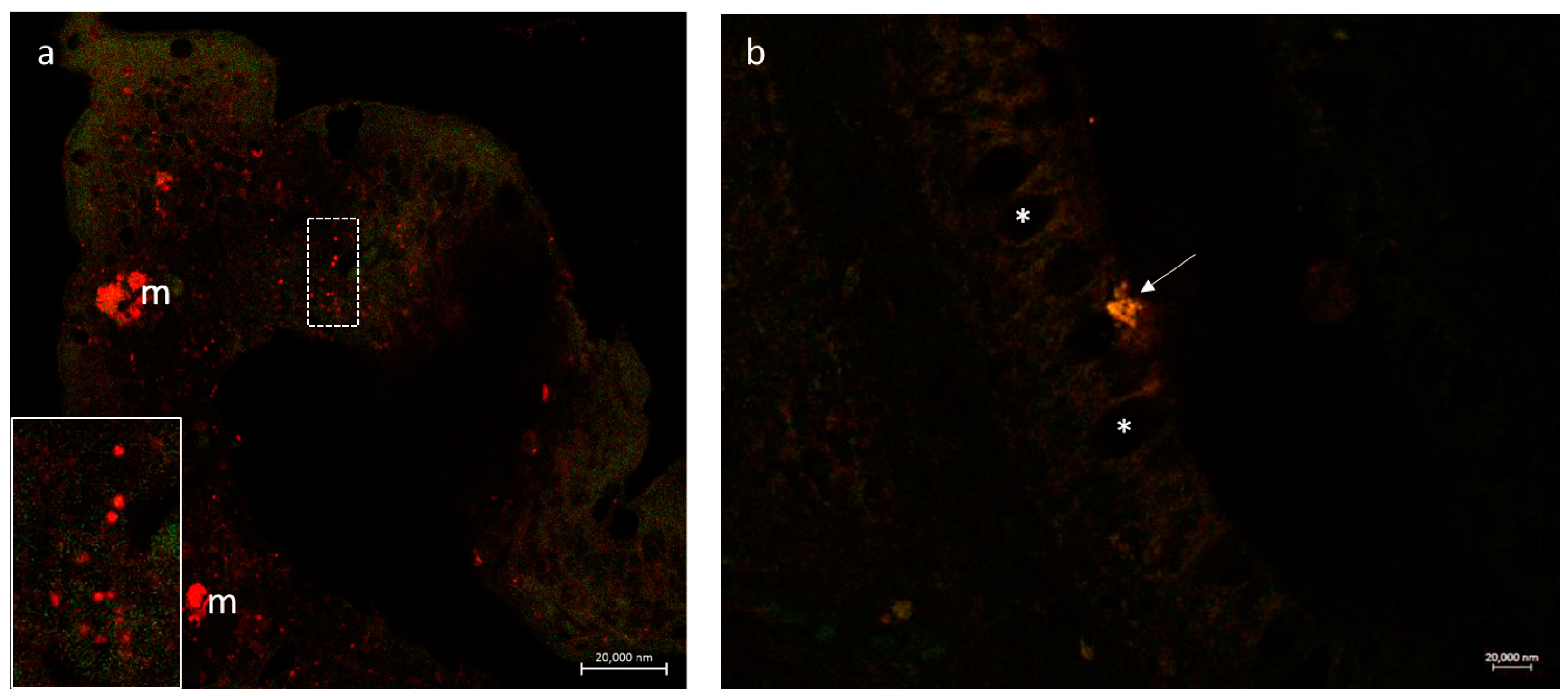
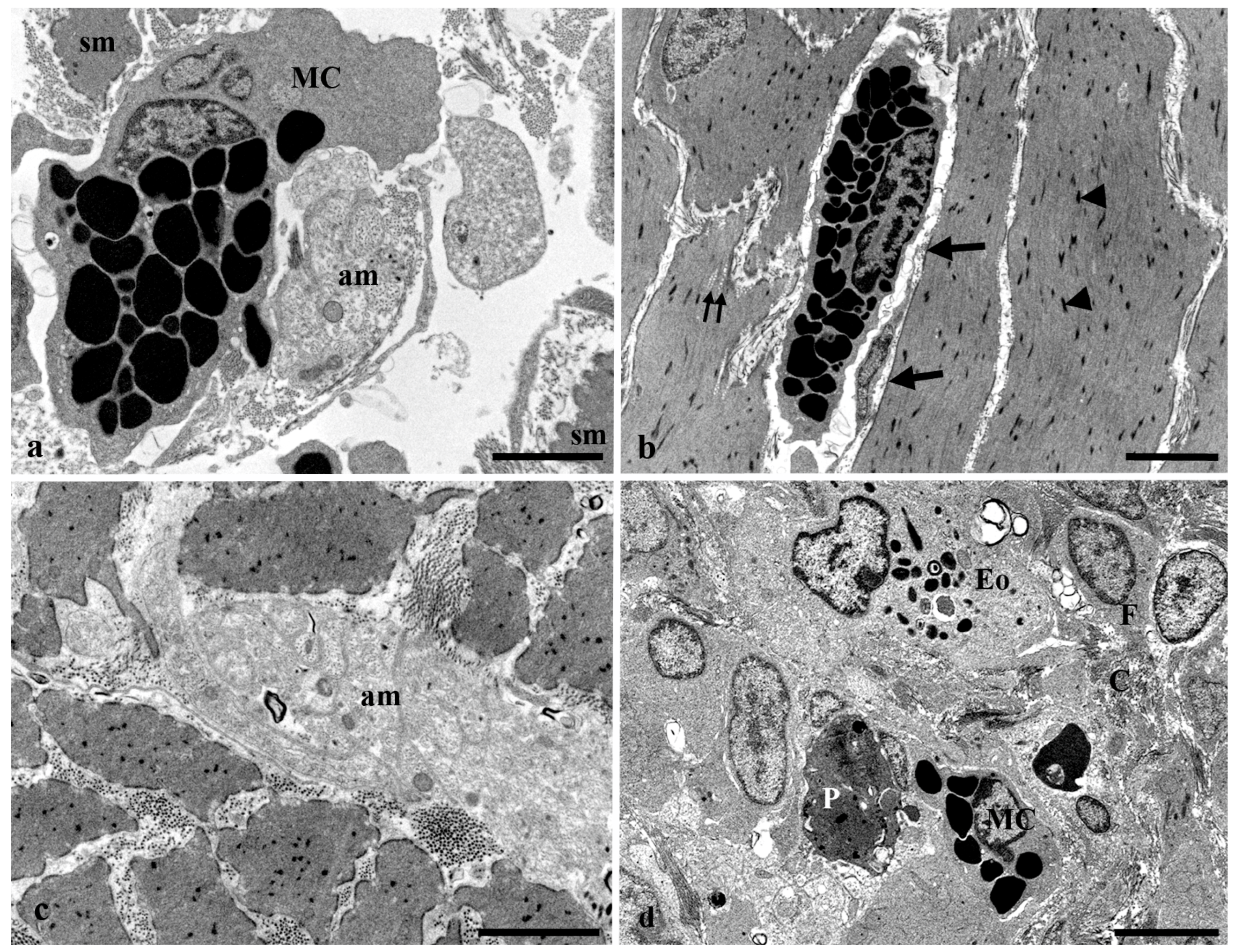
3.3. Transmission Electron Microscopy of the Long Gut of Heterotis Niloticus

The intestinal epithelium consists of columnar epithelial cells intermingled with numerous goblet cells (Figure 7a). A brush border formed by parallel microvilli and the presence of junctional complexes define the apical side of the epithelium (Figure 7a). The basal portion of the epithelium contains the epithelial cell nuclei and numerous death cells (Figure 7a,b). Death cells are characterized by chromatin marginalization and condensation, pyknotic nucleus, shrinkage of cytoplasm, and formation of apoptotic cell fragments (Figure 7b). Cellular fragments are surrounded by macrophage cytoplasmic extensions (Figure 7c) and can be seen incorporated into the macrophage cytoplasm. This occurs in the epithelium and the subepithelium as well (Figure 7d).
The subepithelium contains secretory cells that are similar to the Paneth cells observed in the gut epithelium of other vertebrates. These cells usually occur in small groups and are characterized by the presence of numerous electron-dense granules, dilated cisterna of rough endoplasmic reticulum, and mitochondria (Figure 8a,b). The subepithelium contains also numerous mast cells that appear singly or in small groups. Mast cells appear associated with neuroendocrine cells (Figure 8c), macrophages (Figure 8d), and amyelinic nerve fibers (Figure 9a).
The muscle layer is formed by smooth muscle cells arranged in longitudinal and circular orientations and contains numerous mast cells (Figure 9b) and amyelinic nerve fibers (Figure 9c) scattered among the muscle cells. Finally, the adventitia is a complex layer that contains eosinophils, mast cells, cellular debris, macrophages, and fibroblasts embedded in a collagenous matrix (Figure 9d).
Statistical analysis reveals a growing population of macrophages from the anterior gut to the posterior gut as detected with the tested antibodies (Table 2 and Table 3).

4. Discussion

4.1. Macrophage Diversity and Their Interaction with Enteric Neurons in the African Bonytongue and Catfish Gut

This study provides anatomical evidence for morphological heterogeneity among gut-associated macrophages in the two studied teleost fishes. We also found these macrophages to be associated with gut innervation. Using specific markers, we characterized intestinal macrophage phenotypes in detail along the anterior-to-posterior axis and the concentric muscle layers (longitudinal and circular muscle) of the adult fish.
Label experiments with antibodies against the calbindin and VAChT showed colocalization of enteric neurons and macrophages mainly in the submucosal plexus. Some macrophages are seen also engulfing neuronal somata.

4.2. Cholinergic System Components in the Intestinal Macrophages

Acetylcholine (ACh) is the first identified neurotransmitter that defines the chemical nature of neurotransmission both in the central nervous system and the periphery. Apart from neurotransmission, ACh can play anti-inflammatory roles in immune responses [56] and is synthesized by immune cells, i.e., T/B cells, macrophages, dendritic cells, and neutrophils [57]. Notably, the efferent vagus nerve is being suggested to interplay between the nervous and immune systems, which is known as an anti-inflammatory pathway (CAP). Today, CAP is the most notable cholinergic signaling in regulating immune responses.
Distinct populations of intestinal macrophages are reported in epithelial and subepithelial localizations in the intestines of two teleost species showing immunoreactivity for CD14 and VAChT and the alpha 7 subunit nicotinic receptor of the acetylcholine (alpha 7 nAChR).
All tissue macrophages are derived from bone marrow stem cells through a highly regulated cascade of differentiation events [58,59]. Gut macrophages are derived exclusively from recruited blood monocytes, and, after terminal differentiation, they become resident macrophages in the mucosa of the small intestine and colon. They express CD14 in the inflammatory lesions where recruited pro-inflammatory blood monocytes were found [18].
Macrophage development, polarization, and functional responses were reported in teleost fish and their antimicrobial mechanisms have been extensively studied in various fish models [8]. Similarities and differences have been documented for the regulation of antimicrobial defenses concerning mammalian counterparts including the mechanisms that determine and regulate the highly plastic functional phenotypes and host defenses/repair functions (M1 type-pro-inflammatory, M2 type anti-inflammatory).
CD14 and VAChT/nAChR-alpha7 positive macrophages found in the intestines of the two fish species studied here are to be regarded as distinct phenotypes expressing acetylcholine and its receptor alpha 7 nAChR. The recent discovery that macrophages, lymphocytes, and dendritic cells synthesize ACh and express several types of both muscarinic and nicotinic receptors (mAChRs and nAChRs) supports the existence of a local non-neuronal cholinergic system (NNCS) in these cells and in particular the role of alpha 7 receptor subunit of nAChR in the macrophages to stimulate the survival of the anti-inflammatory M2 phenotype [24,25,27,60,61].
A prominent feature of the intestinal macrophages of the two studied fish species is their interaction with the nervous network. Distinct subsets of resident tissue macrophages, which are associated with peripheral nerves in various tissues, have been only recently studied in more detail [62]. The gastrointestinal tract is innervated by the intrinsic enteric nervous system as well as extrinsic sympathetic and parasympathetic ganglia. We found macrophages associated with nerve fibers immunostained with antibodies against VAChT and acetylated tubulin throughout the resting lamina propria, subepithelial layers, and between the circular and longitudinal muscle layers of the muscularis externa. The macrophages in the circular muscle show an elongated and slender morphology in parallel with the associated nerve fibers. As emphasized by Kolter et al. (2020), macrophages have been characterized as a local surveillance system of nerves and thereby contribute to the integrity of nerve fibers. Additionally, resident macrophages relay inflammatory signals and act as mediators between the cells of the immune system. The macrophages in the muscularis externa are therefore to be regarded as markedly distinct from monocyte-derived macrophages and are defined by tissue and nerve type [62].
Interestingly, we found a close interaction of intestinal epithelial cells and mucous cells with the subset of mucosal macrophages immunostained with the antibodies against VAChT. More cells are seen both surrounding or engulfing mucous cells and have also a positive impact on the epithelial surface. This is in agreement with the recruitment of activated macrophages to the apical membrane of the intestinal epithelial cells and their promotion of the clearance of infection including the improvement of the epithelial barrier integrity [11]. The macrophages are thought to support mucins to trap harmful bacteria by phagocytosing apoptotic cells (secretory) in the intestinal epithelium [6,48]. In the present study, TEM observations show that macrophages delimit and engulf apoptotic cells and apoptotic fragments, presumably epithelial cells to control cell number.
In this study, we found different populations of intestinal macrophages in two air-breathing fish species, but their origin and development and their important roles in intestinal homeostasis and inflammation are not known. Unlike the mammalian models, different cytokines are produced by the immune cells that are responsible for the fate of the distinct macrophage populations.
Here we report for the first time the presence of cholinergic mucous cells (mucous cells immunoreactive to VAChT antibody) in the fish intestinal epithelium. Mucous cells play a vital role in the maintenance of the epithelial barrier function underpinned by rapid stem-cell-driven tissue renewal, and by secretion of a protective mucus layer. It represents a selective barrier between the mucosal immune system and a barrage of microorganisms. A cholinergic input is also implicated in the regulation of mucus secretion [63], but the mechanism of mucus secretion is not known. Acetylcholine is detected in a variety of non-neuronal cells where it acts as a para/autocrine signaling molecule controlling basic cell functions such as proliferation, differentiation, and maintenance of cell-cell contacts [64]. The NNCS is regarded as an ancient network that already existed from the beginning of life before the neural system had developed. The relevance of NNCS for macrophage function is recently emphasized [65]. In addition, the expression of the components involved in the cholinergic system, including choline transferase (ChAT), vesicular ACh transporter (VAChT), and choline transporter (CHT1) have been confirmed in macrophages [27] and the epithelial cells [28]. ACh is reported to participate in epithelial homeostasis [31]. It is tempting to speculate that NNCS of macrophages could interact with cholinergic components of the mucous cells to communicate with each other and maintain gut homeostasis.

4.3. The Piscidin 1 Is Expressed in Macrophages

The expression of antimicrobial peptides such as the cathelicidins and defensins in macrophages has been reported previously [66,67], but the localization of the antimicrobial peptide piscidin 1 has been not found in these cells except for the mast cells of the gills and accessory respiratory organs (ABOs) and the skin in fish [49].

4.4. iNOS and Macrophages

Our immunohistochemical analysis confirms the expression of markers defining a distinct nitrergic cholinergic and a nitrergic noncholinergic macrophage phenotype.
The current classification of macrophages is based on the M1/M2 paradigm, which is related to their involvement in pro- and anti-inflammation. M1 macrophages have been also intimately associated with the expression of inducible nitric oxide synthase (NOS2 or iNOS) to induce the production of NO from L-arginine (see for review [68]). The M1/M2 paradigm is regarded as too simplistic since macrophage populations show a continuum of diverse phenotypes even in the absence of inducers [69]. The role of M2 macrophages has been associated with tissue repair, the promotion of angiogenesis, and tissue remodeling [70]. Furthermore, little is known about the effects of cholinergic receptors on macrophages, except the well-investigated alpha 7-nAChR subunit, which has been shown to induce anti-inflammatory effects, leading to the activation of macrophages toward M2 phenotypes [71].
Our results demonstrate that a cholinergic and nitrergic component coexists in some cell subsets of macrophages as shown by colocalization procedures. Notably, mammalian studies have demonstrated a remarkable plasticity of macrophage phenotypes along with their ability to switch these phenotypes from the pro-inflammatory M1 to the anti-inflammatory M2 and vice versa, depending on the needs of the microenvironment [68]. NO is known to mediate inflammation under normal physiological conditions [72].
Several areas ripe for future experimentation emerge including the gene expression signatures and a need to identify macrophage subpopulations by transcriptome analysis in fish gut.

4.5. Neuro-Immune Axis in Fish Gut

An important aspect of the polarization of the macrophages is the interaction between the nervous and immune systems. CNS is known to regulate immune cell function and differentiation through efferent innervation by the direct effect of nerve fibers on immune cells [71].
Neuroimmune interaction in the intestine has evolved mechanisms for sensing and responding to the intestinal environment. Nerves are opposed to immune cells in the gastrointestinal tract to form neuro-immune cell units [73] that form a neuro-immune axis under the control of the intestinal microbiota [36,74]. Vagal nerves crosstalk with the immune cells and sense environmental cues and adapt to tissue infection [73].
Macrophages and nerves in the studied species reside close to the muscularis externa, which houses the enteric nervous system, the circular and longitudinal muscle, and the submucosal intestinal layers. Macrophage phenotypes characterized by iNOS and VAChT are present in the submucosal layers. A nerve network is seen surrounding the cell subsets that probably exhibit a pro-inflammatory phenotype. In addition, macrophages colocalize with enteric neurons in the submucosal and myenteric plexuses including communication between enteric neurons and glial cells visualized by the antibodies against the tubulin and VAChT. Thus, the macrophages relay inflammatory signals and act as mediators between the immune and the respective nervous system. The enteric glia is a part of the enteric nervous system, which is formed by acetylcholine receptors on glial cells [75]. They serve as a link between the nervous and immune systems of the gut as indicated by their potential to synthesize cytokines [75].
The neuroimmune communication in the fish gut has, however, not yet been known, and mechanisms underlying these interactions and molecular pathways require further investigation as compared with the mammalian counterpart [76,77].

5. Conclusions

Phagocytosis is the ancestral defense mechanism of all metazoan animals and is essential in preventing the dissemination of infectious agents. An inducible antimicrobial response, such as the respiratory burst pathway and production of NO has been demonstrated in fish phagocytes. Current studies point to the elucidation of multiple roles of fish macrophages in disease models as suggested by their function in vivo.
Therefore, our findings of cholinergic markers and antimicrobial substances reflect current thinking on significant similarities found in mammalian macrophages and represent an emerging field of study in fish.

Author Contributions

Conceptualization, G.Z. and J.M.I.; formal analysis, A.A.; investigation, G.Z., A.A., D.M.M., M.A. (Marialuisa Aragona), M.C.G., G.C., M.A. (Marco Albano), J.d.O.F., V.K., R.K.A.S., M.M.H., P.L.C., M.K., K.Z., A.G., J.M.I. and E.R.L.; data curation, A.A., M.A. (Marialuisa Aragona), M.C.G., G.C., M.A. (Marco Albano), J.d.O.F., V.K., R.K.A.S., M.M.H., P.L.C., M.K., K.Z. and A.G.; writing—original draft preparation, G.Z.; writing—review and editing, G.Z., A.A., D.M.M., G.C., J.d.O.F., J.M.I. and E.R.L.; visualization, A.A.; supervision, G.Z., D.M.M., J.M.I. and E.R.L. All authors have read and agreed to the published version of the manuscript.

Funding

This research received no external funding.

Institutional Review Board Statement

Ethical review and approval were waived for this study because no experimental procedures were applied to samples.

Informed Consent Statement

Not applicable.

Data Availability Statement

Not applicable.

Conflicts of Interest

The authors declare no conflict of interest.

References

  1. Amici, S.A.; Dong, J.; Guerau-de-Arellano, M. Molecular Mechanisms Modulating the Phenotype of Macrophages and Microglia. Front. Immunol. 2017, 8, 1520. [Google Scholar] [CrossRef] [PubMed] [Green Version]
  2. Vergadi, E.; Ieronymaki, E.; Lyroni, K.; Vaporidi, K.; Tsatsanis, C. Akt Signaling Pathway in Macrophage Activation and M1/M2 Polarization. J. Immunol. 2017, 198, 1006–1014. [Google Scholar] [CrossRef] [PubMed] [Green Version]
  3. Grayfer, L.; Kerimoglu, B.; Yaparla, A.; Hodgkinson, J.W.; Xie, J.; Belosevic, M. Mechanisms of Fish Macrophage Antimicrobial Immunity. Front. Immunol. 2018, 9, 1105. [Google Scholar] [CrossRef] [PubMed] [Green Version]
  4. Wiegertjes, G.F.; Wentzel, A.S.; Spaink, H.P.; Elks, P.M.; Fink, I.R. Polarization of Immune Responses in Fish: The ‘Macrophages First’Point of View. Mol. Immunol. 2016, 69, 146–156. [Google Scholar] [CrossRef] [PubMed]
  5. Mischopoulou, M.; D’Ambrosio, M.; Bigagli, E.; Luceri, C.; Farrugia, G.; Cipriani, G. Role of Macrophages and Mast Cells as Key Players in the Maintenance of Gastrointestinal Smooth Muscle Homeostasis and Disease. Cell. Mol. Gastroenterol. Hepatol. 2022, 13, 1849–1862. [Google Scholar] [CrossRef]
  6. Alesci, A.; Pergolizzi, S.; Fumia, A.; Calabrò, C.; Lo Cascio, P.; Lauriano, E.R. Mast Cells in Goldfish (Carassius auratus) Gut: Immunohistochemical Characterization. Acta Zool. 2022, azo.12417. [Google Scholar] [CrossRef]
  7. Viola, M.F.; Boeckxstaens, G. Intestinal Resident Macrophages: Multitaskers of the Gut. Neurogastroenterol. Motil. 2020, 32, e13843. [Google Scholar] [CrossRef] [Green Version]
  8. Hodgkinson, J.; Grayfer, L.; Belosevic, M. Biology of Bony Fish Macrophages. Biology 2015, 4, 881–906. [Google Scholar] [CrossRef]
  9. Pastorelli, L.; De Salvo, C.; Mercado, J.R.; Vecchi, M.; Pizarro, T.T. Central Role of the Gut Epithelial Barrier in the Pathogenesis of Chronic Intestinal Inflammation: Lessons Learned from Animal Models and Human Genetics. Front. Immunol. 2013, 4, 280. [Google Scholar] [CrossRef] [Green Version]
  10. Bahar, A.A.; Ren, D. Antimicrobial Peptides. Pharmaceuticals 2013, 6, 1543–1575. [Google Scholar] [CrossRef]
  11. Bording-Jorgensen, M.; Armstrong, H.; Wickenberg, M.; LaPointe, P.; Wine, E. Macrophages and Epithelial Cells Mutually Interact through NLRP3 to Clear Infection and Enhance the Gastrointestinal Barrier. Immuno 2021, 2, 13–25. [Google Scholar] [CrossRef]
  12. Rieger, A.M.; Hall, B.E.; Barreda, D.R. Macrophage Activation Differentially Modulates Particle Binding, Phagocytosis and Downstream Antimicrobial Mechanisms. Dev. Comp. Immunol. 2010, 34, 1144–1159. [Google Scholar] [CrossRef]
  13. Barreda, D. Flow Cytometric Analysis of PKH26-Labeled Goldfish Kidney-Derived Macrophages. Dev. Comp. Immunol. 2000, 24, 395–406. [Google Scholar] [CrossRef] [PubMed]
  14. Neumann, N.F.; Stafford, J.L.; Belosevic, M. Biochemical and Functional Characterisation of Macrophage Stimulating Factors Secreted by Mitogen-Induced Goldfish Kidney Leucocytes. Fish Shellfish Immunol. 2000, 10, 167–186. [Google Scholar] [CrossRef] [PubMed]
  15. Janeway, C.A. Approaching the Asymptote? Evolution and Revolution in Immunology. Cold Spring Harb. Symp. Quant. Biol. 1989, 54, 1–13. [Google Scholar] [CrossRef] [PubMed]
  16. Yoshioka, N.; Taniguchi, Y.; Yoshida, A.; Nakata, K.; Nishizawa, T.; Inagawa, H.; Kohchi, C.; Soma, G.-I. Intracellular Localization of CD14 Protein in Intestinal Macrophages. Anticancer Res. 2009, 29, 865–869. [Google Scholar]
  17. Zanoni, I.; Granucci, F. Role of CD14 in Host Protection against Infections and in Metabolism Regulation. Front. Cell. Infect. Microbiol. 2013, 3, 32. [Google Scholar] [CrossRef] [Green Version]
  18. Smith, P.D.; Smythies, L.E.; Shen, R.; Greenwell-Wild, T.; Gliozzi, M.; Wahl, S.M. Intestinal Macrophages and Response to Microbial Encroachment. Mucosal Immunol. 2011, 4, 31–42. [Google Scholar] [CrossRef] [Green Version]
  19. Alesci, A.; Pergolizzi, S.; Lo Cascio, P.; Fumia, A.; Lauriano, E.R. Neuronal Regeneration: Vertebrates Comparative Overview and New Perspectives for Neurodegenerative Diseases. Acta Zool. 2021, 103, 129–140. [Google Scholar] [CrossRef]
  20. Bi, D.; Wang, Y.; Gao, Y.; Li, X.; Chu, Q.; Cui, J.; Xu, T. Recognition of Lipopolysaccharide and Activation of NF-ΚB by Cytosolic Sensor NOD1 in Teleost Fish. Front. Immunol. 2018, 9, 1413. [Google Scholar] [CrossRef] [Green Version]
  21. Bernardo, D.; Marin, A.C.; Fernández-Tomé, S.; Montalban-Arques, A.; Carrasco, A.; Tristán, E.; Ortega-Moreno, L.; Mora-Gutiérrez, I.; Díaz-Guerra, A.; Caminero-Fernández, R. Human Intestinal Pro-Inflammatory CD11chighCCR2+ CX3CR1+ Macrophages, but Not Their Tolerogenic CD11c- CCR2- CX3CR1- Counterparts, Are Expanded in Inflammatory Bowel Disease. Mucosal Immunol. 2018, 11, 1114–1126. [Google Scholar] [CrossRef]
  22. Rueda Ruzafa, L.; Cedillo, J.L.; Hone, A.J. Nicotinic Acetylcholine Receptor Involvement in Inflammatory Bowel Disease and Interactions with Gut Microbiota. Int. J. Environ. Res. Public Health 2021, 18, 1189. [Google Scholar] [CrossRef] [PubMed]
  23. Mao, D.; Yasuda, R.P.; Fan, H.; Wolfe, B.B.; Kellar, K.J. Heterogeneity of Nicotinic Cholinergic Receptors in Rat Superior Cervical and Nodose Ganglia. Mol. Pharmacol. 2006, 70, 1693–1699. [Google Scholar] [CrossRef] [PubMed]
  24. Lee, R.H.; Vazquez, G. Evidence for a Prosurvival Role of Alpha-7 Nicotinic Acetylcholine Receptor in Alternatively (M 2)-Activated Macrophages. Physiol. Rep. 2013, 1, e00189. [Google Scholar] [CrossRef] [PubMed]
  25. Vieira-Alves, I.; Coimbra-Campos, L.; Sancho, M.; Da Silva, R.F.; Cortes, S.F.; Lemos, V.S. Role of the A7 Nicotinic Acetylcholine Receptor in the Pathophysiology of Atherosclerosis. Front. Physiol. 2020, 1719. [Google Scholar] [CrossRef]
  26. Morris, S.J.; Kunzek, S.; Shore, A.C. The Effect of Acetylcholine on Finger Capillary Pressure and Capillary Flow in Healthy Volunteers. J. Physiol. 1996, 494, 307–313. [Google Scholar] [CrossRef] [PubMed] [Green Version]
  27. Reardon, C.; Duncan, G.S.; Brüstle, A.; Brenner, D.; Tusche, M.W.; Olofsson, P.S.; Rosas-Ballina, M.; Tracey, K.J.; Mak, T.W. Lymphocyte-Derived ACh Regulates Local Innate but Not Adaptive Immunity. Proc. Natl. Acad. Sci. USA 2013, 110, 1410–1415. [Google Scholar] [CrossRef] [Green Version]
  28. Bankova, L.G.; Dwyer, D.F.; Yoshimoto, E.; Ualiyeva, S.; McGinty, J.W.; Raff, H.; von Moltke, J.; Kanaoka, Y.; Frank Austen, K.; Barrett, N.A. The Cysteinyl Leukotriene 3 Receptor Regulates Expansion of IL-25–Producing Airway Brush Cells Leading to Type 2 Inflammation. Sci. Immunol. 2018, 3, eaat9453. [Google Scholar] [CrossRef] [Green Version]
  29. Schütz, B.; Ruppert, A.-L.; Strobel, O.; Lazarus, M.; Urade, Y.; Büchler, M.W.; Weihe, E. Distribution Pattern and Molecular Signature of Cholinergic Tuft Cells in Human Gastro-Intestinal and Pancreatic-Biliary Tract. Sci. Rep. 2019, 9, 17466. [Google Scholar] [CrossRef] [Green Version]
  30. Schneider, C.; O’Leary, C.E.; Locksley, R.M. Regulation of Immune Responses by Tuft Cells. Nat. Rev. Immunol. 2019, 19, 584–593. [Google Scholar] [CrossRef]
  31. Pan, J.; Zhang, L.; Shao, X.; Huang, J. Acetylcholine from Tuft Cells: The Updated Insights beyond Its Immune and Chemosensory Functions. Front. Cell Dev. Biol. 2020, 8, 606. [Google Scholar] [CrossRef] [PubMed]
  32. Capillo, G.; Zaccone, G.; Cupello, C.; Fernandes, J.M.O.; Viswanath, K.; Kuciel, M.; Zuwala, K.; Guerrera, M.C.; Aragona, M.; Icardo, J.M. Expression of Acetylcholine, Its Contribution to Regulation of Immune Function and O2 Sensing and Phylogenetic Interpretations of the African Butterfly Fish Pantodon buchholzi (Osteoglossiformes, Pantodontidae). Fish Shellfish Immunol. 2021, 111, 189–200. [Google Scholar] [CrossRef] [PubMed]
  33. Masini, E.; Fantozzi, R.; Blandina, P.; Brunelleschi, S.; Mannaioni, P.F. Muscarinic Cholinergic Receptor Binding in Rat Mast Cells. Agents Actions 1983, 13, 327–332. [Google Scholar] [CrossRef] [PubMed]
  34. Kageyama-Yahara, N.; Suehiro, Y.; Yamamoto, T.; Kadowaki, M. IgE-Induced Degranulation of Mucosal Mast Cells Is Negatively Regulated via Nicotinic Acetylcholine Receptors. Biochem. Biophys. Res. Commun. 2008, 377, 321–325. [Google Scholar] [CrossRef]
  35. Bosmans, G.; Shimizu Bassi, G.; Florens, M.; Gonzalez-Dominguez, E.; Matteoli, G.; Boeckxstaens, G.E. Cholinergic Modulation of Type 2 Immune Responses. Front. Immunol. 2017, 8, 1873. [Google Scholar] [CrossRef] [PubMed] [Green Version]
  36. Yoo, B.B.; Mazmanian, S.K. The Enteric Network: Interactions between the Immune and Nervous Systems of the Gut. Immunity 2017, 46, 910–926. [Google Scholar] [CrossRef] [Green Version]
  37. Rahman, Z.; Dandekar, M.P. Crosstalk between Gut Microbiome and Immunology in the Management of Ischemic Brain Injury. J. Neuroimmunol. 2021, 353, 577498. [Google Scholar] [CrossRef]
  38. Hirota, C.L.; McKay, D.M. Cholinergic Regulation of Epithelial Ion Transport in the Mammalian Intestine: Cholinergically Mediated Intestinal Ion Transport. Br. J. Pharmacol. 2006, 149, 463–479. [Google Scholar] [CrossRef]
  39. Zaccone, G.; Capillo, G.; Aragona, M.; Alesci, A.; Cupello, C.; Lauriano, E.R.; Guerrera, M.C.; Kuciel, M.; Zuwala, K.; Germana, A. Gill Structure and Neurochemical Markers in the African Bonytongue (Heterotis niloticus): A Preliminary Study. Acta Histochem. 2022, 124, 151954. [Google Scholar] [CrossRef]
  40. Alesci, A.; Pergolizzi, S.; Lo Cascio, P.; Capillo, G.; Lauriano, E.R. Localization of Vasoactive Intestinal Peptide and Toll-like Receptor 2 Immunoreactive Cells in Endostyle of Urochordate Styela plicata (Lesueur, 1823). Microsc. Res. Tech. 2022, jemt.24119. [Google Scholar] [CrossRef]
  41. Alesci, A.; Cicero, N.; Fumia, A.; Petrarca, C.; Mangifesta, R.; Nava, V.; Lo Cascio, P.; Gangemi, S.; Di Gioacchino, M.; Lauriano, E.R. Histological and Chemical Analysis of Heavy Metals in Kidney and Gills of Boops Boops: Melanomacrophages Centers and Rodlet Cells as Environmental Biomarkers. Toxics 2022, 10, 218. [Google Scholar] [CrossRef] [PubMed]
  42. Pergolizzi, S.; Rizzo, G.; Favaloro, A.; Alesci, A.; Pallio, S.; Melita, G.; Cutroneo, G.; Lauriano, E.R. Expression of VAChT and 5-HT in Ulcerative Colitis Dendritic Cells. Acta Histochem. 2021, 123, 151715. [Google Scholar] [CrossRef] [PubMed]
  43. Alesci, A.; Capillo, G.; Mokhtar, D.M.; Fumia, A.; D’Angelo, R.; Lo Cascio, P.; Albano, M.; Guerrera, M.C.; Sayed, R.K.A.; Spanò, N.; et al. Expression of Antimicrobic Peptide Piscidin1 in Gills Mast Cells of Giant Mudskipper Periophthalmodon Schlosseri (Pallas, 1770). Int. J. Mol. Sci. 2022, 23, 13707. [Google Scholar] [CrossRef]
  44. Alesci, A.; Pergolizzi, S.; Savoca, S.; Fumia, A.; Mangano, A.; Albano, M.; Messina, E.; Aragona, M.; Lo Cascio, P.; Capillo, G.; et al. Detecting Intestinal Goblet Cells of the Broadgilled Hagfish Eptatretus Cirrhatus (Forster, 1801): A Confocal Microscopy Evaluation. Biology 2022, 11, 1366. [Google Scholar] [CrossRef] [PubMed]
  45. Alesci, A.; Albano, M.; Savoca, S.; Mokhtar, D.M.; Fumia, A.; Aragona, M.; Lo Cascio, P.; Hussein, M.M.; Capillo, G.; Pergolizzi, S.; et al. Confocal Identification of Immune Molecules in Skin Club Cells of Zebrafish (Danio rerio, Hamilton 1882) and Their Possible Role in Immunity. Biology 2022, 11, 1653. [Google Scholar] [CrossRef]
  46. Alesci, A.; Pergolizzi, S.; Capillo, G.; Lo Cascio, P.; Lauriano, E.R. Rodlet Cells in Kidney of Goldfish (Carassius auratus, Linnaeus 1758): A Light and Confocal Microscopy Study. Acta Histochem. 2022, 124, 151876. [Google Scholar] [CrossRef]
  47. Lauriano, E.R.; Guerrera, M.C.; Laurà, R.; Capillo, G.; Pergolizzi, S.; Aragona, M.; Abbate, F.; Germanà, A. Effect of Light on the Calretinin and Calbindin Expression in Skin Club Cells of Adult Zebrafish. Histochem. Cell Biol. 2020, 154, 495–505. [Google Scholar] [CrossRef]
  48. Alesci, A.; Capillo, G.; Fumia, A.; Messina, E.; Albano, M.; Aragona, M.; Lo Cascio, P.; Spanò, N.; Pergolizzi, S.; Lauriano, E.R. Confocal Characterization of Intestinal Dendritic Cells from Myxines to Teleosts. Biology 2022, 11, 1045. [Google Scholar] [CrossRef]
  49. Zaccone, G.; Capillo, G.; Fernandes, J.M.O.; Kiron, V.; Lauriano, E.R.; Alesci, A.; Lo Cascio, P.; Guerrera, M.C.; Kuciel, M.; Zuwala, K.; et al. Expression of the Antimicrobial Peptide Piscidin 1 and Neuropeptides in Fish Gill and Skin: A Potential Participation in Neuro-Immune Interaction. Mar. Drugs 2022, 20, 145. [Google Scholar] [CrossRef]
  50. Lauriano, E.R.; Pergolizzi, S.; Lo Cascio, P.; Kuciel, M.; Zizzo, N.; Guerrera, M.C.; Aragona, M.; Capillo, G. Expression of Langerin/CD207 in Airways, Lung and Associated Lymph Nodes of a Stranded Striped Dolphin (Stenella coeruleoalba). Acta Histochem. 2020, 122, 151471. [Google Scholar] [CrossRef]
  51. Lauriano, E.R.; Capillo, G.; Icardo, J.M.; Fernandes, J.M.O.; Kiron, V.; Kuciel, M.; Zuwala, K.; Guerrera, M.C.; Aragona, M.; Zaccone, G. Neuroepithelial Cells (NECs) and Mucous Cells Express a Variety of Neurotransmitters and Neurotransmitter Receptors in the Gill and Respiratory Air-Sac of the Catfish Heteropneustes fossilis (Siluriformes, Heteropneustidae): A Possible Role in Local Immune Defence. Zoology 2021, 148, 125958. [Google Scholar] [CrossRef]
  52. Schneider, C.A.; Rasband, W.S.; Eliceiri, K.W. NIH Image to ImageJ: 25 Years of Image Analysis. Nat. Methods 2012, 9, 671–675. [Google Scholar] [CrossRef]
  53. Graves, C.L.; Chen, A.; Kwon, V.; Shiau, C.E. Zebrafish Harbor Diverse Intestinal Macrophage Populations Including a Subset Intimately Associated with Enteric Neural Processes. Iscience 2021, 24, 102496. [Google Scholar] [CrossRef] [PubMed]
  54. Guerrera, M.C.; Aragona, M.; Briglia, M.; Porcino, C.; Mhalhel, K.; Cometa, M.; Abbate, F.; Montalbano, G.; Laurà, R.; Levanti, M. The Alimentary Tract of African Bony-Tongue, Heterotis niloticus (Cuvier, 1829): Morphology Study. Animals 2022, 12, 1565. [Google Scholar] [CrossRef] [PubMed]
  55. Wallace, K.N.; Akhter, S.; Smith, E.M.; Lorent, K.; Pack, M. Intestinal Growth and Differentiation in Zebrafish. Mech. Dev. 2005, 122, 157–173. [Google Scholar] [CrossRef] [PubMed]
  56. Fernández-Cabezudo, M.J.; George, J.A.; Bashir, G.; Mohamed, Y.A.; Al-Mansori, A.; Qureshi, M.M.; Lorke, D.E.; Petroianu, G.; al-Ramadi, B.K. Involvement of Acetylcholine Receptors in Cholinergic Pathway-Mediated Protection Against Autoimmune Diabetes. Front. Immunol. 2019, 10, 1038. [Google Scholar] [CrossRef]
  57. Liu, E.Y.; Xia, Y.; Kong, X.; Guo, M.S.; Anna, X.D.; Zheng, B.Z.; Mak, S.; Xu, M.L.; Tsim, K.W. Interacting with A7 NAChR Is a New Mechanism for AChE to Enhance the Inflammatory Response in Macrophages. Acta Pharm. Sin. B 2020, 10, 1926–1942. [Google Scholar] [CrossRef]
  58. Roach, S.K.; Lee, S.-B.; Schorey, J.S. Differential Activation of the Transcription Factor Cyclic AMP Response Element Binding Protein (CREB) in Macrophages Following Infection with Pathogenic and Nonpathogenic Mycobacteria and Role for CREB in Tumor Necrosis Factor Alpha Production. Infect. Immun. 2005, 73, 514–522. [Google Scholar] [CrossRef] [Green Version]
  59. Valledor, A.F.; Borras, F.E.; Cullell-Young, M.; Celada, A. Transcription Factors That Regulate Monocyte/Macrophage Differentiation. J. Leukoc. Biol. 1998, 63, 405–417. [Google Scholar] [CrossRef]
  60. Borovikova, L.V.; Ivanova, S.; Zhang, M.; Yang, H.; Botchkina, G.I.; Watkins, L.R.; Wang, H.; Abumrad, N.; Eaton, J.W.; Tracey, K.J. Vagus Nerve Stimulation Attenuates the Systemic Inflammatory Response to Endotoxin. Nature 2000, 405, 458–462. [Google Scholar] [CrossRef]
  61. Kiguchi, N.; Kobayashi, D.; Saika, F.; Matsuzaki, S.; Kishioka, S. Inhibition of Peripheral Macrophages by Nicotinic Acetylcholine Receptor Agonists Suppresses Spinal Microglial Activation and Neuropathic Pain in Mice with Peripheral Nerve Injury. J. Neuroinflamm. 2018, 15, 1–13. [Google Scholar] [CrossRef] [PubMed]
  62. Kolter, J.; Kierdorf, K.; Henneke, P. Origin and Differentiation of Nerve-Associated Macrophages. J. Immunol. 2020, 204, 271–279. [Google Scholar] [CrossRef] [PubMed]
  63. Specian, R.D.; Neutra, M.R. Mechanism of Rapid Mucus Secretion in Goblet Cells Stimulated by Acetylcholine. J. Cell Biol. 1980, 85, 626–640. [Google Scholar] [CrossRef] [Green Version]
  64. En-Nosse, M.; Hartmann, S.; Trinkaus, K.; Alt, V.; Stigler, B.; Heiss, C.; Kilian, O.; Schnettler, R.; Lips, K.S. Expression of Non-Neuronal Cholinergic System in Osteoblast-like Cells and Its Involvement in Osteogenesis. Cell Tissue Res. 2009, 338, 203–215. [Google Scholar] [CrossRef] [PubMed]
  65. Reichrath, S.; Reichrath, J.; Moussa, A.-T.; Meier, C.; Tschernig, T. Targeting the Non-Neuronal Cholinergic System in Macrophages for the Management of Infectious Diseases and Cancer: Challenge and Promise. Cell Death Discov. 2016, 2, 1–4. [Google Scholar] [CrossRef] [Green Version]
  66. Selsted, M.E.; Brown, D.M.; DeLange, R.J.; Lehrer, R.I. Primary Structures of MCP-1 and MCP-2, Natural Peptide Antibiotics of Rabbit Lung Macrophages. J. Biol. Chem. 1983, 258, 14485–14489. [Google Scholar] [CrossRef]
  67. Lehrer, R.I.; Selsted, M.E.; Szklarek, D.; Fleischmann, J. Antibacterial Activity of Microbicidal Cationic Proteins 1 and 2, Natural Peptide Antibiotics of Rabbit Lung Macrophages. Infect. Immun. 1983, 42, 10–14. [Google Scholar] [CrossRef] [Green Version]
  68. Poltavets, A.S.; Vishnyakova, P.A.; Elchaninov, A.V.; Sukhikh, G.T.; Fatkhudinov, T.K. Macrophage Modification Strategies for Efficient Cell Therapy. Cells 2020, 9, 1535. [Google Scholar] [CrossRef]
  69. Specht, H.; Emmott, E.; Petelski, A.A.; Huffman, R.G.; Perlman, D.H.; Serra, M.; Kharchenko, P.; Koller, A.; Slavov, N. Single-Cell Proteomic and Transcriptomic Analysis of Macrophage Heterogeneity; Immunology: Singapore, 2019. [Google Scholar]
  70. Mantovani, A.; Sica, A.; Locati, M. Macrophage Polarization Comes of Age. Immunity 2005, 23, 344–346. [Google Scholar] [CrossRef] [Green Version]
  71. Harwani, S.C. Macrophages under Pressure: The Role of Macrophage Polarization in Hypertension. Transl. Res. 2018, 191, 45–63. [Google Scholar] [CrossRef]
  72. Chokshi, N.K.; Guner, Y.S.; Hunter, C.J.; Upperman, J.S.; Grishin, A.; Ford, H.R. The Role of Nitric Oxide in Intestinal Epithelial Injury and Restitution in Neonatal Necrotizing Enterocolitis. In Proceedings of the Seminars in Perinatology; Elsevier: Amsterdam, The Netherlands, 2008; Volume 32, pp. 92–99. [Google Scholar]
  73. Godinho-Silva, C.; Cardoso, F.; Veiga-Fernandes, H. Neuro–Immune Cell Units: A New Paradigm in Physiology. Annu. Rev. Immunol. 2019, 37, 19–46. [Google Scholar] [CrossRef]
  74. Chu, C.; Artis, D.; Chiu, I.M. Neuro-Immune Interactions in the Tissues. Immunity 2020, 52, 464–474. [Google Scholar] [CrossRef] [PubMed]
  75. Rühl, A. Glial Cells in the Gut. Neurogastroenterol. Motil. 2005, 17, 777–790. [Google Scholar] [CrossRef] [PubMed]
  76. Matteoli, G.; Gomez-Pinilla, P.J.; Nemethova, A.; Di Giovangiulio, M.; Cailotto, C.; van Bree, S.H.; Michel, K.; Tracey, K.J.; Schemann, M.; Boesmans, W. A Distinct Vagal Anti-Inflammatory Pathway Modulates Intestinal Muscularis Resident Macrophages Independent of the Spleen. Gut 2014, 63, 938–948. [Google Scholar] [CrossRef] [PubMed]
  77. Kulkarni, S.; Kurapati, S.; Bogunovic, M. Neuro-Innate Immune Interactions in Gut Mucosal Immunity. Curr. Opin. Immunol. 2021, 68, 64–71. [Google Scholar] [CrossRef] [PubMed]
Figure 1. Schematic drawings depicting the morphology of the digestive tract, its segmentation, and macrophage density in Indian catfish (a) and African bonytongue (b). Colorimetric representation of macrophage density in the gastrointestinal tract (c). s (stomach); g (gizzard); ai (anterior intestine); pc (pyloric caeca or blind processes); mi (middle intestine); pi (posterior intestine); r (rectum).
Fishes 08 00043 g001
Figure 2. (ah): Confocal images of African bonytongue and catfish intestines. Double immunolabeling with antibodies against CD14 (green) and VAChT (red). (a) H. niloticus: confocal image highlighting the diversity of macrophage phenotypes containing CD14 (arrowheads) in the subepithelial layer and VAChT (arrows) in the muscularis-associated immunoreactivity in the middle intestine. Cholinergic mucous cells (mc) display VAChT immunoreactivity. (b) H. fossilis. Residing macrophage populations in the intestinal epithelium of pyloric caeca mucosal folds showing CD14 (arrowheads) and VAChT (arrows) immunoreactivity. (c) H. niloticus: distal intestine. VAChT-positive macrophages (arrowheads) with extending processes are observed in the mucosal layer engulfing mucous cells (mc) as compared with the lacking macrophage (arrow) occupation in (d) Large mucous cells (asterisks) are lightly stained. (e) H. niloticus. Distal intestine. Macrophages are visualized by antibody to CD14 (arrowheads) in the subepithelium to highlight their close contact with VAChT-positive nerve endings (arrows). (f) H. niloticus. Distal intestine CD14 positive macrophages (arrowheads) spanning the serosa (S), the longitudinal muscle (l), and the myenteric plexus (m). Note the close vicinity of the macrophages with the VAChT-positive nerve endings (asterisk); circular muscle (cc); macrophage of the enteric system (arrow). (g) H. niloticus. Distal intestine. Macrophages (arrowheads) spanning the circular muscle (cc) of the intestine highlight the diversity of the phenotype. (h) Macrophages (arrows) align with the muscle cells (cc). Scale bar (ah) = 20 nm.
Fishes 08 00043 g002
Figure 3. (ac): African bonytongue distal intestine. Double immunolabeling immunofluorescence with antibodies against CD14 and alpha 7 nAChR subunit demonstrating the presence of alpha 7 nAChR in a subset of macrophages (arrows) found in the lumen of a blood vessel (inset 1 is a region of the upper image in (a)). Inset 2 (that is a region of the image below in (a)) demonstrates a punctate fluorescence pattern for both the two markers in a cell subset of subepithelial macrophages. CD14 antibody labels a macrophage phenotype population in green (arrowheads). Scale bar = 20 nm.
Fishes 08 00043 g003
Figure 4. (a,b): Piscidin immunoreactivity in the intestine of African bonytongue and Indian catfish. (a) A subset of macrophages (m) and mast cells (inset) immunoreactive to the piscidin 1 antibody in the intestine of Indian catfish. (b) A macrophage (arrow) expressing both CD14 and piscidin 1 in the surface layer of African bonytongue intestinal epithelium; mucous cells (asterisks). Scale bar = 20 nm.
Fishes 08 00043 g004
Figure 5. (a) Confocal image of African bonytongue intestine immunolabeled with antibodies against VAChT (red) and iNOS (green). The co-expression of the two markers is noticed in a subset of subepithelial macrophages (red arrowheads). (b) The macrophages (red arrowheads) are imaged by light microscope to reveal their close interaction with nerve endings (arrows in the inset). Scale bar = 20 nm.
Fishes 08 00043 g005
Figure 6. (ad): Enteric neuron–macrophage interaction in the submucosal plexus of African bonytongue distal intestine. (a) Note the colocalization of calbindin (green)-VAChT (red)-positive enteric neurons (E) with VAChT-positive macrophages (arrows). The boxed area is magnified in the inset revealing the close apposition of a macrophage (arrow) with an enteric neuron (E) undergoing apoptosis. (b) Enteric neurons (E) within the submucosal plexus of H. niloticus distal intestine showing anatomical interaction with VAChT-positive macrophages (arrows). Note the presence of macrophages (arrows) positioned along tubulin-positive axons (arrowheads in the inset). (c) Tubulin-positive (green) axons (arrowheads) course through the cholinergic mesenteric neurons (E) within the submucosal plexus. (d) Enteric neuron–glial cell interaction in the submucosal plexus reveals the close apposition of tubulin-positive neuronal fibers (arrowheads) with glial cells (GL). Enteric neurons (E). Scale bar = 20 nm.
Fishes 08 00043 g006
Figure 7. TEM views of the epithelium and subepithelium. (a) The apical side of gut epithelium shows epithelial cell nuclei (n), apical junctional complexes (arrowheads), and brush border. G, goblet cells. (b) Basal epithelium. A death cell shows peripheral chromatin condensation (arrow) and numerous apoptotic bodies. This cell is surrounded by healthy epithelial cells. (c) Cellular debris is surrounded by macrophage cytoplasmic extensions (arrows). G, goblet cells. (d) Muscularis mucosa. A rounded macrophage (n, nucleus) shows engulfed bodies and secondary lysosomes (arrows). sm, smooth muscle cell. Scale bars: A, 5 μm; B, 2 μm; C, D, 1 μm.
Fishes 08 00043 g007
Figure 8. TEM views of the subepithelium. (a) Secretory cells with electron-dense secretory granules and rough endoplasmic reticulum (rER). m, mitochondrion; n, nucleus. (b) Note the abundance of rER and the presence of an immature granule (arrow) at the end of an rER cisterna. (c) Association between mast cell (MC) and neuroendocrine cell (arrow). F, fibroblasts. (d) Cellular debris in the center of the figure are being surrounded by macrophage cytoplasmic extensions (arrows). Note the presence of mast cells (MC) and fibroblasts (F). Scale bars: A, 2 μm; B, 1 μm; C, D, 2 μm.
Fishes 08 00043 g008
Figure 9. TEM views of muscle layers and adventitia. (a) Association of mast cell (MC) and amyelinic nerve fibers (am) in the muscularis mucosa. sm, smooth muscle cells. (b) A mast cell is surrounded by smooth muscle cells in the circular muscle layer. Note subplasmalemmal condensations (arrows), dense bodies (arrowheads), and pinocytotic-like vesicles (double arrow). (c) Amyelinic nerve fibers (am) among smooth muscle cells in the longitudinal muscle layer. (d) The adventitial layer contains eosinophils (Eo), mast cells (MC), pyknotic cells (P), fibroblasts (F), and collagen (C). Scale bars: A, 2 μm; B, 3 μm; C, 2 μm; D, 3 μm.
Fishes 08 00043 g009
Table 1. Antibodies used in this study.
AntibodySupplierDilutionAnimal Source
CD14Santa Cruz Biotechnology, Inc., Dallas, TX, USA1:200Mouse
VAChTSigma-Aldrich, Inc., St. Louis, MO, USA1:300Rabbit
nAChR-alpha7Alomone Labs, Ltd., Jerusalem, Israel1:200Rabbit
iNOSSanta Cruz Biotechnology, Inc., Dallas, TX, USA1:200Mouse
TubulinSanta Cruz Biotechnology, Inc., Dallas, TX, USA1:500Mouse
CalbindinSigma-Aldrich, Inc., St. Louis, MO, USA1:100Mouse
Alexa Fluor 488 Donkey anti-Mouse IgG FITC conjugatedMolecular Probes, Invitrogen1:300Donkey
Alexa Fluor 594 Donkey anti-Rabbit IgG TRITC conjugatedMolecular Probes, Invitrogen1:300Donkey
Table 2. Statistical analysis of the number of macrophages on H. niloticus gut samples (n = 10).
Anterior IntestineMiddle IntestineDistal Intestine
Piscidin1112.45 ± 21.72 *163.56 ± 13.89 **/
CD14102.66 ± 8.37 **154.96 ± 14.75 *294.74 ± 45.16 *
nAChR-alpha799.54 ± 8.93 **//
iNOS105.33 ± 16.04 */217.98 ± 34.47 **
Tubulin//267.46 ± 23.01 *
Calbindin//253.92 ± 35.27 **
VAChT115.18 ± 14.92 **165.75 ± 16,83 *206.67 ± 24.98 *
Piscidin1 + CD1476.23 ± 9.18 *104.65 ± 7.15 **/
nAChR-alpha7 + CD1443.76 ± 6.99 **//
VAChT + CD14/107.63 ± 7.82 **113.79 ± 21.14 **
VAChT + iNOS93.67 ± 15.17 */196.04 ± 29.65 *
VAChT + Tubulin//203.19 ± 19.41 *
VAChT + Calbindin//199.02 ± 16.81 **
** p ≤ 0.01; * p ≤ 0.05.
Table 3. Statistical analysis of the number of macrophages on H. fossilis gut samples (n = 10).
Anterior IntestineMiddle IntestineDistal Intestine
Piscidin1/174.35 ± 17.84 */
CD14116.73 ± 21.34 *146.92 ± 15.44 **315.16 ± 18.65 **
nAChR-alpha7101.58 ± 16.62 **//
iNOS//237.84 ± 26.43 **
VAChT//248.36 ± 25.79 *
Piscidin1 + CD14/112.78 ± 16.77 */
nAChRalpha7 + CD1436.85 ± 3.95 *//
VAChT + CD14//121.87 ± 13.46 *
VAChT + iNOS//221.06 ± 24.93 **
** p ≤ 0.01; * p ≤ 0.05.
Disclaimer/Publisher’s Note: The statements, opinions and data contained in all publications are solely those of the individual author(s) and contributor(s) and not of MDPI and/or the editor(s). MDPI and/or the editor(s) disclaim responsibility for any injury to people or property resulting from any ideas, methods, instructions or products referred to in the content.

