Article

Insights into the Relationship between Intestinal Microbiota of the Aquaculture Worm Sipunculus nudus and Surrounding Sediments

1
South China Sea Fisheries Research Institute, Chinese Academy of Fishery Sciences, Guangzhou 510300, China
2
Key Laboratory of South China Sea Fishery Resources Exploitation & Utilization, Ministry of Agriculture and Rural Affairs, Guangzhou 510300, China
3
Guangdong Provincial Key Laboratory of Fishery Ecology and Environment, Guangzhou 510300, China
4
Institute of Biological and Medical Engineering, Guangdong Academy of Science, Guangzhou 510316, China
*
Authors to whom correspondence should be addressed.
Fishes 2023, 8(1), 32; https://doi.org/10.3390/fishes8010032
Submission received: 27 November 2022 / Revised: 21 December 2022 / Accepted: 29 December 2022 / Published: 3 January 2023
(This article belongs to the Special Issue Gut Microbiota in Fish and Shellfish)

Abstract

:
Sipunculus nudus is an important intertidal aquaculture species that can ingest organic matter from the surface sediment and shows a high transportation capacity in sediment. However, little is known about the influence of intertidal aquaculture species on the sediment microbial community and the exchange of microbiota between the intestine and the surrounding sediment. In this study, the microbial communities in the intestine of S. nudus and three kinds of surrounding sediments were analyzed using high-throughput sequencing of the 16S rRNA gene amplicon, and the relationships between different communities were examined. Principal coordinate analysis and ANOSIM/Adonis analysis showed that the microbial communities of worm intestine samples were significantly different from those of surrounding sediments (p < 0.05). Meanwhile, compared with the sediment samples, the microbial α-diversity was significantly lower in the intestinal samples. Although the relative abundances of Proteobacteria and Cyanobacteria were high in all samples, three phyla (Bacteroidetes, Gemmatimonadetes, and Latescibacteria) showed a great difference between the four groups, as the abundances of the three phyla were significantly lower in the intestinal samples. Moreover, several microbial interactions were found between the worm intestine and surrounding sediments. BugBase functional prediction analysis indicated that the oxygen status of the sediment and the intestine was changed by bioturbation by the worm. Therefore, the microenvironment and microbial community in sediment were affected by the activity of S. nudus in the intertidal aquaculture zone.

1. Introduction

Intertidal zones are important parts of coastal zones. Some intertidal zones can be used for the aquaculture of macrobenthos, such as bivalve filter feeders and worm detritus feeders. Intertidal aquaculture of animals such as Meretrix sp., Scapharca sp., and Sipunculus sp. accounts for a large part of aquaculture production [1], providing large quantities of high-quality protein for human consumption. Some studies focused on the physiological adaptations of intertidal organisms to abiotic factors such as temperature and pH and the underlying genetic mechanisms [2,3,4]. Moreover, some studies about the intestinal microbial diversity of intertidal organisms, such as macrobenthos living in surface sediment, have been conducted [5,6]. However, there are few studies about the interaction between the microbial communities in the intestines of animals living in the bottom sediment and the surrounding sediments.
The sediment provides accommodation for the organisms, and animals ingest or filter feed organic material from the surface sediment [7,8]. The physicochemical characteristics of the sediment significantly affect animals’ survival rates, growth characteristics, and intestinal microbiota [9]. The intestinal microbiota has attracted much attention as it is believed to be a key factor in animal health, growth, and disease [10]. Moreover, the intestinal microbiota not only assists the host in digestion and improves the utilization efficiency of nutrients, but it can also help the host to remove toxins and maintain health, control the colonization of intestinal pathogens and parasites, regulate endocrine function, and improve immune function [5,11]. Previous studies also showed that the intestinal microbiota of aquatic animals could be specialized by the surrounding environments [12,13]. Similarly, most of the intestinal microbiota of earthworms originates from the soil [14]. The organisms aquaculture in the bottom sediment of intertidal zones has similar feeding characteristics to earthworms. They directly ingest organic matter from surface sediment so that their intestinal microbiota might be more likely to be specialized by the sediment of tidal flats. Therefore, the microbial interactions between organisms in the bottom sediment and the surrounding sediment need more study.
Sipunculus nudus (phylum Sipuncula), commonly known as peanut worm, is a marine non-segmented coelomic animal species classified into Annelida [15]. This worm is globally distributed along coasts. It is an important mariculture species in China because of its economic and nutritional values, and its production reaches about 20,000 tons per year in China [16]. Typically, S. nudus is cultured on sand beaches without a supplementary diet [17]. The worms bury themselves into sandy sediment to a maximum depth of about 50 cm. They ingest surface sediment as food, utilize organic matter, and finally excrete through the holes [8,18]. The depth of ingestion and excretion mainly ranges from 20 to 30 cm [8]. They can utilize the sediment efficiently when present at a high density in the sediment for their high transportation capacity. The physical and chemical indices of pore water and sediment can be significantly affected by their bioturbation [19]. Therefore, there might be a noticeable exchange of microbiota between the intestine and surrounding sediments during ingestion and excretion. In the present study, high-throughput sequencing was used to investigate (i) the microbial communities in the intestine of S. nudus and the surrounding sediments and (ii) the interaction between the two. This study provided a deep understanding of the ecological role of S. nudus in reshaping microbial composition and biogeochemical cycles in intertidal zones.

2. Materials and Methods

2.1. Sampling Site and Sample Collection

Samples were collected in the intertidal zones in the eastern region of Beibu Gulf. The area for S. nudus farming was about 1300 ha in the intertidal zones, and the farming zone belonged to middle and low tidal flats. In April, the sediment in the farming zone was cleaned with a high-pressure washing system, and then juvenile S. nudus were bred for about 8 months. We designed four sampling sites in a farming area of 900 m2, and 12 samples of S. nudus and surrounding sediments were collected at each site. Three sediment samples were collected in each replicate, including surface sediment (S; 3 cm around the hole and 0.5 cm depth), sediment in the hole (H; 20–30 cm depth, inner thickness 0.5 cm), and ambient sediment (A; 20–30 cm depth, 10 cm away from the hole). Meanwhile, the worms were collected from the holes and cleaned with pure water for dissection. The intestine of S. nudus was aseptically dissected using sterile scissors. The intestines from the same sampling site were mixed and stored in a centrifuge tube (50 mL), and all the tools were sterilized in the operating process. The intestine of S. nudus was filled with sandy material. The sediment and intestine samples were stored in an insulated incubator with liquid nitrogen for the determination of the microbial composition. The oxidation-reduction potential (ORP) value of the sediment was measured using an oxidation-reduction potentiometer (SX 712; Sanxin Instrument Corporation, Shanghai, China) after the pore water from different layers was filtered.

2.2. DNA Extraction and High-Throughput Sequencing

Total genome DNA of all sediment samples (1 g) and worm intestine samples (1 g) was extracted using the FastDNA®spin kit (MP bio, Carlsbad, CA, USA) and QIAamp® Fast DNA Stool Mini Kit (Qiagen, Germantown, WI, USA), respectively. The V3–V4 region of the microbial 16S rRNA gene was amplified using specific primers with a barcode. The following primers were used: 341F (5′-CCTAYGGGRBGCASCAG-3′) and 806R (5′-GGACTACNNGGGTATCTAAT-3′). The PCR products were sequenced using an Illumina NovaSeq 6000 (Illumina, Inc. San Diego, CA, USA), and 250-bp paired-end reads were generated.
Paired-end reads from the original DNA fragments were merged as raw tags using FLASH (V1.2.11) [20]. Paired-end reads were assigned to each sample according to the individual unique barcode. Raw tags of each sample were processed with QIIME 2 (V2020.2) to get clean tags under specific filtering conditions [21]. The UCHIME algorithm was used to filter clean tags, remove chimeric tags, and obtain effective reads [22]. Finally, the effective reads were clustered into operational taxonomic units (OTUs) of over 97% sequence similarity using UPARSE (V9.2.64) [23]. The sequence with the highest abundance was chosen to represent each OTU, and taxonomic assignments were analyzed using the RDP classifier (V2.2) [24] based on the SILVA database (V128) [25]. BugBase was used to predict and classify the microbial phenotypes, including Gram status, oxygen requirements, and biofilm formation, according to the microbial 16S rRNA gene sequences [26].

2.3. Statistical Analyses

The α-diversity of the sample sequences, including Shannon, Simpson, ACE, Chao1, and coverage indices, was calculated with QIIME 2 (V2020.2) [21]. Principal coordinate analysis (PCoA) was performed to compare the microbial composition based on the unweighted and weighted UniFrac distances of microbial community using R software (R version 3.6.2, Revolution Analytics, Inc., Seattle, WA, USA) [27]. Similarity percentage analysis, analysis of similarity (ANOSIM), and Adonis analysis were further performed to check the microbial community similarities between the intestine and sediment samples. We also used linear discriminant analysis (LDA) effect size (LEfSe) [28] with a threshold logarithmic LDA score of 4.0 to determine the significant differences in microbial composition between the four group samples. In addition, the microbial community structure, including the heatmap analysis of the relative abundances of the dominant genera, the Venn diagram of OTU composition, and the Circos diagram of the dominant genera from the shared OTUs, was visualized using the free online platform OmicShare (http://www.omicshare.com/tools, accessed on 20 March 2022). Statistical differences in microbial α-diversity, microbial composition at the phylum level, and potential microbial phenotypes between the intestine and sediment samples were determined using Wilcoxon tests. Results with p < 0.05 were considered significant.

3. Results

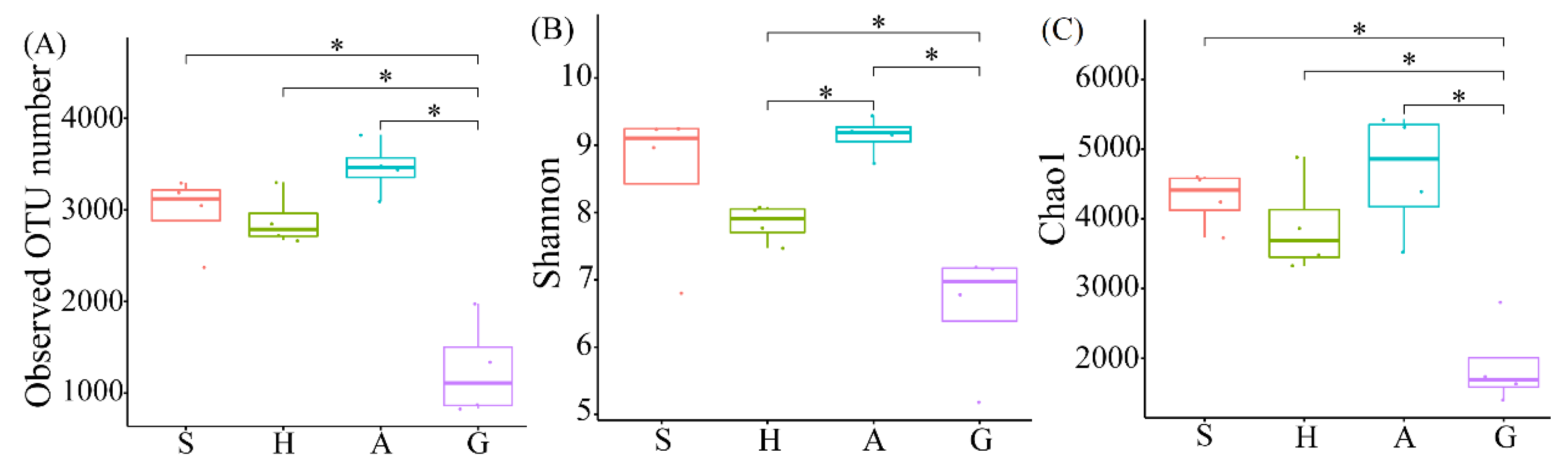
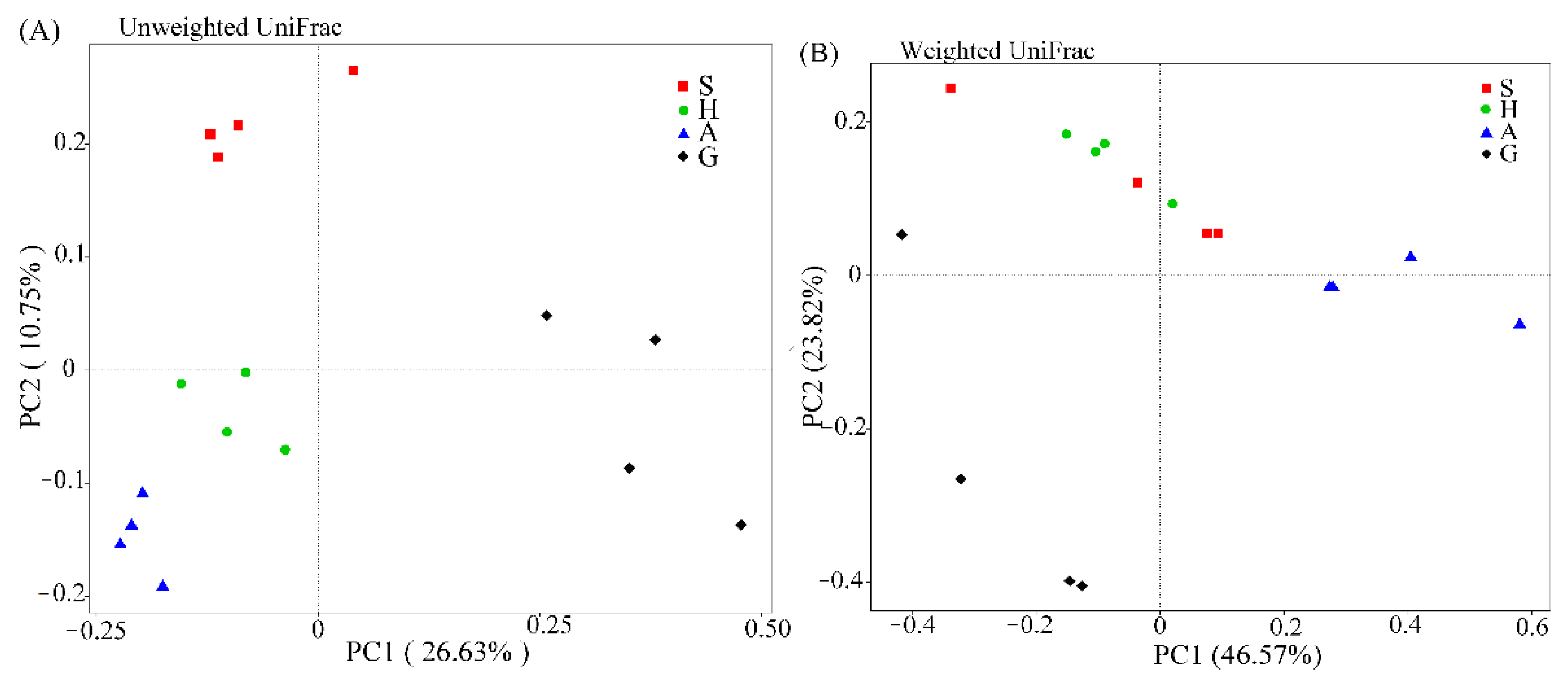
3.1. High-Throughput Sequencing Analysis and Microbial Community Diversity

In total, 1,789,665 raw reads were generated from 16 different intestines and sediment samples from an S. nudus aquaculture area. After filtering, 1,456,113 effective reads (92.99% of the total raw reads) were retrieved, with the number of sequences per sample ranging from 29,600 to 122,177 (Table S1). Finally, a total of 11,933 OTUs were observed at 97% sequence similarity. The coverage of each sample was above 94.5%, indicating that the sequencing depth was sufficient for microbial composition analysis. Compared with microbial communities from the sediments, the α-diversity of microbial richness and diversity were low in intestine samples (Table S1). The number of OTUs in intestine samples (G) was significantly lower than that in surface sediment samples (S), hole sediment samples (H), and ambient sediment samples (A) (p < 0.05). However, there was no significant difference among the three sediment samples (S, H, and A) (Figure 1A). Similarly, the α-diversity (Shannon and Chao1 indices) of group G was significantly lower than that of the sediment samples, except that there was no significant difference in the Shannon index between group G and group S (Figure 1B,C). ANOSIM and Adonis analysis suggested that there was a significant difference between the intestine and sediment samples at the OTU level based on Bray-Curtis dissimilarity; the similarity of microbial community between samples ranged from 29.9% to 36.3% (Table 1). The microbial community overlapped slightly between groups S and H based on the weighted UniFrac distance (Figure 2B). However, PCoA of the unweighted UniFrac distance and the weighted UniFrac distance showed that the intestine microbiota was separated from the sediment samples (Figure 2).

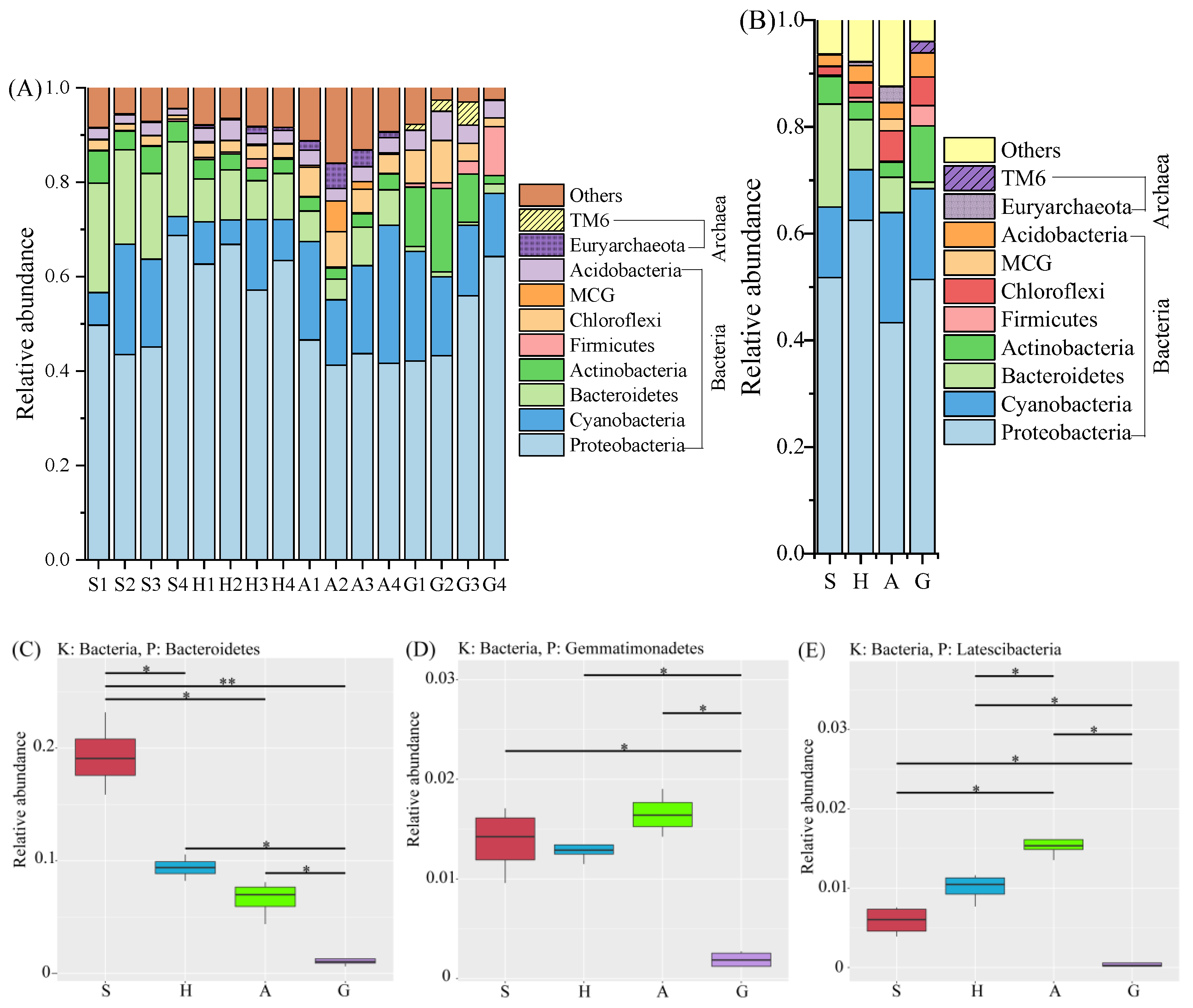
3.2. Microbial Composition and ORP Values among Intestine and Sediment Samples

The top 10 dominant microbial phyla in all samples, including eight phyla belonging to bacteria and two phyla belonging to archaea, are shown in Figure 3. The top 10 phyla from individual samples accounted for over 84.1% of total sequences (Figure 3A). Proteobacteria and Cyanobacteria were the dominant phyla in the four groups, accounting for 63.9–72.0% of all phyla in each group (Figure 3B). Bacteroidetes were significantly more abundant in group S. Chloroflexi, MCG, and Euryarchaeota were much more abundant in group A. However, Actinobacteria, Firmicutes, Acidobacteria, and TM6 were the most abundant taxa in group G (Figure 3B). In addition, three phyla (Bacteroidetes, Gemmatimonadetes, and Latescibacteria) showed a great difference among the four groups, and these three phyla were significantly less abundant in group G (Figure 3C–E). The ORP values of the sediments in groups S, H, and A were −90 ± 29, −137 ± 28, and −206 ± 28 mV, respectively.
At the genus level, differences were observed in the 35 most abundant microbial genera from the worm intestine and different sediments (Figure 4 and Figure S1). Sphingomonas, Methylobacterium, and Ralstonia were more abundant in group H, while Erythrobacter, Illumatobacter, Pseudohaliea, Marinicella, Robiginitalea, and Haliea were the dominant genera in group S. The abundance of eight genera, including Cyanobacterium, Sulfurovum, and Caldithrix was much higher in group A. However, many genera (16 of the top 35 genera) exhibited high abundance in the intestine samples, which indicates that the host may shape the intestinal microbiota.
Furthermore, a LEfSe analysis showed that 53 taxa had a significantly different abundance among the four groups, including six phyla and seven genera (Figure 5). In group H, fewer taxa were distinguished compared to other groups. Three phyla (Actinobacteria, Firmicutes, and TM6) were significantly enriched in group G. The genera Robiginitalea and Marinicella were more abundant in group S. Sphingomonas and Sulfurovum were significantly enriched in group H and group A, respectively. Group G showed a high abundance of genera Escherichia-Shigella, Stenotrophomonas, and Achromobacter.

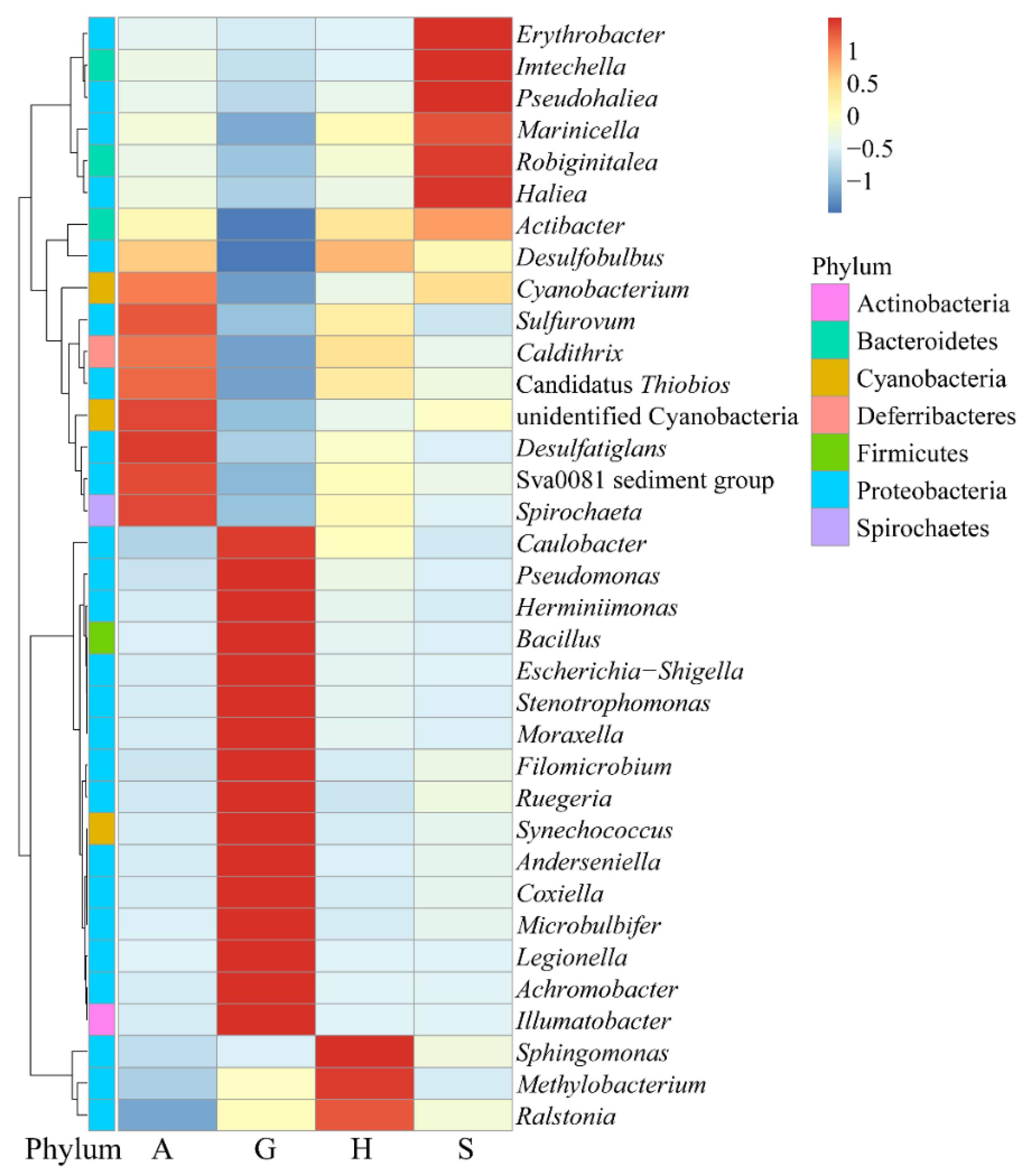
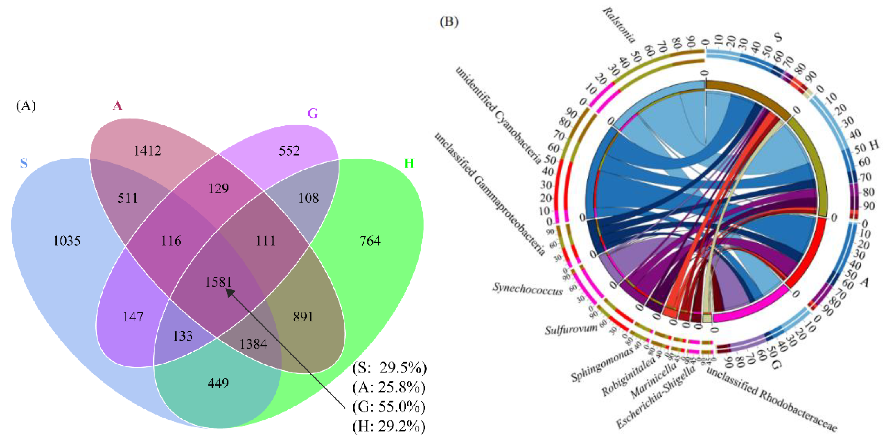
3.3. Core Microbiome of Intestine and Sediment Samples

Relationships between the microbial communities in the intestine and sediment samples at the OTU level are shown in Figure 5. The number of shared OTUs from the four groups was 1581. These OTUs accounted for 25.8–55.0% of total OTUs in the individual groups. A large proportion of OTUs (55.0%) were shared in the intestine samples (Figure 6A). The taxonomic information of shared OTUs was further investigated (Figure 6B). Based on the relative abundance of the dominant genera, unidentified Cyanobacteria and unclassified Gammaproteobacteria were shown to be exchanged more frequently among the four groups. Ralstonia, Sphingomonas, and unclassified Rhodobacteraceae were shared more between group G, group H, and group S. However, Sulfurovum, Robiginitalea, and Marinicella were shared more between the three sediment samples. Synechococcus was frequently exchanged between group G and group S. A larger abundance of Escherichia-Shigella was observed in the intestine samples, and low exchange was found between the intestine and sediment samples.

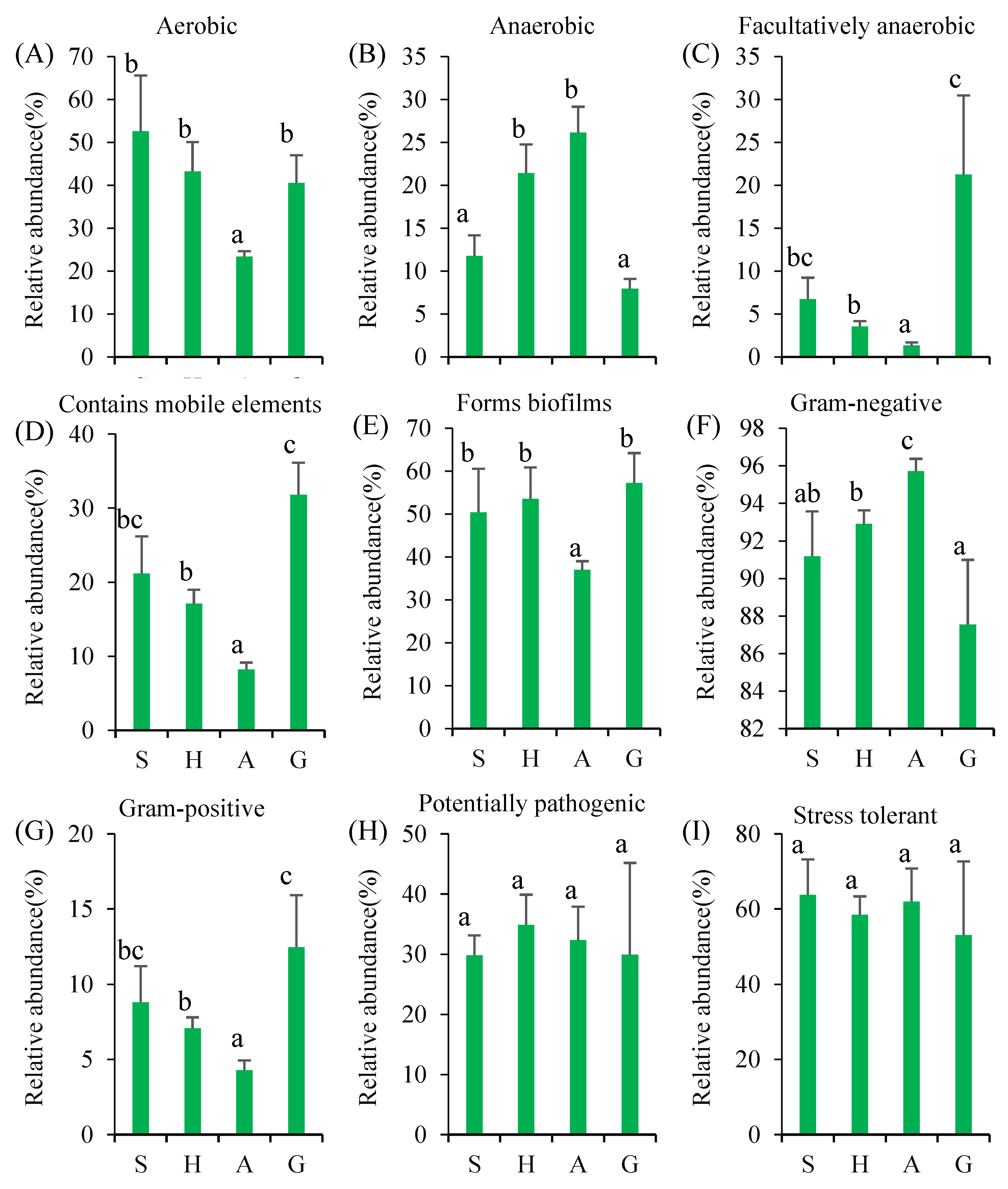
3.4. Microbial Function Changes

Based on 16S rRNA gene sequences, potential phenotypes were predicted by BugBase analysis, including being aerobic, anaerobic, facultatively anaerobic, containing mobile elements, biofilm formation, being Gram-negative or Gram-positive, being potentially pathogenic, and stress tolerance (Figure 7). Sediment bacterial communities showed some differences. The relative abundances of aerobic, facultatively anaerobic microbes containing mobile elements, forms biofilms, and gram-positive microbes were significantly lower in group A than that in groups S and H. There was no significant difference in the abundances of the above microbes between groups S and H. However, low abundances of anaerobic and gram-negative microbes were observed in group S.
In addition, the relative abundances of microbes containing mobile elements, facultatively anaerobic, and gram-positive were significantly higher in group G but significantly lower in groups A and H, while there was no difference between group G and group S. Whereas the opposite phenomenon was observed for Gram-negative microbes. There was no significant difference in the abundance of potentially pathogenic microbes and stress-tolerant microbes among the four groups.

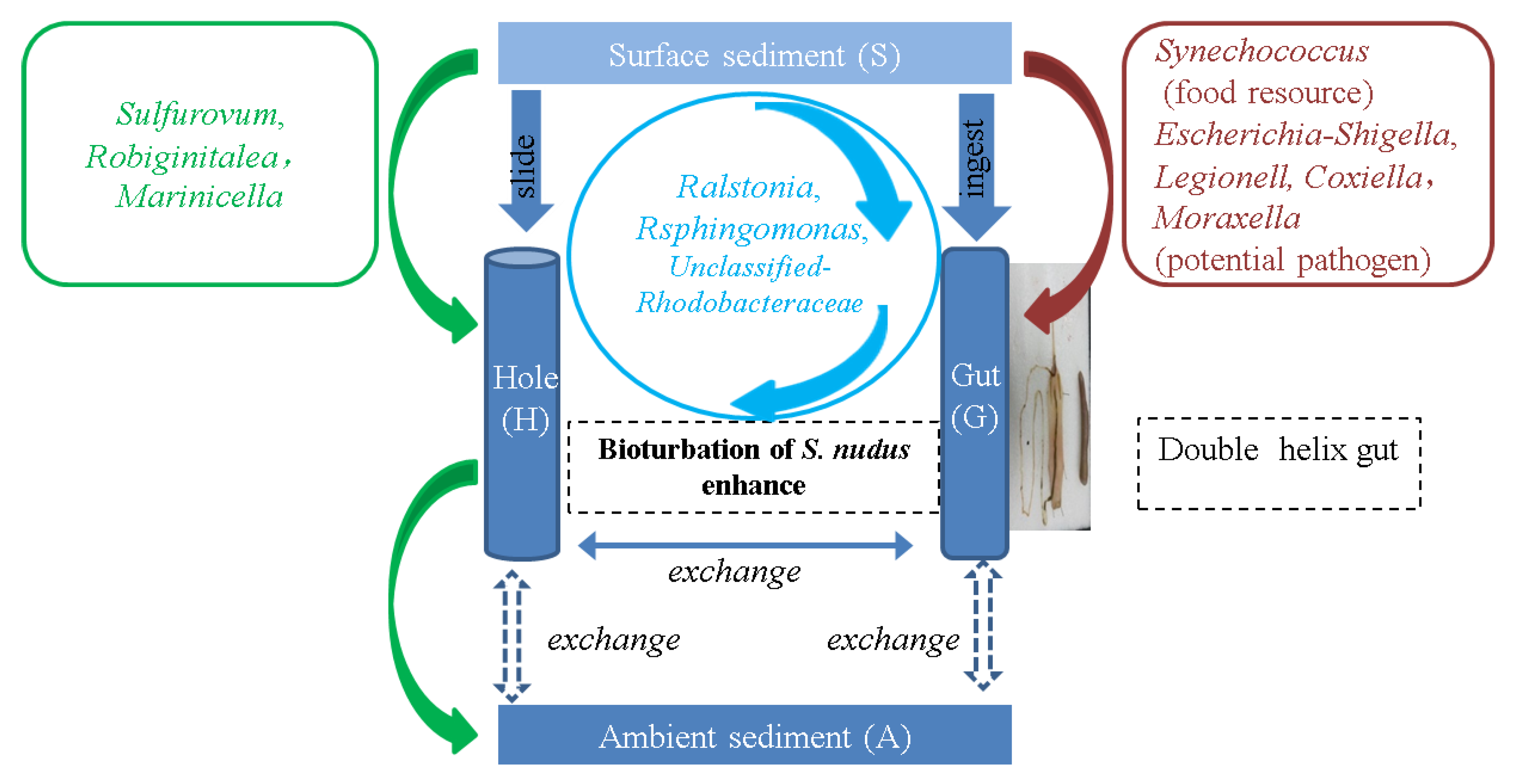
3.5. The Interactions of Microbial Composition between the Intestine of S. nudus and Surrounding Sediment

Several microbial interactions were summarized in the present study. The first interaction was observed between surface sediment and the worm intestine (S–G). The second interaction was found among surface sediment, the worm intestine, and the worm hole (S–G–H). The third was summarized and shown between surface sediment and the worm hole/ambient sediment (S–H–A). Meanwhile, the main genera were found in each interaction pathway. Synechococcus, Escherichia-Shigella, Legionella, Coxiella, and Moraxella were mainly found in the interaction S–G. Ralstonia, Rsphingomonas, and Unclassified Rhodobacteraceae were mainly found in the interaction S–G–H. Sulfurovum, Robiginitalea, and Marinicella were mainly found in the interaction S–H–A.

4. Discussion

The present study provided a detailed description of the microbial composition in the S. nudus intestine and surrounding sediment. There were significant differences in microbial diversity between the intestine and surrounding sediments. Tang et al. (2021) found that OTU numbers and α-diversity in the intestine of Urechis unicinctus were significantly lower than in the sediment [5]. The earthworm had a lower number of OTUs in its intestine than in its diet; however, its vermicompost showed a significant increase in OTU number [29]. Shrimp intestine also had a lower number of OTUs than the surrounding sediment [30]. The microbial compositions of earthworms at the phylum and genus levels are also related to the physicochemical characteristics of the substrate [31]. In the present study, the relative abundances of three phyla (Bacteroidetes, Gemmatimonadetes, and Latescibacteria), OTU number, and α-diversity index in the intestines of S. nudus were significantly lower than in the surface sediment and the hole of S. nudus (Figure 1 and Figure 3C–E), and the different characteristics of the sediments might be responsible for the difference in the microbial community. Meanwhile, some microbial species from sediments cannot easily colonize the intestine of S. nudus for its microenvironment. It is well-known that Bacteroidetes are a group of saprophytic bacteria, and they are increasingly regarded as specialists in the degradation of high-molecular-weight organic matter [32]. Meanwhile, most Bacteroidetes are strictly aerobic bacteria [33]. Therefore, the low abundance of Bacteroidetes in S. nudus might be related to the low content of oxygen in its intestine. The surface sediment had a high abundance of Bacteroidetes and high oxygen levels. Similarly, a lower abundance of Gemmatimonadetes was found in the shrimp intestine than in the surrounding sediment [30]. Liu et al. (2018) found that the abundance of Gemmatimonadetes was correlated with soluble organic carbon, total nitrogen, and total phosphorus in sediment [34]. Latescibacteria appeared to be more profound in the burrow sediment of Upogebia pugettensis [35]. Therefore, species in the phyla Gemmatimonadetes and Latescibacteria from sediments cannot easily colonize the intestine of S. nudus.
At the genus level, several dominant genera exhibited high abundances in the intestine of S. nudus (Figure 4). There were also several dominant genera from other phyla, including Bacillus (Firmicutes), Synechococcus (Cyanobacteria), and Illumatobacter (Actinobacteria). Bacillus can regulate the growth and nonspecific immune parameters of sea cucumbers [36]. Synechococcus is common in the surface sediment of intertidal flats [37]. Illumatobacter is a rare taxon in the environment [38]. Our results showed that the intestines of S. nudus were favorable for the establishment or proliferation of the above microbes. The functions of microbes in different groups were predicted by BugBase analysis. High relative abundances of aerobic and anaerobic microbes were found in the surface sediment and ambient sediment, respectively. The ORP values of the sediments in groups S and A might be responsible for these results. High relative abundances of containing mobile elements and Gram-positive bacteria were found in group G, and there was significant gene exchange among the microbes in the worm intestine. Low abundances of biofilm-forming microbes and Gram-positive bacteria were also found in group A. Therefore, the microenvironments affected by S. nudus had different microbial diversity in aquaculture zones.
There were significant differences at the genus level among the surrounding sediments (S, H, and A). The dominant genera in the hole of S. nudus belonged to the phylum Proteobacteria (Ralstonia, Sphingomonas, and Methylobacterium). However, lower abundances were found for these genera in surface sediment and ambient sediment. Therefore, different microenvironments affected by S. nudus had different microbial compositions. Ralstonia can enhance the bioremediation in sediment polluted by Cd and Zn [39], and Sphingomonas can increase the biodegradation of polycyclic aromatic hydrocarbons [40]. Methylobacterium mesophilicum showed a high capability of degrading monomethyl isophthalate [41]. The results indicated that the microenvironment in the hole of S. nudus might be favorable for colonization by degrading microbes. In addition, the higher relative abundances of Escherichia-Shigella, Legionella, Coxiella, and Moraxella were first found in the intestine of S. nudus. A previous study showed that the potential human pathogenic Salmonella was not a component of the indigenous community in fish intestines but rather was ingested with particulate material [42]. Some pathogens, including Escherichia-Shigella, were generally found in the surrounding environment, and it poses a threat to fish health [43]. We speculated that the potential pathogens were ingested with particulate material, and the pathogen could thrive in the intestine of S. nudus. The health of worms might be threatened by the pathogen. Therefore, some probiotics may possibly be used to shape the intestinal microbiota structure in the aquaculture of S. nudus.
Previous studies showed that the intestinal microbiota of aquatic animals could be specialized to the surrounding environment [12,13]. Moreover, host species, diet, life cycle stage, and rearing water can also affect the intestine microbiota [31,44,45,46,47]. It is widely accepted that most of the intestinal microbes of the earthworm originate from the soil [14,48]. S. nudus has bioturbation and feeding characteristics similar to earthworms. They directly ingest organic matter from surface sediment so that the worm intestine and the ambient sediments share some main microbial species (Figure 3A,B). In the present study, Proteobacteria, Cyanobacteria, and Actinobacteria constituted the majority of microbes in the worm intestine and the surrounding sediments (groups S, H, and A). The results indicated that the intestinal microbiota of S. nudus could be affected by the surrounding sediments. Sediment and S. nudus communities were closely linked, while sediment provided accommodation and food for S. nudus, and the bioturbation by S. nudus could affect the physicochemical characteristic of the surrounding sediments.
Proteobacteria, Firmicutes, and Actinobacteria are the most abundant bacteria in the intestines of fish, shrimp, sea urchins, and earthworms [5,13,30,31,49,50]. In the present study, similar results were obtained in the intestine of S. nudus. These bacterial phyla are associated with the degradation of organic matter, nutrient recycling, and the production of digestive enzymes and vitamins [51,52,53]. Therefore, these phyla might play important roles in the process of digestion and absorption for S. nudus. Previous studies showed that low abundances of Cyanobacteria were found in the intestines of aquaculture animals [13,30,49]. However, Cyanobacteria were found to be an abundant phylum in the intestines of S. nudus. S. nudus mainly feed on the sediment, and sediment is rich in Cyanobacteria [54]. Therefore, we speculated that the intestine was enriched with Cyanobacteria from the surface sediment. Singh et al. (2015) found that Firmicutes was the most abundant phylum in the intestine of Eisenia foetida [55], and the phylum Firmicutes was detected in the earthworm intestine but not in the soil [56]. In the present study, Firmicutes were detected in the intestine and hole of S. nudus but not in the surface sediment and ambient sediment. We speculated that Firmicutes in the hole of S. nudus mainly originated from its excretion. Meanwhile, the abundance of Acidobacteria in sediment was significantly higher than that in the worm intestine. Interestingly, similar variations in the abundance of Firmicutes and Acidobacteria were found in the intestine of earthworms [56,57]. A previous study suggested that the anaerobic environment of the earthworm intestine provided a microenvironment that was favorable for anaerobic and/or facultative anaerobic bacteria [31]. Likewise, the difference in microbial diversity between the intestine of S. nudus and surrounding sediments might be related to the physicochemical characteristics of the microenvironment in the intestine, which are affected by the double helix structure.
At the genus level, there were some differences in microbial community between the intestine of S. nudus and surrounding sediments. Unidentified Cyanobacteria and unclassified Gammaproteobacteria were shown to be exchanged more frequently among the four groups, and the results indicated that the two genera might be more tolerant to different environments, such as sediments and the intestine of S. nudus. Synechococcus is common in the surface sediment of intertidal flats [37], and we found that the genus was frequently present in the intestine of S. nudus and surface sediment and absent from the hole sediment. Therefore, the S. nudus intestine was enriched in Synechococcus, most likely through the ingestion of surface sediment. Moreover, microalgae might be an important food source for S. nudus. In addition, Ralstonia, Sphingomonas, and unclassified Rhodobacteraceae were found to be abundant in the surface sediment, the intestine, and the hole, and there might be a significant interaction between the three environments. Moreover, there might be an interaction network between the surface sediment, the hole, and the ambient sediment for the genera Sulfurovum, Robiginitalea, and Marinicella (Figure 8). The overwhelming abundance of Escherichia-Shigella in the intestine showed that this genus was enriched in the intestine of S. nudus. The significant exchange of microbes between the intestine of S. nudus and sediments might be related to the continuous ingestion and excretion through its hole. Therefore, microbes from surface sediment can pass through the intestine and enter the ambient sediment. Moreover, S. nudus is a typically hydraulically active organism that has significant effects on biogeochemical processes in marine sediments [8]. The top 10 shared microbial genera in the intestine and sediments included anaerobic and aerobic microbes, which indicated that the intestine of S. nudus was not strictly anaerobic (Figure 6B). This might be related to the higher sand content and lower organic content in the intestine. Unlike earthworms, the rhynchodaenm of S. nudus can reach the surface sediment and absorb oxygen during the feeding process [16], so the intestinal microbiota includes a large abundance of facultative anaerobic bacteria.

5. Conclusions

This was the first time the relationship between the intestinal microbes of the cultured edible worm S. nudus and the surrounding sediment in its habitat was determined. Although the microbial community in the intestine of S. nudus could be affected by the surrounding sediments, the worm had the capacity to shape its intestinal microbial structure. Several microbial interactions were explored in the present study. The strongest interactions were observed between surface sediment and the worm intestine, surface sediment and the worm intestine/worm hole, and between surface sediment and the worm hole/ambient sediment. Some potential pathogens (Escherichia-Shigella and Legionella) were found with a high abundance in the intestine of S. nudus. The significant interactions of microbial composition between the intestine of S. nudus and different sediments might be related to the physicochemical characteristics of the microenvironment in the intestine, which was affected by bioturbation and the double helix structure.

Supplementary Materials

The following are available online at https://www.mdpi.com/article/10.3390/fishes8010032/s1, Figure S1: Heat map of the relative abundance of the 35 most abundant microbial genera across all samples, Table S1: Sequencing information and microbial diversity estimates for the worm intestine and different sediments.

Author Contributions

Data curation, J.L., P.W. and R.H.; Funding acquisition, J.L.; Investigation, J.L., S.C., C.Z. and T.L.; Methodology, J.L., C.Z. and R.H.; Project administration, Y.G.; Resources, S.C; Writing—original draft, J.L.; Writing—review & editing, P.W. and R.H. All authors have read and agreed to the published version of the manuscript.

Funding

This work was supported by the Natural Science Foundation of Guangdong province (2021A1515010744); Central Public-interest Scientific Institution Basal Research Fund, South China Sea Fisheries Research Institute, CAFS (2022XK03, 2021SD04, 2021SD16); and the Central Public-interest Scientific Institution Basal Research Fund, CAFS (2020TD16).

Institutional Review Board Statement

All work undertaken in this study complied with the committee on Laboratory Animal Welfare and Ethics of South China Sea Fisheries Research Institute, Chinese Academy of Fishery Sciences (nhdf 2022-15).

Data Availability Statement

Raw sequence data from the surrounding sediment and the worm intestine have been submitted to the NCBI Sequence Read Archive under accession numbers PRJNA835336 and PRJNA835322, respectively.

Conflicts of Interest

The authors declare no conflict of interest.

References

  1. Peng, H.B.; Chan, Y.C.; Compton, T.J.; Cheng, X.F.; Melville, D.S.; Zhang, S.D.; Zhang, Z.W.; Lei, G.C.; Ma, Z.J.; Piersma, T. Mollusc aquaculture homogenizes intertidal soft-sediment communities along the 18,400 km long coastline of China. Divers. Distrib. 2021, 27, 1553–1567. [Google Scholar] [CrossRef]
  2. Wang, J.; Russell, B.D.; Ding, M.W.; Dong, Y.W. Ocean acidification increases the sensitivity of and variability in physiological responses of an intertidal limpet to thermal stress. Biogeosciences 2018, 15, 2803–2817. [Google Scholar] [CrossRef] [Green Version]
  3. Fields, P.A.; Eraso, A.A. Year in the salt marsh: Seasonal changes in gill protein expression in the temperate intertidal mussel Geukensia demissa. Mar. Environ. Res. 2020, 161, 105088. [Google Scholar] [CrossRef]
  4. Liao, M.L.; Dong, Y.W.; Somero, G.N. Thermal adaptation of mRNA secondary structure: Stability versus lability. Proc. Natl. Acad. Sci. USA 2021, 118, e2113324118. [Google Scholar] [CrossRef]
  5. Tang, Y.Z.; Ma, S.; Liu, Y.H.; Pi, Y.R.; Liu, Y.; Zhao, Y. Intestinal microbial diversity and functional analysis of Urechis unicinctus from two different habitats: Pond polycultured with Penaeus japonicas and coastal zone. Aquacult. EnvInterac. 2021, 13, 211–224. [Google Scholar] [CrossRef]
  6. Zhu, Y.J.; Liao, M.L.; Ding, M.W.; Wang, Z.K.; Dong, Y.W. Compositional and functional features of the gut microbiota of the intertidal snail Nerita yoldii along China’s coast. J. Mar. Biol. Assoc. 2021, 101, 1103–1110. [Google Scholar] [CrossRef]
  7. Lemmen, C. North Sea ecosystem-scale model-based quantification of net primary productivity changes by the Benthic filter feeder Mytilus edulis. Water 2018, 10, 1527. [Google Scholar] [CrossRef] [Green Version]
  8. Li, J.W.; Hu, R.P.; Guo, Y.J.; Chen, S.W.; Xie, X.Y.; Qin, J.G.; Ma, Z.H.; Zhu, C.B.; Pei, S.R. Bioturbation of peanut worms Sipunculus nudus on the composition of prokaryotic communities in a tidal flat as revealed by 16S rRNA gene sequences. MicrobiologyOpen 2019, 8, e802. [Google Scholar] [CrossRef] [Green Version]
  9. Sun, F.L.; Wang, Y.S.; Wang, C.Z.; Zhang, L.; Tu, K.; Zheng, Z.P. Insights into the intestinal microbiota of several aquatic organisms and association with the surrounding environment. Aquaculture 2020, 507, 196–202. [Google Scholar] [CrossRef]
  10. Kau, A.L.; Ahern, P.P.; Griffin, N.W.; Goodman, A.L.; Gordon, J.I. Human nutrition, the gut microbiome and the immune system. Nature 2011, 474, 327–336. [Google Scholar] [CrossRef]
  11. Patil, Y.; Gooneratne, R.; Ju, X. Interactions between host and gut microbiota in domestic pigs: A review. Gut Microbes 2020, 11, 310–334. [Google Scholar] [CrossRef] [PubMed]
  12. Sullam, K.E.; Essinger, S.D.; Lozupone, C.A.; O’Connor, M.P.; Rosen, G.L.; Knight, R.; Kilham, S.S.; Russell, J.A. Environmental and ecological factors that shape the gut bacterial communities of fish: A meta-analysis. Mol. Ecol. 2012, 21, 3363–3378. [Google Scholar] [CrossRef] [PubMed] [Green Version]
  13. Dehler, C.E.; Secombes, C.J.; Martin, S.A.M. Environmental and physiological factors shape the gut microbiota of Atlantic salmon parr (Salmo salar L.). Aquaculture 2017, 467, 149–157. [Google Scholar] [CrossRef] [PubMed] [Green Version]
  14. Horn, M.A.; Drake, H.L.; Schramm, A. Nitrous oxide reductase genes (nosZ) of denitrifying microbial populationsinsoiland the earthworm gut are phylogenetically similar. Appl. Environ. Microb. 2006, 72, 1019–1026. [Google Scholar] [CrossRef] [Green Version]
  15. Mwinyi, A.; Meyer, A.; Bleidorn, C.; Lieb, B.; Bartolomaeus, T.; Podsiadlowski, L. Mitochondrial genome sequence and gene order of Sipunculus nudus give additional support for an inclusion of Sipuncula into Annelida. BMC Genom. 2009, 10, 27. [Google Scholar] [CrossRef] [Green Version]
  16. Li, J.W.; Xie, X.Y.; Zhu, C.B.; Guo, Y.J.; Chen, S.W. Edible peanut worm (Sipunculus nudus) in the Beibu Gulf: Resource, aquaculture, ecological impact and counterplan. J. Ocean. Univ. China 2017, 16, 823–830. [Google Scholar] [CrossRef]
  17. Adrianov, A.V.; Maiorova, A.S. Reproduction and development of common species of peanut worms (Sipuncula) from the Sea of Japan. Russ. J. Mar. Biol. 2010, 36, 1–15. [Google Scholar] [CrossRef]
  18. Mark, A.S.; Monika, K. A deep burrowing sipunculan of ecological and geochemical importance. Deep. Sea Res. Part I 2009, 56, 2057–2064. [Google Scholar] [CrossRef]
  19. Li, J.W.; Zhu, C.B.; Guo, Y.J.; Xie, X.Y.; Huang, G.Q.; Chen, S.W. Experimental study of bioturbation by Sipunculus nudus in a polyculture system. Aquaculture 2015, 437, 175–181. [Google Scholar] [CrossRef]
  20. Chen, S.; Zhou, Y.; Chen, Y.; Gu, J. Fastp: An ultra-fast all-in-one FASTQ preprocessor. Bioinformatics 2018, 34, 884–890. [Google Scholar] [CrossRef]
  21. Bolyen, E.; Rideout, J.R.; Dillon, M.R.; Bokulich, N.; Abnet, C.C.; Al-Ghalith, G.A.; Alexander, H.; Alm, E.J.; Arumugam, M.; Asnicar, F.; et al. Reproducible, interactive, scalable and extensible microbiome data science using QIIME 2. Nat. Biotechnol. 2019, 37, 852–857. [Google Scholar] [CrossRef] [PubMed]
  22. Edgar, R.C.; Haas, B.J.; Clemente, J.C.; Quince, C.; Knight, R. UCHIME improves sensitivity and speed of chimera detection. Bioinformatics 2011, 27, 2194–2200. [Google Scholar] [CrossRef] [PubMed] [Green Version]
  23. Edgar, R.C. UPARSE: Highly accurate OTU sequences from microbial amplicon reads. Nat. Methods 2013, 10, 996–998. [Google Scholar] [CrossRef] [PubMed]
  24. Wang, Q.; Garrity, G.M.; Tiedje, J.M.; Cole, J.R. Naive Bayesian classifier for rapid assignment of rRNA sequences into the new bacterial taxonomy. Appl. Environ. Microb. 2007, 73, 5261–5267. [Google Scholar] [CrossRef] [Green Version]
  25. Pruesse, E.; Quast, C.; Knittel, K.; Fuchs, B.M.; Ludwig, W.G.; Peplies, J.; Glockner, F.O. SILVA: A comprehensive online resource for quality checked and aligned ribosomal RNA sequence data compatible with ARB. Nucleic Acids. Res. 2007, 35, 7188–7196. [Google Scholar] [CrossRef] [Green Version]
  26. Ward, T.; Larson, J.; Meulemans, J.; Hillmann, B.; Lynch, J.; Sidiropoulos, D.; Spear, J.R.; Caporaso, G.; Blekhman, R.; Knight, R.; et al. BugBase predicts organism level microbiome phenotypes. BioRxiv 2017, BioRxiv:133462. [Google Scholar] [CrossRef]
  27. Chen, J.; Bittinger, K.; Charlson, E.S.; Hoffmann, C.; Lewis, J.; Wu, G.D.; Collman, R.G.; Bushman, F.D.; Li, H.J. Associating microbiome composition with environmental covariates using generalized UniFrac distances. Bioinformatics 2012, 28, 2106–2113. [Google Scholar] [CrossRef] [Green Version]
  28. Segata, N.; Izard, J.; Waldron, L.; Gevers, D.; Miropolsky, L.; Garrett, W.S.; Huttenhower, C. Metagenomic biomarker discovery and explanation. Genome Biol. 2011, 12, R60. [Google Scholar] [CrossRef] [Green Version]
  29. Wang, Y.; Han, W.; Wang, X.; Chen, H.; Zhu, F.; Wang, X.; Lei, C.L. Speciation of heavy metals and bacteria in cow dung after vermicomposting by the earthworm, Eisenia fetida. Bioresour. Technol. 2017, 245, 411–418. [Google Scholar] [CrossRef]
  30. Jin, R.C.; Jiang, M.; Sun, S.Y.; Dai, X.L.; Wu, H.; Zhou, J.F.; Yu, Z.L.; Zhang, F. Microbial community in Litopenaeus vannamei intestine and its aquaculture environment. J. Fish. China 2020, 44, 2037–2054, (in Chinese abstract). [Google Scholar] [CrossRef]
  31. Sun, M.M.; Chao, H.Z.; Zheng, X.X.; Deng, S.P.; Ye, M.; Hu, F. Ecological role of earthworm intestinal bacteria in terrestrial environments: A review. Sci. Total Environ. 2020, 740, 140008. [Google Scholar] [CrossRef] [PubMed]
  32. Thomas, F.; Hehemann, J.H.; Rebuffet, E.; Michel, G. Environmental and gut bacteroidetes: The food connection. Front. Microbiol. 2011, 2, 93. [Google Scholar] [CrossRef] [PubMed] [Green Version]
  33. Yang, S.J.; Choo, Y.J.; Cho, J.C. Lutimonas vermicola gen. nov., sp. nov., a member of the family Flavobacteriaceae isolated from the marine polychaete Periserrula leucophryna. Int. J. Syst. Evol. Microbiol. 2007, 57, 1679–1684. [Google Scholar] [CrossRef] [PubMed] [Green Version]
  34. Liu, J.L.; Ha, V.N.; Shen, Z.; Zhu, H.L.; Zhao, F.; Zhao, Z. Characteristics of bulk and rhizosphere soil microbial community in an ancient Platycladus orientalis forest. Appl. Soil. Ecol. 2018, 132, 91–98. [Google Scholar] [CrossRef]
  35. Li, C.; Reimers, C.E.; Chapman, J.W.; Li, C.; Reimers, C.E.; Chapman, J.W. Microbiome analyses and presence of cable bacteria in the burrow sediment of Upogebia pugettensis. Mar. Ecol. Prog. Ser. 2020, 648, 79–94. [Google Scholar] [CrossRef]
  36. Liu, B.; Zhou, W.; Wang, H.; Li, C.; Wang, L.; Li, Y.; Wang, J.H. Bacillus baekryungensis MS1 regulates the growth, non-specific immune parameters and gut microbiota of the sea cucumber Apostichopus japonicus. Fish Shellfish Immunol. 2020, 102, 133–139. [Google Scholar] [CrossRef]
  37. Abed, R.M.M.; Kohls, K.; Beer, D.D. Effect of salinity changes on the bacterial diversity, photosynthesis and oxygen consumption of cyanobacterial mats from an intertidal flat of the Arabian Gulf. Environ. Microbiol. 2007, 9, 1384–1392. [Google Scholar] [CrossRef]
  38. Zhang, M.J.; Sun, Q.Y.; Chen, P.X.; Wei, X.H.; Wang, B. How microorganisms tell the truth of potentially toxic elements pollution in environment. J. Hazard. Mater. 2002, 431, 128456. [Google Scholar] [CrossRef]
  39. Park, Y.J.; Ko, J.J.; Yun, S.L.; Lee, E.Y.; Kim, S.J.; Kang, S.W.; Lee, B.C.; Kim, S.K. Enhancement of bioremediation by Ralstonia sp. HM-1 in sediment polluted by Cd and Zn. Bioresour. Technol. 2008, 99, 7458–7463. [Google Scholar] [CrossRef]
  40. Chen, J.L.; Wong, M.H.; Wong, Y.S.; Tam, N.F.Y. Multi-factors on biodegradation kinetics of polycyclic aromatic hydrocarbons (PAHs) by Sphingomonas sp. a bacterial strain isolated from mangrove sediment. Mar. Pollut. Bull. 2008, 57, 695–702. [Google Scholar] [CrossRef]
  41. Li, J.X.; Gu, J.D. Complete degradation of dimethyl isophthalate requires the biochemical cooperation between Klebsiella oxytoca Sc and Methylobacterium mesophilicum Sr Isolated from Wetland sediment. Sci. Total Environ. 2007, 380, 181–187. [Google Scholar] [CrossRef] [PubMed]
  42. Gaertner, J.; Wheeler, P.E.; Obafemi, S.; Valdez, J.; Forstner, M.R.J.; Bonner, T.H.; Dittmar, H. Detection of salmonellae from fish in a natural river system. J. Aquat. Anim. Health 2008, 20, 150–157. [Google Scholar] [CrossRef] [PubMed]
  43. Gainza, O.; Ramírez, C.; Ramos, A.S.; Romero, J. Intestinal microbiota of white shrimp Penaeus vannamei under intensive cultivation conditions in Ecuador. Microb. Ecol. 2018, 75, 562–568. [Google Scholar] [CrossRef]
  44. Navarrete, P.; Magne, F.; Araneda, C.; Fuentes, P.; Barros, L.; Opazo, R.; Espejo, R.; Romero, J. PCR-TTGE analysis 16S rRNA from rainbow trout (Oncorhynchus mykiss) gut microbiota reveals host-specific communities of active bacteria. PLoS ONE 2012, 7, e31335. [Google Scholar] [CrossRef] [PubMed] [Green Version]
  45. Givens, C.E.; Ransom, B.; Bano, N.; Hollibaugh, J.T. Comparison of the gut microbiomes of 12 bony fish and 3 shark species. Mar. Ecol. Prog. Ser. 2015, 518, 209–223. [Google Scholar] [CrossRef] [Green Version]
  46. Zarkasi, K.Z.; Taylor, R.S.; Abell, G.C.J.; Tamplin, M.L.; Glencross, B.D.; Bowman, J.P. Atlantic salmon (Salmo salar L.) gastrointestinal microbial community dynamics in relation to digesta properties and diet. Microb. Ecol. 2016, 71, 589–603. [Google Scholar] [CrossRef]
  47. Duan, Y.F.; Huang, J.H.; Wang, Y.; Zhang, J.S. Characterization of bacterial community in intestinal and rearing water of Penaeus monodon differing growth performances in outdoor and indoor ponds. Aquac. Res. 2020, 51, 4279–4289. [Google Scholar] [CrossRef]
  48. Karsten, G.R.; Drake, H.L. Comparative assessment of the aerobic and anaerobic microfloras of earthworm guts and forest soils. Appl. Environ. Microb. 1995, 67, 1039–1044. [Google Scholar] [CrossRef] [Green Version]
  49. Gajardo, K.; Rodiles, A.; Kortner, T.M.; Krogdahl, A.; Bakke, A.M.; Merrifield, D.L. A high resolution map of the gut microbiota in Atlantic salmon (Salmo salar): A basis for comparative gut microbial research. Sci. Rep. 2016, 6, 30893. [Google Scholar] [CrossRef] [Green Version]
  50. Zheng, Y.; Wu, W.; Hu, G.; Qiu, L.; Meng, S.L.; Song, C. Gut microbiota analysis of juvenile genetically improved farmed tilapia (Oreochromis niloticus) by dietary supplementation of different resveratrol concentrations. Fish Shellfish Immunol. 2018, 77, 200–207. [Google Scholar] [CrossRef]
  51. Stevens, H.; Brinkhoff, T.; Rink, B.; Vollmers, J.; Simon, M. Diversity and abundance of Gram positive bacteria in a tidal flat ecosystem. Environ. Microbiol. 2007, 9, 1810–1822. [Google Scholar] [CrossRef] [PubMed]
  52. Clements, K.D.; Angert, E.R.; Montgomery, W.L. Intestinal microbiota in fishes: What’s known and what’s not. Mol. Ecol. 2014, 23, 1891–1898. [Google Scholar] [CrossRef] [PubMed]
  53. Nishida, A.; Inoue, R.; Inatomi, O.; Bamba, S.; Naito, Y.; Andoh, A. Gut microbiota in the pathogenesis of inflammatory bowel disease. Clin. J. Gastroenterol. 2018, 11, 1–10. [Google Scholar] [CrossRef] [Green Version]
  54. Burja, A.M.; Banaigs, B.; Abou-Mansour, E.; Burgess, J.G.; Wright, P.C. Marine cyanobacteria—A prolific source of natural products. Tetrahedron 2001, 57, 9347–9377. [Google Scholar] [CrossRef]
  55. Singh, A.; Singh, D.P.; Tiwari, R.; Kumar, K.; Singh, R.; Singh, S.; Prasanna, R.; Saxena, A.K.; Nain, L. Taxonomic and functional annotation of gut bacterial communities of Eisenia foetida and Perionyx excavates. Microbiol. Res. 2015, 175, 48–56. [Google Scholar] [CrossRef]
  56. Montagna, M.; Berruti, A.; Bianciotto, V.; Cremonesi, P.; Giannico, R.; Gusmeroli, F. Differential biodiversity responses between kingdoms (plants, fungi, bacteria and metazoa) along an Alpine succession gradient. Mol. Ecol. 2018, 27, 3671–3685. [Google Scholar] [CrossRef]
  57. Pass, D.A.; Morgan, A.J.; Read, D.; Field, D.; Weightman, A.; Kille, P. The effect of anthropogenic arsenic contamination on the earthworm microbiome. Environ. Microbiol. 2015, 17, 1884–1896. [Google Scholar] [CrossRef]
Figure 1. The α-diversity indices of the microbial composition in the worm intestine and different sediments from a Sipunculus nudus aquaculture area. (A) Number of observed OTUs. (B) Shannon diversity index. (C) Chao1 index. Asterisks (*) indicate significant differences (p < 0.05, Wilcoxon test). S—surface sediment; H—sediment in the hole; A—ambient sediment; G—gut of S. nudus.
Fishes 08 00032 g001
Figure 2. Principal coordinate analysis (PCoA) plots of (A) unweighted and (B) weighted UniFrac distances between microbial communities in the worm intestine and different sediments from a Sipunculus nudus aquaculture area. S—surface sediment; H—sediment in the hole; A—ambient sediment; G—gut of S. nudus.
Fishes 08 00032 g002
Figure 3. Relative abundances of main microbes in the worm intestine and different sediments at the phylum level. (A) Each sample. (B) Each group. (CE) Differences in microbial community between intestine and different sediments. Asterisks (*) indicate significant differences (p < 0.05, Wilcoxon test). The two asterisks (**) indicate extremely significant differences (p < 0.01, Wilcoxon test). S—surface sediment; H—sediment in the hole; A—ambient sediment; G—gut of S. nudus.
Fishes 08 00032 g003
Figure 4. Heat map of the relative abundance of the 35 most abundant microbial genera in the worm intestine and different sediments. S—surface sediment; H—sediment in the hole; A—ambient sediment; G—gut of S. nudus.
Fishes 08 00032 g004
Figure 5. Microbial taxa with different abundances in the worm intestine and different sediments identified by LEfSe using an LDA score threshold of >4.0. s, Species; g—genus; f—family; o—order; c—class; p—phylum. S—surface sediment; H—sediment in the hole; A—ambient sediment; G—gut of S. nudus.
Fishes 08 00032 g005
Figure 6. (A) Venn diagram of OTU composition in the worm intestine and different sediments. The percentages in brackets of the Venn diagram indicate the ratios of the number of shared OTUs to the total number of OTUs in each group. (B) Circos diagram of the top 10 microbial genera based on shared OTU analysis for the intestine and sediment samples. The width of the bar of each genus indicates the relative abundance of that genus in the sample. The central circle shows connections between microbial genera and the samples in which they occur. S—surface sediment; H—sediment in the hole; A—ambient sediment; G—gut of S. nudus.
Fishes 08 00032 g006
Figure 7. BugBase functional prediction of the microbial communities from intestine and sediment samples: aerobic (A); anaerobic (B); facultatively anaerobic (C); contains mobile elements (D); forms biofilms (E); Gram-negative (F); Gram-positive (G); potentially pathogenic (H); stress-tolerant (I). Different lowercase letters represent significant differences among the treatments (p < 0.05, Wilcoxon test). S—surface sediment; H—sediment in the hole; A—ambient sediment; G—gut of S. nudus.
Fishes 08 00032 g007
Figure 8. The interactions of microbial composition between the intestine of S. nudus and surrounding sediments. S—surface sediment; H—sediment in the hole; A—ambient sediment; G—gut of S. nudus.
Fishes 08 00032 g008
Table 1. Similarity and ANOSIM/Adonis analyses of microbial communities in intestine and sediment samples based on Bray-Curtis dissimilarity.
Sample GroupsSimilarityANOSIMAdonis
RpR2p
G/S34.8%0.8960.0340.5610.048
G/A29.9%1.00.0280.6310.001
G/H36.3%0.740.0360.5390.001
Disclaimer/Publisher’s Note: The statements, opinions and data contained in all publications are solely those of the individual author(s) and contributor(s) and not of MDPI and/or the editor(s). MDPI and/or the editor(s) disclaim responsibility for any injury to people or property resulting from any ideas, methods, instructions or products referred to in the content.

