Article

Effects of Short-Term Salinity Stress on Ions, Free Amino Acids, Na+/K+-ATPase Activity, and Gill Histology in the Threatened Freshwater Shellfish Solenaia oleivora

1
Key Laboratory of Freshwater Fisheries and Germplasm Resources Utilization, Ministry of Agriculture and Rural Affairs, Freshwater Fisheries Research Center, Chinese Academy of Fishery Sciences, Wuxi 214081, China
2
Key Laboratory of Integrated Rice-Fish Farming Ecology, Ministry of Agriculture and Rural Affairs, Freshwater Fisheries Research Center, Chinese Academy of Fishery Sciences, Wuxi 214081, China
*
Author to whom correspondence should be addressed.
These authors contributed equally to this work.
Fishes 2022, 7(6), 346; https://doi.org/10.3390/fishes7060346
Submission received: 8 October 2022 / Revised: 15 November 2022 / Accepted: 16 November 2022 / Published: 25 November 2022

Abstract

:
Salinity is an important ecological factor affecting the osmolality of aquatic animals. Solenaia oleivora is an endemic and economically important freshwater shellfish in China. However, its osmotic response and osmoregulatory mechanisms under high salinity stress are still unclear. In this study, S. oleivora was exposed to saline water (salinity: 2.2‰) for 3 h, 6 h, 12 h, 24 h, and 48 h, and then the changes in osmolality, ion concentrations, free amino acid (FAA) content, Na+/K+-ATPase (NKA) activity, and gill histology were analyzed. The hemolymph osmolality increased from 3 h after salinity stress and stabilized between 24–48 h. Na+ in the hemolymph increased from 24 h after salinity stress, and Cl increased from 3 h. The content of total FAAs in the hemolymph increased after salinity stress. The content of alanine, glycine, glutamine, proline, and other FAAs increased after salinity stress. NKA activity in the gill, hepatopancreases, adductor muscle, and axe foot decreased during 3–48 h of salinity stress. The gill filament space increased and the number of gill cilia decreased after salinity stress. Principal component analysis (PCA) showed that the first two principal components (PC1 and PC2) cumulatively explained 77.6% of the total variation. The NKA activity was positively associated with PC1, while the ion concentration and most FAAs were negatively associated with PC1. Thus, these results indicated that S. oleivora is an osmoconformer, and inorganic ions, FAA, NKA, and gill structure changes play an important role in its osmoregulation.

1. Introduction

Salinity is an important ecological factor in aquatic ecosystems, and it affects the behavior, growth performance, metabolism, osmoregulation, and circadian rhythm of aquatic organisms, determining their natural distribution [1,2,3]. As the global climate change, aquatic ecosystems are being threatened by many kinds of pollution, acidification, and changes in temperature and salinity. A series of natural factors, such as the weathering of rocks, wind, rain depositing salt, and saltwater intrusion, as well as many artificial factors, such as salt mining, industrial discharges, or road de-icing, lead to increased salinity in the freshwater ecosystems [4,5]. The increased salinity in the ambient aquatic environment directly influences the osmotic pressure of aquatic animals, regulating their growth, reproduction, and survival [6,7,8].
Mollusks have poor mobility and must adapt to salinity fluctuations in their natural habitat through multiple strategies, including osmoregulation. When entering a hypotonic or hypertonic environment, mollusks regulate hemolymph osmolality to remain it in line with the ambient environment [9]. As an important organ responsible for osmoregulation, the gill directly contacts ambient water and is thus the main site of osmotic water loss and salt diffusion gain [10,11]. The histological structures of gills are altered under low or high salinity stress in mollusks, such as Sinonovacula constricta [12] and Saxidomus purpurata [13]. Additionally, inorganic ions are the main osmotic effectors in the hemolymph of mollusks. Previous studies reported that Na+, Cl, and other ions are involved in osmotic adjustment in Pinctada fucata [14], Haliotis Discus [15], and Meretrix lusoria [16]. Likewise, organic substances are also important osmolytes for the osmoregulation of mollusks. Among many organic osmotic osmolytes, free amino acids (FAA) have been proven to play an important role in the osmoregulation of mollusks in coping with salinity challenges [17,18]. Furthermore, Na+-K+-ATPase (NKA), a transmembrane ion transporter, can drive the active transport of ions across the membrane, which is important for osmotic regulation in mollusks and other aquatic animals [19,20,21]. Although many studies have explored osmoregulation mechanisms in mollusks under salinity stress, most studies have focused on marine species, and only a few studies have focused on freshwater species.
Solenaia oleivora (Bivalvia, Unionidae, Gonideinae), a unique rare freshwater bivalve in China, is a type of economically important freshwater mussel, with a high growth rate and high nutritional value [22,23]. This species was previously distributed in relatively large ranges, including Hebei province, Anhui province, Jiangsu province, Jiangxi province, Hubei province, and Zhejiang province [24]. In recent years, the wide population of S. oleivora has declined dramatically, and it is becoming endangered due to water pollution, habitat destruction, and increasing capture pressure [25,26]. Thus, elucidating the responses of S. oleivora to various environmental factors is of great significance for its resource conservation and artificial aquaculture. Our previous study found that S. oleivora is less tolerant to salinity and that high salinity stress affects its survival, physiology, and transcriptome [27]. Therefore, the question of how hemolymph osmolality changes in S. oleivora under salinity stress is of great importance, as it the underlying mechanism of osmoregulation in S. oleivora.
In this study, the hemolymph osmotic response of S. oleivora under salinity stress was investigated. Additionally, the changes in ion concentrations, FAA content, NKA activity, and gill histology were analyzed after high salinity stress. Our results elucidated the osmotic response and osmoregulatory mechanisms of S. oleivora under hypertonic conditions. These findings indicated that S. oleivora has a system to cope with hypertonic stress, which can adjust its osmotic pressure to a certain extent to adapt to changes in environmental salinity. This study fills the gap regarding the study of the osmotic pressure of S. oleivora, accumulated data for the study of the environmental adaptability of S. oleivora, and provided a theoretical basis for its habitat protection and artificial breeding.

2. Materials and Methods

2.1. Experimental Mussels and Rearing Conditions

S. oleivora (wet weight: 2.50 ± 0.4 g; shell length: 4.79 ± 0.55 cm; shell height: 0.97 ± 0.16 cm) were obtained from the Freshwater Fisheries Research Center, Chinese Academy of Fishery Sciences. They were acclimated for 1 week in freshwater with dissolved oxygen >8 mg/L, ammonium nitrogen < 0.06 mg/L, pH 8.07 ± 0.04, and temperature 20 ± 1 °C. During the acclimation period, they were fed with mechanical wall-breaking Nannochloropsis (Reed Mariculture Inc., Watsonville, CA, USA) twice a day.

2.2. Salinity Challenge and Sampling

After 1 week of acclimation, 54 S. oleivora were randomly divided into six groups (0 h, 3 h, 6 h, 12 h, 24 h, and 48 h) with 9 individuals and triplicates in each group. The 0 h group was not subjected to salinity stress and was used as the control group. S. oleivora in the other five groups was transferred to water with a salinity of 2.2‰ (50% of 96 h-LC50) to undergo 3 h, 6 h, 12 h, 24 h, and 48 h of salinity treatment, respectively. The salinity in the water was adjusted by NaCl (AR ≥ 99.5%) and measured with a salinity meter (Smart, China). The gills, hepatopancreas, adductor muscle, axe foot, and hemolymph of S. oleivora (2 individuals from each replicate and 6 individuals in each group) were collected for the detection of hemolymph osmotic pressure, ion concentration, enzyme activity, and FAAs. The gill tissue of the remaining 3 individuals in each group was collected for histological examination. The hemolymph samples were collected from the adductor muscle in the shell posterior using syringes. All samples except those used for histological observation were immediately frozen in liquid nitrogen and then stored at −80 °C.

2.3. Measurement of Hemolymph Osmolality

Hemolymph samples were centrifuged at 14,000× g for 10 min at 4 °C. After that, the supernatant was collected for the measurement of osmolality using the Osmomat 030 Cryoscopic Osmometer (Gonotec, Berlin, Germany).

2.4. Detection of Ion Concentrations and Enzyme Activity

The hepatopancreas, gills, adductor muscle, and axe foot samples were homogenized by adding sterile 0.6% saline solution to prepare 10% (w:v) homogenates and then centrifuged at 4000× g for 10 min at 4 °C. After each tissue was homogenized separately, homogenates of two tissues per replicate in the same group were mixed proportionally. There were three mixed tissue homogenates in each group used for physiological and biochemical detection. The activity of NKA and the concentrations of Na+ and Cl were measured using commercially available kits (NKA: A070-2-2; Na+: C002-1-1; Cl: C003-2-1) (Nanjing Jiancheng Bioengineering Institute, Nanjing, China) according to the manufacturer’s instructions. The concentration of protein was detected using a Bradford Protein Assay Kit (Beyotime, Shanghai, China). The activity of NKA was normalized by protein concentration.

2.5. Detection of FAA Content in Hemolymph

The FAA in the hemolymph was quantified by an amino acid analyzer (Hitachi Model 835) according to the previously described method [28].

2.6. Gill Histology

The gills were fixed in Bouin’s solution for 24 h, embedded into paraffin according to routine procedures, and then cut into slices with a thickness of 5 μm. Next, the paraffin slices were stained with hematoxylin/eosin (H & E) and sealed with neutral gum. Finally, the slices were observed under light microscopy (Olympus, Tokyo, Japan).

2.7. Data Analysis

Data were presented as means ± standard deviation (SD) and analyzed using one-way analysis of variance (ANOVA) in SPSS 17.0. Differences were analyzed via Duncan’s multiple range test when ANOVA identified differences among the groups. The level of significance was set to p < 0.05. The principal component analysis (PCA) of indicators with the significant difference among groups was conformed using an online platform for data analysis and visualization (https://www.bioinformatics.com.cn, accessed on 31 October 2022).

3. Results

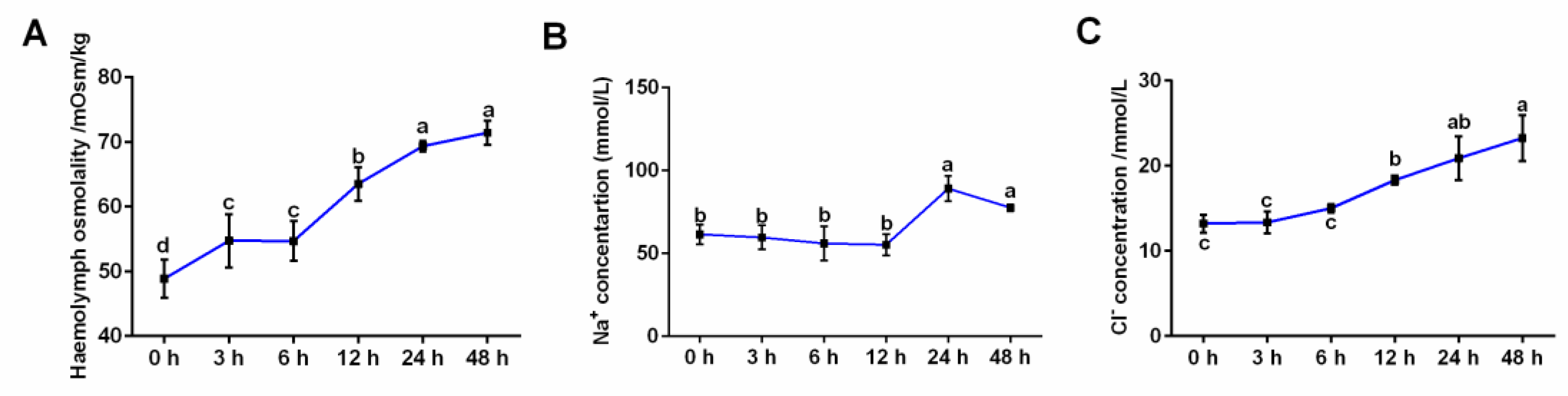
3.1. Effects of Salinity Stress on Hemolymph Osmolality and Ion Concentrations

As shown in Figure 1A, the hemolymph osmolality of S. oleivora significantly increased from 3 h after salinity stress and stabilized between 24–48 h. The concentration of Na+ was significantly elevated between 24–48 h of salinity stress (Figure 1B). The concentration of Cl significantly increased during 12–48 h of salinity stress (Figure 1C).

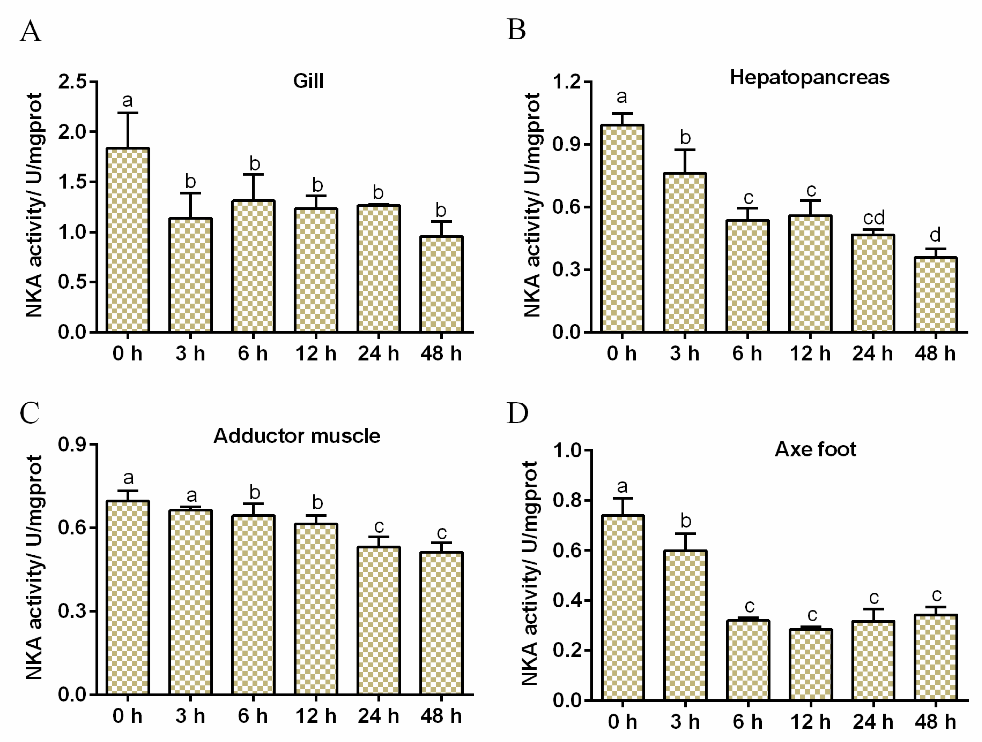
3.2. Effects of Salinity Stress on NKA Activity

The changes in NKA activity in different tissues are shown in Figure 2. In the gill, hepatopancreas, and axe foot, the activity of NKA decreased during 3 h to 48 h of salinity stress (Figure 2A,B,D). In the adductor muscle, NKA activity decreased during 6–48 h of salinity stress (Figure 2C).

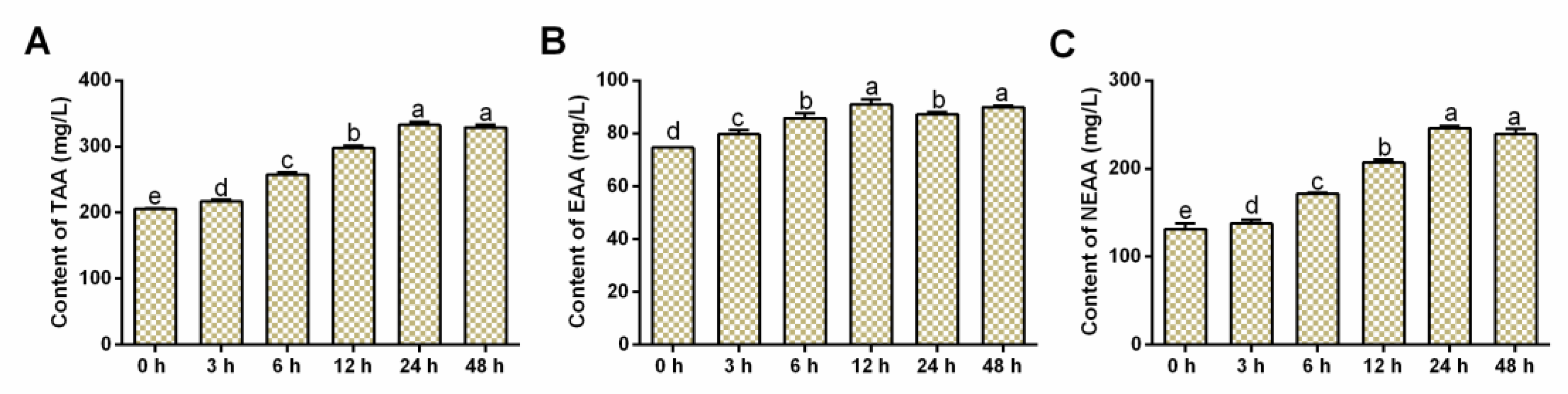
3.3. Effects of Salinity Stress on FAA Contents

As shown in Figure 3A, the contents of total FAA (TFAA) significantly increased from 3 h to 48 h of salinity stress. The content of essential amino acids (EAA) increased during 3–48 h of salinity stress (Figure 3B). The content of nonessential amino acids (NEAA) started to increase from 6 h after salinity stress (Figure 3C). The content alterations of 17 FAA in the hemolymph are shown in Table 1. In the control (0 h) group, the content of glutamic acid (42.23 ± 2.34 mg/L) was the highest, followed by threonine (25.10 ± 1.99 mg/L), while the content of cysteine (1.34 ± 0.26 mg/L) was the lowest. Except for cysteine, isoleucine, phenylalanine, and histidine, the contents of the other 13 FAAs changed after salinity stress. After salinity stress, methionine content reduced at 48 h, and aspartate, threonine, glutamine, valine, leucine, glycine, proline, serine, lysine, arginine, tyrosine, and alanine contents increased during 3–48 h.

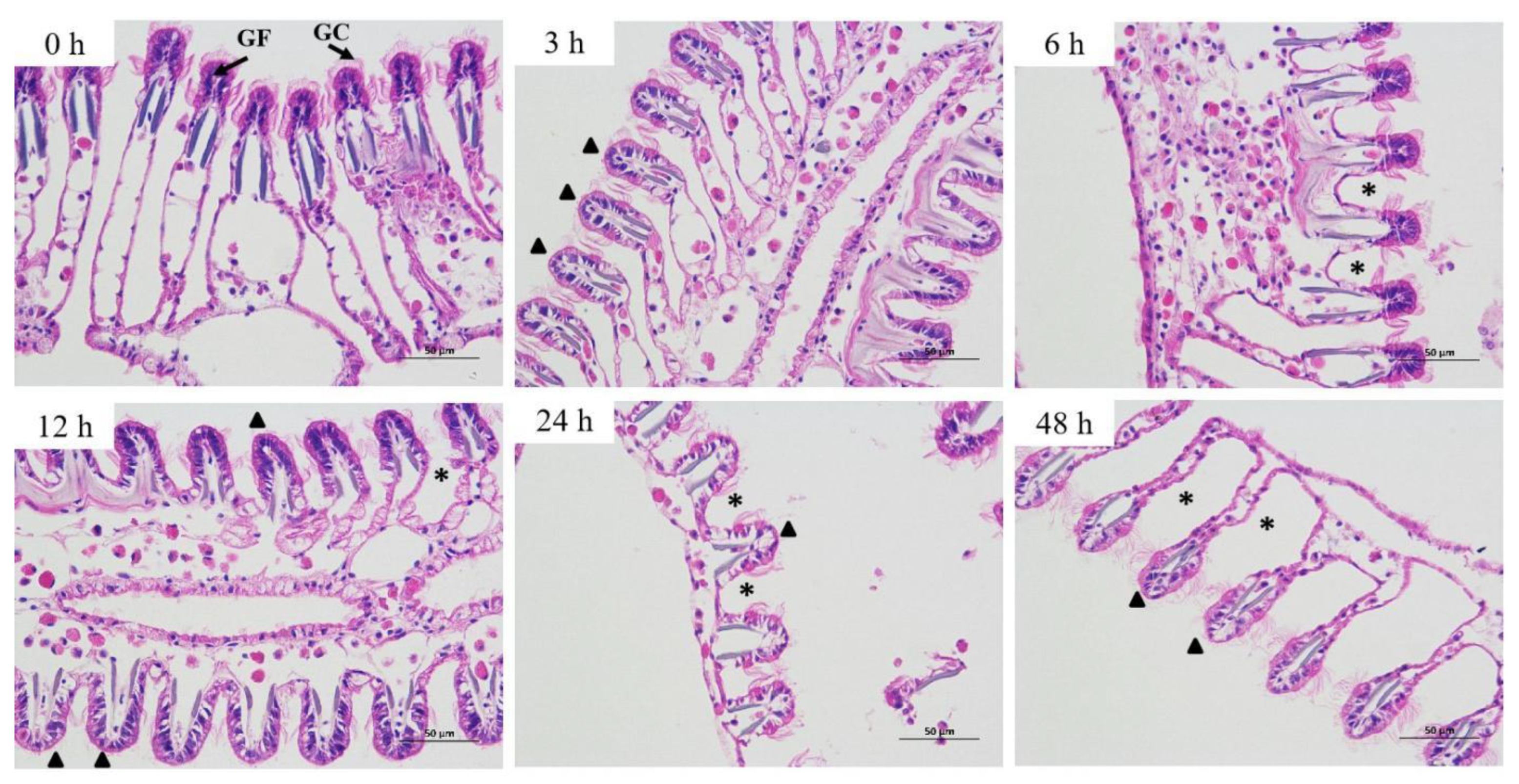
3.4. Effects of Salinity Stress on Gill Histomorphology
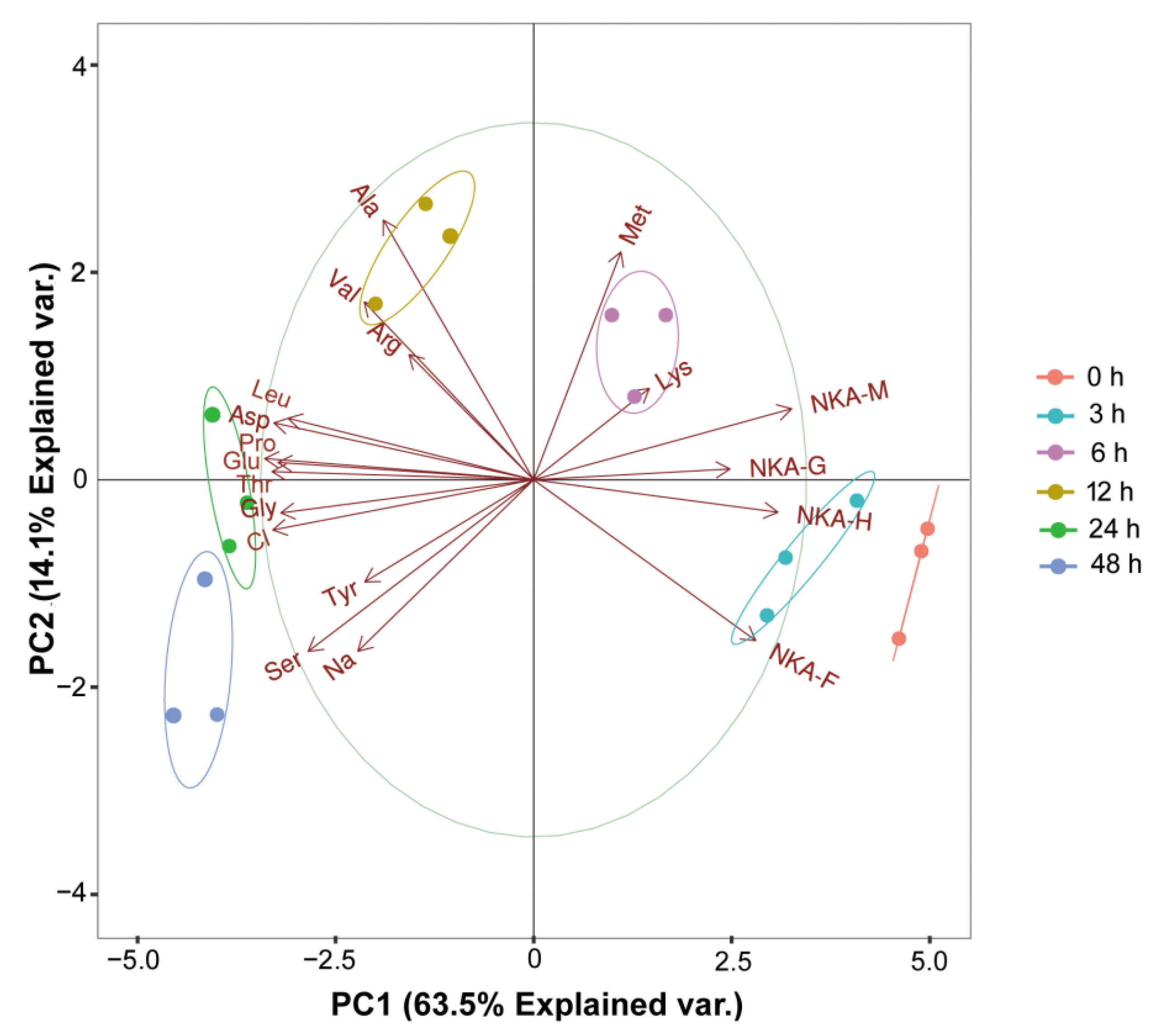
The results showed salinity stress-induced alterations of the gill histomorphology (Figure 4 and Figure S1). In the control group (0 h), the gill filaments of S. oleivora were closely arranged, and the top of the gills expanded into a rod shape. After 3 h of salinity stress, the gill filament space increased and the gill cilia decreased.

3.5. PCA Analysis

Regarding PCA (Figure 5), the first two principal components (PC1 and PC2) cumulatively explained 77.6% of the total variation. The NKA activities in the four tissues were positively associated with PC1. Ion concentration and most FAAs were negatively associated with PC1. Furthermore, all salinity stress groups were clearly separated from the control group. The distance between the control group and the 4 salinity stress groups (6 h, 12 h, 24 h, and 48 h) was greater than that between the control group and the 3 h group. The 0 h and 3 h groups were close to the NKA, and the other groups were close to the FFAs and the inorganic ions.

4. Discussion

Osmoregulation is the basic physiological process of salinity adaptation in most aquatic animals, and the mechanisms of osmoregulation are different in different species [29]. Most marine mollusks are osmoconformers, and their osmolality of body fluid changes in the same direction as the environmental salinity [30]. Interestingly, two previous studies reported that the freshwater mollusks Corbicula fluminea and Corbicula sandai are also osmoconformers [31,32]. In this study, the hemolymph osmolality of freshwater mollusk S. oleivora increased from 3 h after salinity stress and stabilized between 24–48 h, which was consistent with the characteristics of the osmoconformer. Thus, this valuable evidence suggests that, like most marine mollusks, freshwater mollusks are also osmoconformers. Of course, this still needs to be confirmed in more freshwater mollusks.
Inorganic ions, such as Na+, and Cl, are the main osmotic effectors in the hemolymph of mollusks [14,15]. The contribution of Na+ and Cl to hemolymph osmolality is as high as 88.6–90.0%, and the concentrations of these two ions in the hemolymph showed a positive correlation with the hemolymph osmolality in Haliotis diversicolor supertexta [33]. Under hyperosmotic salinity conditions, the passive influx of Na+ and Cl was automatic in the mollusk, which is a defense reaction to protect the stabilization of cell volume [34]. In many species of mollusks, such as the marine mollusks P. fucata and M. lusoria, as well as the freshwater mollusk C. fluminea, the concentration changes of these two ions in the hemolymph were in accordance with the alteration in their ambient water salinity [14,16,31]. In this study, the concentration of Na+ and Cl in the hemolymph of S. oleivora increased after salinity stress, which is consistent with the previously mentioned reports. In addition, in this study, the concentration of Cl increased earlier than that of Na+, indicating that Cl was involved in the osmoregulation of S. oleivora earlier than Na+ under high salt stress.
NKA is a transmembrane protein that drives the extrusion of three Na+ and the uptake of two K+ against their electrochemical gradients to maintain the membrane potential by using energy derived from ATP hydrolysis, which is crucial for osmoregulation [35]. To understand the osmoregulation mechanisms of aquatic organisms in coping with environmental salinity, many studies have investigated the changes in NKA activity in different organs. The changes of NKA in the gills are relatively more frequently reported, but some studies have shown that the kidney, hepatopancreas, and intestine of aquatic organisms are also important sites for NKA function [1,36]. In this study, the activity of NKA was detected in the different tissues, including the gill, hepatopancreas, adductor muscle, and axe foot. In the normal S. oleivora, the highest NKA activity was found in the gill, followed by the hepatopancreas, suggesting a tissue specificity for NKA activity. Many studies have reported the alteration trend of NKA activity in mollusks under osmotic stress. For instance, the activity of NKA in the gill of the freshwater mollusk C. fluminea was decreased within 48 h of salinity (5‰) stress; salinity (35‰ and 40‰) stress decreased NKA activity in the gill of Anadara broughtonii [1]; P. fucata with black shell color showed decreased NKA activity in the gills after 24 or 48 h of salinity (50‰) stress [14]. In the current study, the NKA activity decreased in the gill within 48 h of salinity stress, which was consistent with the results of previous studies. Additionally, NKA activity was also reduced in the other three tissues, including the hepatopancreas, axe foot, and adductor muscle. This suggests that NKA activity in these three tissues may play an important role in osmolality regulation. Given the important role of NKA in ion transport, we hypothesized that the decrease in NKA activity might be a negative reaction to the increase in ion concentrations in the hemolymph. Under hyperosmotic stress, the concentration of ions in the hemolymph increases, and the decreased NKA activity can reduce the cationic transport capacity on the cell membrane.
Organic substances are considered important osmolytes for the osmoregulation of marine invertebrates [37]. The regulation of organic osmolyte content is an important strategy for the adaptation of organisms to hypotonic and hypertonic conditions because of the low effect on cell physiology [37]. As important organic osmolytes, FAAs have been proven to participate in osmoregulation in many marine mollusks under salinity stress, such as Crassostrea gigas [38], M. lusoria [16], and S. constricta [12]. Likewise, a previous study reported that a freshwater mollusk C. sandai uses FAAs as osmolytes under hyperosmotic conditions [38]. In the freshwater clam Lampsilis teres, the FAA content in the gill increased during the acclimation period to hyperosmotic stress [39]. In another freshwater mollusk, Corbicula manilensis, the FAA content in the axe foot increased after being exposed to saline water for 72 h [40]. In the current study, the contents of hemolymph TFAA, EAA, and NEAA increased during 3–48 h of salinity stress, suggesting the vital roles of EAA and NEAA in the osmoregulation of S. oleivora. Moreover, several FAAs, such as alanine, glycine, proline, arginine, and glutamine are the main FAAs that contribute to the osmoconforming process in marine, brackish, and freshwater mollusks, including C. gigas [38], Corbicula japonica [41], and C. sandai [32]. In this study, the content of these five FAAs increased during 3–48 h of salinity stress in S. oleivora, indicating that, similar to their effect in other mollusks, these FAAs played an important role in the osmoregulation of S. oleivora. Different from previous studies, in addition to the above mentioned five free amino acids, in this study, the content of other nine FAAs, including aspartate, threonine, serine, glycine, valine, methionine, leucine, tyrosine, and lysine, also increased after 3–48 of salinity stress. Therefore, these findings suggest that compared to other molluscs, more kinds of FAAs are involved in osmoregulation in S. oleivora under salinity stress.
The gill is an important organ responsible for osmoregulation, and its histopathological changes can be used as markers of salinity adaptation [42]. In S. constricta, the salinity (30‰ and 40‰) stress induced the shrinkage of the gill filaments and decreased gill thickness [12]. In S. purpurata, salinity (35‰) stress resulted in shrunken gill filaments and increased filament spacing [13]. In this study, the gill filament space increased and the gill cilia decreased after salinity stress. These changes in gill tissue structure may help to reduce the contact area between the gills and the hypertonic ambient water, thereby maintaining body water and fluid osmolality.
The PCA analysis showed that NKA activity was positively associated with PC1, and ion concentration, as well as most FAAs, were negatively associated with PC1, suggesting again that NKA, inorganic ions, and FAAs played an important role in the osmoregulation of S. oleivora. The distance between the control group and the 4 salinity stress groups (6 h, 12 h, 24 h, and 48 h) was greater than that between the control group and the 3 h group. This result indicated that with the extension of stress time, the difference between the salinity stress group and the control group was greater. Furthermore, the 0 h and 3 h groups were close to the NKA, and other groups were close to the FFAs and inorganic ions. This finding implied that at the initial stage (0–3 h) of salinity stress, NKA was mainly involved in the osmoregulation of S. oleivora, but with the extension of stress time, inorganic ions and FAAs were mainly responsible for the osmoregulation. In general, the osmoregulation mechanism of S. oleivora in a hypertonic environment is multidimensional.

5. Conclusions

In summary, this study showed that the freshwater mollusk S. oleivora is an osmoconformer, with the hemolymph osmolality varying with alterations in the ambient salinity. Our findings also demonstrated that the osmoregulatory mechanisms of S. oleivora in a hypertonic environment were multidimensional. Specifically, under high salinity stress, S. oleivora regulated hemolymph osmolality by using inorganic ions and potent amino acids as osmotic substances, inhibiting NKA activity in various tissues, and changing the histological structure of gill. This study enhanced the understanding of the osmoregulatory mechanisms of S. oleivora under high salinity stress and provided useful data for conservation and cultivation.

Supplementary Materials

The following supporting information can be downloaded at: https://www.mdpi.com/article/10.3390/fishes7060346/s1, Figure S1: Effects of salinity on gill histomorphology.

Author Contributions

Conceptualization, T.Z., J.Y. and H.W; methodology, T.Z., J.Y. and D.X.; validation, T.Z., J.Y. and H.W.; investigation, T.Z, J.Y. and G.L.; writing—original draft preparation, T.Z. and J.Y.; writing—review and editing, T.Z., J.Y., G.L., D.X. and H.W.; visualization, T.Z. and J.Y.; funding acquisition, D.X. and H.W. All authors have read and agreed to the published version of the manuscript.

Funding

This study was financially supported by the National Key R&D Program of China (2020YFD0900500), the Central Public-Interest Scientific Institution Basal Research Fund, CAFS (2020XT13, 2020TD61, 2021JBFM17, 2020TD62, 2021JBFM07), and the Jiangsu Agricultural Technology Commission Foundation (CX20183026), and the Artificial breeding of Solenaia oleivira and enhancement and releasing project in national-level aquatic germplasm resource protection zone (XTYZB2020-145).

Institutional Review Board Statement

This study was approved by the Care and Use Committee of the Ministry of Freshwater Fisheries Research Center of the Chinese Academy of Fishery Sciences. All animal experimental procedures followed the Guideline for the Care and Use of Laboratory Animals in China.

Data Availability Statement

Data sharing does not apply to this article due to commercial restrictions.

Conflicts of Interest

We declare that we do not have any commercial or associative interest that represents a conflict of interest in connection with the work submitted.

References

  1. Cai, X.; Zhang, X.; Tian, L.; Zhang, P. Effect of salinity stress on hemolymph osmolality and gill Na+/K+-ATPase activity of juvenile ark shell (Anadara broughtonii). South China Fish. Sci. 2015, 11, 12–19. [Google Scholar]
  2. Hossain, F.; Islam, S.M.M.; Islam, M.S.; Shahjahan, M. Behavioral and histo-pathological indices of striped catfish (Pangasionodon hypophthalmus) exposed to different salinities. Aquac. Rep. 2022, 23, 101038. [Google Scholar] [CrossRef]
  3. Wang, T.; Li, Q. Effects of Salinity and Temperature on Growth and Survival of Juvenile Iwagaki Oyster Crassostrea nippona. J. Ocean. Univ. China 2018, 17, 941–946. [Google Scholar] [CrossRef]
  4. Bertrand, C.; Devin, S.; Mouneyrac, C.; Giambérini, L. Eco-physiological responses to salinity changes across the freshwater-marine continuum on two euryhaline bivalves: Corbicula fluminea and Scrobicularia plana. Ecol. Indic. 2017, 74, 334–342. [Google Scholar] [CrossRef]
  5. Velasco, J.; Gutierrez-Canovas, C.; Botella-Cruz, M.; Sanchez-Fernandez, D.; Arribas, P.; Carbonell, J.A.; Millan, A.; Pallares, S. Effects of salinity changes on aquatic organisms in a multiple stressor context. Philos. Trans. R. Soc. B-Biol. Sci. 2019, 374, 20180011. [Google Scholar] [CrossRef] [Green Version]
  6. Beatty, S.J.; Morgan, D.L.; Rashnavadi, M.; Lymbery, A.J. Salinity tolerances of endemic freshwater fishes of south-western Australia: Implications for conservation in a biodiversity hotspot. Mar. Freshw. Res. 2011, 62, 91–100. [Google Scholar] [CrossRef]
  7. Galkanda-Arachchige, H.S.C.; Davis, R.P.; Nazeer, S.; Ibarra-Castro, L.; Davis, D.A. Effect of salinity on growth, survival, and serum osmolality of red snapper, Lutjanus campechanus. Fish Physiol. Biochem. 2021, 47, 1687–1696. [Google Scholar] [CrossRef]
  8. Zhao, F.; Wang, Y.; Zhang, L.; Zhuang, P.; Liu, J. Survival, growth, food conversion efficiency and plasma osmolality of juvenile Siganus guttatus (Bloch, 1787): Experimental analyses of salinity effects. Fish Physiol. Biochem. 2013, 39, 1025–1030. [Google Scholar] [CrossRef]
  9. Berger, V.J.; Kharazova, A.D. Mechanisms of salinity adaptations in marine molluscs. In Interactions and Adaptation Strategies of Marine Organisms; Springer: Dordrecht, The Netherlands, 1997. [Google Scholar]
  10. Luzio, A.; Parra, S.; Costa, B.; Santos, D.; Alvaro, A.R.; Monteiro, S.M. Copper impair autophagy on zebrafish (Danio rerio) gill epithelium. Environ. Toxicol. Pharmacol. 2021, 86, 103674. [Google Scholar] [CrossRef] [PubMed]
  11. Laverty, G.; Skadhauge, E. Adaptation of teleosts to very high salinity. Comp. Biochem. Physiol. Part A Mol. Integr. Physiol. 2012, 163, 1–6. [Google Scholar] [CrossRef]
  12. Cao, W.; Bi, S.; Chi, C.; Dong, Y.; Xia, S.; Liu, Z.; Zhou, L.; Sun, X.; Geng, Y.; Wu, B. Effects of High Salinity Stress on the Survival, Gill Tissue, Enzyme Activity and Free Amino Acid Content in Razor Clam Sinonovacula constricta. Front. Mar. Sci. 2022, 9, 839614. [Google Scholar] [CrossRef]
  13. Wang, Y.; Hu, W.; Li, J.; Huang, X.; Duan, L.; Zhan, Y. Effects of acute salinity stress on gill structure and four enzyme activities in Saxidomus purpurata. J. Agric. Sci. Technol. 2016, 18, 178–186. [Google Scholar]
  14. Sun, J.; Chen, M.; Fu, Z.; Yang, J.; Zhou, S.; Yu, G.; Zhou, W.; Ma, Z. A Comparative Study on Low and High Salinity Tolerance of Two Strains of Pinctada fucata. Front. Mar. Sci. 2021, 8, 704907. [Google Scholar] [CrossRef]
  15. Gao, X.; Li, Y.; Li, X.; Wu, F.; Song, C.; Liu, Y. The response and osmotic pressure regulation mechanism of Haliotis discus hannai (Mollusca, Gastropoda) to sudden salinity changes. Hydrobiologia 2017, 795, 181–198. [Google Scholar] [CrossRef]
  16. Lin, C.-H.; Yeh, P.-L.; Lee, T.-H. Ionic and Amino Acid Regulation in Hard clam (Meretrix Iusoria) in Response to Salinity Challenges. Front. Physiol. 2016, 7, 368. [Google Scholar] [CrossRef] [Green Version]
  17. Zurburg, W.; De Zwaan, A. The role of amino acids in anaerobiosis and osmoregulation in bivalves. J. Exp. Zool. 1981, 215, 315–325. [Google Scholar] [CrossRef]
  18. Deaton, L.E.; Hilbish, T.J.; Koehn, R.K. Hyperosmotic volume regulation in the tissues of the mussel Mytilus edulis. Comp. Biochem. Physiol. Part A Physiol. 1985, 80, 571–574. [Google Scholar] [CrossRef]
  19. Saintsing, D.G.; Towle, D.W. Na++K+-ATPase in the osmoregulating clam Rangia cuneata. J. Exp. Zool. 1978, 206, 435–442. [Google Scholar] [CrossRef]
  20. Evans, A.N.; Lambert, F.N. Na+/K+-ATPase α1 mRNA expression in the gill and rectal gland of the Atlantic stingray, Dasyatis sabina, following acclimation to increased salinity. BMC Res. Notes 2015, 8, 219. [Google Scholar] [CrossRef] [Green Version]
  21. Wang, Y.; Cao, M. Effect of salinity on activities of Na+/K+-ATP enzymes in rock scallop Crassadoma gigantea. Technol. Wind. 2019, 7, 241–243+245. [Google Scholar]
  22. Xu, Q.; Liu, J.; He, L. Flesh content and nutrition component of Solenaia oleivora. Freshw. Fish. 2003, 33, 28–29. [Google Scholar]
  23. Yang, X.; Li, H.; Song, L. Gonadal develpment and growth of freshwater mussel Solenaia oleivora. Freshw. Fish. 2011, 27, 580–582. [Google Scholar]
  24. Wen, H.; Sun, G.; Ding, T.; Xu, P.; Wang, L.; Zhang, C.; Cui, L.; Jin, W.; Hua, D.; Gu, R.; et al. Reproductive type and gonad development in the threatened freshwater mussel Solenaia oleivora (Heude) from the Huaihe River. J. Fish. Sci. China 2020, 27, 1156–1166. [Google Scholar]
  25. Wang, Y.; Zhang, G.; Wei, K.; Gardner, J. Reproductive traits of the threatened freshwater mussel Solenaia oleivora (Bivalvia: Unionidae) from the middle Yangtze River. J. Molluscan Stud. 2015, 81, 522–526. [Google Scholar] [CrossRef] [Green Version]
  26. Zhang, G.; Zhang, W.; Fang, A.; Yang, S. Path analysis of quantitative traits of shellfish Solenaia oleivora population in Ganjiang River. Fish. Sci. 2020, 39, 271–276. [Google Scholar]
  27. Zhang, T.; Yao, J.; Xu, D.; Ma, X.; Jin, W.; Lv, G.; Gu, R.; Wen, H.; Zhou, Y. Gill physiological and transcriptomic response of the threatened freshwater mussel Solenaia oleivora to salinity shift. Comp. Biochem. Physiol. Part D Genom. Proteom. 2021, 40, 100913. [Google Scholar] [CrossRef] [PubMed]
  28. Lee, D.H.; Lim, S.R.; Ra, C.S.; Kim, J.D. Effects of Dietary Garlic Extracts on Whole Body Amino Acid and Fatty Acid Composition, Muscle Free Amino Acid Profiles and Blood Plasma Changes in Juvenile Sterlet Sturgeon, Acipenser ruthenus. Asian-Australas. J. Anim. Sci. 2012, 25, 1419–1429. [Google Scholar] [CrossRef] [PubMed]
  29. Kültz, D. Physiological mechanisms used by fish to cope with salinity stress. J. Exp. Biol. 2015, 218 Pt 12, 1907–1914. [Google Scholar] [CrossRef] [Green Version]
  30. Song, J.A.; Choi, Y.; Choi, C.Y. Effects of salinity changes on the osmoregulatory and stress responses in the bay scallop Argopecten irradians. Fish. Sci. 2022, 88, 275–283. [Google Scholar] [CrossRef]
  31. Ruiz, J.L.; Souza, M.M. Osmotic stress and muscle tissue volume response of a freshwater bivalve. Comp. Biochem. Physiol. Part A Mol. Integr. Physiol. 2008, 151, 399–406. [Google Scholar] [CrossRef]
  32. Hosoi, M.; Yoshinaga, Y.; Toyohara, M.; Shiota, F.; Toyohara, H. Freshwater bivalve Corbicula sandai uses free amino acids as osmolytes under hyperosmotic condition. Fish. Sci. 2008, 74, 1339–1341. [Google Scholar] [CrossRef]
  33. Cheng, W.; Yeh, S.-P.; Wang, C.-S.; Chen, J.-C. Osmotic and ionic changes in Taiwan abalone Haliotis diversicolor supertexta at different salinity levels. Aquaculture 2002, 203, 349–357. [Google Scholar] [CrossRef]
  34. Natochin, Y.V.; Berger, V.Y.; Khlebovich, V.V.; Lavrova, E.A.; Michailova, O.Y. The participation of electrolytes in adaptation mechanisms of intertidal molluscs’ cells to altered salinity. Comp. Biochem. Physiol. Part A Physiol. 1979, 63, 115–119. [Google Scholar] [CrossRef]
  35. Kulac, B.; Atli, G.; Canli, M. Response of ATPases in the osmoregulatory tissues of freshwater fish Oreochromis niloticus exposed to copper in increased salinity. Fish Physiol. Biochem. 2013, 39, 391–401. [Google Scholar] [CrossRef] [PubMed]
  36. Shen, Y.; Ge, X.; Wang, J.; Wang, A.; Lv, F.; Wang, Q. Effects of salinity on Na+/K+-ATPase activity, the osmolality of pericardial cavity fluid and peritoneal fluid and ion comtent in Onchidium struma. J. Fish. Sci. China 2013, 37, 851–857. [Google Scholar] [CrossRef]
  37. Yancey, P.H. Organic osmolytes as compatible, metabolic and counteracting cytoprotectants in high osmolarity and other stresses. J. Exp. Biol. 2005, 208 Pt 15, 2819–2830. [Google Scholar] [CrossRef] [Green Version]
  38. Hosoi, M.; Kubota, S.; Toyohara, M.; Toyohara, H.; Hayashi, I. Effect of salinity change on free amino acid content in Pacific oyster. Fish. Sci. 2003, 69, 395–400. [Google Scholar] [CrossRef]
  39. Jordan, P.J.; Deaton, L.E. Osmotic regulation and salinity tolerance in the freshwater snail Pomacea bridgesi and the freshwater clam Lampsilis teres. Comp. Biochem. Physiol. A-Mol. Integr. Physiol. 1999, 122, 199–205. [Google Scholar] [CrossRef]
  40. Gainey, J.L.F. The Response of the Corbiculidae (Mollusca: Bivalvia) to Osmotic Stress: The Organismal Response. Physiol. Zool. 1978, 51, 68–78. [Google Scholar] [CrossRef]
  41. Matsushima, O.; Hayashi, Y.; Katayama, H.; Yamada, K.; Kado, Y. Effect of metabolic inhibitors on hyperosmotically induced free amino acid accumulation in the isolated foot muscle of the brackish-water bivalve Corbicula japonica. Comp. Biochem. Physiol. Part A Physiol. 1984, 79, 685–690. [Google Scholar] [CrossRef]
  42. DiMaggio, M.A.; Ohs, C.L.; Petty, B.D. Salinity tolerance of the Seminole killifish, Fundulus seminolis, a candidate species for marine baitfish aquaculture. Aquaculture 2009, 293, 74–80. [Google Scholar] [CrossRef]
Figure 1. Effects of salinity stress on hemolymph and ion concentrations. (A) Hemolymph osmolality; (B) Na+ concentration in the hemolymph; (C) Cl concentration in the hemolymph. Data were expressed as mean ± SD (n = 6). Different letters indicate statistical differences (p < 0.05) between groups.
Fishes 07 00346 g001
Figure 2. Effects of salinity stress on NKA activity in different tissues: (A) gill; (B) hepatopancreas; (C) adductor muscle; (D) axe foot. Data were expressed as mean ± SD (n = 6). Different letters indicate statistical differences (p < 0.05) between groups.
Fishes 07 00346 g002
Figure 3. Effects salinity stress on hemolymph protein and FAA contents: (A) total free amino acid (TFAA) content in hemolymph; (B) essential amino acid (EAA) content in hemolymph; (C) Non-essential amino acid content in the hemolymph. Data were expressed as mean ± SD (n = 6). Different letters indicate statistical differences (p < 0.05) between the groups.
Fishes 07 00346 g003
Figure 4. Effects of salinity on gill histomorphology. GF: gill filament; GC: gill cilium. The asterisks represent increased gill filament space, and the triangles represent the decrease in gill cilium number. Scale bars = 50 μm; magnification: 400×.
Fishes 07 00346 g004
Figure 5. PCA analysis of indicators with a significant difference among groups. NKA-G: NKA activity in the gill; NKA-M: NKA activity in the adductor muscle; NKA-F: NKA activity in the axe foot; NKA-H: NKA activity in the hepatopancreas; Lys: lysine; Leu: leucine; Asp: aspartate; Pro: proline; Glu: glutamine; Thr: threonine; Tyr: tyrosine; Ser: serine; Gly: glycine; Arg: arginine; Val: valine; Ala: Alanine; Met: methionine.
Fishes 07 00346 g005
Table 1. Content of 17 free amino acids in the hemolymph (mg/L) of S. oleivora.
Group0 h3 h6 h12 h24 h48 h
Aspartate13.47 ± 0.83 d13.23 ± 0.93 d16.77 ± 1.27 c23.55 ± 0.87 b25.66 ± 1.38 a24.06 ± 1.15 a,b
Threonine25.10 ± 1.99 c26.41 ± 2.07 b,c30.45 ± 2.53 b41.82 ± 2.37 a44.29 ± 2.30 a46.12 ± 3.35 a
Serine10.14 ± 1.33 b12.12 ± 1.48 b9.76 ± 0.93 b11.99 ± 0.99 b20.25 ± 2.06 a22.21 ± 1.76 a
Glutamine42.23 ± 2.34 e39.06 ± 2.69 e52.15 ± 2.93 d58.40 ± 2.66 c72.27 ± 4.43 a65.84 ± 2.94 b
Glycine14.87 ± 1.73 d18.98 ± 1.75 c24.55 ± 1.92 b23.48 ± 1.63 b30.12 ± 2.01 a31.81 ± 1.70 a
Alanine11.56 ± 1.37 c11.17 ± 1.19 c13.59 ± 1.09 b,c17.77 ± 1.16 a15.55 ± 1.68 a,b13.02 ± 1.60 b,c
Cysteine1.34 ± 0.260.99 ± 0.171.06 ± 0.211.43 ± 0.151.24 ± 0.171.33 ± 0.23
Proline12.47 ± 0.41 e15.98 ± 0.95 d23.39 ± 1.38 c29.00 ± 2.77 b36.38 ± 1.61 a34.69 ± 2.29 a
Valine7.89 ± 1.08 c9.93 ± 1.05 b,c11.73 ± 0.89 a,b13.51 ± 0.96 a11.63 ± 1.38 a,b11.65 ± 1.83 a,b
Methionine6.78 ± 0.70 a,b5.67 ± 0.53 b,c7.27 ± 0.77 a6.51 ± 0.58 a,b6.21 ± 0.59 a,b,c5.53 ± 0.59 c
Isoleucine6.15 ± 0.335.97 ± 0.456.27 ± 0.955.25 ± 0.865.33 ± 0.396.24 ± 0.64
Leucine16.85 ± 0.98 c16.93 ± 0.78 c20.98 ± 0.61 b21.62 ± 1.06 b24.53 ± 1.39 a22.59 ± 1.22 b
Tyrosine5.19 ± 0.45 c5.02 ± 0.53 c5.67 ± 0.44 c7.21 ± 1.20 b5.28 ± 0.20 c11.18 ± 0.48 a
Phenylalanine7.05 ± 0.617.16 ± 0.687.60 ± 0.357.85 ± 0.777.42 ± 0.597.07 ± 0.52
Histidine3.69 ± 0.344.78 ± 0.354.34 ± 0.274.57 ± 0.884.01 ± 0.424.65 ± 0.53
Lysine10.24 ± 0.45 c13.27 ± 0.84 a11.96 ± 0.50 a,b11.36 ± 1.07 b,c10.16 ± 0.71 c10.21 ± 0.69 c
Arginine10.75 ± 0.52 b10.98 ± 0.68 b9.93 ± 0.66 b12.97 ± 0.90 a12.64 ± 0.83 a10.84 ± 0.88 b
Notes: Different lower-case letters indicate significant differences (p < 0.05).
Publisher’s Note: MDPI stays neutral with regard to jurisdictional claims in published maps and institutional affiliations.

