Article

Properties of Protein Hydrolysates and Bioinformatics Prediction of Peptides Derived from Thermal and Enzymatic Process of Skipjack Tuna (Katsuwonus pelamis) Roe

by
Thithi Phetchthumrongchai
1,
Viroj Tachapuripunya
2,3,
Sutasinee Chintong
4,
Sittiruk Roytrakul
5,
Teerasak E-kobon
2,3 and
Wanwimol Klaypradit
1,*
1
Department of Fishery Products, Faculty of Fisheries, Kasetsart University, Bangkok 10900, Thailand
2
Department of Genetics, Faculty of Science, Kasetsart University, Bangkok 10900, Thailand
3
Omics Center for Agriculture, Bioresources, Food, and Health, Kasetsart University (OmiKU), Bangkok 10900, Thailand
4
School of Culinary Arts, Suan Dusit University, Bangkok 10300, Thailand
5
Functional Proteomics Technology Laboratory, National Center for Genetic Engineering and Biotechnology (BIOTEC), Pathum Thani 12120, Thailand
*
Author to whom correspondence should be addressed.
Fishes 2022, 7(5), 255; https://doi.org/10.3390/fishes7050255
Submission received: 2 September 2022 / Revised: 21 September 2022 / Accepted: 21 September 2022 / Published: 24 September 2022
(This article belongs to the Section Processing and Comprehensive Utilization of Fishery Products)

Abstract

:
Currently, the use of skipjack tuna (Katsuwonus pelamis) roe to produce hydrolysate is limited, although it is a potentially valuable resource. This study aimed to investigate the physical and chemical characteristics of protein hydrolysates from tuna roe using autoclave and enzymes (alcalase and trypsin at 0.5 and 1.0% w/v). Bioinformatics was also applied to analyze the identified peptides. The hydrolysates were determined for amino acid composition, peptide profile patterns, antioxidant activity, solubility and foaming properties. The proteins were separated by SDS-PAGE before tryptic digestion and peptide identification by nano LC-ESI-MS/MS. The putative bioactivities of the identified peptides were predicted using bioinformatics prediction tools. The main amino acids found in all hydrolysates were cysteine, glycine and arginine (16.26–20.65, 10.67–13.61 and 10.87–12.08 g/100 g protein, respectively). The hydrolysates obtained from autoclaving showed lower molecular weights than those by the enzymatic method. The 0.1 g/mL concentration of hydrolysates provided higher antioxidant activities compared to the others. All hydrolysates had high solubility and exhibited foaming capacity and foam stability. Putative anti-hypertensive, anti-virus and anti-parasite activities were highly abundant within the obtained peptides. Moreover, predicted muti-bioactivity was indicated for seven novel peptides. In the future work, these peptides should be experimentally validated for further applications.

1. Introduction

Fish processing by-products are a potential source of high-quality compounds that could provide essential nutrients for maintaining human health [1]. These by-products account for more than 60% of total biomass, and comprise head, skin, trimmings, fins, frames, bones, liver, viscera and roe [2]. These parts have been considered low in value and are typically used for the production of animal feed, fish meal or fertilizer, or are discarded, creating a great environmental impact [3]. Better utilization of fish waste by-products could recover valuable essential nutrients and bioactive compounds while also reducing environmental pollution and disposal problems. Among fish by-products, fish roe is regarded as a highly nutritious source of protein and essential amino acids [4]. Najafian and Babji (2012) [5] stated that roe protein hydrolysates from defatted fish possesses functional and antioxidant properties with health-promoting benefits, such as anti-inflammatory and antibacterial effects. Fish roe hydrolysate was able to slow lipid oxidation [6] and showed good foaming and emulsification properties [7] in food model systems. In addition, fish roe polypeptide exerted hypoglycemic effects by regulating insulin secretion, which is mediated by Nrf2/ERK signaling [8]. This evidence suggests that it would be beneficial to develop an efficient process to recover the protein and bioactive peptides from fish roe.
Several methods have been developed to produce hydrolysates from fish and fish by-products such as enzymatic hydrolysis, chemical hydrolysis, autolysis and thermal hydrolysis. Enzymatic hydrolysis is currently used to produce potentially bioactive peptides from fish wastes via the addition of proteases including pepsin, trypsin, chymotrypsin and alcalase [4]. Bioactive peptides obtained from hydrolysis contain 3–20 amino acid residues, and their activity depends on sequence and amino acid composition [9]. Previous studies have purified fish by-product hydrolysates and reported sequences of peptides with noticeable potential as high added-value products with functional, biological and nutritional properties. Among these properties, peptides isolated from protein hydrolysate were shown to exert antioxidant effects [10,11,12,13]. They also have an antihypertensive activity by acting as angiotensin-I converting enzyme (ACE) inhibitors [14]. Some researchers demonstrated that fish hydrolysates from half-fin anchovy showed strong anti-proliferative properties [15,16]. In addition, peptides derived from fish protein hydrolysate exhibited other beneficial bioactivities such as cholecystokinin release activity [17], antibacterial activity [18] and immuno-modulatory physiological reactions [19]. High-temperature hydrolysis is a practical method for the complex processing of fish by-products, which allows the separation of fat and extraction of antioxidant peptides from raw material. Protein extraction from sprat (Sprattus sprattus balticus) heads using high-temperature hydrolysis (130 °C for 60 min) was shown to yield valuable nutritional supplements with favorable protein, protein-mineral and fat composition [20]. Yang et al. (2009) [21] reported that antioxidative gelatin hydrolysates from tilapia skin could be obtained by thermal hydrolysis in an autoclave at 121 °C. In another study, tilapia skin hydrolysate was derived from thermal hydrolysis via retorting in an autoclave at 121 °C for 3 h [22]. Apparently, however, no investigations have compared thermal hydrolysis and enzymatic hydrolysis of skipjack tuna roe, and only limited work has been reported on the hydrolysis of fish roe. Thus, this represents a research opportunity, as the production of protein hydrolysate and peptides with antioxidant activities and good functional properties can pave the way for the complete utilization of fish processing by-products, which can be employed in various industrial applications.
Currently, bioinformatics has been used to evaluate bioactive peptides in proteins [23]. It involves computational methods applied to manage, curate, and interpret information related to biological data [24]. After the results of in silico hydrolysis are acquired, the released peptides can be compared with the bioactive peptides reported in the literature and databases [25] that a number of new computational tools have been developed for predicting their putative functions. The number of bioactive peptides varies with different activities. Tachapuripunya et al. (2021) [26] demonstrated that the use of bioinformatics predictions to determine bioactive peptides function from the mucus of seven gastropods species showed more than 95% averaged accuracy and could identify 11 functional categories of putative bioactive peptides which are expected to be experimentally characterized and useful for pharmaceutical and cosmetic applications. For this reason, bioinformatics can be applied to provide a basic understanding of the potential bioactive peptides identified from tuna roe hydrolysates.
The objectives of the present study were to compare thermal hydrolysis using autoclave and enzymatic hydrolysis of skipjack tuna (Katsuwonus pelamis) roe and subsequently examine the antioxidant activities, amino acid profile and functional properties of the resulting protein hydrolysate and peptides. Moreover, peptide functions were predicted using bioinformatics tools to assign putative biological or biochemical roles to the roe peptides.

2. Materials and Methods

2.1. Preparation of Tuna Roe

Skipjack tuna (Katsuwonus pelamis) roes were received from a canned tuna processing plant in Samutsakorn Province, Thailand. The roes were transported to a laboratory within 2 h. The roes were cleaned using running tap water, ground using a blender, then packed in polyethylene bags and stored at −20 °C until use.

2.2. Preparation of Hydrolysate Powder from Tuna Roe

2.2.1. Preparation of Hydrolysate from Tuna Roe by Enzymes

The frozen ground roe was thawed, and then the roe was hydrolyzed using two different enzymes, namely trypsin (activity > 7500 BAEE units/mg, Sigma, Darmstadt, Germany) and alcalase (2.5 Au/g, Value Industrial Products, Bangkok, Thailand) at two different concentrations (0.5 and 1.0% w/v). The roe was mixed with the enzyme at a ratio of 1:2 (w/v) and hydrolyzed using an incubating shaker (SHKE480HP, Thermo Scientific, Waltham, MA, USA) at 55 °C (for alcalase) or at 37 °C (for trypsin) for 4 h, 160 rpm. The hydrolysates were heated at 100 °C for 15 min to deactivate the enzyme and then centrifuged using a centrifuge (SUPREMA 21, Tommy Seiko, Tokyo, Japan) at 10,000× g, 4 °C for 20 min. The supernatants were collected for further drying.

2.2.2. Preparation of Hydrolysate from Tuna Roe Using Autoclave

The ground roe was combined with distilled water at a ratio of 1:2 (w/v) and hydrolyzed using an autoclave (HA-300 MIV, Hirayama, Saitama, Japan) at 121 °C, 15 psi for 4 h. The obtained hydrolysate solution was centrifuged (SUPREMA 21, Tommy Seiko, Tokyo, Japan) at 10,000× g, 4 °C for 20 min. The supernatant was obtained for further drying.

2.3. Preparation of Hydrolysate Powder

The obtained hydrolysate solution from tuna roes was dried using freeze dryer (Coolsafe 95-15, Labogene, Lynge, Denmark), where hydrolysates were pre-frozen at −20 ± 1 °C and then freeze-dried under vacuum at −40 ± 1 °C for 24 h. The yield of dried hydrolysate powder hydrolyzed using autoclave (RAC), alcalase 0.5% (RA0.5), alcalase 1.0% (RA1.0), trypsin 0.5% (RT0.5) and trypsin 1.0% (RT1.0) were found for 8.75 ± 0.09, 7.39 ± 0.10, 7.95 ± 0.10, 8.17 ± 0.17 and 8.53 ± 0.09%, respectively. They were packed in polyethylene bags and stored at −20 °C for further analysis.

2.4. Determination of Properties of Tuna Roe Hydrolysate-Powder

2.4.1. Amino Acid Profile

The hydrolysate powder was analyzed for protein content, following Latimer (2016) [27] before being subjected to determine the amino acid profile. The content of protein found in RAC, RA0.5, RA1.0, RT0.5 and RT1.0 were 81.31 ± 1.26, 80.40 ± 1.19, 81.85 ± 1.53, 82.67 ± 1.26 and 83.13 ± 1.13%, respectively.
Hydrolysate powder (0.5 g) was hydrolyzed with 6 M HCl (5 mL) or with 4.2 M NaOH (for tryptophan analysis) using a heating bath (B-300, Buchi, Switzerland) at 110 °C for 24 h. The hydrolyzed solution was diluted with water (HPLC grade, 50 mL), filtered through a 0.22 μm nylon membrane filter, and the filtrate (1 μL) was used for injection. Each amino acid was derivatized with o-phthaldialdehyde (OPA) and analyzed by high-performance liquid chromatography (HPLC) (1200 infinity series, Agilent, CA, United States) equipped with poroshell-120 column (HPH-C18, 4.6 × 100 mm, 2.7 μm). The condition of HPLC analysis for amino acid profile followed [28]. The content of amino acid was reported as g/100 g protein.

2.4.2. Antioxidant Activity

DPPH Radical Scavenging Activity

DPPH (2,2 diphenyl-1-picrylhydracil) radical scavenging assay was assessed following the method of Kedare and Singh [29] with some modifications. DPPH (0.1 mM) was prepared in absolute ethanol and hydrolysate solutions were prepared at concentrations of 0.001, 0.01 and 0.1 g/mL. The DPPH (250 μL) was added to each hydrolysate solution (250 μL) in a microtube and centrifuged at 10,000× g for 30 min using a centrifuge (1610 Hettich Universal, Hettich, Tuttlingen, Germany). The supernatant (200 μL) was placed into a 96-well plate. The absorbance was measured at 517 nm using a microplate reader (Spectro-star nano, BMG LABTECH, Ortenberg, Germany). The DPPH radical scavenging activity was calculated and reported as a percentage according to Formula (1):
DPPH scavenging activity (%) = [Acontrol − Asample/Acontrol] × 100
where Acontrol and Asample are absorbances of the DPPH without hydrolysates, and DPPH with hydrolysates, respectively.

ABTS Radical Scavenging Activity

ABTS (2,2′-azinobis(3-ethylbenzothiazoline-6-sulfonic acid)) radical scavenging activity was assessed following the method of RE et al. [30] with some modifications. The ABTS (7 mM) was prepared in absolute 2.45 mM potassium persulfate, kept in the dark at room temperature for 14–16 h, and then diluted with phosphate buffer pH 7.4 to obtain absorbance of 0.7 before use.
Hydrolysate solutions were prepared at three concentrations of 0.001, 0.01 and 0.1 g/mL. ABTS solutions (100 μL) were added to each hydrolysate solution (50 μL) in a 96-well plate and kept away from light for 15 min. The absorbance was measured at 734 nm using a microplate reader. ABTS radical scavenging activity was calculated and reported as a percentage following Formula (2):
ABTS scavenging activity (%) = [Acontrol − Asample/Acontrol] × 100
where Acontrol and Asample are absorbances of the ABTS without hydrolysates, and ABTS with hydrolysates, respectively.

Metal Chelating Activity

Determination of the effects of peptides at different concentrations (0.001, 0.01 and 0.1 g/mL) to reduce iron (II) was modified from the method of Zhou et al. [31]. Hydrolysate solution (100 μL) was added with 2 mM FeCl2 (10 μL), distilled water (170 μL) and 5 mM ferrozine (20 μL) in a 96-well plate, and then kept away from light at room temperature. After 20 min, the absorbance was measured at 562 nm using a microplate reader. The percentage of metal chelating activity was calculated using Formula (3):
Metal chelating activity (%) = [Acontrol − Asample/Acontrol] × 100
where Acontrol and Asample are absorbances of the FeCl2 without peptides, and FeCl2 with peptides, respectively.

2.4.3. Protein Solubility

Determination of protein solubility of hydrolysate powder was conducted according to method of Park et al. [32]. The hydrolysates (10 mg) were dispersed in distilled water (1 mL). The mixture was allowed to solubilize for 30 min, and then centrifuged at 10,000× g for 15 min using a centrifuge. The protein content in the supernatant and also total protein content in the hydrolysate pellet were measured. Protein solubility was calculated using Formula (4):
Solubility (%) = [(Protein content in supernatant)/(Total protein content in peptide pellet)] × 100

2.4.4. Foaming Capacity and Foam Stability

Determination of foaming capacity (FC) and foam stability (FS) of hydrolysate solution (1% w/v) was slightly modified from the method of Park et al. [32]. The solution (10 mL) was transferred into a volumetric cylinder (25 mL) and homogenized at 12,000 rpm for 1 min at room temperature. The homogenized solutions were allowed to stand for 0, 15, 30 and 45 min. The FC and FS were calculated according to Formulas (5) and (6), respectively:
Foaming capacity (FC) (%) = VT/V0 × 100
Foam stability (FS) (%) = (Ft/Vt)/(FT/VT) × 100
where VT is total volume after homogenizing; V0 is the original total volume before homogenizing; FT is foam volume after homogenizing; Ft and Vt are foam and total volume after leaving at room temperature for different times (t = 15, 30, and 45 min).

2.5. Proteomic Analysis of the Hydrolysate Peptides

2.5.1. Protein and Peptide Separation Using 1D SDS-PAGE

The 1D SDS-PAGE (sodium dodecyl sulfate polyacrylamide gel electrophoresis) method was used for the separation of the peptide and protein from the tuna roe hydrolysates following the method of Tachapuripunya et al. [26]. Peptide powder was mixed with tricine sample buffer before incubating at 100 °C for 3 min. The mixtures were centrifuged at 11,000 rpm for 5 min, then supernatants were separated on SDS-PAGE using the Tris/tricine buffer system. Each sample (20 μL) was loaded with 4% stacking gel and 12% separating gel, and then electrophoresed using the Bio-Rad power supply (Bio-Rad, Hercules, CA, USA) at 10 mA 70 min for stacking gel, and 15 mA 270 min for separating gel under room temperature. After separation, the gel was stained by 0.25% Coomassie brilliant blue G-250 overnight and de-stained with a destaining solution before washing in distilled water for 15 min.
Coomassie brilliant blue-stained gels were then counterstained with silver nitrate following the method of Tachapuripunya et al. [26] before visualization with Bio-Rad Gel Doc XR+ with Image Lab TM software (Bio-Rad, Hercules, CA, USA). The protein and peptide band sizes were determined using protein molecular marker standards (AccuProtein Chroma range 16–250 kDa, Enzmart Biotech, Bangkok, Thailand).

2.5.2. LC-ESI-MS/MS Analysis

For the mass spectrometric analysis, the samples were run at only 1 cm depth into the resolving gel followed by the same staining methods. The obtained protein bands were excised from the gels, and the gel plugs proceeded through in-gel tryptic digestion according to the protocol of Tachapuripunya et al. [26]. Five microliters of the cleaned peptide solutions were subjected to the Nano-liquid chromatography system (EASY-nLC II, Bruker) equipped with EASY-Column, 10 cm, ID 75 μm, 3 μm, C18-A2 (Thermo Fisher Scientific, Waltham, MA, USA) at a flow rate of 400 nL/min. Mobile phase A was water with 0.1% formic acid, and phase B was acetonitrile with 0.1% formic acid. The solution gradient was between 5–35% B for 50 min followed by 80% B for 10 min. A standard control was tryptic-digested bovine serum albumin (BSA) (100 fmol). The peptide samples were analyzed by tandem mass spectrometry using HCT ultra PTM Discovery System (Bruker Daltonics Ltd., Billerica, MA, USA). Peptide sequences and protein identification were identified by the MASCOT MS/MS Ion Search program (Matrix Science, London, UK) and with searching against the SwissProt protein database specific to Actinopterygii.

2.5.3. Bioinformatics Prediction of the Identified Peptides

The identified peptides from the tuna roe were predicted for their putative bioactivities following the method of Tachapuripunya et al. [26]. Briefly, peptides with known bioactive properties from 13 bioactive peptide databases were used to develop 20 machine-learning classifiers (anti-hypertensive, anti-cancer, anti-toxic, anti-proinflammatory, anti-parasite, anti-virus, anti-fungi, anti-microbial, anti-bacteria, tumor homing, drug delivering, cell communicating and anti-quorum sensing) based on k-nearest neighbor (k-NN) and random forest (RF) algorithms using R language. Four-hundred and seven features were extracted from these known peptides and peptides without the interested properties to develop and train the kNN and RF prediction models for each bioactive peptide property. After testing the models, the prediction models with the best values of accuracy, sensitivity, specificity, 95% confident interval, no information rate and p-values parameters were applied to the tuna roe hydrolysate peptides. Predicted results from both predictors were integrated to generate the consensus results and compared across different treatments.

2.6. Statistical Analysis

The experiments were conducted at least three times and values are shown as mean and standard deviation (SD). We used one-way analysis of variance (ANOVA) and Duncan’s multiple range test (p < 0.05) to identify significant differences among treatments, using the statistical software SPSS (ver. 23, IBM, Armonk, NY, USA).

3. Results and Discussion

3.1. Amino Acid Composition of Hydrolysates from Tuna Roes

Amino acid composition of protein is a typical parameter measured for its nutritional quality and functionality [33]. Table 1 shows amino acid compositions found for the hydrolysates produced using autoclaving and the two enzymes (alcalase and trypsin). Overall, the hydrolysates had non-essential amino acids (NEAA) and essential amino acids (EAA) in the range of 64.12–69.76% and 30.24–35.88%, respectively. Major amino acids found in the hydrolysates were cysteine (16.26–20.65 g/100 g protein), glycine (10.67–13.61 g/100 g protein), arginine (10.87–12.08 g/100 g protein) and glutamic acid (8.97–9.83 g/100 g protein). Similar results were reported by Kiettiolarn et al. (2022) [34], who found that dominant amino acids of the protein hydrolysate from tuna cooking juice concentrate were cysteine, glycine, glutamic acid and arginine. (19.40–20.67, 14.69–15.61, 10.25–10.80 and 7.75–7.85 g/100 g protein, respectively). However, the amino acids found in this study were different from the report by Phetchthumrongchai et al. (2022) [28], who studied dried skipjack tuna roe without hydrolysis and reported that glutamic acid, leucine and aspartic acid (13.58–14.61, 8.06–8.42, and 7.81–8.39 g/100 g of protein, respectively) were the three dominant amino acids. Lee et al. (2021) [35] also mentioned that mainly glutamic acid, taurine and arginine were found in raw viscera of comb pen shell, while taurine and alanine were the predominant amino acids found in hydrolyzed samples (using bath reactor at 140 °C). These studies suggest that hydrolysis conditions have an effect on amino acid profile. In this study, cysteine was dominant for all hydrolysates from tuna roes (16.26–20.65 g/100 g protein). This amino acid is an important factor for growth and as a constituent of muscles. Kohlmeier (2015) [36] reported that prolonged lack of cysteine and methionine caused growth failure, loss of muscle mass and organ damage. Papet et al. (2019) [37] also indicated that cysteine was an important constituent of skeletal muscles.
When amino acids were classified as hydrophobic amino acid (HPB) and hydrophilic amino acid (HPL) groups, HPB was in the range of 48.02–49.67% and HPL was 50.33%–51.98%. In this study, glycine, leucine and alanine were the most common HPB. It was in accordance to the study of He et al. (2015) [38] reported that high HPB composition of Paphia undulata meat improved the efficiency of antioxidant. Similarly, Chi et al. (2015) [39] indicated that protein hydrolysates from dark muscle of skipjack tuna had high content of HPB and aromatic amino acid residues, and that these resulted in antioxidant activity.
Functional properties of protein hydrolysates such as solubility, emulsifying and foaming properties normally depend on surface hydrophobicity, size, isoelectric point (pI) and pH. Klompong et al. (2007) [40] reported that hydrolysates of yellow stripe trevally muscle had high solubility over a wide pH range so it was presumed that the products were hydrophilic amino acids and had a low molecular weight. Nalinanon et al. (2011) [41] also mentioned that the functional properties of protein hydrolysates from ornate threadfin bream (Nemipterus hexodon) muscle might be due to their HPB and HPL groups and also their charge.

3.2. Protein and Peptide Profiles of Hydrolysates from Tuna Roes

The molecular weight (MW) of the protein hydrolysates produced from tuna roes using different hydrolysis methods were compared by 1D SDS-PAGE (Figure 1). The results showed protein bands distribution ranging from 51 kDa to lower than 16 kDa.
Generally, the protein hydrolysate resulted from the conversion of high-molecular-weight proteins into low-molecular-weight proteins. RAC presents the smear band starting from 51 kDa to lower than 16 kDa, and clearly indicated that autoclave hydrolysis (high temperature and pressure) was able to degrade the proteins or fragmented proteins in our samples. This result was similar to Lee et al. (2021) [35], who reported that viscera from the comb pen shell hydrolyzed by using batch reactor at different temperatures (ranging from 140 to 290 °C, with pressure) produced a protein hydrolysate with a maximum molecular weight of 5 kDa. Ismael et al. (2016) [42] also explained that the subcritical water microenvironment was highly influenced by the temperature, which directly affects the molecular size of the peptides. The major protein bands of RA0.5, RA1.0, RT0.5 and RT1.0 were observed in the range of 51–41 kDa, which were associated with actin and troponin-T. This observation was supported by Lee et al. (2016) [43] who indicated that protein bands in MW ranges of 50–37 kDa could be actin and troponin-T in protein concentrates from yellowfin tuna roe prepared by either incubator (70 °C, 15 h) or freeze dryer. Phetchthumrongchai et al. (2022) [28] also indicated that proteins in raw and dried skipjack tuna roe in the range of 57–31 kDa might be subunits of actin and troponin-T. Proteins in the range of 27–24 kDa were also observed in the enzymatic hydrolysates, and these might be ovomucoid or phosvitin. However, the bands of RA0.5 and RA1.0 were more pronounced than RT0.5 and RT1.0. Al-Holy & Rasco (2006) [44] reported that proteins with MW of 27 kDa in sturgeon caviar may possibly represent ovomucoid or phosvitin, which are glycoproteins with MW around of 27–29 kDa. Peptide fragments below 24 kDa were also observed in all hydrolysates, and might be subunits of myosin light chain (15 kDa) and possibly lysozyme or phosvitin (10 kDa). The study of Lee et al. (2016) [43] indicated that a band at MW of 15 kDa could be the myosin light chain in protein concentrate from yellowfin tuna roe. Al-Holy and Rasco (2006) [44] also predicted that a band with MW of 10 kDa represented small proteins such as lysozyme or phosvitin in salmon roe. Chalamaiah et al. (2013) [45] reported that the protein pattern of pepsin and trypsin produced by rohu roe hydrolysates showed the highest level of low-molecular-mass peptides below 10 kDa. Our results showed different MW ranges among the hydrolysate samples, and indicated that high temperature and pressure caused more degradation of the proteins than that by enzymatic hydrolysis. The molecular weight profile of peptides is an important property that might influence the functional activity of hydrolysates.

3.3. Antioxidant Activity of Hydrolysates from Tuna Roes

3.3.1. DPPH Radical Scavenging Activity

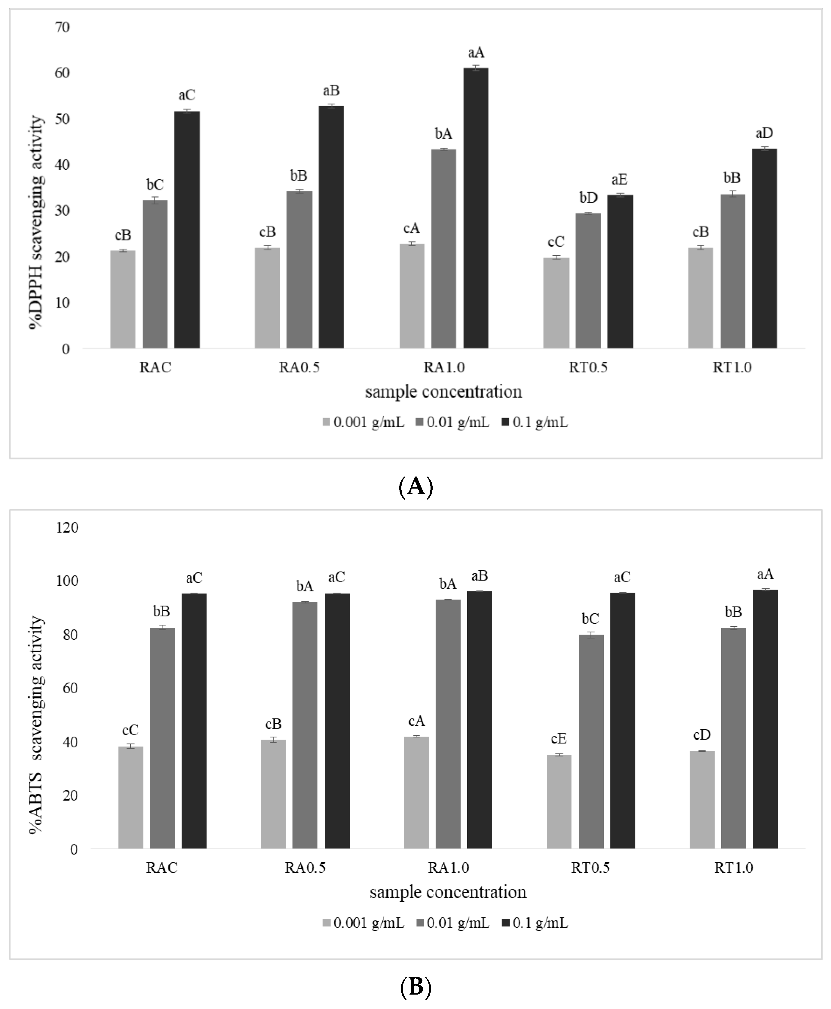
The 2,2-diphenylpicrylhydrazyl (DPPH) radical scavenging activity assay was conducted to evaluate properties of protein hydrolysates based on electron transfer as the hydrogen donor [46]. All hydrolysates showed increased antioxidant activity as the concentration increased (Figure 2A). RA0.5 and RA1.0 at concentrations of 0.1 g/mL had higher activity (52.74% and 61.04%, respectively) compared to others at the same concentration (p < 0.05), indicating higher hydrogen donating ability. These results could be explained by alcalase and trypsin producing different peptides due to different protein cleavage specificity. In contrast, hydrolysis using autoclave with high temperature and pressure randomized the protein cleavage, so that various peptide sizes of hydrolysates were obtained. However, RAC showed higher activity than those by RT0.5 and RT1.0. Several factors, including protease type, processing conditions and initial protein substrate could affect the antioxidative peptides of the product [47]. These results were supported the study by Yang et al. (2011) [48], who reported that different concentrations of protein hydrolysate from bigeye tuna heads (1.25 mg/mL and 10 mg/mL) produced using alcalase had DPPH radical scavenging activity of 25.63% and 82.85%, respectively. Wang et al. (2022) [49] also showed by DPPH scavenging activity that hydrolysates from skipjack tuna roes (0.01 g/mL) produced using alcalase (30.58%) had higher antioxidant activity than using trypsin (25.65%).Moreover, DPPH scavenging activity was found in protein hydrolysate of yellow stripe trevally meat [40] and alcalase-digested egg-yolk protein hydrolysates [50].

3.3.2. ABTS Radical Scavenging Activity

ABTS radical scavenging activity of all hydrolysates is presented in Figure 2B. This measurement is used to determine the interaction between an antioxidant and the pre-generated ABTS+ radical cation based on the donation of a hydrogen atom or an electron to stabilize radicals [51,52]. The ABTS activity showed a trend similar to DPPH, in that the activity increased as the concentration of hydrolysates increased. However, it was observed that percentage of ABTS activity of all hydrolysates was higher than DPPH activity, because ABTS measurement is normally effective for both hydrophilic and lipophilic compounds, while DPPH assay is used only for lipophilic compounds [53]. Klomklao et al. (2018) [54] also reported that antioxidative compounds with the most hydrophilic constituents had high ABTS radical scavenging activity. Moreover, Intarasirisawat et al. (2012) [6] reported that the difference they found in scavenging activity of DPPH and ABTS radicals among protein hydrolysates from skipjack tuna roe might result from a difference in chain length, side chain and hydrophobicity of amino acids. Interestingly, cysteine, which was found predominantly in all hydrolysates for this study, might play a role in ABTS+ scavenging. This is supported by Gómez-Ruiz et al. (2008) [55], who indicated that cysteine was the most active amino acid, followed by tryptophan, tyrosine and histidine in an ABTS+ scavenging assay of ovine casein hydrolysates. All of our hydrolysates at 0.1 g/mL had very high ABTS activity (more than 95%). RA0.5 and RA1.0 at 0.01 g/mL showed higher activity than the others at the same concentrations. However, hydrolysates at concentration of 0.001 g/mL had the lowest activity (p < 0.05) compared to the other two concentrations. Overall, hydrolysates from tuna roe were able to donate a hydrogen atom to radicals, thus stopping the radical chain reaction.

3.3.3. Metal Chelating Activity

Metal chelating activity of all hydrolysates was assayed to determine the ability of antioxidants in chelating the prooxidative metal (ferrous (Fe2+); results are shown in Figure 2C. Chelating activity was quite similar to ABTS activity, in that hydrolysates at concentrations of 0.01 and 0.1 g/mL had higher activity than those at 0.001 g/mL. RAC had lower metal activity than RA and RT at every concentration. The difference in metal activity might be due to amino acid sequences and chain length of peptides. Figure 1 shows that RAC had smaller sized peptides compared to RA and RT, resulting in lower ability to form complexes with Fe2+. These results agree with Intarasirisawat et al. (2012) [6], who indicated that small peptides were not able to form a complex with ferrous (Fe2+) ions. Klomklao et al. (2018) [54] also explained that limited hydrolysis (degree of hydrolysate (DH) at 20%) of protein hydrolysates from skipjack tuna viscera enhanced chelating activity when compared with over-hydrolysis (DH at 30%) (57.03% and 43.33%, respectively).

3.4. Functional Properties of Hydrolysates from Tuna Roes

3.4.1. Solubility

Good solubility of protein hydrolysates is one of the most important characteristics for many food applications, as it influences the physiochemical and functional properties, especially foaming, emulsion and gel properties [56,57]. The solubility of hydrolysates from tuna roes are shown in Table 2. All hydrolysates had high solubility (more than 89%) because of their high content of hydrophilic acids (Table 1). The results support the studies by Mc Clements et al. (2021) [58] and Wouters et al. (2016) [59], who explained that the structure of proteins (polar and non-polar groups) affects solubility: hydrophilic amino acids promote solubility, while hydrophobic residues promote aggregation between proteins, thereby reducing solubility. However, RAC in our study showed the highest solubility (95.15%) compared to others (p < 0.05), which might be due to RAC having smaller molecular size than hydrolysates from alcalase and trypsin digestion (Figure 1). These results support Chalamaiah et al. (2015) [4], who explained that protein hydrolysates from common carp roe obtained by enzymatic hydrolysis with reduced molecular size and increased hydrophilic amino acid groups showed high solubility (from 62.8 to 100%). When comparing hydrolysates obtained from alcalase and trypsin, the solubility was similar except for RT0.5. These results are supported by Vogelsang-O’Dwyer et al. (2022) [60], who explained that solubility of protein hydrolysate might depend on several factors, including the hydrolysis conditions, protease, pH, hydrolysis time and degree of hydrolysis. Mokni Ghribi et al. (2015) [61] reported that solubility of protein hydrolysate from chickpeas treated with alcalase increased with an increasing degree of hydrolysis, which was similar to the finding of García Arteaga et al. (2020) [62], who indicated that the solubility of pea protein hydrolysates obtained by proteases was dependent on the protease applied.

3.4.2. Foaming Properties

Foaming can be described as a two-phase system consisting of air cells separated by a thin continuous liquid layer, typically measured in terms of foaming capacity (FC) and foam stability (FS) [63,64]. The foaming properties of proteins are influenced by the source of the protein, methods and thermal parameters of processing, structure stability of proteins, surface hydrophobicity and also solubility [65]. Overall, the hydrolysates had high FC because heating or enzymatic treatment caused partial denaturation of the protein, increasing foaming properties. As the protein structure unfolds and exposes hydrophobic sites, it may be able to lower interfacial tension by adsorbing more quickly to air-water interfaces with polar groups directed toward water and nonpolar groups directed toward the air, thereby increasing the foaming capacity [66]. Our results agree with Chalamaiah et al. (2015) [4], who reported that protein hydrolysate from carp roe showed higher FC (71–188%) than untreated carp roe protein (41–78%). Nongonierma et al. (2011) [67] also indicated that low molecular weight peptides appeared to exhibit higher FC than native proteins. All hydrolysates rapidly decreased in FS after standing for 15 min, but RA0.5, RA1.0, RT0.5 and RT1.0 showed slower decreases at 30 and 45 min compared to RAC. Klompong et al. (2007) [40] explained that larger peptides can form flexible films around air bubbles, as evidenced by a higher FS. Intarasirisawat et al. (2012) [6] also reported that longer-chain peptides could form a thicker and stronger film surrounding air bubbles in tuna roe hydrolysate. The results of this study suggested that longer-chain peptides had an effect in reducing FC but increasing FS compared to shorter peptides.

3.5. Putative Bioactive Peptides of Hydrolysates from Tuna Roes

Peptides were predicted for thirteen bio-functionalities based on machine-learning-derived classifications, including anti-hypertensive (AH), anti-cancer (AC), anti-toxic (AT), anti-proinflammatory (AI), anti-parasite (AP), anti-virus (AV), anti-fungi (AF), anti-microbial (MI), anti-bacteria (AB), tumor homing (TH), drug delivering (DD), cell communicating (CC) and anti-quorum sensing (QS). Putative bioactive peptides obtained from autoclave and enzymatic hydrolysis methods are presented in Figure 3. Putative anti-hypertensive, anti-virus and anti-parasite peptides were highly abundant within the tuna roe hydrolysates. Anti-hypertensive peptides had the highest probability score, and might be important in regulating osmotic balance to prevent tissue dehydration. Daliri et al. (2017) [68] reported that the anti-hypertensive peptides from various food sources that help regulate blood pressure can inhibit angiotensin converting enzyme (ACE) and reduce high blood pressure or osmotic pressure of interstitial fluid. Tachapuripunya et al. (2021) [26] also reported that these peptides may play a role in maintaining osmotic balance between the epithelial layer and the mucus covering it to prevent tissue dehydration. Recently, anti-hypertensive peptides were also found in marine seafood by-products such as seabass skin, tuna frame, giant grouper roe and squid by-products [69,70]. However, the antihypertensive function of the tuna roe peptides will need to be further tested.
Tuna roe peptides also presented putative evidence of anti-microbials including anti-bacteria, anti-fungi and anti-virus that the reason might be due to these peptides play a role in protecting against harmful microorganisms in the natural environment. Lys and Arg were also frequently found in the tuna roe peptides, and Song et al. (2011) [71] explained that the majority of anti-microbial peptides were shown to be cationic with positively charged amino acid groups, i.e., Lys and Arg. These have been previously reported in fish, such as in the study by Bergsson et al. (2005) [72], who found anti-microbial peptides in the skin mucus of healthy Atlantic cod. This finding supports Ucak et al. (2021) [69] who reported that anti-microbial bioactive peptides work as general protective mechanisms in fish skin. Seo et al. (2014) [73] also reported that peptides of skipjack tuna skin showed activity of anti-microbials to against bacteria including fish pathogens such as Vibrio parahaemolyticus, Aeromonas hydrophila and Streptococcus iniae. Ucak et al. [69] mentioned that the presence of anti-microbials is different among fish species. Anti-parasite peptides shown in the tuna roe peptides may be a means of protection from infection caused by parasites in the natural environment.
In addition, putative anti-quorum sensing also found in tuna roe peptides indicated the potential to disrupt or block quorum sensing activity against bacterial infections [74]. Moreover, tuna roe peptides also showed putative evidence of tumor-homing peptides, drug-delivering peptides and cell-communicating peptides. Tumor-homing bioactive peptides are short peptides or oligopeptides usually consisting of 3–30 amino acids, which specifically bind onto target tumor cells; they are useful bio-tools for regulating anti-tumor payloads in vivo and are also linked to anti-cancer-based drug delivery [75,76]. According to Parn et al. (2015) [77], the drug-delivering peptides share similar properties with tumor-homing, cell-communicating and cell-penetrating peptides in that they are positively charged, hydrophobic and short peptides. Athira et al. (2022) [78] also showed that peptides from Indian major carp presented tumor-homing and anti-cancer peptide. Peptides from many marine species were reported as bioactive natural products with high potential broad spectra of bioactivities such as anti-tumor, anti-microbial or anti-hypertensive [79].
Tuna roe peptides also showed putative evidence of anti-cancer properties, probably due to the presence of lysine. This finding supports the work of Roomi et al. (2015) [80], who reported that nutrients containing lysine and proline proved to be highly toxic to prostate cancer (DU-145) cells. Hsu et al. (2011) [81] reported that two peptides isolated from tuna dark muscle (Leu-Pro-His-Val-Leu-Thr-Pro-Glu-Ala-Gly-Ala-Thr and Pro-Thr-Ala-Glu-Gly-Gly-Val-Tyr-Met-Val-Thr) showed inhibitory activity on the anti-proliferation of breast cancer (MCF-7) cells. Ishak et al. (2018) [82] reported that Pro, Gly, Lys, Arg and Tyr were responsible for anti-cancer activity, and also explained that the lower molecular weight (300–1950 Da) of peptides imparted anti-cancerous activity, due to higher mobility and diffusion. Nurdiani et al. (2016) [83] also reported a high number of anti-cancer peptides from fish by-products. These peptides had not been previously identified or characterized, so their anti-cancer activity remains largely unknown.
Interestingly, anti-proinflammatory properties were also found in the tuna roe peptides. This is important in promoting wound healing, which involves cellular immune response and homeostasis [84,85]. Generally, peptides having positively charged amino acids, (i.e., Lys, Arg, and His) in the C- or N-terminus show anti-inflammatory properties [86]. Phadke et al. (2021) [87] reported that lower molecular weight proteins with hydrophobic amino acids hydrolyzed from fish waste showed anti-inflammatory properties. Recently, anti-proinflammatory peptides were found in the salmon pectoral fin [88]. The autoclaving and enzymatic hydrolysis methods used in this study would also provide low molecular weight and short-chain amino acid sequences rich with those residues, allowing them to confer their anti-inflammatory activity.
Table 3 shows the top three putative properties, anti-hypertensive, anti-virus and anti-parasite, which were abundant in the tuna roe peptides of all hydrolysates. Hydrolysates produced from trypsin (0.5% and 1.0%) gave a higher number of the peptide chain than those by alcalase and autoclaving. The results of peptides number found in RT0.5 and RT1.0 were 184 (34.78%) and 192 (36.29%) for anti-hypertensive, 75 (14.18%) and 80 (15.12%) for anti-virus and 49 (9.26%) and 46 (8.70%) for anti-parasite, respectively. Tachapuripunya et al. (2021) [26] reported that top three bioactive peptides found in the mucous of seven gastropod species were anti-hypertensive (839 peptides on average), drug-delivering (65 peptides on average) and anti-parasitic (40 peptides on average). Moreover, bioactive peptides with at least five to seven bioactive properties were also reported for 268 (9.5%).
Type of peptide and amino acid sequence with high probability score (>0.75) of all hydrolysates are show in Table 4. Interestingly, the length of the peptides was found from octa-peptides to docosa-peptides and seven amino acid sequences found in tuna roe peptides presented multi-properties such as anti-hypertensive, anti-parasite, cell communicating and drug delivering qualities. Type of peptide and amino acid sequence caused the differences in bioactive properties. Suo et al. (2022) [89] reported that 10 novel peptides prepared from milts of skipjack tuna such as YDD, TRE or KLYALF, etc. had angiotensin-converting enzyme (ACE) inhibitory (ACEi) activity. Cai et al. (2022) [90] indicated that amino acid sequence (ACGSDGK) obtained from yellowfin tuna protein hydrolysate showed anti-oxidant activity. Zheng et al. (2022) [91] also reported that ACEi peptides derived from muscle of skipjack tuna, especially SP and VDRYF, are beneficial for functional food against hypertension. Behera et al. (2022) [92] reviewed that the bioactive peptides isolated and characterized from different fish waste (head, skin, frames or viscerals, etc.) have been successfully employed as anti-microbial, anti-oxidant, anti-hypertensive, and anti-carcinogenic agents.

4. Conclusions

Hydrolysates of skipjack tuna roe obtained from enzymatic hydrolysis (alcalase and trypsin) and autoclave provided a good source of protein with abundant cysteine, glycine and arginine. Enzymatic hydrolysates showed higher molecular weight of peptides and also higher antioxidant activities (DPPH activity, ABTS activity and metal chelating activity) than hydrolysates produced by autoclave. All hydrolysates had high solubility and also presented foaming capacity and foam stability. Bioinformatics analysis suggested 13 putative bioactive properties of the tuna roe peptides. Seven peptides with high probability score (>0.75) including MLRASAMR, CDSTSTLCLR, SENQDAQMEK, EHKSLTGTAR, ADVIFKNESLYSHR, MNLGDATTRPPVGRR and LHIQWLEAQEQHQQQEAQLSSR showed multi-bioactive properties. These potential bioactive peptides are expected to be experimentally characterized and will be useful for further application of tuna roe.

Author Contributions

Conceptualization, W.K.; methodology, T.P., V.T. and S.C.; software, T.E.-k.; validation, W.K., S.R. and T.E.-k.; formal analysis, T.P. and V.T.; investigation, T.P.; resources, W.K. and T.E.-k.; data curation, W.K., T.P. and T.E.-k.; writing—original draft preparation, W.K., T.P. and S.C.; writing—review and editing, W.K., S.R. and T.E.-k.; visualization, W.K.; supervision, W.K.; project administration, W.K.; funding acquisition, W.K. All authors have read and agreed to the published version of the manuscript.

Funding

This research was funded by Kasetsart University Research and Development Institute (KURDI), FF (KU) 7.65 under the research program “Development of functional ingredients from by-products of canned tuna processing” and also Kasetsart University through the Graduate School Fellowship Program.

Institutional Review Board Statement

Not applicable.

Informed Consent Statement

Not applicable.

Data Availability Statement

Not applicable.

Conflicts of Interest

The authors declare no conflict of interest.

References

  1. Hsu, K.-C. Purification of antioxidative peptides prepared from enzymatic hydrolysates of tuna dark muscle by-product. Food Chem. 2010, 122, 42–48. [Google Scholar] [CrossRef]
  2. Stevens, J.R.; Newton, R.W.; Tlusty, M.; Little, D.C. The rise of aquaculture by-products: Increasing food production, value, and sustainability through strategic utilisation. Mar. Policy 2018, 90, 115–124. [Google Scholar] [CrossRef]
  3. Klomklao, S.; Benjakul, S. Utilization of Tuna Processing Byproducts: Protein Hydrolysate from Skipjack Tuna (Katsuwonus pelamis) Viscera. J. Food Process. Preserv. 2017, 41, e12970. [Google Scholar] [CrossRef]
  4. Chalamaiah, M.; Jyothirmayi, T.; Diwan, P.V.; Dinesh Kumar, B. Antioxidant activity and functional properties of enzymatic protein hydrolysates from common carp (Cyprinus carpio) roe (egg). J. Food Sci. Technol. 2015, 52, 5817–5825. [Google Scholar] [CrossRef]
  5. Najafian, L.; Babji, A.S. A review of fish-derived antioxidant and antimicrobial peptides: Their production, assessment, and applications. Peptides 2012, 33, 178–185. [Google Scholar] [CrossRef] [PubMed]
  6. Intarasirisawat, R.; Benjakul, S.; Visessanguan, W.; Wu, J. Antioxidative and functional properties of protein hydrolysate from defatted skipjack (Katsuwonous pelamis) roe. Food Chem. 2012, 135, 3039–3048. [Google Scholar] [CrossRef] [PubMed]
  7. Rajabzadeh, M.; Pourashouri, P.; Shabanpour, B.; Alishahi, A. Amino acid composition, antioxidant and functional properties of protein hydrolysates from the roe of rainbow trout (Oncorhynchus mykiss). Int. J. Food Sci. Technol. 2018, 53, 313–319. [Google Scholar] [CrossRef]
  8. Chen, W.; Wei, J.; Zhang, L.; Chen, J.; Li, Y.; Pei, D.; Wang, N.; Liu, Y.; Di, D. Fish Roe Polypeptide Exerts Hypoglycemia Activity via Regulating Insulin Secretion Mediated by Nrf2/ERK Signaling. Int. J. Pept. Res. Ther. 2020, 27, 543–553. [Google Scholar] [CrossRef]
  9. Kim, S.Y.; Je, J.Y.; Kim, S.K. Purification and characterization of antioxidant peptide from hoki (Johnius belengerii) frame protein by gastrointestinal digestion. J. Nutr. Biochem. 2007, 18, 31–38. [Google Scholar] [CrossRef]
  10. Ahn, C.B.; Kim, J.G.; Je, J.Y. Purification and antioxidant properties of octapeptide from salmon byproduct protein hydrolysate by gastrointestinal digestion. Food Chem. 2014, 147, 78–83. [Google Scholar] [CrossRef]
  11. Cai, L.; Wu, X.; Zhang, Y.; Li, X.; Ma, S.; Li, J. Purification and characterization of three antioxidant peptides from protein hydrolysate of grass carp (Ctenopharyngodon idella) skin. J. Funct. Foods 2015, 16, 234–242. [Google Scholar] [CrossRef]
  12. Chi, C.-F.; Wang, B.; Hu, F.-Y.; Wang, Y.-M.; Zhang, B.; Deng, S.-G.; Wu, C.-W. Purification and identification of three novel antioxidant peptides from protein hydrolysate of bluefin leatherjacket (Navodon septentrionalis) skin. Food Res. Int. 2015, 73, 124–129. [Google Scholar] [CrossRef]
  13. Chi, C.-F.; Wang, B.; Wang, Y.-M.; Zhang, B.; Deng, S.-G. Isolation and characterization of three antioxidant peptides from protein hydrolysate of bluefin leatherjacket (Navodon septentrionalis) heads. J. Funct. Foods 2015, 12, 1–10. [Google Scholar] [CrossRef]
  14. Gajanan, P.G.; Elavarasan, K.; Shamasundar, B.A. Bioactive and functional properties of protein hydrolysates from fish frame processing waste using plant proteases. Environ. Sci. Pollut Res. Int. 2016, 23, 24901–24911. [Google Scholar] [CrossRef]
  15. Picot, L.; Bordenave, S.; Didelot, S.; Fruitier-Arnaudin, I.; Sannier, F.; Thorkelsson, G.; Bergé, J.P.; Guérard, F.; Chabeaud, A.; Piot, J.M. Antiproliferative activity of fish protein hydrolysates on human breast cancer cell lines. Process Biochem. 2006, 41, 1217–1222. [Google Scholar] [CrossRef]
  16. Song, R.; Wei, R.; Zhang, B.; Yang, Z.; Wang, D. Antioxidant and antiproliferative activities of heated sterilized pepsin hydrolysate derived from half-fin anchovy (Setipinna taty). Mar. Drugs 2011, 9, 1142–1156. [Google Scholar] [CrossRef] [PubMed]
  17. Cudennec, B.; Ravallec-Plé, R.; Courois, E.; Fouchereau-Peron, M. Peptides from fish and crustacean by-products hydrolysates stimulate cholecystokinin release in STC-1 cells. Food Chem. 2008, 111, 970–975. [Google Scholar] [CrossRef]
  18. Ennaas, N.; Hammami, R.; Beaulieu, L.; Fliss, I. Purification and characterization of four antibacterial peptides from protamex hydrolysate of Atlantic mackerel (Scomber scombrus) by-products. Biochem. Biophys. Res. Commun. 2015, 462, 195–200. [Google Scholar] [CrossRef]
  19. Danquah, M.K.; Agyei, D. Pharmaceutical applications of bioactive peptides. OA Biotechnol. 2012, 1, 1–7. [Google Scholar] [CrossRef]
  20. Olga, M.; Larisa, B.; Natalya, M.; Svetlana, A.; Vladimir, V.; Vasilij, V.; Thomas, G.; Axel, H. High-temperature Hydrolysis As A Method For Complex Processing Of Sprat (sprattus Sprattus Balticus) By-products. JNTM 2022, 12, 8–14. [Google Scholar]
  21. Yang, J.-I.; Liang, W.-S.; Chow, C.-J.; Siebert, K.J. Process for the production of tilapia retorted skin gelatin hydrolysates with optimized antioxidative properties. Process Biochem. 2009, 44, 1152–1157. [Google Scholar] [CrossRef]
  22. Wang, W.; Li, Z.; Liu, J.; Wang, Y.; Liu, S.; Sun, M. Comparison between Thermal Hydrolysis and Enzymatic Proteolysis Processes for the Preparation of Tilapia Skin Collagen Hydrolysates. Czech J. Food Sci. 2013, 31, 1–4. [Google Scholar] [CrossRef]
  23. Ji, C.; Han, J.; Zhang, J.; Hu, J.; Fu, Y.; Qi, H.; Sun, Y.; Yu, C. Omics-prediction of bioactive peptides from the edible cyanobacterium Arthrospira platensis proteome. J. Sci. Food Agric. 2018, 98, 984–990. [Google Scholar] [CrossRef] [PubMed]
  24. Li-Chan, E.C.Y. Bioactive peptides and protein hydrolysates: Research trends and challenges for application as nutraceuticals and functional food ingredients. Curr. Opin. Food Sci. 2015, 1, 28–37. [Google Scholar] [CrossRef]
  25. Cottage, Y.J.K.E.A. Bioinformatics methods to predict protein structure and function. A practical approach. Mol. Biotechnol. 2003, 23, 139–166. [Google Scholar] [CrossRef]
  26. Tachapuripunya, V.; Roytrakul, S.; Chumnanpuen, P. Unveiling Putative Functions of Mucus Proteins and Their Tryptic Peptides in Seven Gastropod Species Using Comparative Proteomics and Machine Learning-Based Bioinformatics Predictions. Molecules 2021, 26, 3475. [Google Scholar] [CrossRef] [PubMed]
  27. Latimer, G.W., Jr. Official Methods of Analysis of AOAC International, 20th ed.; AOAC: Rockville, ML, USA, 2016. [Google Scholar]
  28. Phetchthumrongchai, T.; Chuchird, N.; Roytrakul, S.; Chintong, S.; Klaypradit, W. Physical, chemical composition and umami compound of dried immature and mature roes of skipjack tuna (Katsuwonus pelamis). Fish. Aquat. Sci. 2022, 25, 390–402. [Google Scholar] [CrossRef]
  29. Kedare, S.B.; Singh, R.P. Genesis and development of DPPH method of antioxidant assay. J. Food Sci. Technol. 2011, 48, 412–422. [Google Scholar] [CrossRef]
  30. Re, R.; Pellegrini, N.; Proteggente, A.; Pannala, A.; Yang, M.; Rice-evans, C. Antioxidant activity applying an improved abts radical cation decolorization assay. Free Radic. Biol. Med. 1999, 26, 1231–1237. [Google Scholar] [CrossRef]
  31. Zhou, J.; Yang, Q.; Zhu, X.; Lin, T.; Hao, D.; Xu, J. Antioxidant activities of Clerodendrum cyrtophyllum Turcz leaf extracts and their major components. PLoS ONE 2020, 15, e0234435. [Google Scholar] [CrossRef]
  32. Park, S.H.; Lee, H.J.; Yoon, I.S.; Lee, G.W.; Kim, J.S.; Heu, M.S. Protein functionality of concentrates prepared from yellowfin tuna (Thunnus albacares) roe by cook-dried process. Food Sci. Biotechnol. 2016, 25, 1569–1575. [Google Scholar] [CrossRef] [PubMed]
  33. Wubshet, S.G.; Lindberg, D.; Veiseth-Kent, E.; Kristoffersen, K.A.; Böcker, U.; Washburn, K.E.; Afseth, N.K. Bioanalytical Aspects in Enzymatic Protein Hydrolysis of By-Products. In Proteins: Sustainable Source, Processing and Applications; Academic Press: Cambridge, MA, USA, 2019; pp. 225–258. [Google Scholar]
  34. Kiettiolarn, M.; Kitsanayanyong, L.; Maneerote, J.; Unajak, S.; Tepwong, P. Optimization and production of protein hydrolysate containing antioxidant activity from tuna cooking juice concentrate by response surface methodology. Fish. Aquat. Sci. 2022, 25, 335–349. [Google Scholar] [CrossRef]
  35. Lee, H.J.; Roy, V.C.; Ho, T.C.; Park, J.S.; Jeong, Y.R.; Lee, S.C.; Kim, S.Y.; Chun, B.S. Amino Acid Profiles and Biopotentiality of Hydrolysates Obtained from Comb Penshell (Atrina pectinata) Viscera Using Subcritical Water Hydrolysis. Mar. Drugs 2021, 19, 137. [Google Scholar] [CrossRef] [PubMed]
  36. Kohlmeier, M. Amino Acids and Nitrogen Compounds. In Nutrient Metabolism; Academic Press: Cambridge, MA, USA, 2015; pp. 265–477. [Google Scholar]
  37. Papet, I.; Rémond, D.; Dardevet, D.; Mosoni, L.; Polakof, S.; Peyron, M.-A.; Savary-Auzeloux, I. Sulfur Amino Acids and Skeletal Muscle. In Nutrition and Skeletal Muscle; Academic Press: Cambridge, MA, USA, 2019; pp. 335–363. [Google Scholar]
  38. He, X.Q.; Cao, W.H.; Pan, G.K.; Yang, L.; Zhang, C.H. Enzymatic hydrolysis optimization of Paphia undulata and lymphocyte proliferation activity of the isolated peptide fractions. J. Sci. Food Agric. 2015, 95, 1544–1553. [Google Scholar] [CrossRef]
  39. Chi, C.F.; Hu, F.Y.; Wang, B.; Li, Z.R.; Luo, H.Y. Influence of Amino Acid Compositions and Peptide Profiles on Antioxidant Capacities of Two Protein Hydrolysates from Skipjack Tuna (Katsuwonus pelamis) Dark Muscle. Mar. Drugs 2015, 13, 2580–2601. [Google Scholar] [CrossRef]
  40. Klompong, V.; Benjakul, S.; Kantachote, D.; Shahidi, F. Antioxidative activity and functional properties of protein hydrolysate of yellow stripe trevally (Selaroides leptolepis) as influenced by the degree of hydrolysis and enzyme type. Food Chem. 2007, 102, 1317–1327. [Google Scholar] [CrossRef]
  41. Nalinanon, S.; Benjakul, S.; Kishimura, H.; Shahidi, F. Functionalities and antioxidant properties of protein hydrolysates from the muscle of ornate threadfin bream treated with pepsin from skipjack tuna. Food Chem. 2011, 124, 1354–1362. [Google Scholar] [CrossRef]
  42. Marcet, I.; Alvarez, C.; Paredes, B.; Diaz, M. The use of sub-critical water hydrolysis for the recovery of peptides and free amino acids from food processing wastes. Review of sources and main parameters. Waste Manag. 2016, 49, 364–371. [Google Scholar] [CrossRef]
  43. Lee, H.J.; Park, S.H.; Yoon, I.S.; Lee, G.-W.; Kim, Y.J.; Kim, J.-S.; Heu, M.S. Chemical composition of protein concentrate prepared from Yellowfin tuna Thunnus albacares roe by cook-dried process. Fish. Aquat. Sci. 2016, 19, 12. [Google Scholar] [CrossRef]
  44. Al-holy, M.A.; Rasco, B.A. Characterization of salmon (Oncorhynchus keta) and sturgeon (Acipenser transmontanus) caviar proteins. J. Food Biochem. 2006, 30, 422–428. [Google Scholar] [CrossRef]
  45. Chalamaiah, M.; Jyothirmayi, T.; Bhaskarachary, K.; Vajreswari, A.; Hemalatha, R.; Dinesh Kumar, B. Chemical composition, molecular mass distribution and antioxidant capacity of rohu (Labeo rohita) roe (egg) protein hydrolysates prepared by gastrointestinal proteases. Food Res. Int. 2013, 52, 221–229. [Google Scholar] [CrossRef]
  46. Thiansilakul, Y.; Benjakul, S.; Shahidi, F. Compositions, functional properties and antioxidative activity of protein hydrolysates prepared from round scad (Decapterus maruadsi). Food Chem. 2007, 103, 1385–1394. [Google Scholar] [CrossRef]
  47. Benjakul, S.; Yarnpakdee, S.; Senphan, T.; Halldorsdottir, S.M.; Kristinsson, H.G. Fish protein hydrolysates: Production, bioactivities, and applications. In Antioxidants and Functional Components in Aquatic Foods; John Wiley & Sons, Ltd.: Hoboken, NJ, USA, 2014. [Google Scholar]
  48. Yang, P.; Ke, H.; Hong, P.; Zeng, S.; Cao, W. Antioxidant activity of bigeye tuna (Thunnus obesus) head protein hydrolysate prepared with Alcalase. Int. J. Food Sci. Technol. 2011, 46, 2460–2466. [Google Scholar] [CrossRef]
  49. Wang, J.; Wang, Y.M.; Li, L.Y.; Chi, C.F.; Wang, B. Twelve Antioxidant Peptides From Protein Hydrolysate of Skipjack Tuna (Katsuwonus pelamis) Roe Prepared by Flavourzyme: Purification, Sequence Identification, and Activity Evaluation. Front Nutr. 2021, 8, 813780. [Google Scholar] [CrossRef] [PubMed]
  50. Sakanaka, S.; Tachibana, Y. Active oxygen scavenging activity of egg-yolk protein hydrolysates and their effects on lipid oxidation in beef and tuna homogenates. Food Chem. 2006, 95, 243–249. [Google Scholar] [CrossRef]
  51. Binsan, W.; Benjakul, S.; Visessanguan, W.; Roytrakul, S.; Tanaka, M.; Kishimura, H. Antioxidative activity of Mungoong, an extract paste, from the cephalothorax of white shrimp (Litopenaeus vannamei). Food Chem. 2008, 106, 185–193. [Google Scholar] [CrossRef]
  52. Senphan, T.; Benjakul, S. Antioxidative activities of hydrolysates from seabass skin prepared using protease from hepatopancreas of Pacific white shrimp. J. Funct. Foods 2014, 6, 147–156. [Google Scholar] [CrossRef]
  53. Idowu, A.T.; Benjakul, S.; Sinthusamran, S.; Sookchoo, P.; Kishimura, H. Protein hydrolysate from salmon frames: Production, characteristics and antioxidative activity. J. Food Biochem. 2019, 43, e12734. [Google Scholar] [CrossRef] [PubMed]
  54. Klomklao, S.; Benjakul, S. Protein Hydrolysates Prepared from the Viscera of Skipjack Tuna (Katsuwonus pelmamis): Antioxidative Activity and Functional Properties. Turk. J. Fish. Aquat. Sci. 2018, 18, 69–79. [Google Scholar] [CrossRef]
  55. Gómez-Ruiz, J.Á.; López-Expósito, I.; Pihlanto, A.; Ramos, M.; Recio, I. Antioxidant activity of ovine casein hydrolysates: Identification of active peptides by HPLC–MS/MS. Eur. Food Res. Technol. 2008, 227, 1061–1067. [Google Scholar] [CrossRef]
  56. Avelar, Z.; Vicente, A.A.; Saraiva, J.A.; Rodrigues, R.M. The role of emergent processing technologies in tailoring plant protein functionality: New insights. Trends Food Sci. Technol. 2021, 113, 219–231. [Google Scholar] [CrossRef]
  57. Klomklao, S.; Benjakul, S.; Kishimura, H. Functional properties and antioxidative activity of protein hydrolysates from toothed ponyfish muscle treated with viscera extract from hybrid catfish. Int. J. Food Sci. Technol. 2013, 48, 1483–1489. [Google Scholar] [CrossRef]
  58. McClements, D.J.; Grossmann, L. The science of plant-based foods: Constructing next-generation meat, fish, milk, and egg analogs. Compr. Rev. Food Sci. Food Saf. 2021, 20, 4049–4100. [Google Scholar] [CrossRef] [PubMed]
  59. Wouters, A.G.B.; Rombouts, I.; Fierens, E.; Brijs, K.; Delcour, J.A. Relevance of the Functional Properties of Enzymatic Plant Protein Hydrolysates in Food Systems. Compr. Rev. Food Sci. Food Saf. 2016, 15, 786–800. [Google Scholar] [CrossRef] [PubMed]
  60. Vogelsang-O’Dwyer, M.; Sahin, A.W.; Arendt, E.K.; Zannini, E. Enzymatic Hydrolysis of Pulse Proteins as a Tool to Improve Techno-Functional Properties. Foods 2022, 11, 1307. [Google Scholar] [CrossRef]
  61. Mokni Ghribi, A.; Maklouf Gafsi, I.; Sila, A.; Blecker, C.; Danthine, S.; Attia, H.; Bougatef, A.; Besbes, S. Effects of enzymatic hydrolysis on conformational and functional properties of chickpea protein isolate. Food Chem. 2015, 187, 322–330. [Google Scholar] [CrossRef] [PubMed]
  62. García Arteaga, V.; Apéstegui Guardia, M.; Muranyi, I.; Eisner, P.; Schweiggert-Weisz, U. Effect of enzymatic hydrolysis on molecular weight distribution, techno-functional properties and sensory perception of pea protein isolates. Innov. Food Sci. Emerg. Technol. 2020, 65, 102449. [Google Scholar] [CrossRef]
  63. Foegeding, E.A.; Davis, J.P. Food protein functionality: A comprehensive approach. Food Hydrocoll. 2011, 25, 1853–1864. [Google Scholar] [CrossRef]
  64. Liu, Q.; Kong, B.; Xiong, Y.L.; Xia, X. Antioxidant activity and functional properties of porcine plasma protein hydrolysate as influenced by the degree of hydrolysis. Food Chem. 2010, 118, 403–410. [Google Scholar] [CrossRef]
  65. Zayas, J.F. Foaming Properties of Proteins. In Functionality of Proteins in Food; Springer: Berlin/Heidelberg, Germany, 1997. [Google Scholar]
  66. Mauer, L. PROTEIN|Heat Treatment for Food Proteins. In Encyclopedia of Food Science and Nutrition, 2nd ed.; Caballero, B., Ed.; Academic Press: Oxford, UK, 2003; pp. 4868–4872. [Google Scholar]
  67. Nongonierma, A.; FitzGerald, R.; Nongonierma, A.B.; FitzGerald, R.J. Enzymes exogenous to milk in dairy technology|Proteinases. In Encyclopedia of Dairy Sciences, 2nd ed.; Fuquay, J.W., Ed.; Academic Press: San Diego, CA, USA, 2011; pp. 289–296. [Google Scholar]
  68. Daliri, E.B.; Oh, D.H.; Lee, B.H. Bioactive Peptides. Foods 2017, 6, 32. [Google Scholar] [CrossRef] [PubMed]
  69. Ucak, I.; Afreen, M.; Montesano, D.; Carrillo, C.; Tomasevic, I.; Simal-Gandara, J.; Barba, F.J. Functional and Bioactive Properties of Peptides Derived from Marine Side Streams. Mar. Drugs 2021, 19, 71. [Google Scholar] [CrossRef]
  70. Panjaitan, F.C.A.; Gomez, H.L.R.; Chang, Y.W. In Silico Analysis of Bioactive Peptides Released from Giant Grouper (Epinephelus lanceolatus) Roe Proteins Identified by Proteomics Approach. Molecules 2018, 23, 2910. [Google Scholar] [CrossRef]
  71. Song, R.; Wei, R.; Zhang, B.; Wang, D. Optimization of the Antibacterial Activity of Half-Fin Anchovy (Setipinna taty) Hydrolysates. Food Bioproc. Technol. 2011, 5, 1979–1989. [Google Scholar] [CrossRef]
  72. Bergsson, G.; Agerberth, B.; Jornvall, H.; Gudmundsson, G.H. Isolation and identification of antimicrobial components from the epidermal mucus of Atlantic cod (Gadus morhua). FEBS J. 2005, 272, 4960–4969. [Google Scholar] [CrossRef] [PubMed]
  73. Seo, J.K.; Lee, M.J.; Go, H.J.; Kim, Y.J.; Park, N.G. Antimicrobial function of the GAPDH-related antimicrobial peptide in the skin of skipjack tuna, Katsuwonus pelamis. Fish Shellfish Immunol. 2014, 36, 571–581. [Google Scholar] [CrossRef] [PubMed]
  74. Natrah, F.M.; Defoirdt, T.; Sorgeloos, P.; Bossier, P. Disruption of bacterial cell-to-cell communication by marine organisms and its relevance to aquaculture. Mar. Biotechnol. 2011, 13, 109–126. [Google Scholar] [CrossRef] [PubMed]
  75. Sharma, A.; Kapoor, P.; Gautam, A.; Chaudhary, K.; Kumar, R.; Chauhan, J.S.; Tyagi, A.; Raghava, G.P. Computational approach for designing tumor homing peptides. Sci. Rep. 2013, 3, 1607. [Google Scholar] [CrossRef]
  76. Kondo, E.; Iioka, H.; Saito, K. Tumor-homing peptide and its utility for advanced cancer medicine. Cancer Sci. 2021, 112, 2118–2125. [Google Scholar] [CrossRef] [PubMed]
  77. Parn, K.; Eriste, E.; Langel, U. The Antimicrobial and Antiviral Applications of Cell-Penetrating Peptides. Methods Mol. Biol. 2015, 1324, 223–245. [Google Scholar] [CrossRef]
  78. Athira, P.P.; Anooja, V.V.; Anju, M.V.; Neelima, S.; Archana, K.; Muhammed Musthafa, S.; Antony, S.P.; Bright Singh, I.S.; Philip, R. A hepatic antimicrobial peptide, hepcidin from Indian major carp, Catla catla: Molecular identification and functional characterization. J. Genet. Eng. Biotechnol. 2022, 20, 49. [Google Scholar] [CrossRef]
  79. Cheung, R.C.; Ng, T.B.; Wong, J.H. Marine Peptides: Bioactivities and Applications. Mar. Drugs 2015, 13, 4006–4043. [Google Scholar] [CrossRef]
  80. Roomi, M.W.; Shanker, N.; Niedzwiecki, A.; Rath, M. Induction of Apoptosis in the Human Prostate Cancer Cell Line DU-145 by a Novel Micronutrient Formulation. Open J. Apoptosis 2015, 4, 11–21. [Google Scholar] [CrossRef]
  81. Hsu, K.-C.; Li-Chan, E.C.Y.; Jao, C.-L. Antiproliferative activity of peptides prepared from enzymatic hydrolysates of tuna dark muscle on human breast cancer cell line MCF-7. Food Chem. 2011, 126, 617–622. [Google Scholar] [CrossRef]
  82. Ishak, N.H.; Sarbon, N.M. A Review of Protein Hydrolysates and Bioactive Peptides Deriving from Wastes Generated by Fish Processing. Food Bioproc. Technol. 2017, 11, 2–16. [Google Scholar] [CrossRef]
  83. Nurdiani, R.; Vasiljevic, T.; Singh, T.K.; Donkor, O.N. Bioactive peptides from fish by-products with anticarcinogenic potential. Int. Food Res. J. 2016, 24, 1840–1849. [Google Scholar]
  84. Kemp, D.C.; Kwon, J.Y. Fish and Shellfish-Derived Anti-Inflammatory Protein Products: Properties and Mechanisms. Molecules 2021, 26, 3225. [Google Scholar] [CrossRef]
  85. Calder, P.C.; Ahluwalia, N.; Albers, R.; Bosco, N.; Bourdet-Sicard, R.; Haller, D.; Holgate, S.T.; Jonsson, L.S.; Latulippe, M.E.; Marcos, A.; et al. A consideration of biomarkers to be used for evaluation of inflammation in human nutritional studies. Br. J. Nutr. 2013, 109 (Suppl. S1), S1–S34. [Google Scholar] [CrossRef] [PubMed]
  86. Gao, R.; Shu, W.; Shen, Y.; Sun, Q.; Bai, F.; Wang, J.; Li, D.; Li, Y.; Jin, W.; Yuan, L. Sturgeon protein-derived peptides exert anti-inflammatory effects in LPS-stimulated RAW264.7 macrophages via the MAPK pathway. J. Funct. Foods 2020, 72, 104044. [Google Scholar] [CrossRef]
  87. Phadke, G.G.; Rathod, N.B.; Ozogul, F.; Elavarasan, K.; Karthikeyan, M.; Shin, K.H.; Kim, S.K. Exploiting of Secondary Raw Materials from Fish Processing Industry as a Source of Bioactive Peptide-Rich Protein Hydrolysates. Mar. Drugs 2021, 19, 480. [Google Scholar] [CrossRef]
  88. Ahn, C.B.; Cho, Y.S.; Je, J.Y. Purification and anti-inflammatory action of tripeptide from salmon pectoral fin byproduct protein hydrolysate. Food Chem. 2015, 168, 151–156. [Google Scholar] [CrossRef]
  89. Suo, S.K.; Zheng, S.L.; Chi, C.F.; Luo, H.Y.; Wang, B. Novel angiotensin-converting enzyme inhibitory peptides from tuna byproducts-milts: Preparation, characterization, molecular docking study, and antioxidant function on H2O2-damaged human umbilical vein endothelial cells. Front. Nutr. 2022, 9, 957778. [Google Scholar] [CrossRef] [PubMed]
  90. Cai, B.; Wan, P.; Chen, H.; Huang, J.; Ye, Z.; Chen, D.; Pan, J. Purification and Identification of Novel Myeloperoxidase Inhibitory Antioxidant Peptides from Tuna (Thunnas albacares) Protein Hydrolysates. Molecules 2022, 27, 2681. [Google Scholar] [CrossRef] [PubMed]
  91. Zheng, S.L.; Luo, Q.B.; Suo, S.K.; Zhao, Y.Q.; Chi, C.F.; Wang, B. Preparation, Identification, Molecular Docking Study and Protective Function on HUVECs of Novel ACE Inhibitory Peptides from Protein Hydrolysate of Skipjack Tuna Muscle. Mar. Drugs 2022, 20, 176. [Google Scholar] [CrossRef] [PubMed]
  92. Behera, A.; Das, R.; Patnaik, P.; Mohanty, J.; Mohanty, G. A review on fish peptides isolated from fish waste with their potent bioactivities. J. Appl. Biol. Biotechnol. 2022, 10, 195–209. [Google Scholar] [CrossRef]
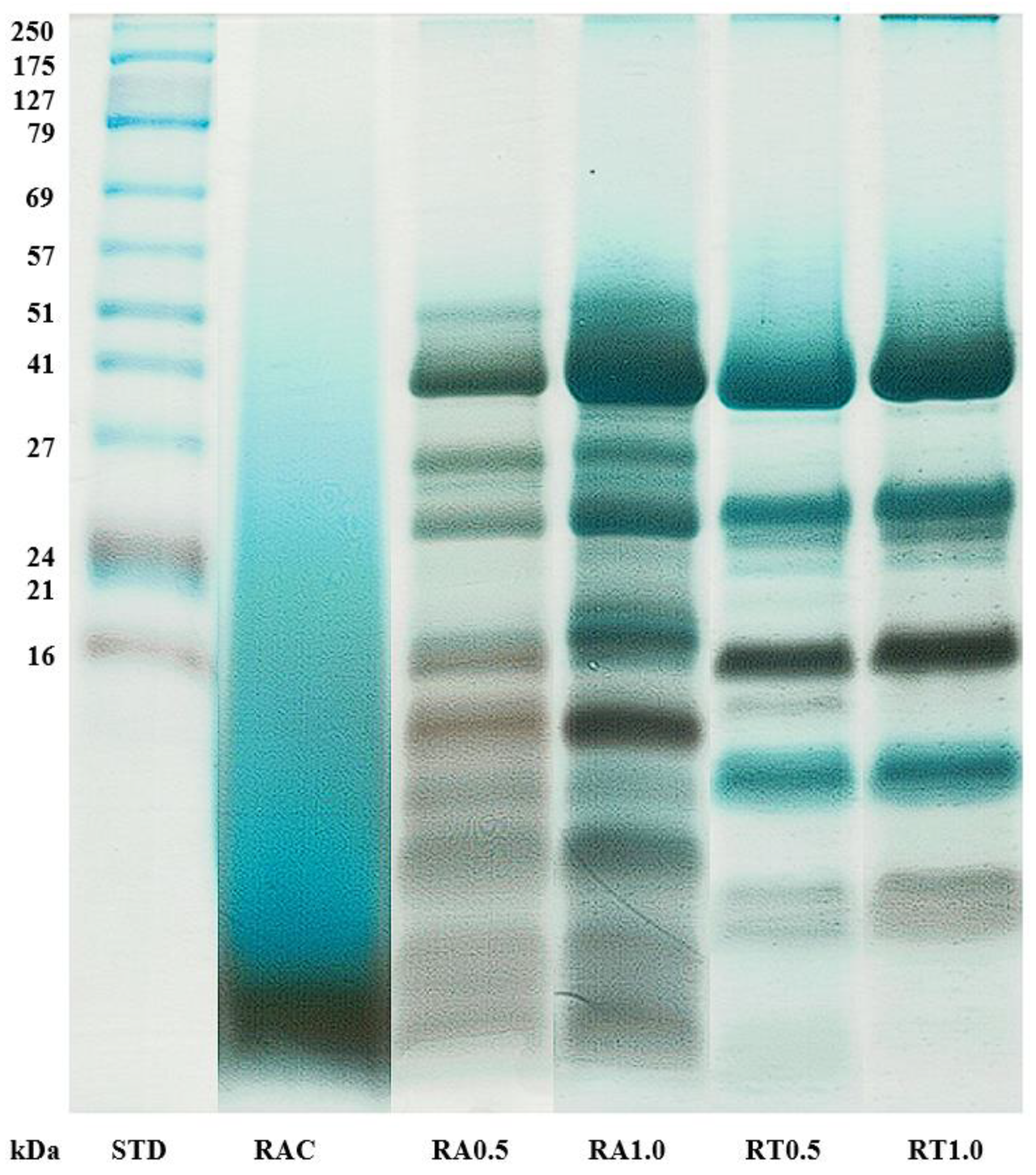
Figure 1. SDS-PAGE patterns of hydrolysates from skipjack tuna roes using autoclave and enzymes (alcalase and trypsin) at different concentrations. Caption; see Table 1.
Fishes 07 00255 g001
Figure 2. Antioxidant activities of hydrolysates from skipjack tuna roes using autoclave and enzymes (alcalase and trypsin) at different concentrations. (A) DPPH radical scavenging activity; (B) ABTS radical scavenging activity; (C) metal chelating activity. Different uppercase letters in the same sample concentration indicate significant difference. Different lowercase letters in the same sample indicate significant difference. Caption; see Table 1.
Fishes 07 00255 g002aFishes 07 00255 g002b
Figure 3. Bioinformatically predicted bioactivity of peptides in protein hydrolysates of skipjack tuna roes using autoclave and enzymes (alcalase and trypsin) at different concentrations. AH, anti-hypertensive; AC, anti-cancer; AT, anti-toxic; AI, anti-proinflammatory; AP, anti-parasite; AV, anti-virus; AF, anti-fungi; MI, anti-microbial; AB, anti-bacteria; TH, tumor homing; DD, drug delivering; CC, cell communicating; QS, anti-quorum sensing.
Fishes 07 00255 g003
Table 1. Amino acid composition of hydrolysates from skipjack tuna roes using autoclave and enzymes (alcalase and trypsin) at different concentrations.
Amino AcidAmino Acid Content (g/100 g Protein)Group
RACRA0.5RA1.0RT0.5RT1.0
Essential amino acids
(EAA)
HistidineHIS2.98 ± 0.033.47 ± 0.103.52 ± 0.073.22 ± 0.133.26 ± 0.09HPL
IsoleucineILE1.69 ± 0.013.11 ± 0.013.05 ± 0.032.56 ± 0.033.18 ± 0.15HPB
LeucineLEU5.30 ± 0.037.81 ± 0.018.03 ± 0.027.17 ± 0.057.71 ± 0.29HPB
LysineLYS5.46 ± 0.036.23 ± 0.096.01 ± 0.046.27 ± 0.056.02 ± 0.03HPL
MethionineMET2.19 ± 0.012.76 ± 0.012.84 ± 0.082.62 ± 0.042.65 ± 0.06HPB
PhenylalaninePHE2.96 ± 0.004.04 ± 0.014.28 ± 0.034.24 ± 0.064.26 ± 0.05HPB
ThreonineTHR2.07 ± 0.022.44 ± 0.012.29 ± 0.192.09 ± 0.032.09 ± 0.08HPL
ValineVAL5.44 ± 0.023.79 ± 0.063.64 ± 0.063.84 ± 0.033.64 ± 0.01HPB
TryptophanTRP2.15 ± 0.011.94 ± 0.092.22 ± 0.091.95 ± 0.132.31 ± 0.19HPB
Non-essential amino acids
(NEAA)
AlanineALA5.19 ± 0.015.90 ± 0.015.98 ± 0.015.58 ± 0.055.85 ± 0.17HPB
ArginineARG10.87 ± 0.0511.20 ± 0.0711.18 ± 0.0712.08 ± 0.0911.82 ± 0.06HPL
AsparticASP2.70 ± 0.052.80 ± 0.002.89 ± 0.022.81 ± 0.043.10 ± 0.02HPL
CysteineCYS20.65 ± 0.1418.14 ± 0.2017.53 ± 0.2218.13 ± 0.2316.26 ± 0.24HPB
GlutamicGLU9.04 ± 0.039.03 ± 0.019.03 ± 0.038.97 ± 0.109.83 ± 0.01HPL
GlycineGLY13.61 ± 0.0410.88 ± 0.0310.67 ± 0.1310.97 ± 0.0510.80 ± 0.04HPL
SerineSER5.25 ± 0.014.61 ± 0.004.74 ± 0.024.98 ± 0.054.77 ± 0.08HPL
TyrosineTYR2.45 ± 0.001.85 ± 0.012.10 ± 0.022.52 ± 0.032.45 ± 0.08HPB
Total (%)100.00100.00100.00100.00100.00
EAA (%)30.2435.5935.8833.9635.12
NEAA (%)69.7664.4164.1266.0464.88
HPB (%)48.0249.3449.6748.6148.31
HPL (%)51.9850.6650.3351.3951.69
Values are presented as mean ± SD (n = 3). Abbreviations: RAC, hydrolysates from tuna roe using autoclave; RA0.5 and RA1.0, hydrolysates from tuna roe using alcalase 0.5% and 1.0%, respectively; RT0.5 and TR1.0, hydrolysates from tuna roe using trypsin 0.5% and 1.0%, respectively; EAA, essential amino acids; NEAA, non-essential amino acids; HPB, hydrophobic amino acids; HPL, hydrophilic amino acids.
Table 2. Functional properties of hydrolysates from skipjack tuna roes using autoclave and enzymes (alcalase and trypsin) at different concentrations.
SampleSolubility
(%)
Foaming Capacity (FC)
(%)
Foam Stability (FS) (%)
FS 15 (min)FS 30 (min)FS 45 (min)
RAC95.15 ± 0.83 a148.44 ± 1.02 a123.80 ± 0.65 aA99.03 ± 1.43 aB57.05 ± 1.57 bC
RA0.591.57 ± 0.77 b141.78 ± 1.02 b77.84 ± 0.78 bA77.16 ± 1.03 bA69.52 ± 1.64 aB
RA1.092.72 ± 0.65 b147.33 ± 0.67 a79.36 ± 0.66 bA78.59 ± 0.67 bA70.18 ± 1.43 aB
RT0.589.65 ± 1.26 c120.89 ± 1.68 c41.80 ± 1.92 cA41.24 ± 1.92 cA38.45 ± 1.95 cA
RT1.091.73 ± 0.94 b119.33 ± 1.33 c41.57 ± 2.74 cA40.97 ± 2.08 cA38.58 ± 0.00 cA
Values are presented as mean ± SD (n = 3). Different uppercase letters in the same row indicate significant difference. Different lowercase letters in the same column indicate significant difference. Caption; see Table 1.
Table 3. Number of peptides with putative anti-hypertensive, anti-virus and anti-parasite qua lities in protein hydrolysates of skipjack tuna roes using autoclave and enzymes (alcalase and trypsin) at different concentrations.
SamplePropertyNumber of Peptide
RACAnti-hypertensive111(20.98%)
RA0.5 95(17.96%)
RA1.0 92(17.39%)
RT0.5 184(34.78%)
RT1.0 192(36.29%)
RACAnti-virus46(8.70%)
RA0.5 43(8.13%)
RA1.0 40(7.56%)
RT0.5 75(14.18%)
RT1.0 80(15.12%)
RACAnti-parasite18(3.40%)
RA0.5 26(4.91%)
RA1.0 18(3.40%)
RT0.5 49(9.26%)
RT1.0 46(8.70%)
Caption; see Table 1.
Table 4. Type of peptide and amino acid sequence with high probability score (>0.75) of predicted bioactivity of peptides in protein hydrolysates of skipjack tuna roes using autoclave and enzymes (alcalase and trypsin) at different concentrations.
Type of PeptideAmino Acid SequenceProperty
Octa-peptidesMLRASAMRAnti-hypertensive
Anti-parasite
Drug delivering
Deca-peptidesCDSTSTLCLRAnti-hypertensive
Cell communicating
Tumor homing
SENQDAQMEKAnti-hypertensive
Anti-parasite
Cell communicating
EHKSLTGTARAnti-hypertensive
Drug delivering
Anti-quorum sensing
Tetradeca-peptidesADVIFKNESLYSHRAnti-cancer
Anti-hypertensive
Anti-parasite
Pentadeca-peptidesMNLGDATTRPPVGRRAnti-hypertensive
Cell communicating
Anti-quorum sensing
Docosa-peptidesLHIQWLEAQEQHQQQEAQLSSRAnti-hypertensive
Drug delivering
Tumor homing
Publisher’s Note: MDPI stays neutral with regard to jurisdictional claims in published maps and institutional affiliations.