Article

Development and Validation of a Quantitative Polymerase Chain Reaction Assay for the Detection of Red Sea Bream Iridovirus

1
Department of Marine Biology and Aquaculture, College of Marine Science, Gyeongsang National University, 2, Tongyeonghaean-ro, Tongyeong 53064, Korea
2
PCR Reagent Development Group, Bioneer, 8-11, Munpyeongseo-ro, Daedeok-gu, Daejeon 34302, Korea
3
Department of Aquatic Life Medicine, College of Fisheries Science, Pukyong National University, 45, Yongso-ro, Nam-Gu, Busan 48513, Korea
*
Authors to whom correspondence should be addressed.
Fishes 2022, 7(5), 236; https://doi.org/10.3390/fishes7050236
Submission received: 10 August 2022 / Revised: 26 August 2022 / Accepted: 1 September 2022 / Published: 5 September 2022

Abstract

:
The analytical and diagnostic performances of methods for detecting red sea bream iridovirus (RSIV), which infects marine fish, have not been evaluated. As disease management and transmission control depend on early and reliable pathogen detection, rapid virus detection techniques are crucial. Herein, we evaluated the diagnostic performance of a TaqMan-based real-time polymerase chain reaction (PCR) assay that detects RSIV rapidly and accurately. The assay amplified the RSIV, infectious spleen and kidney necrosis virus, and turbot reddish body iridovirus genotypes of Megalocytivirus and the detection limit was 10.96 copies/reaction. The assay’s performance remained uncompromised even in the presence of nine potential PCR inhibitors, including compounds commonly used in aquaculture. The variation of the cycle threshold values between assays performed by three technicians was evaluated using a plasmid DNA containing the major capsid protein gene sequence. The variation between replicates was low. The diagnostic sensitivity and specificity of the developed assay were evaluated using fish samples (n = 510) and were found to be 100% and 99.60%, respectively. Two technicians evaluated the reproducibility of the assay using fish samples (n = 90), finding a high correlation of 0.998 (p < 0.0001). Therefore, the newly developed real-time PCR assay detects RSIV both accurately and rapidly.

1. Introduction

Red sea bream iridovirus (RSIV), belonging to the family Iridoviridae and genus Megalocytivirus [1], has been associated with severe economic losses in marine fisheries. Megalocytivirus is divided into three genotypes based on the major capsid protein (MCP) and adenosine triphosphatase (ATPase) gene sequences, namely, RSIV, infectious spleen and kidney necrosis virus (ISKNV), and turbot reddish body iridovirus (TRBIV). Among these, RSIV and ISKNV cause red sea bream iridoviral disease (RSIVD) [2]. RSIV has been detected in Asian and American countries since 1990 when it was first isolated from farmed red sea bream (Pagrus major) in Japan [3]. Several studies have detected RSIV in over 30 species of fish, thus indicating a very broad host range [4]. In addition, it has been detected primarily in seawaters above 25 °C during summer and causes 60–100% mortality in rock bream (Oplegnathus fasciatus) [5,6,7]. Therefore, the diseases caused by RSIV have been registered as “reportable viral diseases” by the World Organization for Animal Health (OIE) Aquatic Animal Health Code since 2000, and are currently managed by the OIE [4]. Currently, no treatment exists to reduce the burden of disease caused by RSIV. Although a formalin-killed commercial vaccine developed to protect against RSIV has been found to be effective in fish species such as red sea bream, striped jack (Pseudocaranx dentex), Malabar grouper (Epinephelus malabaricus), and orange-spotted grouper (E. coioides), it is not very effective in protecting against diseases in fish belonging to the genus Oplegnathus, the most susceptible species [4]. Therefore, to prevent outbreaks of RSIVD in fish farms, hatcheries, and quarantine stations, it is necessary to implement effective disease control strategies through systematic and routine surveillance programs that use appropriate diagnostic tests.
To date, data on the quantitative evaluation of RSIV risk factors are limited. A sensitive and reliable quantitative detection method is essential to monitor the presence of viruses, assess RSIV infection in Korean rock bream, and improve production in aquaculture. Several methods have previously been reported for the detection of RSIV, including cell culture [8], antibody-based antigen detection methods [9], loop-mediated isothermal amplification (LAMP) [10], histopathology [11], polymerase chain reaction (PCR) [12,13], and nested PCR [14]. Other diagnostic methods, such as electron microscopy, antibody-based detection, virus isolation through cell lines, and isothermal amplification using LAMP, have also been reported. However, these techniques are time-consuming and require sensitive cell lines, expensive equipment, and skilled technicians. Although the OIE recommends the use of conventional PCR for the molecular detection of RSIV and ISKNV genotypes [4], this method cannot be used to quantify the viral load and is relatively less sensitive than the real-time PCR assay.
Several real-time PCR-based assays have been reported for the rapid detection of RSIV [15,16,17]. However, the performance of these assays has not been fully evaluated for the various analytical and diagnostic factors. In this study, we developed, optimized, and evaluated the various performance characteristics of a high-sensitivity TaqMan-based real-time PCR assay for the rapid detection of RSIV. In particular, we were able to detect RSIV within a short period by minimizing the reaction time of the real-time PCR assay.

2. Materials and Methods

2.1. Primers, Hydrolysis Probe, and the Real-Time PCR Assay

The primers and hydrolysis probe were designed based on the consensus sequences of the MCP in 157 Megalocytiviruses (RSIV, ISKNV, and TRBIV types) retrieved from the GenBank database of the National Center for Biotechnology Information. The target specificity of the primer was verified through in silico cross-reaction analysis (http://genome.ucsc.edu/cgi-bin/hgPcr?command=start) and using primer-BLAST software [18]. The sequences were aligned using Jalview Ver. 2.11.2.2 software [19]. The nucleotide sequence of the forward primer, Meg 1041F, was: 5′-CCA CCA GAT GGG AGT AGA C-3′, whereas that of the reverse primer, Meg 1139R, was: 5′-GGT TGA TAT TGC CCA TGT CCA-3′. The nucleotide sequence of the probe, Meg 1079P, was: 5′-[FAM]-CCT ACT A[i-EBQ]CT TTG CGC CCA GCA TG-[phosphate]-3′. The probe was labeled with 6-carboxyfluorescein (FAM) at the 5′ end, which functioned as the reporter. Internal-Excellent Bioneer Quencher (i-EBQ) was used as the quencher. The 3′ end was blocked using phosphate. The primers and the probe were ordered from Bioneer Corporation, Daejeon, South Korea.
The final volume of the reaction mixture was 25 μL, and it consisted of 900 nM of each primer (Meg 1041F and Meg 1139R) and 250 nM of probe (Meg 1079P), and 12.5 μL of the HS Prime qPCR Premix with UDG (2×) (Genetbio, Daejeon, South Korea), and 5 μL nucleic acid. The real-time PCR assay was performed in a Dice® Real Time System III (Takara, Kusatsu, Japan). The thermal profile used for DNA amplification consisted of one cycle of 95 °C for 1 min (pre-denaturation), 45 cycles of 95 °C for 5 s (denaturation), and 60 °C for 10 s (annealing and extension).

2.2. Fish Populations

To evaluate diagnostic performance, a total of 510 rock bream (farmed and experimentally infected fish) were collected from eleven fish farms and one hatchery (n = 400) (Table S1 in Supplementary Materials), and an additional 110 were experimentally infected with RSIV. In 2021, 70 healthy fish with weights of 10.5–46.6 g were randomly collected from a rock bream hatchery with no RSIVD history (Table S1), and there were no specific histopathological clinical signs. Another 330 fish (20.5–396.4 g) were randomly collected from eleven fish farms where RSIVD occurred in 2019 (Table S1). The rock bream from the farms was cultured in an under-net pens enclosure, and those from the hatchery were cultured in a land tank of a flow-through aquaculture system. None of the fish collected had a history of vaccination against RSIVD. After sampling, the fish were euthanized, placed in an ice box at 4 °C, and transported to the laboratory within 1 h. The spleen was aseptically excised with the same weight (15 mg), divided into two independent tubes, and stored at −80 °C. Collected spleens were randomly coded with identification numbers to avoid bias in the technician’s test review. The spleen tissue (two independent tubes) was homogenized, and DNA was extracted using an AccuPrep® Genomic DNA Extraction kit (Bioneer, Daejeon, South Korea) according to the manufacturer’s instructions. The total extracted DNA was measured for purity and concentration using a NanoVue spectrophotometer (GE Healthcare, New York, NY, USA), diluted at 50 ng/μL, and stored at −20 °C. The RSIVD status of the collected fish samples was determined by nested PCR (Table S1) [14].

2.3. Experimental Infection

The virus for experimental infection was obtained from the spleen and kidney of RSIV-infected rock bream at a farm located in Tongyeong in August 2019, and the tissue was stored at −80 °C. The presence of RSIV was confirmed by PCR and sequencing analysis methods provided by OIE. RSIV was classified as RSIV genotype II (accession number: AY532608) by phylogenetic analysis of the MCP sequence. The RSIV-infected tissues were homogenized with PBS (phosphate-buffered saline) and centrifuged at 3000× g at 4 °C for 20 min. The virus-containing supernatant was filtered through a 0.45-μm syringe filter, and the filtrate was used for infection experiments.
Rock bream were purchased from a hatchery in Namhae (Gyeongsangnam-do) where RSIVD had not been reported and transported to the Gyeongsang National University. A total of 20 rock bream were randomly selected and confirmed to be free of bacterial, parasitic, and viral diseases. To confirm the presence of pathogenic bacteria, the fish spleens were collected and smeared on the brain and heart infusion agar medium, colonies developed, and the presence of parasites was confirmed by observing the gills and body surface under a microscope. The presence of RSIV was confirmed by nested PCR [14]. Approximately 200 rock bream (total length: 9.6 ± 1.7 cm, weight: 20.1 ± 4.7 g) were acclimated for 1 month in a 1600-L tank. The tank was a flow-through aquaculture system (500–1000 L/h), and was continuously supplied with sand-filtered, 50 μm filter-housed, and UV-treated (>30 mW/cm2) seawater. During the acclimation period, seawater was maintained at a temperature of 15 ± 2 °C, dissolved oxygen at >6 mg/L, salinity at 28–30 psu, pH at 7.8–8.6, and NH3+ at <0.1 mg/L. In addition, rock bream individuals were fed a pellet/extruded type commercial diet of 2–3% of the fish body weight per day.
RSIV experimental infection was performed in a 600-L tank (n = 118). The water temperature in the tank was raised by 1 °C every 5 days until it reached 25 °C where it was maintained for three weeks. A total of 110 rock bream were used in the diagnostic performance evaluation experiment, and the remaining 8 were used to confirm the tissue distribution of RSIV-infected rock bream. Then, 0.1 mL tissue homogenate containing 106 RSIV genome copies/fish at 25 °C was intraperitoneally (IP) injected into the fish (n = 118). RSIV-infected rock bream were anesthetized with benzocaine at 1, 3, 5, and 7 dpi (n = 20 on each day, randomly), and spleen tissue (15 mg) was aseptically collected. Then, the spleen (15 mg) of the remaining rock bream (n = 30) that died at 10–14 dpi was aseptically collected. All spleens collected were divided into two tubes and stored at −80 °C. Tube labeling, DNA extraction, DNA quantification, and nested PCR were performed as described in Section 2.2.
After inoculating the fish with RSIV, moribund fish (n = 8, for 8–14 dpi) were collected to determine the tissue distribution of RSIV. Tissue extracts from the spleen, kidney, heart, gill, eye, intestine, liver, stomach, skin, brain, and muscle were collected and stored at −80 °C until further use. All experimental protocols followed the guidelines of the Institutional Animal Care and Use Committee of the Gyeongsang National University (approval number: GNU-220526-E0056).

2.4. Nucleic Acid Extraction

DNA from fish tissues, viruses, and bacteria used in this study were extracted using an AccuPrep® Genomic DNA extraction kit (Bioneer, Daejeon, South Korea). For RNA viruses, total RNA extraction and cDNA synthesis were performed according to the manufacturer’s instructions, using an easy-spinTM Total RNA Extraction Kit (iNtRON Biotechnology, Seongnam, South Korea), and a PrimeScript 1st strand cDNA Synthesis Kit (TaKaRa) [20].

2.5. Real-Time PCR Assay Validation: Analytical Evaluation

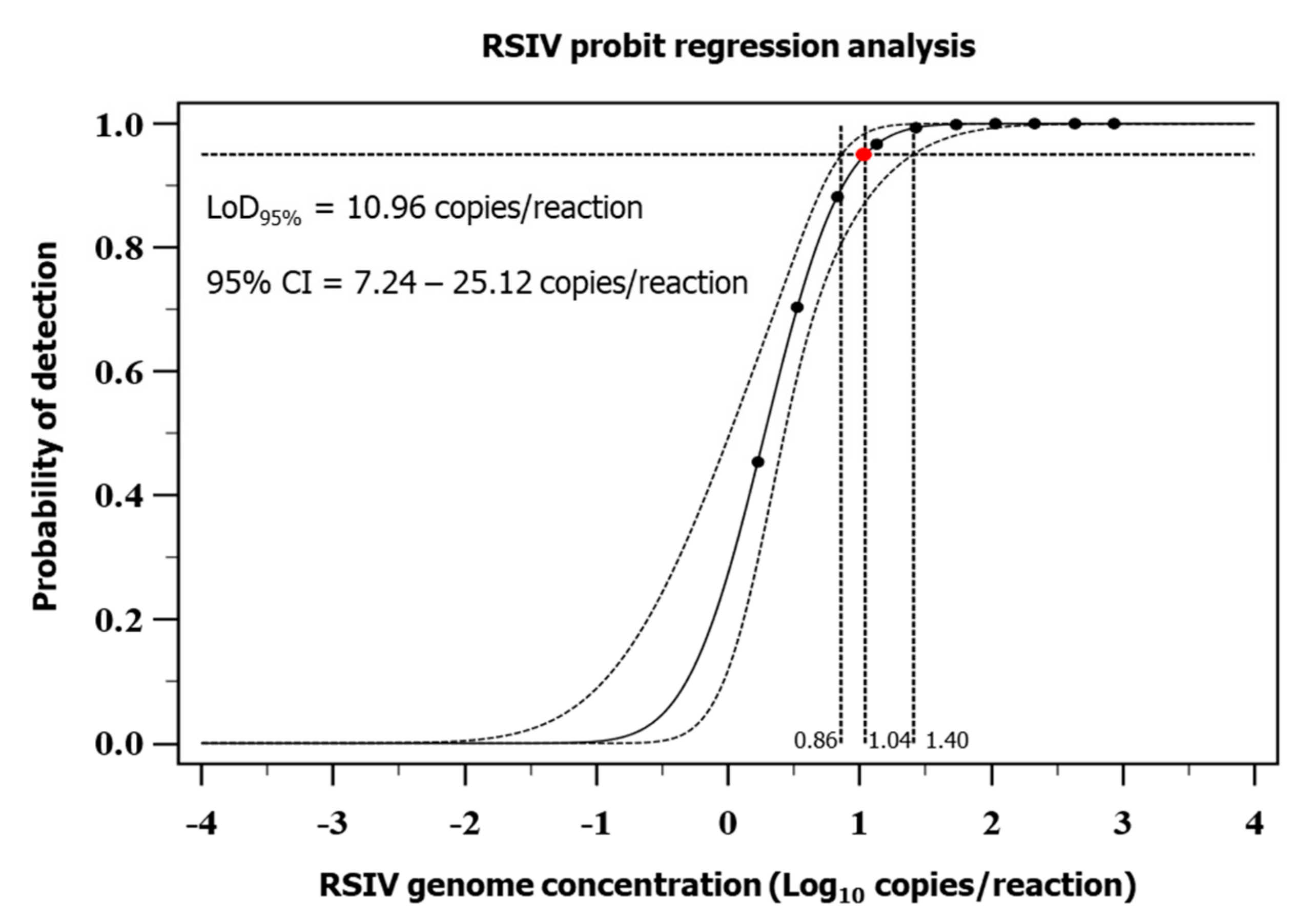
2.5.1. Analytical Sensitivity (ASe)

The MCP amplicon-specific pDNA was generated as follows. DNA was extracted from the RSIV-positive tissue samples, and the 1362 bp PCR amplicon of MCP was ligated to the pGEM-T easy vector (Promega, Madison, WI, USA) [21]. Transformation of the pDNA containing MCP was carried out in competent Escherichia coli (JM109) cells, which were cultured on an LB agar plate containing 100 mg/L ampicillin at 37 °C. Multiple colonies were tested for the success or failure of ligation and transformation using a PCR assay consisting of M13 vector-specific primers; those colonies with inserts were grown in LB broth containing ampicillin, at 37 °C. Thereafter, the pDNA was purified using the GeneAll® Exprep Plasmid SV Kit (GeneAll Biotechnology, Seoul, South Korea), and the number of copies of the purified pDNA was calculated using the formula described in a previous study [22]. This pDNA was used for the evaluation of the analytical performance of all real-time PCR assays.
The ASe of the real-time PCR assay was evaluated using the MCP amplicon-specific plasmid DNA (pDNA) as a template. The dynamic range (standard curve) was obtained using each dilution of a 10-fold serial dilution of the pDNA (8.6 × 10⁷–8.6 copies/reaction) as a template; the experiment was performed in triplicate. The efficiency of the real-time PCR assay was calculated using the cycle threshold (Ct) method (efficiency = [10(−1/slope) − 1] × 100), and the linearity was demonstrated using the coefficient of determination (R2). To determine the 95% limit of detection (LoD95%), a 2-fold serial dilution (860, 430, 215, 107.5, 53.75, 26.88, 13.44, 6.72, 3.36, and 1.68 copies/reaction) of the pDNA was performed with 24 replicates at each dilution [23]. The LoD95% analysis of the real-time PCR assay was performed using the MedCalc statistical software ver. 20.109. The Ct values obtained in each assay were used to determine the standard deviation (SD) and the coefficient of variation (%CV), which were used to assess the reliability of the real-time PCR assay. The Ct value corresponding to the lower limit of detection (Ct value = 39.75) was chosen as the analytical cut-off limit of the real-time PCR assay. Ct values that exceeded this defined limit (indicating very low amounts of the target) were considered unreliable [24]. To revalidate the detection limit of the real-time PCR assay, we tested pDNA (approximately 20 or 10 copies/reaction) at a concentration twice or equivalent to that of the LoD95% concentration, and this analysis was performed 96 times for both concentrations.

2.5.2. Analytical Specificity (ASp)

The ASp of the real-time PCR assay was assessed by performing the assay on certain viral and bacterial pathogens of fish. Eighteen fish viral and bacterial pathogens were used in this study to independently extract nucleic acid to evaluate the cross-reactivity of the real-time PCR assay in triplicate. Three different types of Megalocytivirus were used: red sea bream iridovirus (RSIV; genotype II; accession number, AY532608), pearl gourami iridovirus (PGIV; ISKNV type) [25], and flounder iridovirus (FLIV; TRBIV type) [26]. The other viral pathogens used were lymphocystis disease virus (LCDV), viral nervous necrosis virus (VNNV; BFNNV type), and viral hemorrhagic septicemia virus (VHSV; type IVa). LCDV and VHSV were isolated from the skin tumors and kidney of olive flounder (Paralichthys olivaceus) at a fish farm in Jeju, respectively. VNNV was isolated from the brain of red sea bream cultured at the Fisheries Research Institute in Gyeongsangnam-do, and nucleic acids were extracted from each tissue and cDNA was synthesized and then cross-reaction tests were performed. The bacterial pathogens used were Vibrio harveyi (FP8370), V. ichthyoenteri (FP8487), V. ordalii, V. campbellii, V. alginolyticus, V. anguillarum (TR14001NF), Streptococcus iniae (FP5228), S. parauberis (FP3287), Photobacterium damselae (FP4101), Lactococcus garvieae (FP5245), Edwardsiella tarda (FP5060), and E. coli (JM109). The PGIV and FLIV, which were used for the cross-reactivity testing of the real-time PCR assay, were provided by Professor Kwang-il Kim of Pukyong National University in South Korea; all bacterial strains were provided by the pathology division of the National Institute of Fisheries Science, Busan, South Korea.

2.5.3. Effect of Interfering Substances

To confirm the effect of compounds other than the target virus on the measurement accuracy of the analyte in the real-time PCR assay, we added compounds that were potentially found in fish in aquaculture and evaluated the assay [27,28]. Nine compounds were used, including L-ascorbic acid (10 mM), fucoidan (50 μg/mL), β-glucan (50 mg/mL), enrofloxacin (5 mg/mL), ampicillin (10 mg/mL), kanamycin (10 mg/mL), amoxicillin (10 mg/mL), florfenicol (10 mg/mL), and trimethoprim (10 mg/mL). These compounds were spiked into a solution containing the RSIV DNA (103 copies/reaction). The real-time PCR mix (total volume of 25 μL) consisted of 2.5 μL of DNA, 2.5 μL of interfering material, 900 nM of the forward and reverse primers and 250 nM probe, and 12.5 μL of HS Prime qPCR Premix with UDG (2×) (Genetbio). For the RSIV-positive sample, DNA extracted from the spleen tissue of RSIV-infected rock bream was used, and for the RSIV-negative sample, DNA from healthy rock bream was used. The inhibitory effect of each compound on the real-time PCR assay was identified by testing in triplicate. The Ct values of the assay were analyzed using a t-test to identify the effect of the potential interfering substances on the real-time PCR assay. Statistical analysis was performed using SPSS software version 20.0 (IBM, New York, NY, USA).

2.5.4. Analytical Repeatability

The analytical repeatability of the real-time PCR assay was evaluated at three concentrations of the pDNA (8.6 × 10⁵, 8.6 × 10³, and 8.6 × 101 copies/reaction). For each replicate, a new dilution series for the pDNA was prepared from a concentrated aliquot to be used as a template. Real-time PCR analysis was performed once a day, every 3 days, for a total of 31 days. The experiment was performed in triplicate for each pDNA concentration, and three laboratory technicians independently performed the real-time PCR assay at the three concentrations.

2.6. Real-Time PCR Assay Validation: Diagnostic Performance Evaluation

2.6.1. Diagnostic Sensitivity (DSe) and Diagnostic Specificity (DSp)

The DSe and DSp of the real-time PCR assay were validated using rock bream spleen tissues (n = 510). The spleen tissues included spleen tissues (n = 400) collected from rock bream farms and hatcheries and those obtained through RSIV experimental infection (n = 110). Each sample was confirmed for RSIV status by nested PCR as described previously, and DNA was extracted from replicate spleen pieces (15 mg) collected from the same fish and subjected to real-time PCR analysis. All PCR confirmed that there is no non-specific amplification by adding DEPC-D.W. as a template. Spleen samples used in real-time PCR were randomly coded with identification numbers to avoid bias in the technician’s test review, and the RSIV status of samples confirmed by nested PCR was blinded. A real-time PCR assay was used to determine the performance parameters such as diagnostic sensitivity, specificity, positive predictive value, negative predictive value, and accuracy. All analyses were performed using the MedCalc statistical software version 20.109. The samples were identified as positive and negative based on a defined cut-off value (Ct value of 39.75).

2.6.2. Comparison of the Reproducibility of the Real-Time PCR Assay between Technicians

Two technicians from the same laboratory evaluated the reproducibility of the Ct values on sets of duplicate samples (n = 90). Each technician performed real-time PCR using DNA extracted from replicate pieces of spleen from the same fish. The extracted DNA was quantified at 50 ng/μL after measuring the concentration and purity with a spectrophotometer (GE Healthcare), and the samples (n = 90) were blinded from the two technicians. Correlation between the data was evaluated by plotting the Ct values of one test with that of the other test. Only samples with Ct values ≤ 39.75 for both samples were included in the analyses. Concordance of the Ct values was calculated using the Passing—Bablok regression analysis and the Bland and Altman method (MedCalc statistical software version 20.109) [29,30].

3. Results

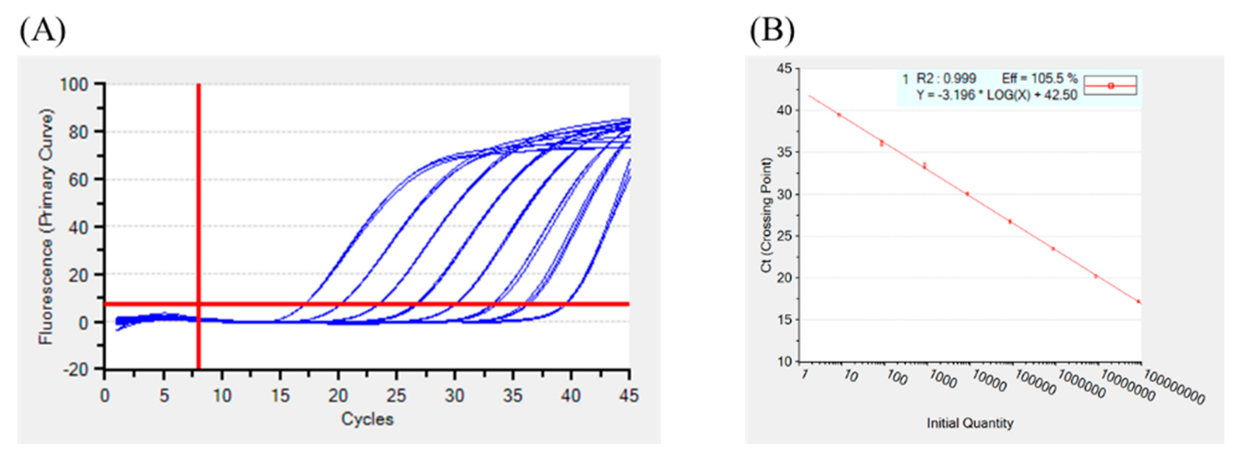
3.1. Analytical Sensitivity of the Real-Time PCR Assay

The primers and the hydrolysis probe of the real-time PCR assay that were evaluated in this study were designed based on the highly conserved region of MCP in Megalocytiviruses. The linear dynamic range of the assay was evaluated using a pDNA containing the MCP insert (8.6 × 10⁷ to 8.6 copies/reaction) (Table S2). The standard curve revealed a linear response (R2 = 0.999). A regression analysis of the linear part of the standard curve revealed a slope of -3.196 and a real-time PCR amplification efficiency of 105.5% (Figure 1). The LoD95% of the real-time PCR assay, determined using probit regression analysis, was 10.96 copies/reaction (95% confidence interval, CI 7.24 to 25.12 copies/reaction), and the cut-off Ct value was 39.75 (Figure 2) (Table S3). Validation of the measured LoD95% revealed a detection rate of 100% (n = 96) and 97.9% (n = 96) for 20 copies/reaction (approximately 2× LoD95%) and 10 copies/reaction (approximately 1× LoD95%), respectively; the mean Ct values for the 2× LoD95% and the 1× LoD95% were 38.66 (SD, 0.63; %CV, 1.62) and 39.22 (SD, 0.8; %CV, 2.04), respectively (Table S4). This shows that the measured LoD95% is reproducible.

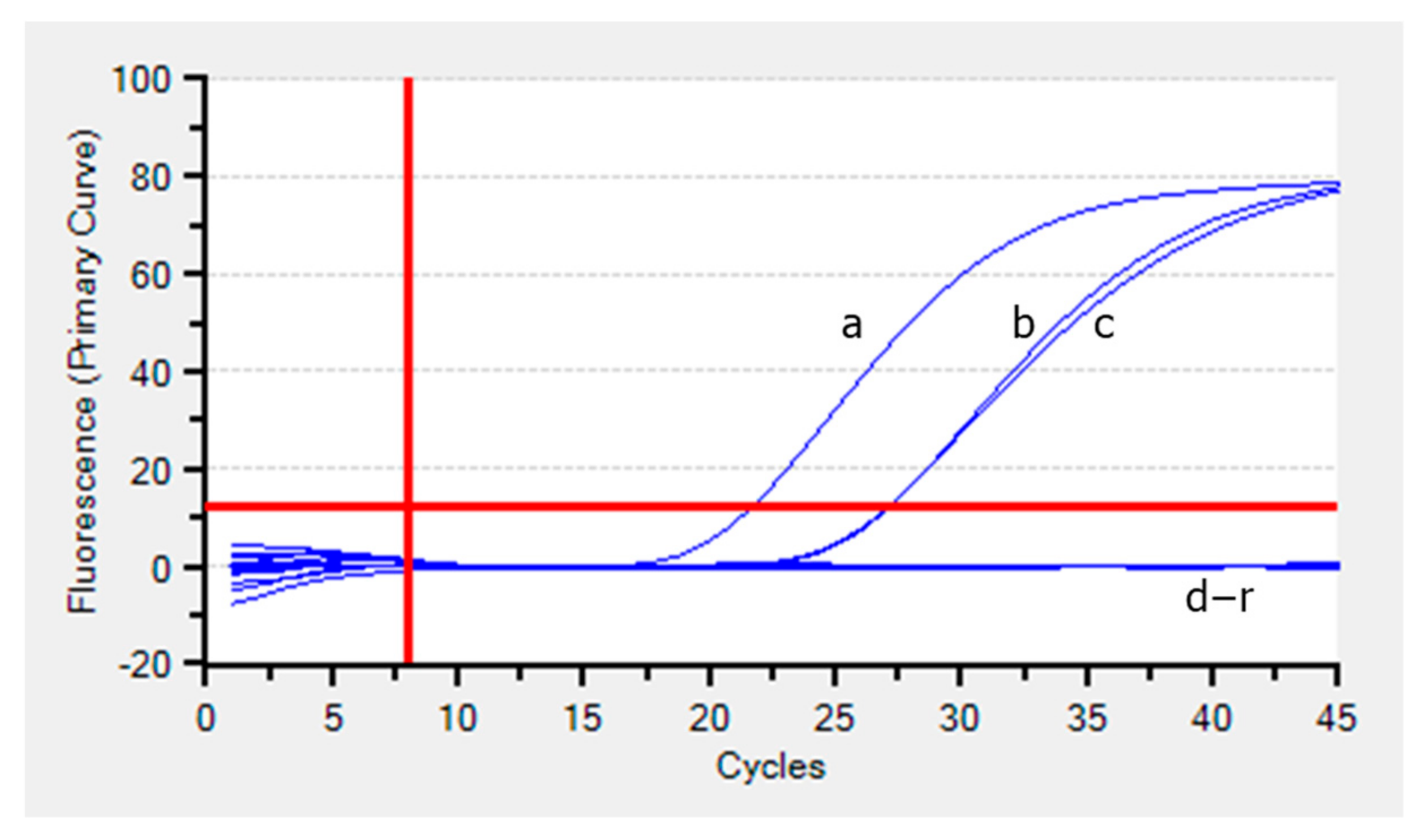
3.2. Analytical Specificity of the Real-Time PCR Assay

The PCR assay specifically amplified the RSIV, ISKNV, and TRBIV genotypes of Megalocytivirus but failed to amplify the nucleotide sequences of the other viral and bacterial fish pathogens described in Section 2.5.2. (Figure 3). In addition, no organisms other than the targeted Megalocytiviruses were detected in the in silico cross-reactivity assay.

3.3. Interfering Substances

The effect of L-ascorbic acid (10 mM), fucoidan (50 μg/mL), β-glucan (50 mg/mL), enrofloxacin (5 mg/mL), ampicillin (10 mg/mL), kanamycin (10 mg/mL), amoxicillin (10 mg/mL), florfenicol (10 mg/mL), and trimethoprim (10 mg/mL), which are potential PCR inhibitors, was investigated. We compared the difference in the mean Ct value (from three replicates) of the RSIV-positive amplicon and that of the RSIV-positive amplicon in the presence of the interfering substance. In addition, no non-specific reactions were observed when the PCR assay was carried out with RSIV-free DNA as a template in the presence of interfering substances. The Ct values obtained during the real-time PCR analysis of the RSIV DNA (103 copies/reaction) performed in the presence of the nine inhibitor compounds were not significantly different. Therefore, the presence of potential PCR inhibitors had no significant effect on the real-time PCR assay (p > 0.05) (Table 1).

3.4. Analytical Repeatability

Three independent technicians performed the real-time PCR assay in triplicate once every 3 days for a total of 31 days to evaluate the repeatability of the assay. The arithmetic mean, SD, and %CV of the three independent experiments were calculated to evaluate the intra- and inter-assay repeatability of the technicians. We found that the variability between the replicates analyzed increased with decreasing pDNA concentration; however, the SD and %CV remained low (i.e., <2). We observed good repeatability at all pDNA concentrations (Table 2).

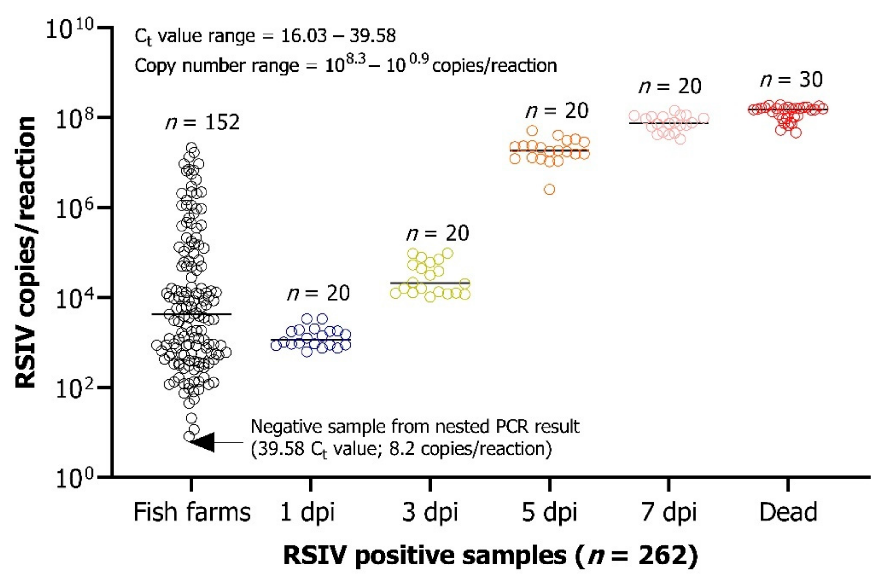
3.5. Diagnostic Performance of the Real-Time PCR Assay on Fish Samples

The performance of the real-time PCR assay was evaluated using 510 spleen samples as described in Section 2.6.1. When tested using a nested PCR assay, 261 of the 510 fish samples were positive for RSIV and 249 were negative. Compared to that of the reference nested PCR assay, the diagnostic sensitivity, specificity, positive predictive value, negative predictive value, and accuracy of the developed real-time PCR assay were 100% (CI 98.60 to 100%), 99.60% (CI 97.78 to 99.99%), 99.62% (CI 97.36 to 99.95%), 100%, and 99.80% (CI 98.91 to 100%), respectively, when a cut-off Ct value of 39.75 was used for analysis (Table 3). Of the 249 samples confirmed to be RSIV-negative by nested PCR, one turned positive in the real-time PCR with a Ct value of 39.58 (8.2 copies/reaction) (Figure 4; Table S1). To confirm the result of the discordant sample using the nested PCR assay, we increased the number of amplification cycles in the 2-step PCR to 45 cycles and found that the sample was positive for RSIV (data not shown).

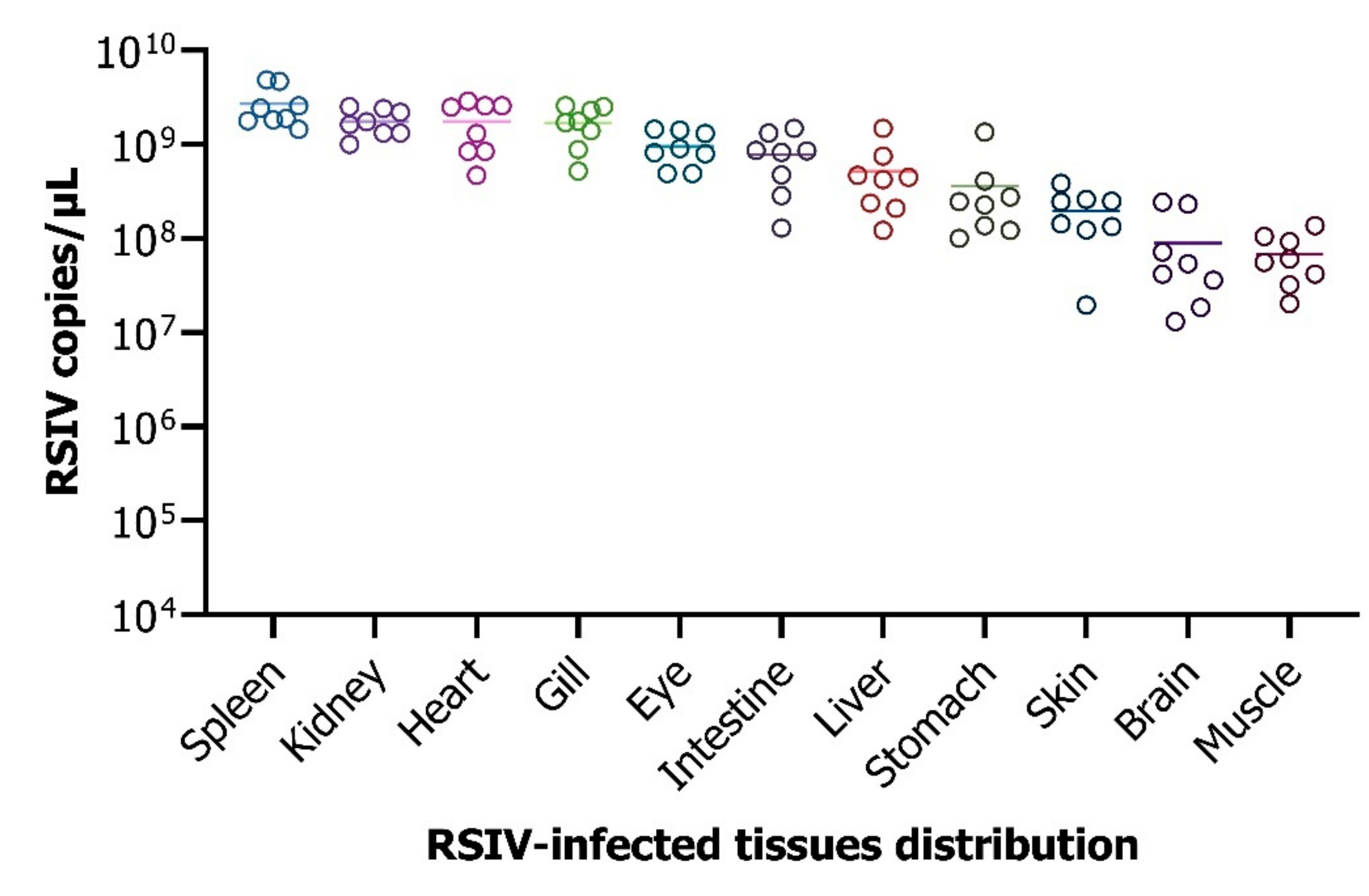
3.6. Analysis of the Viral Load in the Different Tissues Infected by the Virus

Fish that were challenged with RSIV showed signs of RSIV infection within 8 days of inoculation. For real-time PCR analysis, 11 tissues were individually collected from eight RSIV-challenged moribund fish. Analysis of the viral load in the tissues (n = 8) using the real-time PCR assay revealed that the spleen and kidneys had the highest viral loads (109.4 and 109.2 copies/μL, respectively), whereas the muscles had the lowest (107.8 copies/μL) (Figure 5).

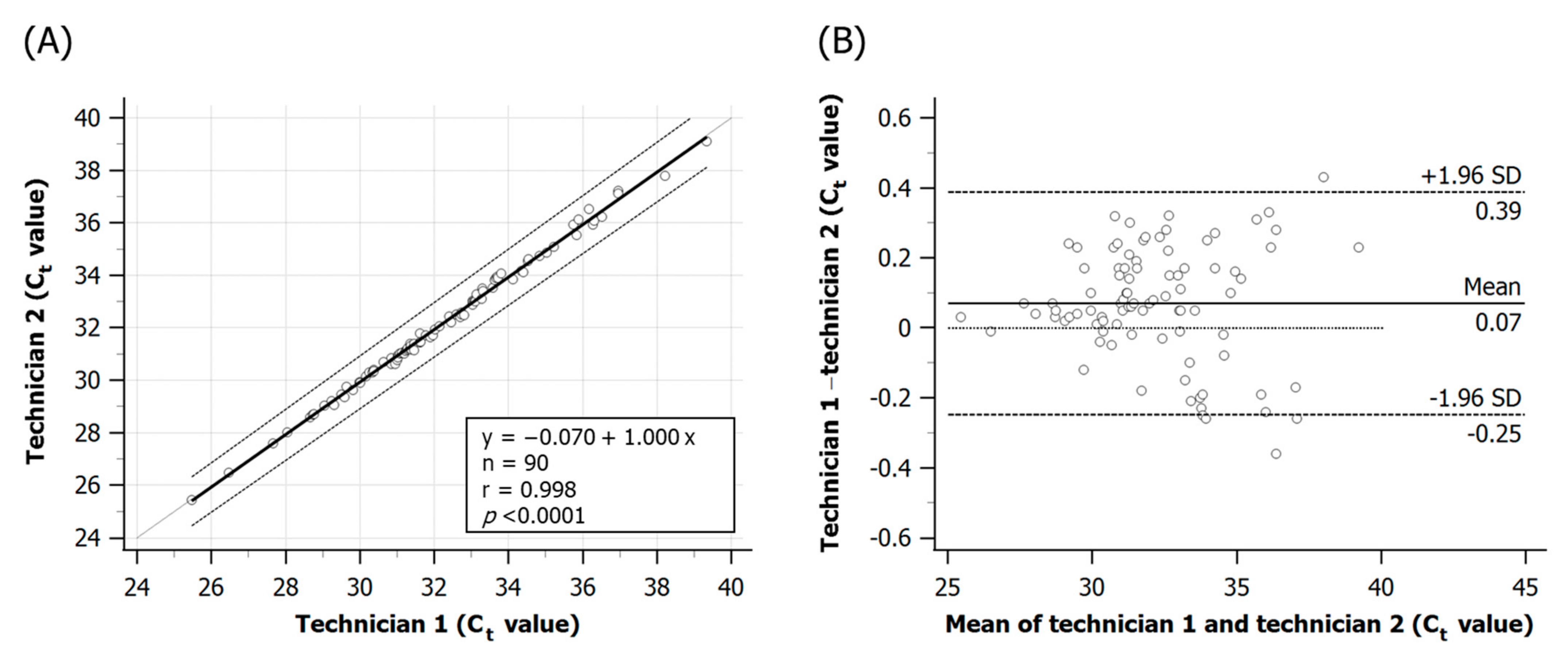
3.7. Within-Laboratory Reproducibility of the Real-Time PCR Analysis of Fish Samples

The real-time PCR analysis of the RSIV-infected tissues, performed by two technicians, showed a very high correlation (r = 0.998, 95% CI 0.997 to 0.999, p < 0.0001) with a slope of 1.000 (Passing–Bablok regression, 95% CI, 0.9850 to 1.0178, regression equation y = −0.070 + 1.000 x) (Figure 6). The mean difference (bias) between the Ct values generated in the real-time PCR assays performed by the two technicians was assessed by the Bland–Altman test and was found to be 0.07 ± 0.16 (Bland-Altman, 95% CI, −0.25 to 0.39 Ct value) (Figure 6). Although there were differences in the Ct values between the two technicians, the concordance was good in most fish samples with a low viral load.

4. Discussion

The real-time PCR assay developed and validated in this study is the optimal quantitative assay for the detection of RSIV and the other Megalocytivirus genotypes. Although real-time PCR assays for the detection of RSIV are available, in most cases, the diagnostic performance of the assay has not been well evaluated. In our study, we tried various methods for the diagnostic validation of the developed assay, in compliance with the validation methods for infectious diseases recommended by the OIE [31]. In this study, we developed a real-time PCR assay with high specificity and sensitivity for the detection of RSIV.
This was achieved by targeting the MCP of RSIV. MCP of Megalocytivirus is present in most iridoviruses and because it has a conserved nucleotide sequence, it has been widely used for the diagnosis or identification of iridovirus infection [2,32,33]. In addition, MCP is a major structural component of Megalocytivirus particles, constituting 40–45% of the total particle polypeptide. The viral envelope protein encoded by MCP plays a role in protecting the viral genome and recognizing host cells [32,34]. Thus, the important role and the conserved properties of the gene encoding MCP in several viral isolates belonging to Megalocytivirus make it a sensitive and appropriate target for RSIV detection using a real-time PCR assay.
To facilitate the detection of low viral copy numbers, it is important to reduce the background signal in the real-time PCR assay. The background fluorescence signal is primarily determined by the design of the hydrolysis probe; the shorter the distance between the fluorescent dye and the quencher, the higher the quenching performance because of the fluorescence resonance energy transfer [35]. In this study, we reduced the distance between the dye and the quencher to seven nucleotides by labeling the inside of the probe with a quencher (i-EBQ), to markedly reduce the background fluorescence of the real-time PCR assay and improve the amplification sensitivity [36]. The 3′ end of the probe was blocked with phosphate to prevent extension. In the primers and the hydrolysis probe that we designed, a reduction in the background signal improved the dynamic range of fluorescence detection and slightly decreased the Ct value (data not shown). Thus, in addition to being faster and more sensitive than conventional PCR assays, the TaqMan-based real-time PCR assay target-specific DNA regions in the species of interest using specially designed hydrolysis probes [37,38].
The LoD95% of the real-time PCR assay was 10.96 copies/reaction, indicating no cross-reactivity with other fish-infecting viruses and bacteria. In addition, we evaluated the performance of our novel real-time PCR assay against a published real-time PCR assay [16]. In the analyses, the Ct values of the two assays performed on 90 RSIV-infected tissues were compared; no statistically significant difference was found in the Ct values between the two methods (p > 0.05) (Figure S1). However, the reference real-time PCR had not been evaluated for LoD95%, presence of PCR inhibitory substances, precision, diagnostic performance using fish spleen samples, various types of fish tissue samples, and reproducibility between technicians. Our results show that our novel real-time PCR assay is reliable for the quantitative detection of RSIV.
The OIE recommends “PCR” as the standard method for diagnosing quarantined aquatic animals and for monitoring RSIVD. A previous study reported a detection limit of 104 copies/reaction for the RSIVD PCR method stated in the OIE manual [39]. These high detection limits may not identify samples infected with a low viral load [39]. The assay may not detect pathogens present in aquatic animals imported/exported between countries, thereby leading to the introduction of the disease in a country where it has not been found before, resulting in serious economic damage in the country. In addition, when estimating the prevalence of disease through the monitoring of aquatic animals in the country, the presence of the disease may be underestimated. In the OIE, nested PCR has been reported as a tentative method for the detection of RSIVD caused by a low viral load [4,14]. In our study, we measured the detection limit using a protocol that was slightly different from the nested PCR protocol reported previously [14]. After the 1-step PCR using the pDNA as a template, the amplicon generated was immediately subjected to a 2-step PCR without diluting 1/100-fold; the detection limit of the assay was confirmed to be approximately 10 copies/reaction (Figure S2). Although the detection limits of the real-time PCR and nested PCR assays developed in this study were similar, the nested PCR took a relatively longer time to complete. Moreover, as the nested PCR amplicon needs to be analyzed by gel electrophoresis, a risk of contamination exists. Additionally, quantitative analysis is impossible. The novel real-time PCR assay can be applied to samples with a low viral load (approximately 10 copies/reaction), and can be used to detect RSIV in seawater, in fish at an early stage of infection, and in low-susceptibility fish.
For performing the developed RSIV real-time PCR assay on fish samples, an appropriate fish target organ is required. According to the OIE (2012), the spleen or kidney is the best organ for diagnosing RSIVD [4]. In our study, we detected a high viral load in the spleen and kidney, and the spleen was used for all subsequent diagnostic evaluations of fish samples. We do not have information on the tissue distribution of RSIV during the early stages of RSIVD infection. Although our results suggest that the spleen tissue has a higher abundance of RSIV, more detailed studies are needed in the future to evaluate the applicability of our findings. This can be done by testing all organs showing signs of different levels of infection in a greater number of fish samples obtained through aquaculture.
The diagnostic performance of the real-time PCR assay was validated using 510 rock bream fish samples, comprising 261 RSIV-positive and 249 RSIV-negative samples validated using nested PCR. The real-time PCR assay showed high DSe (100%) and DSp (99.60%). Among the samples tested, one RSIV-negative fish sample with a low viral load of 8.2 copies/reaction showed a discrepancy in the real-time PCR assay, as the viral load in this sample was below the detection limit of the nested PCR, which was used to determine the positive/negative status of the samples. This sample, collected from a rock bream farm, was a healthy fish without any RSIVD symptoms; however, our real-time PCR assay detected the presence of RSIV in the sample. Therefore, our real-time PCR assay can be used to identify symptomatic as well as asymptomatic RSIV-infected fish.
We investigated nine compounds that are frequently used in fish farms and that can remain in fish bodies. Although PCR failure (false negatives) because of the presence of PCR inhibitors that naturally occur in fish is well known, only a few of the compounds tested inhibited our developed real-time PCR assay [40,41]. To prevent false negative results, we recommend using an internal control (IC), which comprises a nucleotide sequence from an endogenous housekeeping gene that naturally occurs in the test sample [40]. Co-amplification of the test sample and the IC can increase the reliability of the PCR results and enable validation of the negative results with greater precision. In this study, it was confirmed that they do not contain IC but are normally amplified in fish tissues obtained from fish farms. These results suggest that the developed real-time PCR assay can be used as a diagnostic tool for RSIV in the field of aquaculture.
In summary, the real-time PCR assay developed in this study targeted MCP, which is conserved in Megalocytivirus, and accurately detected RSIV in fish from aquaculture farms. We believe that this real-time PCR assay developed for the rapid diagnostic testing of RSIV infection in fish will help control RSIVD in aquaculture farms and quarantine stations.

5. Conclusions

We developed a real-time PCR assay for RSIV detection. DSe and DSp of the developed real-time PCR are 100% and 99.60%, respectively, and RSIV can be detected quickly by minimizing the reaction time. A rapid and accurate detection method is potentially helpful for RSIVD testing of large numbers of specimens in fish farms.

Supplementary Materials

The following supporting information can be downloaded at: https://www.mdpi.com/article/10.3390/fishes7050236/s1. Figure S1: Comparison of the probity viral copy numbers of red sea bream iridovirus (RSIV) in RSIV-positive DNA samples subjected to two TaqMan-based real-time PCR assays (n = 90). The result obtained using the real-time PCR assay developed in this study is shown on the left side, whereas the result obtained using the reference real-time PCR assay is shown on the right side. The bars represent the median viral copy numbers. Figure S2: Determination of the detection limit of the nested PCR assay using a plasmid DNA (pDNA) containing the Megalocytivirus major capsid protein (MCP) gene insert. (A) Sensitivity of the nested PCR assay identified using 10-fold serial dilutions of the pDNA. (B) Sensitivity of the nested PCR assay identified using 2-fold serial dilutions of low concentrations of the pDNA. The nested PCR could detect up to 10 copies/reaction. Negative control, DEPC D.W.; Table S1: Red sea bream iridoviral disease (RSIVD) detection results in the spleens of rock bream (n = 400) collected from eleven farms and one hatchery.; Table S2: TaqMan-based real-time PCR analysis (n = 3) of 10-fold serial dilutions (8.6 × 10⁷ to 8.6 copies/reaction) of plasmid DNA (pDNA) containing the Megalocytivirus major capsid protein (MCP) gene.; Table S3: Limit of detection of the developed TaqMan-based real-time PCR assay identified by testing 2-fold serial dilutions of the plasmid DNA containing the MCP gene, obtained by in vitro transcription.; Table S4: Re-validation of the limit of detection (LoD95%) of the developed TaqMan-based real-time PCR assay.

Author Contributions

Conceptualization, K.-H.K. and C.-I.P.; methodology, K.-H.K.; formal analysis, K.-H.K., K.-M.C., G.K., W.-S.W., M.-Y.S. and H.-J.S.; investigation, K.-H.K.; resources, C.-I.P.; writing—original draft preparation, K.-H.K.; writing—review and editing, D.Y. and D.-H.K.; supervision, C.-I.P.; project administration, C.-I.P.; funding acquisition, C.-I.P. All authors have read and agreed to the published version of the manuscript.

Funding

Ministry of Oceans and Fisheries, Korea (201903922).

Institutional Review Board Statement

All experimental protocols followed the guidelines of the Institutional Animal Care and Use Committee of the Gyeongsang National University (approval number: GNU-220526-E0056).

Acknowledgments

This research was supported by Korea Institute of Marine Science & Technology Promotion (KIMST) funded by the Ministry of Oceans and Fisheries, Korea (201903922).

Conflicts of Interest

The authors declare no conflict of interest.

References

  1. Jancovich, J.K.; Chinchar, V.G.; Hyatt, A.; Miyazaki, T.; Williams, T.; Zhang, Q.Y. Iridoviridae. In Virus Taxonomy: 9th Report of the International Committee on Taxonomy of Viruses; King, A.M.Q., Adams, M.J., Carstens, E.B., Lefkowitz, E.J., Eds.; Academic Press: London, UK, 2012; pp. 207–210. [Google Scholar]
  2. Kurita, J.; Nakajima, K. Megalocytiviruses. Viruses 2012, 4, 521–538. [Google Scholar] [CrossRef] [PubMed]
  3. Inouye, K.; Yamano, K.; Maeno, Y.; Nakajima, K.; Matsuoka, M.; Wada, Y.; Sorimachi, M. Iridovirus infection of cultured red sea bream, Pagrus major. Fish Pathol. 1992, 27, 19–27. [Google Scholar] [CrossRef]
  4. OIE (World Organisation for Animal Health). Red sea bream iridoviral disease. In Manual of Diagnostic Tests for Aquatic Animals; OIE: Paris, France, 2012; Chapter 2.3.7; Available online: https://www.oie.int/fileadmin/Home/eng/Health_standards/aahm/current/2.3.07_RSIVD.pdf (accessed on 29 March 2022).
  5. Jun, L.J.; Jeong, J.B.; Kim, J.H.; Nam, J.H.; Shin, K.W.; Kim, J.K.; Kang, J.C.; Jeong, H.D. Influence of temperature shifts on the onset and development of red sea bream iridoviral disease in rock bream Oplegnathus fasciatus. Dis. Aquat. Org. 2009, 84, 201–208. [Google Scholar] [CrossRef]
  6. Jung, M.H.; Jung, S.J.; Vinay, T.N.; Nikapitiya, C.; Kim, J.O.; Lee, J.H.; Lee, J.; Oh, M.J. Effects of water temperature on mortality in Megalocytivirus-infected rock bream Oplegnathus fasciatus (Temminck et Schlegel) and development of protective immunity. J. Fish Dis. 2015, 38, 729–737. [Google Scholar] [CrossRef] [PubMed]
  7. Jung, M.H.; Nikapitiya, C.; Vinay, T.N.; Lee, J.H.; Jung, S.J. Rock bream iridovirus (RBIV) replication in rock bream (Oplegnathus fasciatus) exposed for different time periods to susceptible water temperatures. Fish Shellfish Immunol. 2017, 70, 731–735. [Google Scholar] [CrossRef]
  8. Kawato, Y.; Yamashita, H.; Yuasa, K.; Miwa, S.; Nakajima, K. Development of a highly permissive cell line from spotted knifejaw (Oplegnathus punctatus) for red sea bream iridovirus. Aquaculture 2017, 473, 291–298. [Google Scholar] [CrossRef]
  9. Nakajima, K.; Maeno, Y.; Fukudome, M.; Fukuda, Y.; Tanaka, S.; Matsuoka, S.; Sorimachi, M. Immunofluorescence test for the rapid diagnosis of red sea bream iridovirus infection using monoclonal antibody. Fish Pathol. 1995, 30, 115–119. [Google Scholar] [CrossRef]
  10. Caipang, C.M.; Haraguchi, I.; Ohira, T.; Hirono, I.; Aoki, T. Rapid detection of a fish iridovirus using loop-mediated isothermal amplification (LAMP). J. Virol. Methods 2004, 121, 155–161. [Google Scholar] [CrossRef]
  11. He, J.G.; Zeng, K.; Weng, S.P.; Chan, S.M. Systemic disease caused by an iridovirus-like agent in cultured mandarinfish, Siniperca chuatsi (Basilewsky), in China. J. Fish. Dis. 2000, 23, 219–222. [Google Scholar] [CrossRef]
  12. Kurita, J.; Nakajima, K.; Hirono, I.; Aoki, T. Polymerase chain reaction (PCR) amplification of DNA of red sea bream iridovirus (RSIV). Fish Pathol. 1998, 33, 17–23. [Google Scholar] [CrossRef] [Green Version]
  13. Oshima, S.; Hata, J.; Hirasawa, N.; Ohtaka, T.; Hirono, I.; Aoki, T.; Yamashita, S. Rapid diagnosis of red sea bream iridovirus infection using the polymerase chain reaction. Dis. Aquat. Org. 1998, 32, 87–90. [Google Scholar] [CrossRef] [PubMed]
  14. Choi, S.K.; Kwon, S.R.; Nam, Y.K.; Kim, S.K.; Kim, K.H. Organ distribution of red sea bream iridovirus (RSIV) DNA in asymptomatic yearling and fingerling rock bream (Oplegnathus fasciatus) and effects of water temperature on transition of RSIV into acute phase. Aquaculture 2006, 256, 23–26. [Google Scholar] [CrossRef]
  15. Caipang, C.M.; Hirono, I.; Aoki, T. Development of a Real-time PCR Assay for the Detection and Quantification of Red Seabream Iridovirus (RSIV). Fish Pathol. 2003, 38, 1–7. [Google Scholar] [CrossRef]
  16. Mohr, P.G.; Moody, N.J.; Williams, L.M.; Hoad, J.; Cummins, D.M.; Davies, K.R.; StJ Crane, M. Molecular confirmation of infectious spleen and kidney necrosis virus (ISKNV) in farmed and imported ornamental fish in Australia. Dis. Aquat. Org. 2015, 116, 103–110. [Google Scholar] [CrossRef]
  17. Lee, E.S.; Cho, M.; Min, E.Y.; Jung, S.H.; Kim, K.I. Novel peptide nucleic acid-based real-time PCR assay for detection and genotyping of Megalocytivirus. Aquaculture 2020, 518, 734818. [Google Scholar] [CrossRef]
  18. Ye, J.; Coulouris, G.; Zaretskaya, I.; Cutcutache, I.; Rozen, S.; Madden, T.L. Primer-BLAST: A tool to design target-specific primers for polymerase chain reaction. BMC Bioinform. 2012, 13, 134. [Google Scholar] [CrossRef]
  19. Waterhouse, A.M.; Procter, J.B.; Martin, D.M.A.; Clamp, M.; Barton, G.J. Jalview Version 2-a multiple sequence alignment editor and analysis workbench. Bioinformatics 2009, 25, 1189–1191. [Google Scholar] [CrossRef]
  20. Choi, K.M.; Joo, M.S.; Kang, G.; Woo, W.S.; Kim, K.H.; Jeong, S.H.; Son, M.Y.; Kim, D.H.; Park, C.I. First report of eosinophil peroxidase in starry flounder (Platichthys stellatus): Gene identification and gene expression profiling. Fish Shellfish Immunol. 2021, 118, 155–159. [Google Scholar] [CrossRef]
  21. Kim, K.I.; Hwang, S.D.; Cho, M.Y.; Jung, S.H.; Kim, Y.C.; Jeong, H.D. A natural infection by the red sea bream iridovirus-type Megalocytivirus in the golden mandarin fish Siniperca scherzeri. J. Fish. Dis. 2018, 41, 1229–1233. [Google Scholar] [CrossRef]
  22. Godornes, C.; Leader, B.T.; Molini, B.J.; Centurion-Lara, A.; Lukehart, S.A. Quantitation of rabbit cytokine mRNA by real-time RT-PCR. Cytokine 2007, 38, 1–7. [Google Scholar] [CrossRef] [Green Version]
  23. Kralik, P.; Ricchi, M. A Basic Guide to Real Time PCR in Microbial Diagnostics: Definitions, Parameters, and Everything. Front. Microbiol. 2017, 8, 108. [Google Scholar] [CrossRef] [PubMed]
  24. Caraguel, C.G.; Stryhn, H.; Gagné, N.; Dohoo, I.R.; Hammell, K.L. Selection of a cutoff value for real-time polymerase chain reaction results to fit a diagnostic purpose: Analytical and epidemiologic approaches. J. Vet. Diagn. Investig. 2011, 23, 2–15. [Google Scholar] [CrossRef] [PubMed]
  25. Jeong, J.B.; Kim, H.Y.; Jun, L.J.; Lyu, J.H.; Park, N.G.; Kim, J.K.; Jeong, H.D. Outbreaks and risks of infectious spleen and kidney necrosis virus disease in freshwater ornamental fishes. Dis. Aquat. Org. 2008, 78, 209–215. [Google Scholar] [CrossRef] [PubMed]
  26. Kim, K.I.; Jin, J.W.; Kim, Y.C.; Jeong, H.D. Detection and genetic differentiation of megalocytiviruses in shellfish, via high-resolution melting (HRM) analysis. Korean J. Fish Aquat. Sci. 2014, 47, 241–246. [Google Scholar]
  27. Burd, E.M. Validation of Laboratory-Developed Molecular Assays for Infectious Diseases. Clin. Microbiol. Rev. 2010, 23, 550–576. [Google Scholar] [CrossRef]
  28. Schrader, C.; Schielke, A.; Ellerbroek, L.; Johne, R. PCR inhibitors–occurrence, properties and removal. J. Appl. Microbiol. 2012, 113, 1014–1026. [Google Scholar] [CrossRef]
  29. Bilić-Zulle, L. Comparison of methods: Passing and Bablok regression. Biochem. Med. 2011, 21, 49–52. [Google Scholar] [CrossRef]
  30. Kirsch, K.; Detilleux, J.; Serteyn, D.; Sandersen, C. Comparison of two portable clinical analyzers to one stationary analyzer for the determination of blood gas partial pressures and blood electrolyte concentrations in horses. PLoS ONE 2019, 14, e0211104. [Google Scholar] [CrossRef]
  31. OIE (World Organisation for Animal Health). Principles and methods of validation of diagnostic assays for infectious diseases. In Manual of Diagnostic Tests and Vaccines for Terrestrial Animals; OIE: Paris, France, 2013; Chapter 1.1.6; Available online: https://www.oie.int/fileadmin/Home/eng/Health_standards/tahm/1.01.06_VALIDATION.pdf (accessed on 24 November 2021).
  32. Tidona, C.A.; Schnitzler, P.; Kehm, R.; Darai, G. Is the major capsid protein of iridoviruses a suitable target for study of viral Evolution? Virus Genes 1998, 16, 59–66. [Google Scholar] [CrossRef]
  33. Eaton, H.E.; Metcalf, J.; Penny, E.; Tcherepanov, V.; Upton, C.; Brunetti, C.R. Comparative genomic analysis of the family Iridoviridae: Re-annotating and defining the core set of iridovirus genes. Virol. J. 2007, 4, 11. [Google Scholar] [CrossRef]
  34. Fu, X.; Li, N.; Lai, Y.; Liu, L.; Lin, Q.; Shi, C.; Huang, Z.; Wu, S. Protective immunity against iridovirus disease in mandarin fish induced by recombinant major capsid protein of infectious spleen and kidney necrosis virus. Fish Shellfish Immunol. 2012, 33, 880–885. [Google Scholar] [CrossRef] [PubMed]
  35. Hirotsu, Y.; Mochizuki, H.; Omata, M. Double-quencher probes improve detection sensitivity toward Severe Acute Respiratory Syndrome Coronavirus 2 (SARS-CoV-2) in a reverse-transcription polymerase chain reaction (RT-PCR) assay. J. Virol. Methods 2020, 284, 113926. [Google Scholar] [CrossRef] [PubMed]
  36. Xia, H.; Gravelsina, S.; Öhrmalm, C.; Ottoson, J.; Blomberg, J. Development of single-tube nested real-time PCR assays with long internally quenched probes for detection of norovirus genogroup II. Biotechniques 2016, 60, 28–34. [Google Scholar] [CrossRef] [PubMed]
  37. Holland, P.M.; Abramson, R.D.; Watson, R.; Gelfand, D.H. Detection of specific polymerase chain reaction product by utilizing the 5′–3′ exonuclease activity of Thermus aquaticus DNA polymerase. Proc. Natl. Acad. Sci. USA 1991, 88, 7276–7280. [Google Scholar] [CrossRef]
  38. Wilhelm, J.; Pingoud, A. Real-time polymerase chain reaction. ChemBioChem 2003, 4, 1120–1128. [Google Scholar] [CrossRef]
  39. Rimmer, A.E.; Becker, J.A.; Tweedie, A.; Whittington, R.J. Development of a quantitative polymerase chain reaction (qPCR) assay for the detection of dwarf gourami iridovirus (DGIV) and other megalocytiviruses and comparison with the Office International des Epizooties (OIE) reference PCR protocol. Aquaculture 2012, 358, 155–163. [Google Scholar] [CrossRef]
  40. Hoffmann, B.; Beer, M.; Reid, S.M.; Mertens, P.; Oura, C.A.; van Rijn, P.A.; Slomka, M.J.; Banks, J.; Brown, I.H.; Alexander, D.J.; et al. A review of RT-PCR technologies used in veterinary virology and disease control: Sensitive and specific diagnosis of five livestock diseases notifiable to the World Organisation for Animal Health. Vet. Microbiol. 2009, 139, 1–23. [Google Scholar] [CrossRef]
  41. Sidstedt, M.; Rådström, P.; Hedman, J. PCR inhibition in qPCR, dPCR and MPS-mechanisms and solutions. Anal. Bioanal. Chem. 2020, 412, 2009–2023. [Google Scholar] [CrossRef] [Green Version]
Figure 1. Analysis of a TaqMan-based real-time PCR assay performed in triplicate on aliquots of plasmid DNA (pDNA) containing the Megalocytivirus major capsid protein (MCP) gene, serially diluted 10-fold (8.6 × 10⁷ to 8.6 copies/reaction). (A) Amplification plots of the real-time PCR assay. (B) Standard curve derived by plotting the threshold cycle (Ct) values against the pDNA copy number.
Fishes 07 00236 g001
Figure 2. Analytical limit of detection (LoD95%) of the TaqMan-based real-time PCR assay. The plasmid DNA (pDNA) containing the Megalocytivirus major capsid protein (MCP) gene was serially diluted (from 860 to 1.68 copies/reaction) and tested. The probability of detection of each concentration of the pDNA was calculated using 24 independent experiments and the LoD95% was determined by probit regression analysis.
Fishes 07 00236 g002
Figure 3. Analytical specificity of the TaqMan-based real-time PCR assay. a—Red sea bream iridovirus (RSIV) genotype II; b—infectious spleen and kidney necrosis virus (ISKNV) type; c—turbot reddish body iridovirus (TRBIV) type; d–r—the three viral and 12 bacterial species found in the fish (see Section 2.5.2 for pathogen details).
Fishes 07 00236 g003
Figure 4. Quantification of the viral copy numbers of the red sea bream iridovirus (RSIV)-positive DNA samples using TaqMan-based real-time PCR analysis. The bars represent the median viral copy numbers. The black arrows indicate the sample that tested RSIV-negative in the nested PCR, and positive (8.2 copies/reaction) in the real-time PCR.
Fishes 07 00236 g004
Figure 5. Tissue distribution of the red sea bream iridovirus (RSIV) in RSIV-infected rock bream fish (Oplegnathus fasciatus). The bars represent the mean viral copy numbers (n = 8).
Fishes 07 00236 g005
Figure 6. Comparison of the Ct values obtained during the real-time PCR analysis, performed by two technicians, using the spleen sample of red sea bream iridovirus (RSIV)-infected rock bream fish (Oplegnathus fasciatus). (A) Passing–Bablok regression analysis of the Ct values obtained by technician 1 and 2 in RSIV-positive samples. (B) Bland–Altman analysis of the differences in the Ct values obtained by technician 1 and 2. The mean Ct values obtained by technicians 1 and 2 plotted against the difference.
Fishes 07 00236 g006
Table 1. TaqMan-based real-time PCR analysis in the presence of nine potential PCR inhibitors. All experiments were performed in triplicate. The ΔCt value is the difference between the mean Ct value (from three replicas) of the RSIV-positive amplicon and that of the RSIV-positive amplicon in the presence of an interfering substance.
MaterialsStock ConcentrationMean CtSDCV (%)ΔCtt-Test
Only Positive SamplePositive Sample + Interfering SubstanceNegative Sample + Interfering Substance
L-Ascorbic acid10 mM32.7732.99N.D.0.110.32−0.22p = 0.054
Fucoidan50 μg/mL32.7732.84N.D.0.080.25−0.07p = 0.4
Beta-glucan50 mg/mL32.7732.72N.D.0.150.460.05p = 0.701
Enrofloxacin5 mg/mL32.7732.71N.D.0.060.200.06p = 0.481
Ampicillin10 mg/mL32.7732.64N.D.0.200.620.13p = 0.386
Kanamycin10 mg/mL32.7732.72N.D.0.150.470.05p = 0.702
Amoxicillin10 mg/mL32.7732.57N.D.0.150.450.2p = 0.129
Florfenicol10 mg/mL32.7732.80N.D.0.100.30−0.03p = 0.704
Trimethoprim10 mg/mL32.7732.71N.D.0.100.290.06p = 0.519
Table 2. Repeatability analysis of the TaqMan-based real-time PCR assay using three concentrations of plasmid DNA (pDNA) containing the Megalocytivirus major capsid protein (MCP) gene insert.
Concentrations (Copies/Reaction)Intra-Assay RepeatabilityInter-Assay Repeatability
Technician 1Technician 2Technician 3Technicians 1–3
Mean Ct
(n = 33)
SDCV (%)Mean Ct
(n = 33)
SDCV (%)Mean Ct
(n = 33)
SDCV (%)Mean Ct
(n = 99)
SDCV (%)
8.60 × 10523.270.070.323.380.170.7323.370.180.7823.340.160.67
8.60 × 10329.880.080.2829.980.220.7229.920.361.2129.930.250.83
8.60 × 10136.390.391.0836.550.491.3436.390.391.0836.460.441.2
Table 3. Diagnostic performance of the TaqMan-based real-time PCR assay in comparison with the reference nested PCR assay conducted on fish samples.
No. of Samples with the Following Result in RSIV Nested PCR Assay
Known PositiveKnown NegativeTotal
TaqMan qPCR assay
(Ct cut-off value 39.75)
Positive2611262
Negative0248248
Total261249
Performance parameterValue95% CI
Sensitivity (%)10098.60 to 100
Specificity (%)99.697.78 to 99.99
Positive predictive value (%)99.6297.36 to 99.95
Negative predictive value (%)100
Accuracy (%)99.898.91 to 100
Publisher’s Note: MDPI stays neutral with regard to jurisdictional claims in published maps and institutional affiliations.

