Article

Effect of Estradiol on Estrogen Nuclear Receptors Genes Expression on Embryonic Development Stages in Chinese Soft-Shelled Turtle (Pelodiscus sinensis)

1
Yangtze River Fisheries Research Institute, Chinese Academy of Fisheries Science, Wuhan 430223, China
2
College of Fisheries and Life Science, Shanghai Ocean University, Shanghai 201306, China
*
Authors to whom correspondence should be addressed.
Fishes 2022, 7(5), 223; https://doi.org/10.3390/fishes7050223
Submission received: 8 July 2022 / Revised: 24 August 2022 / Accepted: 24 August 2022 / Published: 27 August 2022

Abstract

:
Among Chinese soft-shelled turtles, Pelodiscus sinensis, males have a richer nutritional value and higher market price than females. All-male offspring were obtained by 17β-estradiol (E2). However, the molecular mechanisms of E2 inducing sexual reversal remain unclear. In this study, we cloned estrogen nuclear receptors (ERs) from P. sinensis and investigated their expression profiles. We examined the responses of ERα and ERβ after treatment with different concentrations of 1.0, 5.0, and 10 mg/mL E2. ERs showed abundant expressions in the adult gonad, ERα for ovary, and ERβ for testis. E2 can up-regulate the expression of ERα, which showed a remarkable increase while the promotion of ERβ was unobvious. They reached a high level at stage 17 after the treatment of E2, genes of the female-related genes Rspo1, Wnt4, β-catenin, Foxl2, Cyp19a1, and Sox3 exhibited a significant raise at stage 17 with the increase in the concentration of E2 while the male-related genes Sox9, Dmrt1, and Amh were significantly inhibited. Our study cloned the full length of ERs and analyzed their structures and expressions, laying a foundation for the further study of the effect of estrogen on sex determination.

Graphical Abstract

1. Introduction

The Chinese soft-shelled turtle, Pelodiscus sinensis, is an economically important aquatic species that is widely distributed in China [1]. It has typical features of sexual growth dimorphism which mean males grow significantly faster and have richer nutritional value and higher market price than females [2]. In order to expand the breeding efficiency of breeding enterprises, the all-male breeding of P. sinensis using sex control approaches has become a practical requirement. Sex determination in reptiles can be divided into two categories. One is genotypic sex determination (GSD), in which sex determination genes on sex chromosomes first regulate and induce the cascade reaction of related sex determination genes, and finally regulate the development of primordial gonads towards the testis or ovary [3]. The other type is environment-dependent sex determination (ESD). The most typical ESD model is temperature-dependent sex determination (TSD), and the sex determination of embryos is affected by the environmental temperature during embryonic development, rather than genetic material. Researchers have confirmed that P. sinensis is a GSD species with ZZ/ZW heteromorphic micro-sex chromosomes [4], which was significantly different from the typical TSD in Trachemys scripta [5].
To obtain all-male offspring, E2 treatment is administered to male embryos (ZZ), E2 can induce gonads to develop into ovaries, but does not change the genotype, thus forming an individual with a female phenotype and male genotype called pseudo-female turtles (ΔZZ). These individuals could be used as the female parent while the male turtle was used as the male parent (ZZ) for breeding work, and their offspring would all be male [6,7]. In vertebrates, the sexual reversal process was co-regulated by the female-related genes (Rspo1/Wnt/β-catenin/Foxl2/Cyp19a1/Sox3) and male-related genes (Sox9/Amh/Dmrt1), and the male-to-female sexual reversal could be caused after estrogen treatment during embryonic development [8]. Rspo1, known as a female-determining factor, functions upstream of the female sex determination pathway to activate the Wnt/β-catenin signaling pathway in mammals [9]. In humans, the deletion or mutation of Rspo1 could lead to female-to-male sexual reversal [10]. In mice, the over-expression of β-catenin reversed the sex of male XY individuals to female XY individuals [11]. The Sry-related gene Sox9 was involved in the differentiation of Sertoli cells in male gonads of vertebrates with different kinds of sex determination which could be inhibited by estrogen resulting in male-to-female sexual reversal [12]. The medaka, Oryzias latipes, the pattern of male-to-female sexual reversal could be achieved by estrogen treatment and its special sex determination gene named DM-domain gene on the Y chromosome (Dmy) was clearly suppressed during the embryonic development stage [13]. In Gulf pipefish, Syngnathus scovelli, males were exposed to estrogen, which led to the feminization of the male liver transcriptome in a pipefish having undergone sexual reversal [14].
The estrogen receptors expressed in vertebrates and a few invertebrates undertake the effect of estrogen on sex-related genes [15]. Estrogen receptors were mainly divided into two categories: classical estrogen nuclear receptors (ERs) and novel membrane receptors. The first estrogen binding protein discovered is known as estrogen receptor α (ERα, also known as ER1 or Esr1) [16]. The second nuclear estrogen receptor was named estrogen receptor β (ERβ, also known as ER2 or Esr2) [17]. Both of them are nuclear transcription factors that were involved in the regulation of many complex physiological processes [18,19]. In the nuclear, ERs act as ligand-dependent transcription factors, when the hormone signal enters the cytoplasm, it binds to the estrogen nuclear receptor, forming the hormone–nuclear receptor complex. It was transported through the nuclear envelope to the nucleus which regulated the transcription factor activity by recognizing specific DNA fragments, thereby achieving the transcriptional regulation of sex-related genes [20]. ERs also regulate gene expression via binding to other transcription factors such as nuclear transcription factor-κB (NF-κB) [21]. Researchers found that the other is novel membrane receptors, that estrogen could rapidly up-regulate endometrial cyclic adenosine monophosphate (cAMP) levels through the cell membrane binding site, and thus speculated the existence of the membrane estrogen receptor (mER) [22], which was later discovered as G protein-coupled estrogen receptor 30 (GPER30) [23]. By way of membrane receptors, G-protein-coupled receptors were activated after binding with ligands, and their coupled proteins linked hormones to cAMP in the cytoplasm, thus playing a regulatory role [8].
Exogenous estradiol (E2) is critical in the process of all-male offspring; however, the molecular mechanism between E2 and estrogen receptors in sex determination remains unclear. In the present study, we cloned the ERs from P. sinensis and characterized their expression in normal adults and embryos. We further investigated the effects of E2 on the expression of nuclear receptors in male embryos. The results will contribute to further investigations of sex determination, sex differentiation, and the breeding work in Chinese soft-shelled turtles.

2. Materials and Methods

2.1. Maintenance of the Chinese Soft-Shelled Turtles

One-year-old healthy turtles (3 males and 3 females, mean weight 1100 ± 100 g) and fertilized eggs were obtained by random sampling from Anhui Xijia Agricultural Development Company (Bengbu, Anhui, China). Following euthanasia by MS-222 (600 mg/kg), the turtles were dissected and eight tissues including the heart, liver, spleen, lung, kidney, brain, ovary, or testis were collected and stored in liquid nitrogen for RNA isolation. All animal handling and experimental procedures were conducted under the guidelines for the care and use of the laboratory and approved by the Yangtze River Fisheries Research Institute Animal Care Committee.

2.2. Estradiol Treatment

The fertilized eggs were incubated in the constant temperature humidity incubator (Xinmiao, Shanghai, China) and kept at 30 ± 0.5 °C and 80–85% humidity for 15 days. We diluted E2 (MedChemExpress, Wuhan, China) with ethanol (Xilong, Guangdong, China) into a reagent of 1 mg/mL, 5 mg/mL, and 10 mg/mL which were the experimental groups. We dipped a cotton swab in a small amount of hydrochloric acid (HCl) (Xilong, Guangdong, China), and gently smeared it on the soft-shelled turtle fertilized eggs to make the egg-shell soft at stage 12 (15 days) of embryo development which was the critical period of sex differentiation. A micro-syringe (Gaoge, Shanghai, China) was used to inject 5 μL of one of the E2 solutions into each fertilized egg, the control group was injected with an equal volume of ethanol, and the blank group does not do any processing [24]. Two hundred fertilized eggs were inoculated at each concentration, and the blank control group raised 200 fertilized eggs for sampling. The injected site was then disinfected with 50 mg/mL of ampicillin and sealed with paraffin, putting the eggs back in the constant temperature humidity incubator.

2.3. Samples Collection

Samples, i.e., the embryos, were collected from stages 13 to 20, the fertilized eggs were broken up with tweezers and the embryos were removed and placed in cryogenic tubes, first in liquid nitrogen, and at the end of sampling, all samples were placed at −80 °C. Twelve samples were collected at each stage. The sex of all embryos was detected using sex-related markers (Ps4085-F/R, COI-F/R) developed by our laboratory [2]. After sex determination, the number of male embryos at each stage varied from 3 to 8. In order to ensure the consistency of the number of samples at each stage, we randomly selected 3 male embryonic samples from each stage for further analysis. Male samples of the experimental group and control group were used to explore the expressions of ERs during the embryonic development stage after the E2 treatment of adult turtle samples was used to detect the tissue distributions of ERs. Blank group samples were used to detect the expression of ERs during the normal embryonic development stage. All adult turtles were targeted for RNA extraction from organs (i.e., heart, liver, spleen, lung, kidney, brain, ovary, or testis), while total embryos after sex determination were targeted for RNA extraction.

2.4. RNA Extraction

The samples were taken out from −80 °C and thawed on ice, and then the samples were quickly decomposed. The gonads of the embryonic samples and part of the tissue samples of the adult turtles were placed into a 2 mL EP tube with 1 mL Trizol kit (Life Technologies, Shanghai, China) and three grinding beads. Then, the tissue crushing instrument was used at 120 Hz for 60 s, the solution was absorbed into a new 1.5 mL EP tube, and then chloroform was added and shaken for 5 min and centrifuged for 10 min. The supernatant was absorbed into a new 1.5 mL EP tube after centrifugation and blended with an equal volume of pre-cooled isopropyl alcohol, and centrifuged for another 10 min. After the supernatant was aspirated, 1 mL of pre-cooled 75% ethanol was added to 1.5 mL EP for centrifuging for 5 min to wash the RNA precipitate. The washing procedure was repeated three times, and then the liquid was poured out and dried on the ultra-clean table, and sterile water was added to dissolve for 10 min. Then, the concentration of the extracted RNA was detected by a Nanophotometer NP60 spectrophotometer (Implen, Munich, Germany) and the integrity of the RNA was detected by 1% agarose gel electrophoresis.

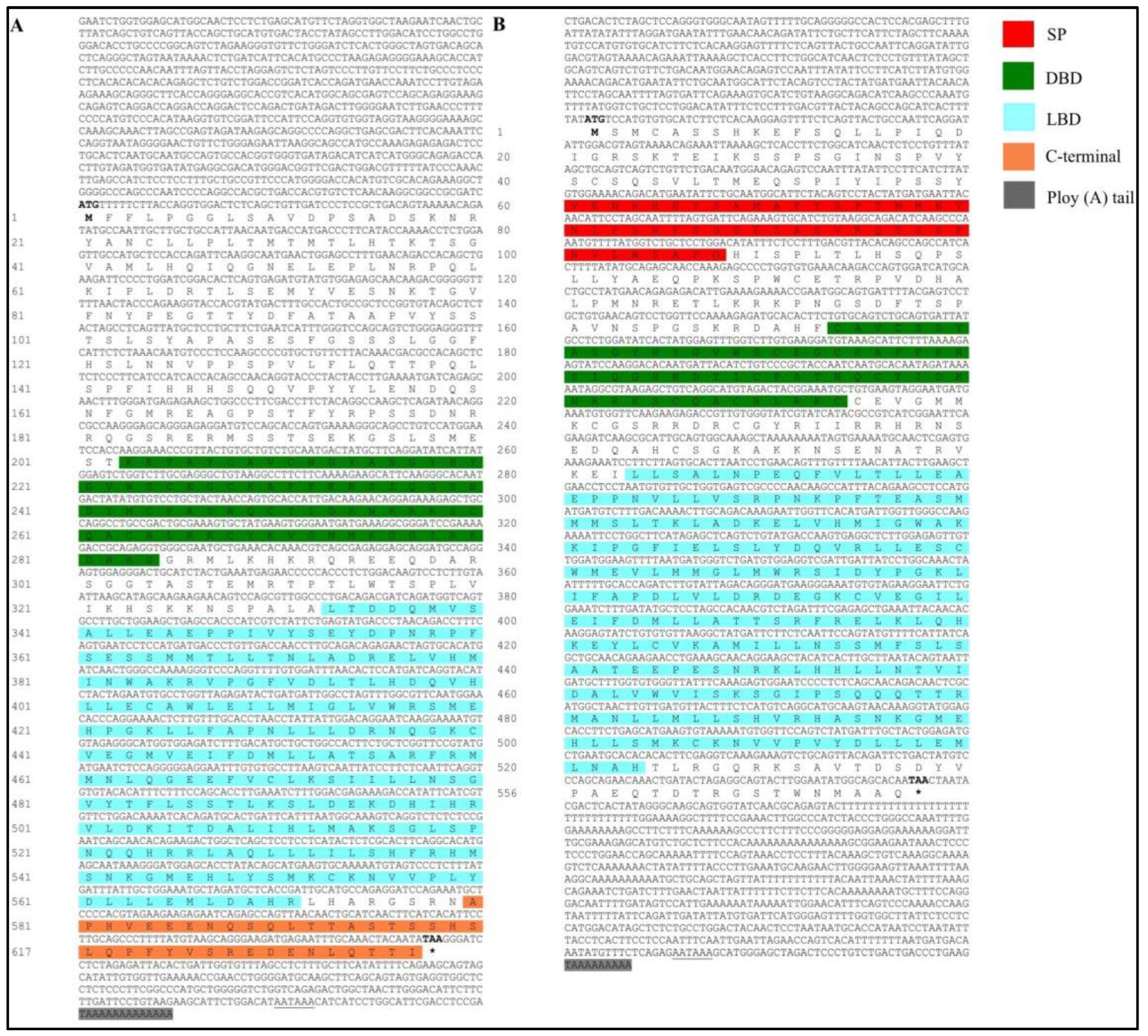
2.5. Full-Length cDNA Cloning of ERs

The cDNAs were synthesized using Hiscript®III 1st Strand cDNA Synthesis Kit (+gDNA wiper) (Vazyme, Nanjing, China) according to the manufacturer’s instructions. Sequences from conserved domain amplification, 3′ RACE, and 5′ RACE were assembled to generate the full-length cDNA. Then, polymerase chain reaction (PCR) amplification for the conserved domain was carried out in the following programs: 95 °C for 2 min; 35 cycles of 95 °C for 30 s; 60 °C for 30 s and 72 °C for 30 s; and a final extension at 72 °C for 5 min. The PCR products were detected by 1.2% agarose gel electrophoresis and sequenced by the Tianyihuiyuan Biotech Company (Wuhan, China). The programs of 5′ and 3′ RACE were set as 94 °C for 5 min; followed by 5 cycles of 94 °C for 30 s and 72 °C for 3 min; 5 cycles of 94 °C for 30 s, 70 °C for 30 s, and 72 °C for 3 min; and 25 cycles of 94 °C for 30 s, 68 °C for 30 s, and 72 °C for 3 min. The PCR products were purified using a gel extraction kit (Omega, Wuhan, China) and the purified products were ligated to a pMD18T vector (Takara, Dalian, China), and the recombinant plasmids were subsequently transformed into competent Escherichia coli DH5α cells (Biomed, Beijing, China). Positive clones were screened and sequenced by the Tianyihuiyuan Biotech Company. Primers based on the transcriptome sequences (https://ngdc.cncb.ac.cn/gsa/, accessed on 10 November 2021 with accession number CRA005737) were designed and other primers for related genes were used (Table 1).

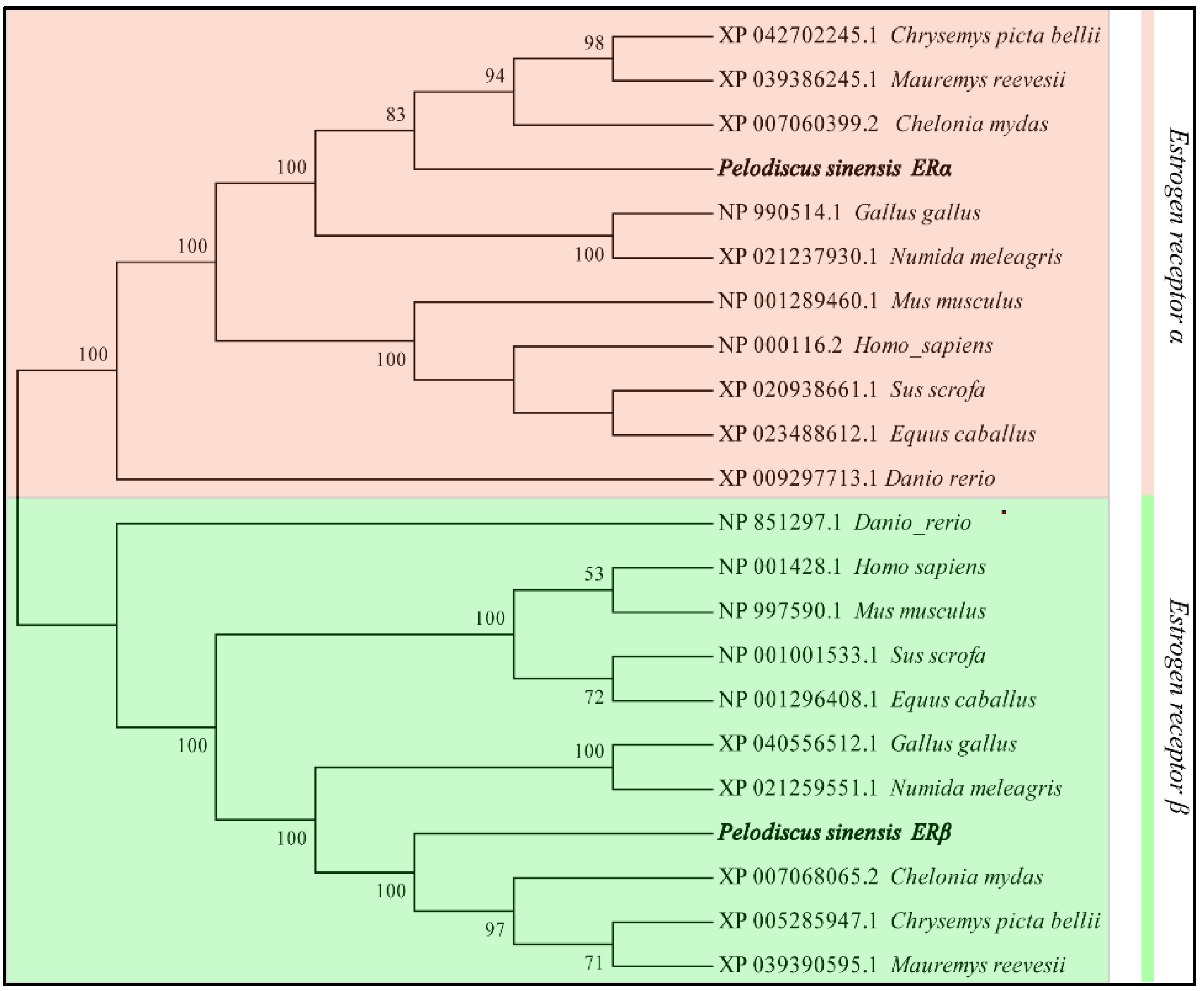
2.6. Sequence and Phylogenetic Analysis

Sequences of ER protein from different species were downloaded from the NCBI database and aligned using DNAMAN. The protein domains were predicted using online software. A phylogenetic tree was then constructed by the neighbor-joining (NJ) method, with 1000 bootstrap replicates using MEGA 7.0 (http://www.megasoftware.net, accessed 13 December 2021).

2.7. Gene Expression Analysis by Quantitative Real-Time Reverse Transcription-PCR

The expression patterns of ERs genes and the transcriptional response to E2 were investigated based on quantitative real-time reverse transcription-polymerase chain reaction (qRT-PCR). Total RNAs were isolated from each tissue of the Chinese soft-shelled turtles according to the Trizol kit, 1500 ng/µL of total RNA was reverse-transcribed to single-strand cDNA using a HiScript®II Q RT SuperMix for qPCR (+gDNA wiper) (Vazyme) according to the manufacturer’s instructions. The primers used for qRT-PCR (Table 1) were designed according to the ERs sequences. The Gapdh gene was used as an internal control. The qRT-PCR was performed using SYBR Select Master Mix (Vazyme) with the following thermal cycling conditions: 95 °C for 1 min, 40 cycles of denaturation at 95 °C for 15 s, annealing at 60 °C for 30 s. Each group was performed for three replicates to reduce the error of the experiment. The relative expression levels of ERs were normalized to that of Gapdh quantification by the 2ΔΔCt method. The same method was used to examine the expression levels of other genes.

2.8. Statistical Analysis

The qRT-PCR data were expressed as mean ± SD and statistical analysis (one-way ANOVA) was performed by one-way analysis of variance (ANOVA) followed by Duncan’s multiple comparison test. The level of statistical analysis was set at p < 0.05 and was considered significant.

3. Results

3.1. Sequence Analysis of P. sinensis ERs Gene

The full-length cDNA sequence of ERα was 3014 bp, with an 900 bp 5′ untranslated region (UTR), a 260 bp 3′ UTR with poly (A) tail, and an open reading frame (ORF) of 1854 bp, coding 617-amino acid. ERα protein contained a DNA-binding domain (DBD; amino acids 203–284), a ligand-binding domain (LBD; amino acids 333–570), and a final C-terminal (amino acids 580–617). (Figure 1). The amino acid sequence of P. sinensis ERα shared 88.33, 82.80, and 79.77% identity with that of Mauremys mutica, Chelonia mydas, and Chrysemys picta bellii, respectively (Table 2). The phylogenetic analysis showed that P. sinensis formed a clade with other turtle species with high similarity (Figure 2).
Likewise, the full-length cDNA sequence of ERβ was 2950bp and comprised a 483-bp 5′ UTR, a 1671-bp open reading frame, and a 796-bp 3′ UTR. The 556-amino acid ERβ protein with an N-terminal (NH2) signal peptide (SP; amino acids 60–107), a DBD (amino acids 173–233), and an LBD (amino acids 305–523). The blast results show that the ERβ protein sequence had a high identity with, M. mutica, C. bellii, and C. mydas of 93.17, 87.54, and 86.35%, respectively (Table 2).

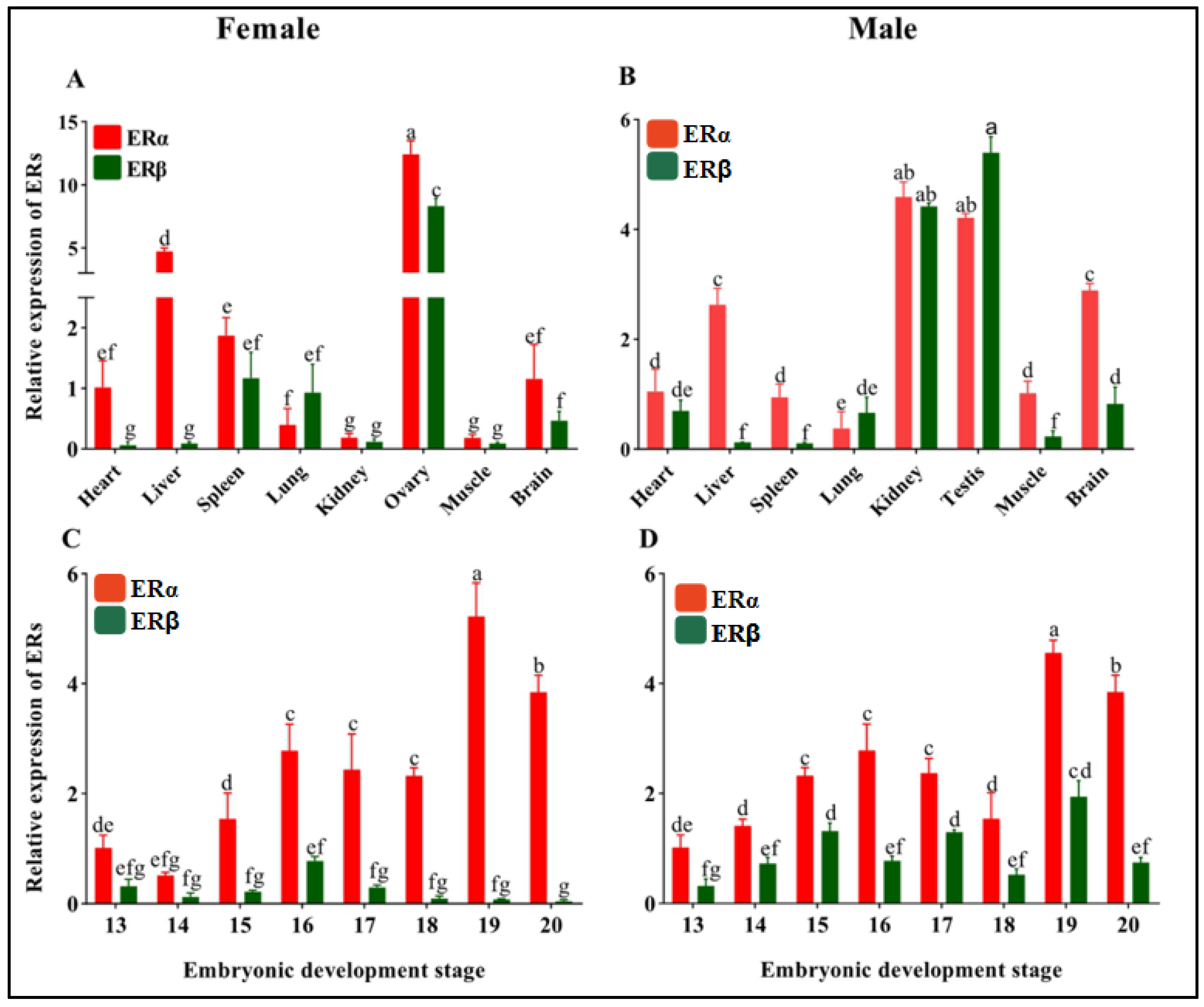
3.2. Tissue and Embryonic Development Stage Distribution of ERs

The expression of ERs in different tissues and embryonic development stages between ZZ-male and ZW-female were evaluated by qRT-PCR. In normal adults, ERs were detected in the heart, liver, spleen, lung, kidney, brain, muscle, and gonad. Both ERα and ERβ had the highest expression in the gonad while the muscle had the lowest level for ERα, the heart for ERβ in females (Figure 3A), the lung for ERα, and the spleen for ERβ in males. ERβ had a higher expression value than ERα only in the testis and kidney (Figure 3B). The ERs expression of embryonic development stages between ZZ-male and ZW-female in the blank group held a prominent discrepancy that ERα had higher expression levels than ERβ across all developmental stages for both sexes in the blank group, suggesting that ERα may play a greater role during these developmental stages (Figure 3C,D).

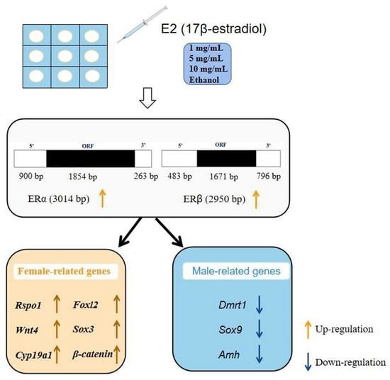
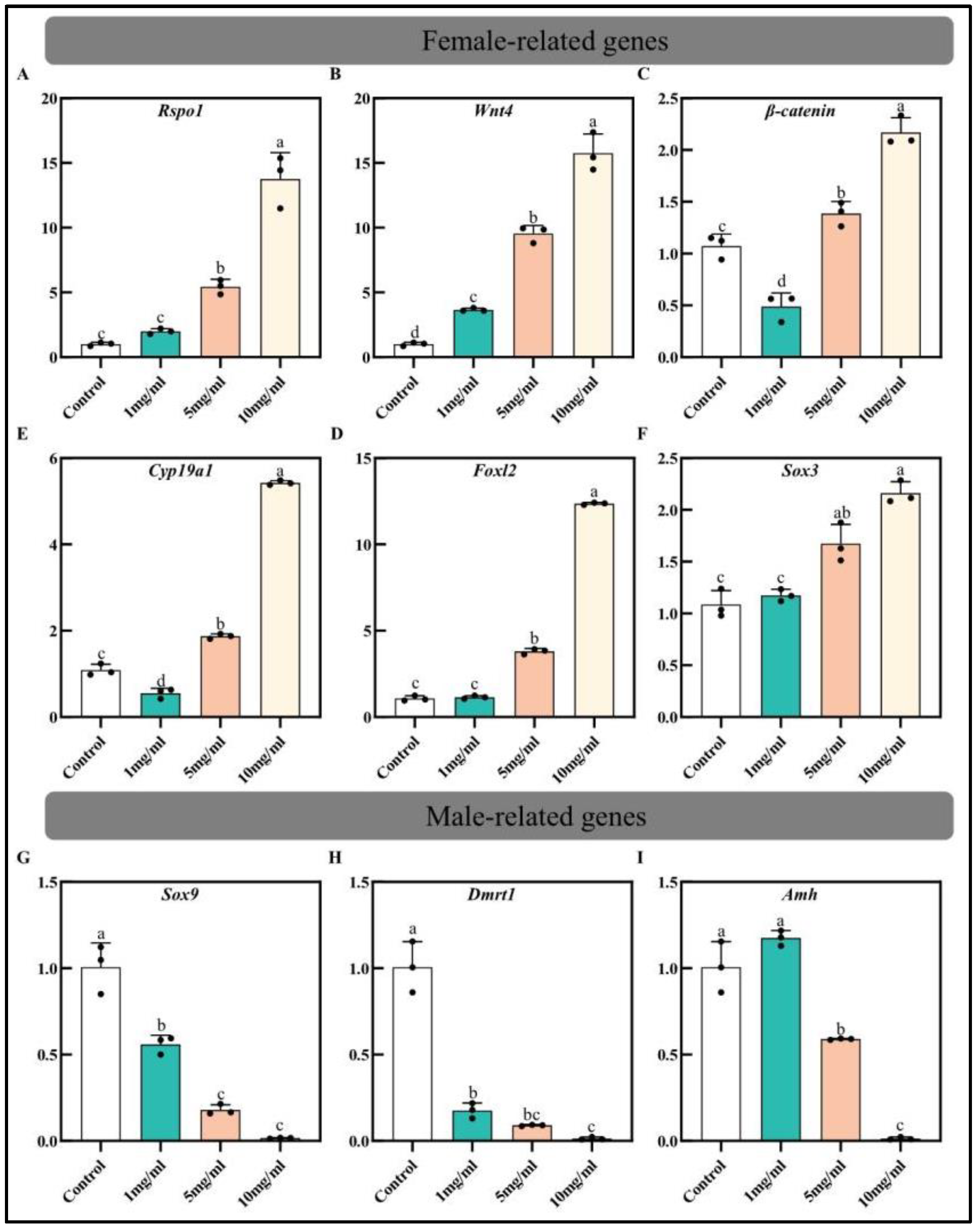
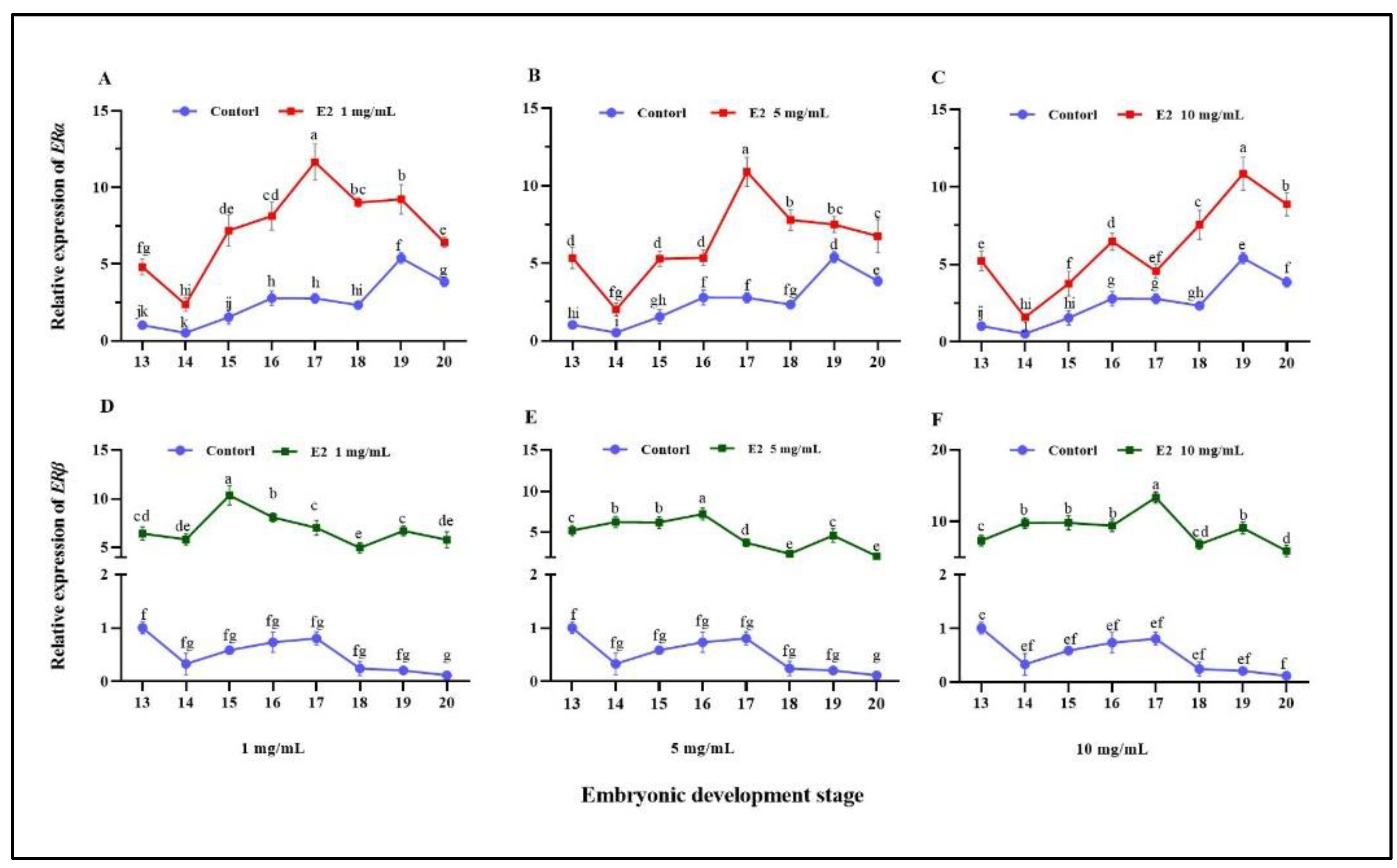
3.3. Effect of Estradiol on ERs Expression and Sex-Related Genes

To analyze the influence of E2 on ERs, their expressions were investigated in the experimental group during embryonic development in the Chinese soft-shelled turtles throughout qRT-PCR. The relative expressions of ERs after treatment with different concentrations of E2 were measured (Figure 4). Compared with the control group, the expression of ERα gradually increased from stage 13 to 20, which was more obvious after stage 14. The expression of ERα was markedly up-regulated and peaked at stage 17 after the injection of 1 mg/mL and 5 mg/mL E2 (Figure 4A,B) while the expression of ERβ owed a stable increase (Figure 4D,E). The expression of ERα was enhanced and peaked at stage 19 after treatment with 10 mg/mL E2, and ERβ expression was significantly increased and peaked at stage 19 during the embryonic development stage (Figure 4C,F). Additionally, the expression level of ERα peaked at stage 17 with the concentration of E2 for 1 mg/mL, and 5 mg/mL and it would continue to grow to stage 19 for 10 mg/mL. Meanwhile, ERβ also reached its highest expression level at stage 17 for 10 mg/mL. Thus, to further explore the effect of ERs on sex differentiation, sex-related genes that responded to E2 were measured at stage 17 (Figure 5). The expression levels of Rspo1, Wnt4, β-catenin, Foxl2, Cyp19a1, and Sox3 had an increased expression with increasing concentrations while Sox9, Dmrt1, and Amh were significantly inhibited.

4. Discussion

Sex determination and the gonadal development mechanism in P. sinensis were of concern because of the economic characteristics associated with significant sexual dimorphism [28]. To obtain all-male offspring, the pseudo-females (ΔZZ) could be obtained by the treatment of E2 to male embryos, then the pseudo-females (ΔZZ) and male (ZZ) can produce all-male individuals [6,24]. In the present study, the full-length cDNA sequences of ERα and ERβ were cloned and their expressions in normal adult tissues and embryonic development stages were investigated.
RT-qPCR analysis in this study revealed that ERs exhibited different expression profiles in the tissues of females and males, indicating their different physiological functions [29]. Both of them were detected in the gonads with high expression levels, suggesting that they were related to the development of gonads and sex differentiation. Even knocking down estrogen receptor-related factor (ERRF) in Drosophila led to improperly developed testis [30]. In the present study, the expression of ERα was higher than ERβ in the ovary, suggesting that ERα may play a critical role in early female sex differentiation and ovary development. There have been articles that suggest ERα plays a major role in the differentiation of the gonad of P. sinensis [31]. ERβ was mostly distributed in the ovary and testis which was similar to Mandarin fish [32]. In P. sinensis, it has been shown that ERα agonists could induce ovarian differentiation and sexual reversal [33]. In mammals, the expression of ERα was located in the interstitial cells and germinal epithelium, and ERβ was expressed in mesenchymal–epithelial cells of the early ovarian development phase, indicating that both of those were indispensable for germ cell differentiation and ovarian lumen development [34,35]. The expression level of ERβ in the testis was higher than that of ERα and showed a high level in the kidney. Previous research showed that the expression level of ERβ represented obvious sex differences which were 4–8 times higher in males than in females [36]. In teleost fishes, there is another type of ER named ER-γ whose DBD (ERβ for amino acids 155–234, ERβ2 for amino acids 171–252) shared 88.4% identity and had similar expression levels in the pituitary [20] and has been re-named ERβ2 [37]. The similarity between ERβ and ERβ2 seems to be specific for teleost fishes likely due to a gene replication event causing the receptor’s emergence. In tilapia, the homozygous mutants of ERβ resulted in decreasing spermatogonia and an abnormal increase in spermatozoa, and the mutation of ERβ2 could cause reproductive tract malformations, indicating that ERβ was critical for spermatogenesis while ERβ2 was indispensable for the development of a male reproductive organ [38]. Additionally, the high expressions of ERα and ERβ were observed in the spleen, brain, and liver which were similar to the results in mice and humans [39,40].
Estrogen exerted a variety of important physiological effects, which have been suggested to be mediated via the two known ERs [41]. In this study, ERα played a major role during the embryonic development stages of both sexes and had a more marked change than ERβ after the stimulation of E2, which suggested that ERα and ERβ may play different roles and even have antagonistic effects. In American alligators, studies have shown that it was ERα, rather than ERβ, that regulated the sexual reversal induced by estrogen [42]. In mice, Erα-mediated estrogen effects in female reproductive tracts while ERβ antagonized ERα-mediated estrogenic action [43]. Moreover, ERβ inhibited ERα-mediated gene transcription in the presence of ERα, whereas, in the absence of ERα, it could partially replace ERα and even regulate the transcriptional activity of ERα [44]. In Branchiostoma belcheri, the staining of the related cells of gonads revealed that the ERα and ERβ co-existed in the same cell, but the target cell localization was different, suggesting that ERα and ERβ may have different roles in the mediated estrogen signaling pathway and the mechanism of gene transcription [45]. In tilapia, mutations in ERα caused a loss of reproductive function in both males and females, while ERβ mutants showed significantly delayed ovarian development and follicle growth in females while males showed fewer spermatogonia and more abnormal sperms. The ERβ2 mutants displayed the abnormal development of the ovary and testis, resulting in infertility in females and males, respectively, although they produced gametes in their gonads [38]. These mutation results of ER subtypes further suggest that different ERs may play different roles during embryonic development stages [46].
Furthermore, the sex-related genes were detected at stages 17 after the E2 treatment to male embryos, but the expression of genes showed a different expression change with ERs. With the increase in E2 concentration, the expression levels of female-related genes Rspo1/Wnt4/β-catenin/Foxl2/Cyp19a1 were clearly increased. Conversely, the expression levels of male pathway genes such as Sox9/Dmrt1/Amh were significantly inhibited. Compared with ERs, the expression levels of genes of male and female pathways changed differently after E2 treatment. At stage 17, with the increase in E2 concentration, the expression level of ERα compared with ERα under 1 mg/mL and 5 mg/mL E2 treatment was decreased, this is different from the higher expression of female sex-related genes and the lower expression of male sex-related genes with increasing concentration. Moreover, the peak value stage of ERs clearly shifted backward, which was ERα for stage 17, stage 17, and stage 19 while ERβ for stage 15, stage 16, and stage 17 under 1 mg/mL, 5 mg/mL and 10 mg/mL E2, respectively. This may be because sex-related genes were not only regulated by ERs but also undertook the effect of some other factors such as nuclear transcription factor-κB (NF-κB) [21,47] which was generally referred to as “transcriptional cross-talk” [48]. When the ERs bond to the NF-κB, inhibition of NF-kB activity by ERα and ERβ would be caused if the AF-1 of ER was activated [49,50]. The ability of ERα to activate cooperatively transcription with NF-kB required the AF-1 domain [51]. Thus, due to the combination of NF-kB and ERs, the signal entering the nucleus to bind to ERs would be reduced, resulting in different expression trends of ERs and sex-related genes.
The sex determination and sex differentiation of animals are very complex physiological processes, focusing on the mechanisms underlying sex differentiation by the ERs will aid in the development of effective breeding strategies. ERα and ERβ are necessary for mediating the effects of E2; in the present study, ERα may play a more important role than ERβ during embryonic development stages. Their detailed molecular mechanisms in the sexual reversal process and reproduction deserve further study. Subsequent studies can specifically explore their functions by knocking down individual nuclear receptors and the experimental research results will hopefully serve as useful feedback information for improvements for the Chinese soft-shelled turtle’s gonadal differentiation and breeding efforts.

5. Conclusions

In this study, the pseudo-females could be obtained by injecting estradiol, in which the sex was reversed with the male genotype and female phenotype. The full-length of ERα was 3014 bp, while ERβ was 2095 bp. After treatment with different concentrations of E2, the expression levels of ERα and ERβ were enhanced clearly. Besides, female sex-related genes Rspo1/Wnt4/β-catenin/Foxl2/Cyp19a1 were also increased while male sex-related genes Sox9/Dmrt1/Amh were down-regulated significantly. This study provides a reference for further investigations of the molecular mechanism of sex determination and all-male breeding of P. sinensis.

Author Contributions

G.C.: operated the experiments, data curation, and writing—original draft preparation. T.Z.: assisted sample collection and revised the manuscript. M.C.: performed the PCR experiments. G.Z.: provided the experimental consumables. H.L.: devised the experiment and revised the manuscript. All authors have read and agreed to the published version of the manuscript.

Funding

This work was supported by the Central Public-Interest Scientific Institution Basal Research Fund; CAFS (No. YFI202212 and No. 2020TD33); and the National Freshwater Aquatic Germplasm Resource Center (FGRC18537).

Institutional Review Board Statement

This study was conducted according to the appropriate Animal Experimental Ethical Inspection of Laboratory Animal Centre of the Yangtze River Fisheries Research Institute, Chinese Academy of Fishery Sciences (Wuhan, China) (ID Number: 2022YFI-ZT-01).

Conflicts of Interest

The authors report no declarations of interest.

References

  1. Gong, S.P.; Vamberger, M.; Auer, M.; Praschag, P.; Fritz, U. Millennium-old farm breeding of Chinese softshell turtles (Pelodiscus sinensis) results in massive erosion of biodiversity. Sci. Nat. 2018, 105, 34. [Google Scholar] [CrossRef]
  2. Liang, H.W.; Wang, L.H.; Sha, H.; Zou, G.W. Development and Validation of Sex-Specific Markers in Pelodiscus Sinensis Using Restriction Site-Associated DNA Sequencing. Genes 2019, 10, 302. [Google Scholar] [CrossRef]
  3. Cornejo-Paramo, P.; Lira-Noriega, A.; Ramirez-Suastegui, C.; Mendez-de-la-Cruz, F.R.; Szekely, T.; Urrutia, A.O.; Cortez, D. Sex determination systems in reptiles are related to ambient temperature but not to the level of climatic fluctuation. BMC Evol. Biol. 2020, 20, 103. [Google Scholar] [CrossRef] [PubMed]
  4. Mu, Y.; Zhao, B.; Tang, W.Q.; Sun, B.J.; Zeng, Z.G.; Valenzuela, N.; Du, W.G. Temperature-Dependent Sex Determination Ruled Out in the Chinese Soft-Shelled Turtle (Pelodiscus sinensis) via Molecular Cytogenetics and Incubation Experiments across Populations. Sex. Dev. 2015, 9, 111–117. [Google Scholar] [CrossRef] [PubMed]
  5. Kim, S.S.; Kettlewell, J.R.; Anderson, R.C.; Bardwell, V.J.; Zarkower, D. Sexually dimorphic expression of multiple doublesex-related genes in the embryonic mouse gonad. Gene Expr. Patterns 2003, 3, 77–82. [Google Scholar] [CrossRef]
  6. Wang, Y.; Luo, X.; Qu, C.; Xu, T.; Zou, G.; Liang, H. The Important Role of Sex-Related Sox Family Genes in the Sex Reversal of the Chinese Soft-Shelled Turtle (Pelodiscus sinensis). Biology 2022, 11, 83. [Google Scholar] [CrossRef]
  7. Liang, H.; Meng, Y.; Cao, L.; Li, X.; Zou, G. Effect of exogenous hormones on R-spondin 1 (RSPO1) gene expression and embryo development in Pelodiscus sinensis. Reprod. Fertil. Dev. 2019, 31, 1425–1433. [Google Scholar] [CrossRef]
  8. Cheng, S.B.; Dong, J.; Pang, Y.F.; LaRocca, J.; Hixon, M.; Thomas, P.; Filardo, E.J. Anatomical location and redistribution of G protein-coupled estrogen receptor-1 during the estrus cycle in mouse kidney and specific binding to estrogens but not aldosterone. Mol. Cell. Endocrinol. 2014, 382, 950–959. [Google Scholar] [CrossRef]
  9. Zhou, L.Y.; Charkraborty, T.; Yu, X.G.; Wu, L.M.; Liu, G.; Mohapatra, S.; Wang, D.S.; Nagahama, Y. R-spondins are involved in the ovarian differentiation in a teleost, medaka (Oryzias latipes). BMC Dev. Biol. 2012, 12, 36. [Google Scholar] [CrossRef]
  10. Parma, P.; Radi, O.; Vidal, V.; Chaboissier, M.C.; Dellambra, E.; Valentini, S.; Guerra, L.; Schedl, A.; Camerino, G. R-spondin1 is essential in sex determination, skin differentiation and malignancy. Nat. Genet. 2006, 38, 1304–1309. [Google Scholar] [CrossRef]
  11. Maatouk, D.M.; DiNapoli, L.; Alvers, A.; Parker, K.L.; Taketo, M.M.; Capel, B. Stabilization of beta-catenin in XY gonads causes male-to-female sex-reversal. Hum. Mol. Genet. 2008, 17, 2949–2955. [Google Scholar] [CrossRef] [PubMed]
  12. Stewart, M.K.; Mattiske, D.M.; Pask, A.J. Estrogen suppresses SOX9 and activates markers of female development in a human testis-derived cell line. BMC Mol. Cell Biol. 2020, 21, 66. [Google Scholar] [CrossRef] [PubMed]
  13. Iwamatsu, T.; Kobayashi, H. Sex reversal in the medaka treated in vitro 17 alpha-methyldihydrotestosterone during oocyte maturation. Zool. Sci. 2005, 22, 1505. [Google Scholar]
  14. Rose, E.; Flanagan, S.P.; Jones, A.G. The Effects of Synthetic Estrogen Exposure on the Sexually Dimorphic Liver Transcriptome of the Sex-Role-Reversed Gulf Pipefish. PLoS ONE 2015, 10, e0139401. [Google Scholar] [CrossRef]
  15. Eick, G.N.; Thornton, J.W. Evolution of steroid receptors from an estrogen-sensitive ancestral receptor. Mol. Cell. Endocrinol. 2011, 334, 31–38. [Google Scholar] [CrossRef]
  16. Jensen, E.V.; Jacobson, H.I.; Walf, A.A.; Frye, C.A. Estrogen action: A historic perspective on the implications of considering alternative approaches. Physiol. Behav. 2010, 99, 151–162. [Google Scholar] [CrossRef]
  17. Kowalski, A.A.; Graddy, L.G.; Vale-Cruz, D.S.; Choi, I.; Katzenellenbogen, B.S.; Simmen, F.A.; Simmen, R.C.M. Molecular cloning of porcine estrogen receptor-beta complementary DNAs and developmental expression in periimplantation embryos. Biol. Reprod. 2002, 66, 760–769. [Google Scholar] [CrossRef]
  18. Edwards, D.P. Regulation of signal transduction pathways by estrogen and progesterone. Annu. Rev. Physiol. 2005, 67, 335–376. [Google Scholar] [CrossRef]
  19. Hewitt, S.C.; Korach, K.S. Estrogen Receptors: New Directions in the New Millennium. Endocr. Rev. 2018, 39, 664–675. [Google Scholar] [CrossRef]
  20. Qin, X.; Xiao, Y.; Ye, C.; Jia, J.; Liu, X.; Liang, H.; Zou, G.; Hu, G. Pituitary Action of E2 in Prepubertal Grass Carp: Receptor Specificity and Signal Transduction for Luteinizing Hormone and Follicle-Stimulating Hormone Regulation. Front. Endocrinol. 2018, 9, 308. [Google Scholar] [CrossRef]
  21. Marino, M.; Galluzzo, P.; Ascenzi, P. Estrogen signaling multiple pathways to impact gene transcription. Curr. Genom. 2006, 7, 497–508. [Google Scholar] [CrossRef] [PubMed] [Green Version]
  22. Pietras, R.J.; Marquez-Garban, D.C. Membrane-associated estrogen receptor signaling pathways in human cancers. Clin. Cancer Res. 2007, 13, 4672–4676. [Google Scholar] [CrossRef] [PubMed]
  23. Barton, M.; Filardo, E.J.; Lolait, S.J.; Thomas, P.; Maggiolini, M.; Prossnitz, E.R. Twenty years of the G protein-coupled estrogen receptor GPER: Historical and personal perspectives. J. Steroid Biochem. Mol. Biol. 2018, 176, 4–15. [Google Scholar] [CrossRef] [PubMed]
  24. Zhou, T.; Sha, H.; Chen, M.; Chen, G.; Zou, G.; Liang, H. MicroRNAs May Play an Important Role in Sexual Reversal Process of Chinese Soft-Shelled Turtle, Pelodiscus sinensis. Genes 2021, 12, 1696. [Google Scholar] [CrossRef]
  25. Sun, J.-J.; Sun, Z.-H.; Wei, J.-L.; Ding, J.; Song, J.; Chang, Y.-Q. Identification and functional analysis of foxl2 and nodal in sea cucumber, Apostichopus japonicus. Gene. Expr. Patterns GEP 2022, 44, 119245. [Google Scholar] [CrossRef]
  26. Liang, H.W.; Meng, Y.; Cao, L.H.; Li, X.; Zou, G.W. Expression and characterization of the cyp19a gene and its responses to estradiol/letrozole exposure in Chinese soft-shelled turtle (Pelodiscus sinensis). Mol. Reprod. Dev. 2019, 86, 480–490. [Google Scholar] [CrossRef] [PubMed]
  27. Sun, W.; Cai, H.; Zhang, G.; Zhang, H.; Bao, H.; Wang, L.; Ye, J.; Qian, G.; Ge, C. Dmrt1 is required for primary male sexual differentiation in Chinese soft-shelled turtle Pelodiscus sinensis. Sci. Rep. 2018, 8, 4433. [Google Scholar] [CrossRef] [PubMed]
  28. Dean, R.; Mank, J.E. The role of sex chromosomes in sexual dimorphism: Discordance between molecular and phenotypic data. J. Evol. Biol. 2014, 27, 1443–1453. [Google Scholar] [CrossRef]
  29. Luo, A.; Zhang, X. ERRF is essential for Estrogen-Estrogen Receptor alpha signaling pathway in ER positive breast cancer cells. Biochem. Biophys. Res. Commun. 2016, 474, 400–405. [Google Scholar] [CrossRef]
  30. Misra, S.; Pandey, A.K.; Gupta, S.; Kumar, A.; Khanna, P.; Shankar, J.; Ram, K.R. Estrogen related receptor is required for the testicular development and for the normal sperm axoneme/mitochondrial derivatives in Drosophila males. Sci. Rep. 2017, 7, 40372. [Google Scholar] [CrossRef]
  31. Li, P.; Guo, Y.; Jin, L.; Liang, X.; Chen, G.A.; Sun, W.; Xiao, L.; Qian, G.Y.; Ge, C.T. ESR1 mediates estrogen-induced feminization of genetic male Chinese soft-shelled turtle. Biol. Reprod. 2022, ioac088. [Google Scholar] [CrossRef] [PubMed]
  32. Toyota, K.; Masuda, S.; Sugita, S.; Miyaoku, K.; Yamagishi, G.; Akashi, H.; Miyagawa, S. Estrogen Receptor 1 (ESR1) Agonist Induces Ovarian Differentiation and Aberrant Mullerian Duct Development in the Chinese Soft-shelled Turtle, Pelodiscus sinensi. Zool. Stud. 2020, 59, e54. [Google Scholar] [PubMed]
  33. Ouyang, H.; Han, C.; Zhu, Q.; Xu, L.; Huang, J.; Li, S.; Li, G.; Lin, H.; Zhang, Y. Molecular cloning and characterization of estrogen and androgen receptors in Mandarin fish, Siniperca chuatsi. Aquac. Rep. 2021, 21, 100834. [Google Scholar] [CrossRef]
  34. Pelletier, G.; Labrie, C.; Labrie, F. Localization of oestrogen receptor alpha, oestrogen receptor beta and androgen receptors in the rat reproductive organs. J. Endocrinol. 2000, 165, 359–370. [Google Scholar] [CrossRef] [PubMed]
  35. Schoen, J.; Neumann, S.; Wildt, D.E.; Pukazhenthi, B.S.; Jewgenow, K. Localization of Oestrogen Receptors in the Epididymis During Sexual Maturation of the Domestic Cat. Reprod. Domest. Anim. 2009, 44, 294–301. [Google Scholar] [CrossRef]
  36. Blazquez, M.; Gonzalez, A.; Papadaki, M.; Mylonas, C.; Piferrer, F. Sex-related changes in estrogen receptors and aromatase gene expression and enzymatic activity during early development and sex differentiation in the European sea bass (Dicentrarchus labrax). Gen. Comp. Endocrinol. 2008, 158, 95–101. [Google Scholar] [CrossRef]
  37. Wojnarowski, K.; Cholewinska, P.; Palic, D.; Bednarska, M.; Jarosz, M.; Wisniewska, I. Estrogen Receptors Mediated Negative Effects of Estrogens and Xenoestrogens in Teleost Fishes-Review. Int. J. Mol. Sci. 2022, 23, 2605. [Google Scholar] [CrossRef]
  38. Yan, L.; Feng, H.; Wang, F.; Lu, B.; Liu, X.; Sun, L.; Wang, D. Establishment of three estrogen receptors (esr1, esr2a, esr2b) knockout lines for functional study in Nile tilapia. J. Steroid Biochem. Mol. Biol. 2019, 191, 105379. [Google Scholar]
  39. Saito, K.; Cui, H. Emerging Roles of Estrogen-Related Receptors in the Brain: Potential Interactions with Estrogen Signaling. Int. J. Mol. Sci. 2018, 19, 1091. [Google Scholar] [CrossRef]
  40. O’Brien, M.H.; Pitot, H.C.; Chung, S.-H.; Lambert, P.F.; Drinkwater, N.R.; Bilger, A. Estrogen Receptor-alpha Suppresses Liver Carcinogenesis and Establishes Sex-Specific Gene Expression. Cancers 2021, 13, 2355. [Google Scholar] [CrossRef]
  41. Hwang, W.J.; Lee, T.Y.; Kim, N.S.; Kwon, J.S. The Role of Estrogen Receptors and Their Signaling across Psychiatric Disorders. Int. J. Mol. Sci. 2021, 22, 373. [Google Scholar] [CrossRef]
  42. Guillette, L.J.; Iguchi, T.; Doheny, B.M.; Bryan, T.A.; Zhu, J.; Katsu, Y.; Bernhard, M.C.; Kohno, S. Estrogen Receptor 1 (ESR1; ERα), not ESR2 (ERβ), Modulates Estrogen-Induced Sex Reversal in the American Alligator, a Species with Temperature-Dependent Sex Determination. Endocrinology 2015, 156, 1887–1899. [Google Scholar]
  43. Gustafsson, J.-A. Historical overview of nuclear receptors. J. Steroid Biochem. Mol. Biol. 2016, 157, 3–6. [Google Scholar] [CrossRef] [PubMed]
  44. Lindberg, M.K.; Moverare, S.; Skrtic, S.; Gao, H.; Dahlman-Wright, K.; Gustafsson, J.A.; Ohlsson, C. Estrogen receptor (ER)-beta reduces ER alpha-regulated gene transcription, supporting a “Ying Yang” relationship between ER alpha and ER beta in mice. Mol. Endocrinol. 2003, 17, 203–208. [Google Scholar] [CrossRef] [PubMed]
  45. Fang, Y.Q.; Weng, Y.Z.; Ye, R.Z.; Liu, L.L. Immunolocalization of aromatase, estrogen and estrogen receptor alpha and beta in the epithelium of digestive tract and enteric neurons of amphioxus (Branchiostoma belcheri). Prog. Nat. Sci.-Mater. Int. 2005, 15, 157–161. [Google Scholar]
  46. Wang, L.; Cen, S.; Shi, X.; Zhang, H.; Wu, L.; Tian, X.; Ma, W.; Li, X.; Ma, X. Molecular characterization and functional analysis of Esr1 and Esr2 in gonads of Chinese soft-shelled turtle (Pelodiscus sinensis). J. Steroid Biochem. Mol. Biol. 2022, 222, 106147. [Google Scholar] [CrossRef] [PubMed]
  47. O’Lone, R.; Frith, M.C.; Karlsson, E.K.; Hansen, U. Genomic targets of nuclear estrogen receptors. Mol. Endocrinol. 2004, 18, 1859–1875. [Google Scholar] [CrossRef]
  48. Agrawal, R.; Sahoo, B.K.; Saini, D.K. Cross-talk and specificity in two-component signal transduction pathways. Future Microbiol. 2016, 11, 685–697. [Google Scholar] [CrossRef]
  49. Chen, D.; Li, Q.; Chen, H.; Huang, Q.; Zeng, M. Estrogen receptor regulates immune defense by suppressing NF-kappa B signaling in the Crassostrea hongkongensis. Fish Shellfish. Immunol. 2020, 106, 796–803. [Google Scholar] [CrossRef]
  50. Hollingshead, B.D.; Beischlag, T.V.; DiNatale, B.C.; Ramadoss, P.; Perdew, G.H. Inflammatory signaling and aryl hydrocarbon receptor mediate synergistic induction of interleukin 6 in MCF-7 cells. Cancer Res. 2008, 68, 3609–3617. [Google Scholar] [CrossRef]
  51. Wissink, S.; van der Burg, B.; Katzenellenbogen, B.S.; van der Saag, P.T. Synergistic activation of the serotonin-1A receptor by nuclear factor-kappa B and estrogen. Mol. Endocrinol. 2001, 15, 543–552. [Google Scholar] [PubMed] [Green Version]
Figure 1. The cDNA sequence and amino acid of ERs of P. sinensis. The start codon and end codon are in bold black, * denotes the termination codon, and the underline denotes the 3′ untranslated region with the tailing signal AATAAA. SP: signal peptide. DBD: DNA-binding domain. LBD: ligand-binding domain. (A) The cDNA sequence and amino acid of ERα. (B) The cDNA sequence and amino acid of ERβ.
Fishes 07 00223 g001
Figure 2. Gene tree of ER amino acid sequences.
Fishes 07 00223 g002
Figure 3. Expression profiles of ERs cloned from P. sinensis in normal adult females (A) and male tissues (B), and expression analysis in blank group female embryos (C) and male embryos (D) at different embryonic development stages. Data are mean ± SD (n = 3). Significant differences at p < 0.05 are labeled with different letters.
Fishes 07 00223 g003
Figure 4. Expression of ERs in E2-treated male embryos at embryonic development stages after injection with different concentrations of E2. Data are the mean ± SD (n = 3). Different letters indicate significant differences between the control group and the experimental group, respectively (p < 0.05): (A) ERα for 1 mg/mL E2; (B) ERα for 5 mg/mL E2; (C) ERα for 10 mg/mL E2; (D) ERβ for 1 mg/mL E2; (E) ERβ for 5 mg/mL E2; and (F) ERβ for 10 mg/mL E2.
Fishes 07 00223 g004
Figure 5. The expressions of sex-related genes at stage 17. Female-related genes: (A) Rspo1; (B) Wnt4; (C) β-catenin; (D) Foxl2; (E) Cyp19a1; and (F) Sox3. Male-related genes: (G) Sox9. (H) Dmrt1 (I) Amh. Data are the mean ± SD (n = 3). Significant differences at p < 0.05 are labeled with different letters.
Fishes 07 00223 g005
Table 1. Primers are used for PCR.
Primer NamePrimer Sequence (5′–3′)ApplicationRelated References
ERα-FGTTGATCCCTCCGCTGACAGTCDS amplification
ERα-RCTCGCAAGACCAGACTCCATAAT
ERβ-FTGACGTTACTACAGCCAGCATCAC
ERβ-RCGACCTCCACATCAGACCCATC
ERα-GSP5-1ATCTGGTGGAGCATGGCAACTC5′ RACE
ERα-GSP5-2GAATCTGGTGGAGCATGGCAAC
ERβ-GSP5-1TAATCAAAGCTCGTGGAGTGGC
ERβ-GSP5-2GCGTACGTGTATTTGTCGGTCA
ERα-GSP3-1GCCAGTTAACAACTGCATCAACTT3′ RACE
ERα-GSP3-2AAGCAGGGAAGATGAGAATTTGC
ERβ-GSP3-1ATGCTAGATGCTCACCGATTGC
ERβ-GSP3-2CAGGCACATGAGCAATAAAGGG
UPM shortCTAATACGACTCACTATAGGGC5′ and 3′ RACE
UPM long CTAATACGACTCACTATAGGGCAAGCA
ERα-FCCGACTGCGAAAGTGCTATGAqPCR
ERα-RACGCTGGACTGTTCTTCTTGCTA
ERβ-FGCAACAGACAACTCGCATGG
ERβ-RGTGTGTGCATTCAGCATCTCC
Rspo1-FCCTGCTGGAGAGGAATGACA[7]
Rspo1-RCCCACTCGCTCATTTCACA
Wnt4-FGAGGTGATGGACTCGGTGCG
Wnt4-RCCCGTTCTTGAGGTCGTGGTC
β-catenin-FGCTTTGGGACTCCACCTTACAG
β-catenin-RATCACCAGCCCGAAGAACAGT
Foxl2-FATCTGTTTTTATTAGCACGGTT[25]
Foxl2-RCCTTCTCAGGAGGAGTTTCGT
Cyp19a1-FTCGTGGCTGTACAAGAAATACGAA[26]
Cyp19a1-RCCAGTCATATCTCCACGGCTCT
Sox3-FGAGTGTAGAGGTGGAATGGAAACG
Sox3-RAAACCCTCAAGCAGGATACGG
Sox9-FTACGACTACACCGACCACCA
Sox9-RGTAGTGTCTGCAATGGGCGT
Dmrt1-FCCGCCTCGGGAAAGAAGTC[27]
Dmrt1-RTGCTGGATGCCGTAGTTGC
Amh-FCGGCTACTCCTCCCACACG
Amh-RCCTGGCTGGAGTATTTGACGG
Ps4085-FGTTTGAAGTGCTGCTGGGAAGSex identification [2]
Ps4085-RTTCCCCGTATAAAGCCAGGG
COI-FCAACCAACCACAAAGACATTGGCAC
COI-RACCTCAGGGTGTCCGAAAATCAAA
Gapdh-FAGAACATCATTCCAGCATCCAInternal control
Gapdh-RCTTCATCACCTTCTTAATGTCGTC
Table 2. Comparative identity of the amino acid sequence of ERs.
ERα ERβ
SpeciesAccession NumberIdentity (%)Accession NumberIdentity (%)
Mauremys muticaXP 039386245.188.33XP 039390595.193.17
Chelonia mydasXP 007060399.282.80XP 007068065.286.35
Chrysemys picta belliiXP 042702245.179.77XP 005285947.187.54
Numida meleagrisXP 021237930.176.01XP 021259551.180.03
GallusNP 990514.174.28XP 040556512.180.38
Homo sapiensNP 000116.267.49NP 001428.169.11
Sus scrofaXP 020938661.167.49NP 001001533.170.31
Mus musculusNP 001289460.166.47NP 997590.172.01
Equus caballusXP 023488612.164.88NP 001296408.170.31
Danio rerioXP 009297713.141.18NP 851297.149.66
Publisher’s Note: MDPI stays neutral with regard to jurisdictional claims in published maps and institutional affiliations.

