Article

Effects of Microplastics on Reproduction and Growth of Freshwater Live Feeds Daphnia magna

1
College of Management, National Kaohsiung University of Science and Technology, Kaohsiung 807618, Taiwan
2
Department of Fisheries Production and Management, National Kaohsiung University of Science and Technology, Kaohsiung 811213, Taiwan
3
Department and Graduate Institute of Aquaculture, College of Hydrosphere, National Kaohsiung University of Science and Technology, Kaohsiung 811213, Taiwan
4
Institute of Aquatic Science and Technology, College of Hydrosphere, National Kaohsiung University of Science and Technology, Kaohsiung 811213, Taiwan
5
Natural Sciences, General Education Center, Aletheia University, New Taipei City 25103, Taiwan
*
Author to whom correspondence should be addressed.
Fishes 2022, 7(4), 181; https://doi.org/10.3390/fishes7040181
Submission received: 27 May 2022 / Revised: 3 July 2022 / Accepted: 20 July 2022 / Published: 22 July 2022
(This article belongs to the Special Issue Live Feeds for Sustainable Aquaculture)

Abstract

:
In recent years, much research has focused on studying the damage caused by microplastics to the ecological environment and human health. Indeed, MPs are often consumed by shellfishes and zooplanktons due to their similarity in size to POM (particular organic matter). Especially in zooplankton, the accumulation of MPs in the body affects the reproductive system and the growth rate of juveniles. Moreover, toxins derived from MPs are continuously accumulated in predators of zooplankton and impact the whole ecosystem across the food chain. In this work, we found that even though MPs were internalized by and adherent around Daphnia magna, there were no significant differences in the survival rate of their adults and offspring. However, the population of ovigerous adults under high MPs exposure for 7 days decreased significantly, suggesting an extension of the days of sexual maturity in D. magna. The removal of MPs after 7 days’ MPs treatment resulted in an increase in D. magna juveniles and neonates which indicated their growth was reduced or inhibited in the MPs environment. Overall, the uptake of MPs led to negative effects on population reproduction and the growth of offspring in D. magna.

1. Introduction

In 2008, the NAOO held a meeting on marine plastic pollution and defined MPs (microplastics) as plastic particles found in oceans with sizes of less than 5 mm or 1 μm, and NPs (nanoplastics) as plastic particles with a size range of 1 μm to 100 nm [1,2,3,4,5]. There are two major sources of plastic particles [6], including primary MPs and secondary MPs. Primary MPs are plastic particles directly or indirectly released into the ocean, which may derive from the laundry process, cosmetic pearls (Microbeads), factory manufacturing processes, or particles generated by the tires of vehicles when braking, etc. Secondary MPs in oceans are derived from plastic material degraded and broken down by UV rays of the sun and other physical processes [7,8,9]. About 90% of the world’s plastic materials are made up of polyethylene (PE), polypropylene (PP), polyvinyl chloride (PVC), polystyrene (PS), or polyethylene terephthalate (PET) [10]. These materials are widely used around the world because they are cheap, easy to manufacture, and have alternative properties to natural products including wood, stone, and glass [3]. However, concerns of the acute and chronic effects of environmental microplastics (MPs) on aquatic organisms are rapidly increasing, especially in the absorption of heavy metals, dioxin and other highly toxic compounds. According to SAPEA (2019) [11], the risk of environmental MPs is not currently high but could become widespread across the whole ocean in future.
The use of plastic materials increased dramatically over the past few years, with a total global production of 1.7 million tons of plastic materials in 1950 [10]. From 1950 to 2014, annual plastic production increased at least 20-fold to 300 million metric tons [10]. Large amounts of plastic materials enter water environments from land, with an estimated 4.8–12.7 million tons of plastic materials entering the ocean each year [12]. In addition, the potential degradation of large plastic pellets increases the risk posed to marine life and the environment [13,14,15,16,17,18]. A growing number of studies focus on the behavior and effects on marine life under the exposure of MPs [19,20]. MPs also enter freshwater ecosystems through land-based wastewater [21,22,23,24]. Therefore, in recent years, an increasing number of studies revealed the effects of plastic particles on freshwater organisms [25,26,27], providing data on an increasingly important environmental issue in freshwater ecosystems [25,26,28].
Zooplankton organisms are the primary consumers in the food chain and are important bait for aquaculture and fisheries. Some studies revealed the possible hazards of plastic particles in the food chain, such as plankton acting as potential carriers of toxic substances derived from the ingestion of plastic particles [29]. Additionally, the toxic substances are accumulated in higher order consumers through transfer in the food chain, leading to harmful impacts on mammals and birds [30]. Similarly, MPs are also ingested by live feed organisms in aquaculture and are transferred and accumulated in fish bodies, resulting in delayed growth and decreased production or harmful effects on human health when toxic seafood is consumed.
In freshwater aquaculture, Daphnia spp. plays an important role as a kind of live feed. D. magna constitutes a major component of food webs in the freshwater ecosystem, being not only the main food item of fish but also the main herbivore of algae [31]. The MPs ingested by Daphnia magna depend on the type, size and shape of the particles [30,32,33]. Jemec et al. [29] found that D. magna ingests the fibers of MPs, which are up to 1400 μm in length and up to 528 μm or 106 μm in width. MPs have a high chance of being eaten by large zooplankton, such as juvenile fish [33,34]. MP intake depends on the ability of zooplankton to ingest food and their preservation. Studies have shown that spiky particles (e.g., fragments, fibers) present a greater threat as they are more difficult to digest than smooth particles (i.e., spherical beads) [35]. In D. magna, MPs and NPs were found to cause a wide range of adverse effects, such as mortality, feeding inhibition, and a decrease in the reproduction rate, among others [24,30,33,36,37,38,39,40]. Here, D. magna were immersed in an MP-enriched environment to evaluate potential effects on their physiological responses and understand the potential impacts on their survival, reproduction, body size and production of offspring after long-term immersion with MPs, which may provide important information for future human aquaculture of feed organisms.

2. Materials and Methods

2.1. Animals

D. magna is a small filter feeder freshwater crustacean with cyclic solitary reproduction and the population is usually dominated by females [41]. D. magna were collected from a co-aquaculture pond with milkfish and white shrimp in Maito, Taiwan. D. magna with a body length of about 0.5–1.5 mm were collected using a 100-mesh plankton net. The water condition was as follows: 6‰ salinity, pH 7.8, 5.6 ppm DO, 1 ppm NH3, and 0.5 ppm NO2 and the water was refreshed once per 2 days. D. magna were collected, with 200 individuals/per treatment, kept in a 200-L FRP tank for 7 days, and disinfected with 20 ppm formalin and 10% florfenicol for 3 days. The water quality in a 200-L tank was maintained by using freshwater within the specific range of pH 7.18~7.90, water temperature of 27~28 °C, 7 ppm DO, less than 0.5 ppm NH3 and 0.1 ppm NO2 under 5000 lux of light intensity at a photoperiod of 16:8 light/darkness. Additionally, they were fed Chlorella sp. 20 mL per day (concentration was 106 cell/mL). The D. magna population were selected without considering male and female individuals.

2.2. Microplastics (MPs, Composed of Polystyrene)

The MPs purchased from Sigma-Aldrich (cat#59769) were carboxylate-modified polystyrene particles with a latex bead-like shape and an average particle size of 0.5 μm, and shared a yellow-green fluorescence at 470~505 nm. During the period of MP treatment, all the freshwater was filtered using 0.1 µm filters to completely remove residue plastic particles that ensured all responses of D. magna resulted from MPs provided from the study. The stock of MPs solution (1.05 g/mL with 10% solids) were diluted to filtered water to prepare the working reagents of MPs (1.5 mg/mL). Additionally, all treatments including 0 (control), 0.01, 0.02, 0.05 and 0.1 mg/L MPs were derived from the working reagents of MPs under 3 repetitions. The MPs (ingested or not) were determined by fluorescent signals observed using a fluorescence microscope (Euromex). The images of MPs internalization were verified using a digital camera (SCMOS CCD) equipped on the microscope.

2.3. Determining Sexual Maturation and Offspring Survival

During 7 or 30 days incubation with MPs or MP-free freshwater, the ovigerous behavior of D. magna females was determined as an indicator of first sexual maturation. The number of D. magna eggs per 20 females and the survival rate of their offspring were recorded after 7 days MPs treatment. After 7 days MPs treatment, all of the D. magna including adults, juveniles and neonates produced from 20 females were harvested to examine the residual MPs uptake inside their body. Additionally, the D. magna parents were removed and the offspring were transferred to MP-free freshwater for 30 days cultivation to evaluate their sexual maturation. After 30 days of MP-free freshwater incubation, 500 mL of water containing well-mixed D. magna offspring was harvested to evaluate body length and the population ratio of adults (with clutched eggs), juveniles (without clutched eggs and over 2.5 mm in length) and neonates (less than 2.5 mm in length). All the tests were performed in triplicate.

2.4. Statistics

The microplastic measurement method was based on the fluorescence signal observed via a Fluorescence microscope (Euromex, Mataró, Spain). The images of MPs internalization were verified using a digital camera (SCMOS CCD) equipped on the microscope. The number of MPs in D. magna, the number of eggs, survival rate and survival rate of offspring in the 7 days immersion were calculated and the differences among treatment groups were tested using One Way ANOVA. Tukey’s post hoc tests were performed with stocking and the ratio of adults, juveniles and neonates in the total number of D. magna after 30 days was calculated.

3. Results

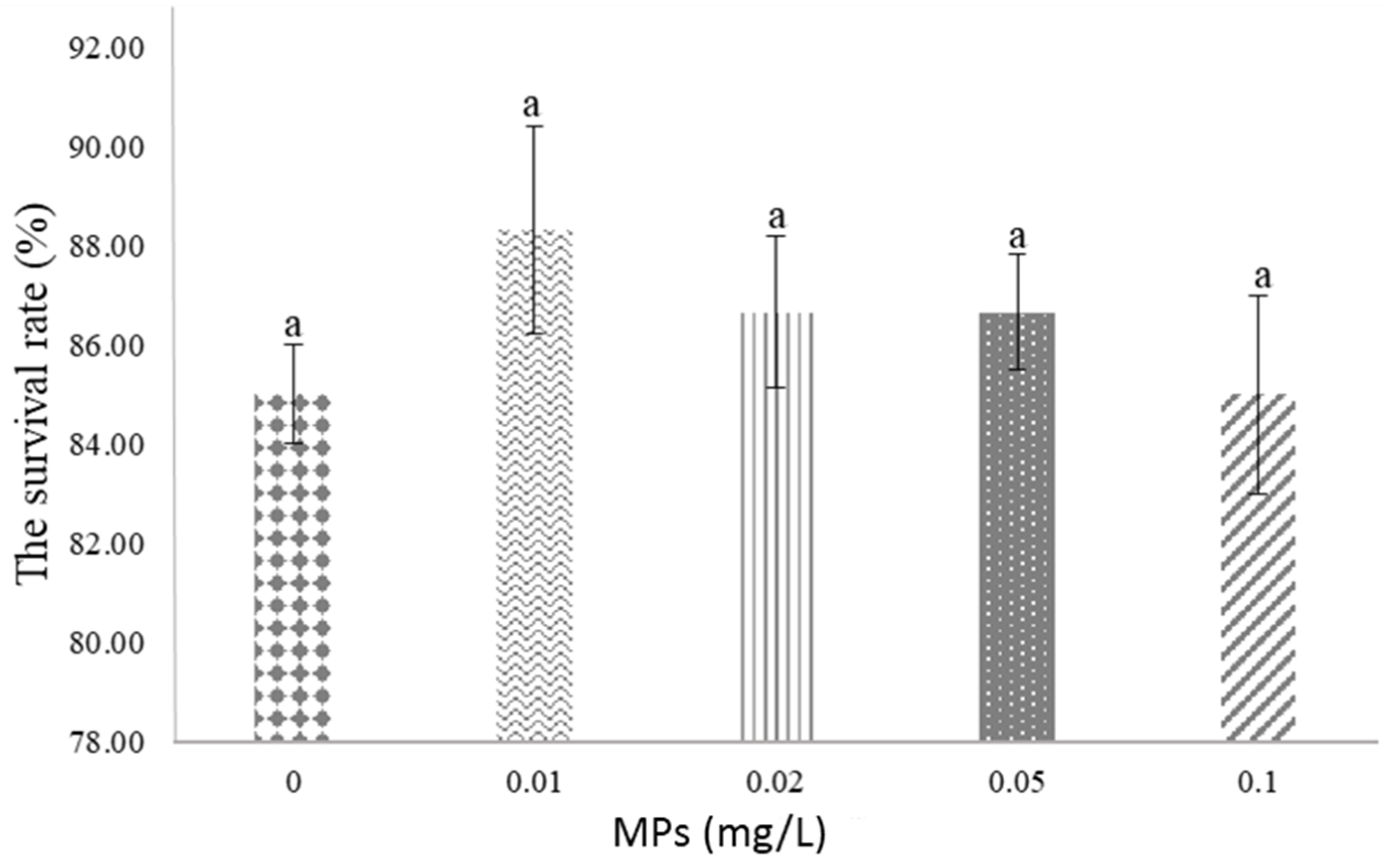
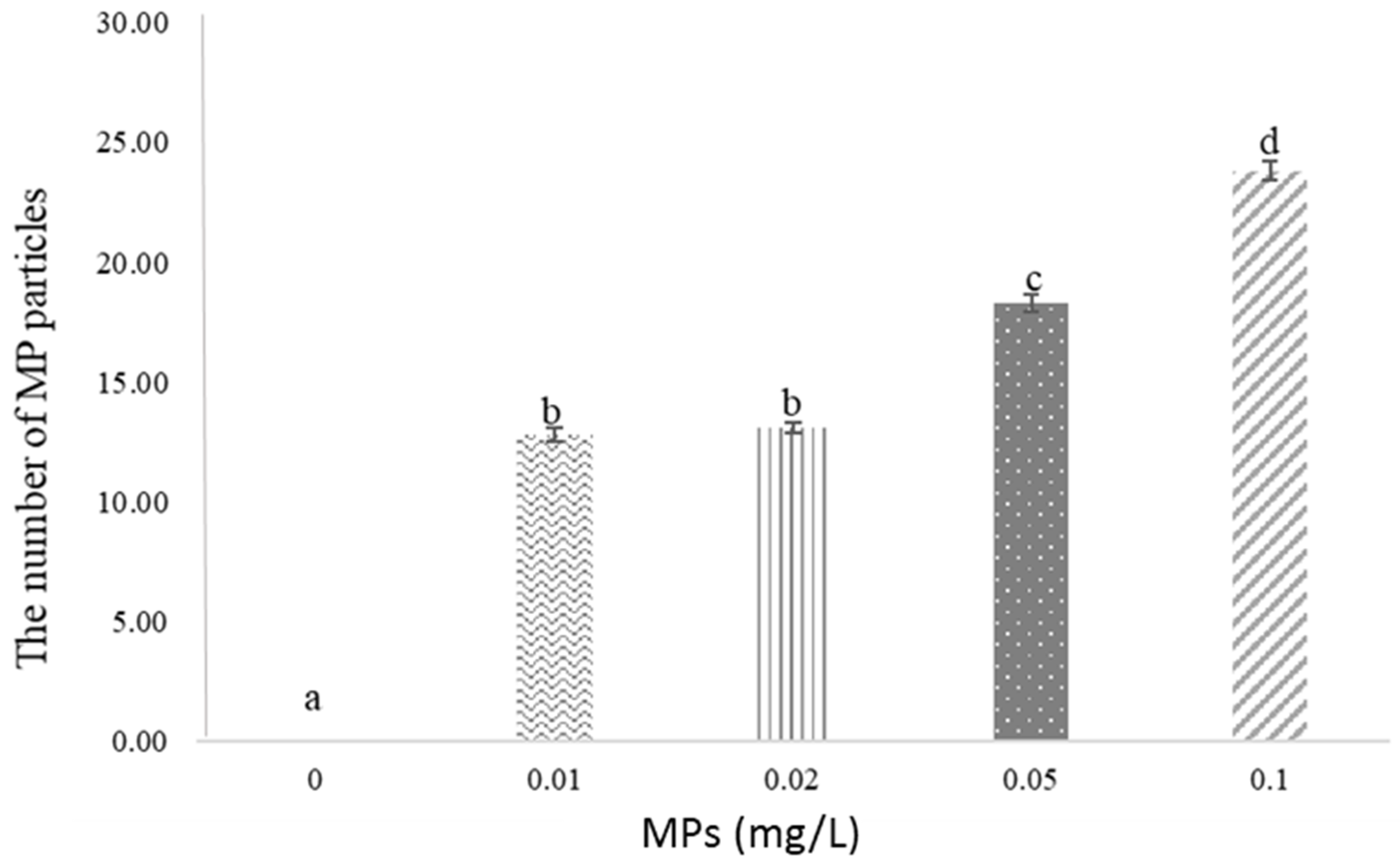
3.1. No Effluences on the Survival Rate of D. magna Even with Higher MPs Accumulation

The survival rate of D. magna exposed to MPs for 7 days was not significantly different in all groups (p = 0.984, Figure 1). After 7 days of exposure to plastic particles, the amount of residual MPs in D. magna bodies increased significantly followed by an increasing intensity of MPs incubation (p < 0.001). The amount of residual MPs in D. magna bodies was in the range of 0, 12.82 ± 0.28, 13.07 ± 0.21, 18.28 ± 0.38 and 23.82 ± 0.38 particles in the groups of control, 0.01, 0.02, 0.05 and 0.1 mg/L MPs, respectively (Figure 2). The Tukey test revealed significant differences between groups (p < 0.001), except for the groups of 0.01 vs. 0.02 mg/L (p = 0.82).

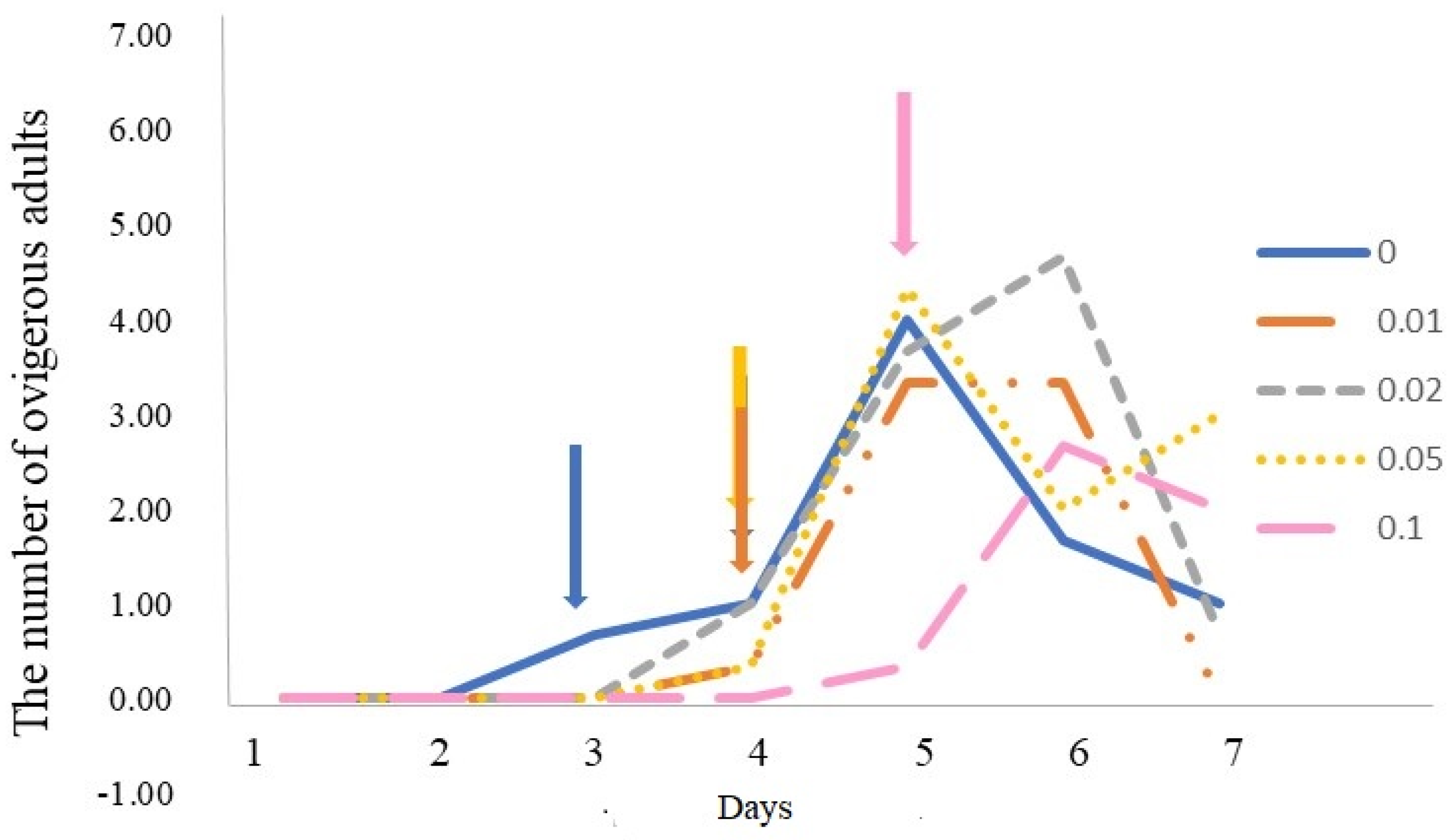
3.2. MPs Ingestion Induced Delayed Sexual Maturity in D. magna

The effect of exposure to MPs led to an increase in the number of days of birth to clutching of eggs in D. magna (p < 0.05) compared to the control group. In the control group, the occurrence of egg clutches was found on the third day, but on the fourth day in the 0.01, 0.02 and 0.05 mg/L groups and the fifth day in the group of 0.1 mg/L (Figure 3). During MP incubation, the number of D. magna with clutched eggs in the control group was 0.67 ± 0.58, 1.00 ± 1.00, 4.00 ± 1.00, 1.67 ± 1.53 and 1.00 ± 1.00 from the 3rd day to 7th day, and 0.33 ± 0.58, 3.33 ± 1.53 and 3.33 ± 0.58 in the 0.01 mg/L group from the 4th day to 6th day, respectively. From the 4th day to 7th day, an average number of ovigerous D. magna (1.00 ± 1.00, 3.67 ± 0.58, 4.67 ± 4.62 and 0.67 ± 0.58) was observed in the 0.02 mg/L group and 0.33 ± 0.58, 4.33 ± 4.93, 2.00 ± 1.00 and 3.00 ± 1.00 in the 0.05 mg/L group, respectively. The 0.1 mg/L 0.1 mg/L treatment of MPs possessed an average of 0.33 ± 0.58, 2.67 ± 0.58 and 2.00 ± 1.00 of D. magna bearing eggs from the 5th day to 7th day (Figure 3). After 7 days of MP exposure, the eggs held by D. magna decreased significantly with an increasing intensity of MPs (p < 0.05). After 7 days of MPs incubation, the results showed that the number of eggs in 0.1 and 0.05 mg/L groups (2.07 ± 0.06 and 4.70 ± 0.60) was less than in the 0.02, 0.01 mg/L and control groups (6.43 ± 0.08, 7.431 ± 0.81 and 7.93 ± 0.47) (Figure 4).

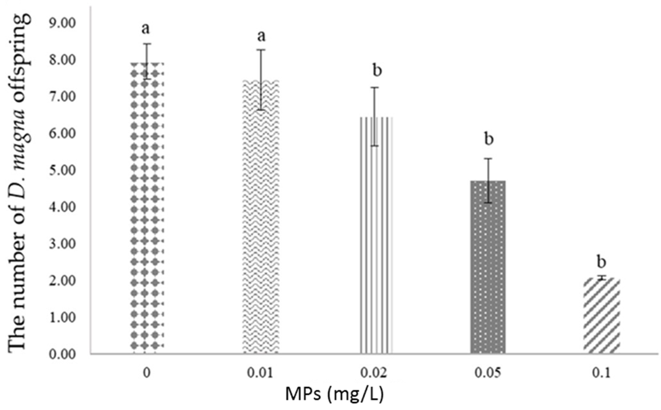
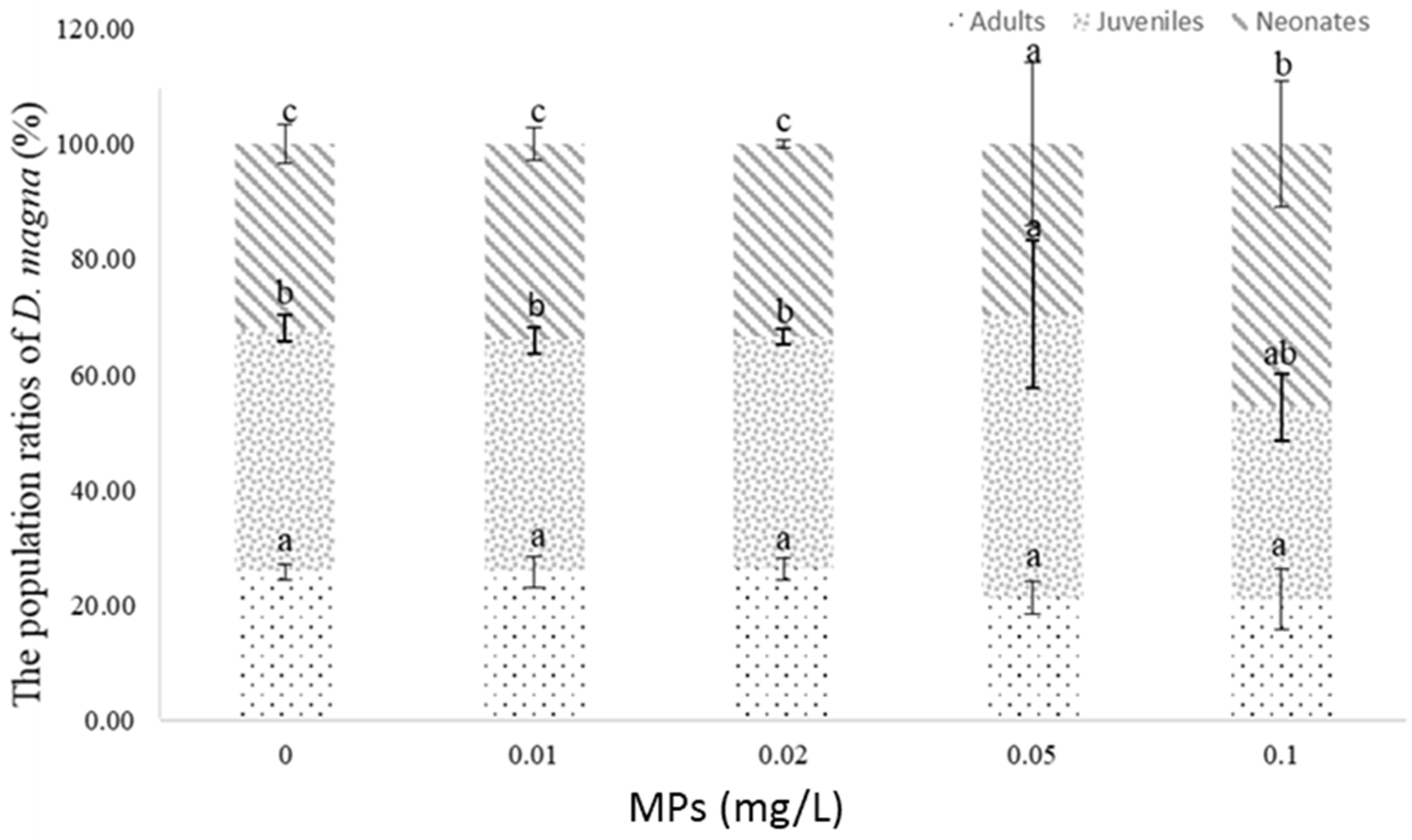
3.3. Greater MPs Enhanced the Number of Neonates in D. magna Offspring

After 7 days MP treatment, the D. magna offspring were transferred to MP-free tanks for 30days. After 30 days, the results indicated that the body size of the newborn offspring had no significant differences. The body length of the offspring in groups of control, 0.01, 0.02, 0.05 and 0.1 mg/L were 3.11 ± 0.07, 3.02 ± 0.02, 3.07 ± 0.01, 2.92 ± 0.06 and 3.03 ± 0.05 mm, respectively (Table 1). However, the population number of D. magna offspring increased. There were 32.02 ± 3.31, 33.99 ± 02.83, 33.40 ± 0.71, 29.55 ± 14.06 and 45.68 ± 11.00 individuals in the control and other groups with increased MPs.
In D. magna offspring, the number of adults was 25.77 ± 1.39, 25.73 ± 2.74, 26.32 ± 1.82, 21.27 ± 2.79 and 20.94 ± 5.22 and numbers were not significantly different in all groups. The population of juveniles in the groups of 0.05 and 0.1 mg/L MPs accounted for 49.17 ± 12.90 and 33.36 ± 5.82, which was significantly different from the other groups (p < 0.05, Figure 5). Similarly, the treatment with 0.1 mg/L MPs showed 45.68 ± 11.00 individuals of D. magna in the neonate stage which was significantly different compared with other treatments (p < 0.05, Figure 5). It may be suggested that the removal of MPs after 7 days MPs treatment resulted in an increasing number of D. magna juveniles and neonates that indicated their growth was initially reduced or inhibited in an MPs environment.

4. Discussion

Plankton are an important source of protein, lipid and carbohydrates in marine and freshwater ecosystems [42,43,44]. MPs may induce the death of zooplankton populations due to a delay in sexual maturity [45], the nutrient depletion of juvenile and neonates [46] and toxin accumulation [47], leading to harmful impacts for higher consumers through the food chain of the ecosystem and human aquaculture [48]. Elgarahy et al. [49] pointed out that MPs present a high potential of adsorption of a variety of pollutants on their surfaces. In this study, it was found that MPs easily attached to the surface of plankton and ingested into their body. This is important for D. magna, as they have complex life cycles [50,51]. The current result is similar to that of the study of Wang et al. [52]. MPs are able to pose not only direct harm to organisms but also act as vectors to transport an indirect hazard, leading to implications in the function and resilience of aquatic ecosystems. Adult D. magna attach themselves to rocks on the bottom of the water near the shore but their juveniles are planktonic [39,53,54]. The absorption of a large amount of MPs may result in harmful effluence on their juveniles, thereby reducing the population.
The results indicated that the survival, reproduction and body size of offspring were defective, which may indirectly lead to the production of live feeds, D. magna, and provide important implications in future aquaculture. Here, we found that a high dose of MPs did not have any significant effect on the survival rate of D. magna, and the same result was shown for their offspring population, which is consistent with the study of Canniff and Hoang [55]. Schür et al. [56] also proposed that exposure to wastewater-incubated MPs resulted in a low mortality of zooplankton. However, this result is not consistent with that of Bosker et al. [57], who observed a significant decline in D. magna population biomass due to microplastics exposure. However, the results revealed MPs had a significant effect in delaying the days to sexual maturity and the number of ovigerous D. magna, which is not consistent with the findings of Canniff and Hoang [58], which presented that the reproduction of D. magna was not affected by MPs. However, the study of Ogonowski et al. [24] proposed that exposure to SMPs (secondary MPs) caused elevated mortality, an increased inter-brood period and decreased reproduction [24]. Furthermore, for long-term effluences, Eltemsah and Bøhn [59] found that MPs caused increasing mortality to D. magna within 120 h but not within 48 h. Additionally, the juveniles demonstrated higher sensitivity to MPs exposure than the adults. Under MPs exposure, the D. magna juveniles showed higher sensitivity (>50%) which slightly increased their mortality, and reduced the growth and stimulation of early reproduction [59]. Moreover, our results showed that the number of newborn offspring continued to increase followed by an increase in MPs in water. Bosker et al. [56] obtained a similar result, in that no significant impact was observed on the number and body length of newborn D. magna offspring following treatment with high MPs. The damage caused to microvilli by NPs aggregates provides an example to explain the negative effluences of food consumption and growth in Chironomus riparius larvae [60]. The same result regarding the correlation of D. magna body length with MP content was found in the current study.
MPs reportedly accelerate the aggregation of MPs. MPs stimulate marine phytoplankton to increase sticky protein secretions, which accelerates the aggregation of MPs [61,62,63,64,65,66] that are commonly referred to as “marine plastic snowflakes”. MPs are more likely to adhere to extracellular polymers and affect the normal interaction of marine ecosystems. A large amount of MPs in the ocean leads to the death of phytoplankton due to a change in the composition of their extracellular secretions [67]. In this study, we found that D. magna was able to adhere to and adsorb MPs on its body surface after 7 days incubation with abundant MPs. However, MPs agglomeration, occurring at higher MPs concentrations, yielded a lower ingestion rate [24,33,38]. When a predator ingests D. magna in this situation, a high number of MPs enter into the food chain, which increases the bioaccumulation and risk posed to the ecosystem. These planktonic organisms are considered to be carriers and transboundary transporters of MPs, which may disrupt community structure and ecological functions [68,69,70]. The ingestion of these nano and microplastic particles by a variety of organisms, including bivalves, mussels, shrimp, oysters, radiopods, and silkworm, affects their fertility, metabolism, and mortality [71,72,73].

5. Conclusions

Overall, the incubation of D. magna in the presence of MPs (0.01 mg/L, 0.02 mg/L, 0.05 mg/L and 0.1 mg/L) for 7 days indicated that MPs lead to offspring with delayed physiological development in terms of egg-breeding behavior, growth in body length, and morphology, which may result in potential impacts on survival in large fleas. Fortunately, the removal of MPs was found to be an effective way to recover their delayed physiological and morphological development, which provides important clues to further understand the interactions between MPs and the survival, sexual maturity, and offspring of D. magna. Furthermore, these findings contribute to risk evaluations of marine and freshwater MPs.

Author Contributions

B.C. conceived of the idea and developed the theory and experimental design. C.-H.H. and T.-W.C. co-contributed to the measurements and planning and supervised the work. B.C. processed the experimental data, performed the analysis, drafted the manuscript and designed the figures. C.-H.K. and Y.-Y.C. manufactured the samples and characterized them with statistics. M.-C.H. aided in interpreting the results and worked on the manuscript. All authors have read and agreed to the published version of the manuscript.

Funding

This research received no external funding.

Institutional Review Board Statement

Studies with invertebrates (such as Daphnia magma) do not currently require ethical approval.

Data Availability Statement

All data were presented in this manuscript.

Acknowledgments

We acknowledge the private aquaculture farm of Hsueh Hsin-Yao in Taiwan for kindly providing of the source of D. magna. We also thank M.-C.H. for technical and equipment support as well as for providing comments that greatly improved the manuscript.

Conflicts of Interest

The authors declare no conflict of interest.

References

  1. Moore, C.J.; Lattin, G.L.; Zellers, A.F. Quantity and type of plastic debris flowing from two urban rivers to coastal waters and beaches of Southern California. J. Integr. Coast. Zone Manag. 2011, 11, 65–73. [Google Scholar] [CrossRef]
  2. Andrady, A.L.; Neal, M.A. Applications and societal benefits of plastics. Philos. Trans. R. Soc. B Biol. Sci. 2009, 364, 1977–1984. [Google Scholar] [CrossRef]
  3. Cole, M.; Lindeque, P.; Halsband, C.; Galloway, T.S. Microplastics as contaminants in the marine environment: A review. Mar. Pollut. Bull. 2011, 62, 2588–2597. [Google Scholar] [CrossRef] [PubMed]
  4. Wright, S.L.; Thompson, R.C.; Galloway, T.S. The physical impacts of microplastics on marine organisms: A review. Environ. Pollut. 2013, 178, 483–492. [Google Scholar] [PubMed]
  5. Rochman, C.M.; Anna-Marie Cook, A.M.; Koelmans, A.A. Plastic debris and policy: Using current scientific understanding to invoke positive change. Environ. Toxicol. Chem. 2016, 35, 1617–1626. [Google Scholar] [CrossRef]
  6. Sundt, P.; Schulze, P.; Syversen, F. Sources of Microplastics-Pollution to the Marine Environment Mepex for the Norwegian Environment Agency; Report No. M-321/2015; Norwegian Environment Agency: Trondheim, Norway, 2014.
  7. Shan, A.A.; Hasan, F.; Hameed, A.; Ahmed, A. Biological degradation of plastics: A comprehensive review. Biotechnol. Adv. 2008, 26, 246–265. [Google Scholar]
  8. Costa, J.P.D.; Santos, P.S.M.; Duarte, A.C.; Santos, T.R. Nanoplastics in the environment Sources, Fates and effects. Sci. Total Environ. 2010, 566–567, 15–26. [Google Scholar]
  9. Andrady, A.L. Microplastics in the marine environment. Mar. Pollut. Bull. 2011, 62, 1596–1605. [Google Scholar] [CrossRef]
  10. Plastics Europe. Plastics-the Facts 2014/2015, an Analysis of European Plastics Production, Demand and Waste Data; Plastics Europe: Brussels, Belgium.
  11. Koelmans, B.; Pahl, S.; Backhaus, T.; Bessa, F.; van Calster, G.; Contzen, N.; Cronin, R.; Galloway, T.; Hart, A.; Henderson, L.; et al. Science Advice for Policy by European Academies. In A Scientific Perspective on Microplastics in Nature and Society; SAPEA: Berlin, Germany, 2019. [Google Scholar]
  12. Jambeck, J.R.; Geyer, R.; Wilcox, C.; Siegler, T.R.; Perryman, M.; Andrady, A.; Narayan, R.; Law, K.L. Plastic waste inputs from land into the ocean. Science 2015, 347, 768–771. [Google Scholar] [CrossRef]
  13. Wegner, A.; Besseling, E.; Foekema, E.M.; Kamermans, P.; Koelmans, A.A. Effects of nanopolystyrene on the feeding behavior of the blue mussel (Mytilus edulis L.). Environ. Toxicol. Chem. 2012, 31, 2490–2497. [Google Scholar] [CrossRef]
  14. Farrell, P.; Nelson, K. Trophic level transfer of microplastic: Mytilus edulis (L.) to Carcinus maenas (L.). Environ. Pollut. 2013, 77, 1–3. [Google Scholar] [CrossRef] [PubMed]
  15. Baulch, S.; Perry, C. Evaluating the impacts of marine debris on cetaceans. Mar. Pollut. Bull. 2014, 80, 210–221. [Google Scholar] [CrossRef] [PubMed]
  16. Chua, E.M.; Shimeta, J.; Nugegoda, D.; Morrison, P.D.; Clarke, B.O. Assimilation of polybrominated diphenyl ethers from microplastics by the marine amphipod, Allorchestes compressa. Environ. Sci. Technol. 2014, 48, 8127–8134. [Google Scholar] [CrossRef] [PubMed]
  17. Watts, A.J.R.; Lewis, C.; Goodhead, R.M.; Beckett, S.J.; Moger, J.; Tyler, C.R.; Galloway, T.S. Uptake and retention of microplastics by the shore crab Carcinus maenas. Environ. Semicond. Sci. Technol. 2014, 48, 8823–8830. [Google Scholar] [CrossRef]
  18. Van Cauwenberghe, L.; Claessens, M.; Vandegehuchte, M.B.; Janssen, C.R. Microplastics are taken up by mussels (Mytilus edulis) and lugworms (Arenicola marina) living in natural habitats. Environ. Pollut. 2015, 199, 10–17. [Google Scholar] [CrossRef]
  19. Laist, D.W. Impacts of Marine Debris: Entanglement of Marine Life in Marine Debris Including a Comprehensive List of Species with Entanglement and Ingestion Records. In Marine Debris: Sources, Impacts and Solutions; Coe, J.M., Rogers, D.B., Eds.; Springer: New York, USA, 1997; pp. 99–139. [Google Scholar]
  20. Derraik, J.G.B. The pollution of the marine environment by plastic debris: A Review. Mar. Pollut. Bull. 2002, 44, 842–852. [Google Scholar] [CrossRef]
  21. Rochman, C.M.; Hoh, E.; Kurobe, T.; Teh, S.J. Ingested plastic transfers hazardous chemicals to fish and induces hepatic stress. Sci. Rep. 2013, 3, 3263. [Google Scholar] [CrossRef]
  22. Greven, A.C.; Merk, T.; Karagöz, F.; Mohr, K.; Klapper, M.; Jovanovi, B.; Palic, D. Polycarbonate and polystyrene nanoplastic particles act as stressors to the innate immune system of fathead minnow (Pimephales promelas). Environ. Toxicol. Chem. 2016, 35, 3093–3100. [Google Scholar] [CrossRef]
  23. Mason, S.A.; Garneau, D.; Sutton, R.; Chu, Y.; Ehmann, K.; Barnes, J.; Fink, P.; Papazissimos, D.; Rogers, D.L. Microplastic pollution is widely detected in US municipal wastewater treatment plant effluent. Environ. Pollut. 2016, 218, 1045–1054. [Google Scholar] [CrossRef]
  24. Ogonowski, M.; Schür, C.; Jarsen, Å.; Gorokhova, E. The effects of natural and anthropogenic microparticles on individual fitness in Daphnia magna. PLoS ONE 2016, 11, e0155063. [Google Scholar] [CrossRef]
  25. Wagner, M.; Engwall, M.; Hollert, H. Editorial: (Micro)Plastics and the environment. Environ. Sci. Eur. 2014, 26, 16. [Google Scholar] [CrossRef] [PubMed] [Green Version]
  26. Dris, R.; Imhof, H.; Sanchez, W.; Gasperi, J.; Galgani, F.; Tassin, B.; Laforsch, C. Beyond the ocean: Contamination of freshwater ecosystems with (micro-) plastic particles. Environ. Chem. 2015, 12, 539–550. [Google Scholar] [CrossRef]
  27. Horton, A.A.; Walton, A.; Spurgeon, D.J.; Lahive, E.; Svendsen, C. Microplastics in freshwater and terrestrial environments: Evaluating the current under standing to identify the knowledge gaps and future research priorities. Sci. Total Environ. 2017, 586, 127–141. [Google Scholar] [CrossRef] [PubMed] [Green Version]
  28. Besseling, E.; Quik, J.T.K.; Sun, M.; Koelmans, A.A. Fate of nano- and microplastic in freshwater systems: A modeling study. Environ. Pollut. 2017, 220, 540–548. [Google Scholar] [CrossRef]
  29. Teuten, E.L.; Saquing, J.M.; Knappe, D.R.U.; Barlaz, M.A.; Jonsson, S.; Björn, A.; Takada, H. Transport and release of chemicals from plastics to the environment and to wildlife. Philos. Trans. R. Soc. B 2009, 364, 2027–2045. [Google Scholar] [CrossRef] [PubMed] [Green Version]
  30. Jemec, A.; Horvat, P.; Kunej, U.; Bele, M.; Kržan, A. Uptake and effects of microplastic textile fibers on freshwater crustacean Daphnia magna. Environ. Pollut. 2016, 219, 201–209. [Google Scholar] [CrossRef]
  31. Lampert, W. Daphnia: Development of a Model Organism in Ecology and Evolution; International Ecology Institute: Oldendorf/Luhe, Germany, 2011; Volume 70, pp. 337–344. [Google Scholar]
  32. Imhof, H.K.; Rusek, J.; Thiel, M.; Wolinska, J.; Laforsch, C. Do microplastic particles affect Daphnia magna at the morphological, life history and molecular level? PLoS ONE 2017, 12, e0187590. [Google Scholar] [CrossRef] [Green Version]
  33. Frydkjær, C.K.; Iversen, N.; Roslev, P. Ingestion and egestion of microplastics by the cladoceran Daphnia magna: Effects of regular and irregular shaped plastic and sorbed phenanthrene. Bull. Environ. Contam. Toxicol. 2017, 99, 655–661. [Google Scholar] [CrossRef]
  34. Juchelka, C.M.; Snell, T.W. Rapid toxicity assessment using ingestion rate of cladocerans and ciliates. Arch. Environ. Contam. Toxicol. 1995, 28, 508–512. [Google Scholar] [CrossRef]
  35. Lopiz, J.K.; Cowen, R.K. Variability in the trophic role of coral reef fish larvae in the oceanic plankton. Mar. Ecol. Prog. Ser. 2009, 381, 259–272. [Google Scholar] [CrossRef] [Green Version]
  36. Besseling, E.; Wang, B.; Lürling, M.; Koelmans, A.A. Nanoplastic affects growth of S. obliquus and reproduction of D. Magna. Environ. Sci. Technol. 2014, 48, 12336–12343. [Google Scholar] [CrossRef]
  37. Nasser, F.; Lynch, I. Secreted protein eco-corona mediates uptake and impacts of polystyrene nanoparticles on Daphnia magna. J. Proteome Res. 2016, 137, 45–51. [Google Scholar] [CrossRef] [Green Version]
  38. Ma, Y.; Huang, A.; Cao, S.; Sun, F.; Wang, L.; Guo, H.; Ji, R. Effects of nanoplastics and microplastics on toxicity, bioaccumulation, and environmental fate of phenanthrene in fresh water. Environ. Pollut. 2016, 219, 166–173. [Google Scholar] [CrossRef]
  39. Rehse, S.; Kloas, W.; Zarfl, C. Short-term exposure with high concentrations of pristine microplastic particles leads to immobilisation of Daphnia magna. Chemosphere 2016, 153, 91–99. [Google Scholar] [CrossRef]
  40. Kim, D.; Chae, Y.; An, Y.J. Mixture toxicity of nickel and microplastics with different functional groups on Daphnia magna. Environ. Sci. Technol. 2017, 51, 12852–12858. [Google Scholar] [CrossRef]
  41. Forró, L.; Korovchinsky, N.M.; Kotov, A.A.; Petrusek, A. Global diversity of cladocerans (Cladocera; Crustacea) in freshwater. In Freshwater Animal Diversity Assessment; Springer: Dordrecht, The Netherlands, 2008; Volume 595, pp. 177–184. [Google Scholar]
  42. Taipale, S.J.; Galloway, A.W.E.; Aalto, A.L.; Kahilainen, K.K.; Strandberg, U.; Kankaala, P. Terrestrial carbohydrates support freshwater zooplankton during phytoplankton deficiency. Sci. Rep. 2016, 6, 30897. [Google Scholar] [CrossRef] [Green Version]
  43. Arts, M.T. Lipids in freshwater zooplankton: Selected ecological and physiological aspects. In Lipids in Freshwater Ecosystems; Springer: New York, USA, 1999; pp. 71–90. [Google Scholar]
  44. Napiórkowska-Krzebietke, A. Phytoplankton as a basic nutritional source in diets of fish. J. Elem. 2017, 22, 831–841. [Google Scholar] [CrossRef]
  45. Sridharan, S.; Kumar, M.; Bolan, N.S.; Singh, L.; Kumar, S.; Kumar, R.; You, S. Are microplastics destabilizing the global network of terrestrial and aquatic ecosystem services? Environ. Res. 2021, 198, 111243. [Google Scholar] [CrossRef]
  46. Yu, S.P.Y.; Cole, M.C.; Chan, B.K.K. Effects of microplastic on zooplankton survival and sublethal responses. Oceanogr. Mar. Biol. 2020, 58, 359–408, in press. [Google Scholar]
  47. Yin, K.; Wang, Y.; Zhao, H.; Wang, D.; Guo, M.; Mu, M.; Liu, Y.; Nie, X.; Li, B.; Li, J.; et al. A comparative review of microplastics and nanoplastics: Toxicity hazards on digestive, reproductive and nervous system. Sci. Total Environ. 2021, 774, 145758. [Google Scholar] [CrossRef]
  48. Zhou, A.; Zhang, Y.; Xie, S.; Chen, Y.; Li, X.; Wang, J.; Zou, J. Microplastics and their potential effects on the aquaculture systems: A critical review. Rev. Aquac. 2020, 13, 719–733. [Google Scholar] [CrossRef]
  49. Elgarahy, A.M.; Akhdhar, A.; Elwakeel, K.Z. Microplastics prevalence, interactions, and remediation in the aquatic environment: A critical review. J. Environ. Chem. Eng. 2021, 9, 106224. [Google Scholar] [CrossRef]
  50. Canniff, P.M.; Hoang, T.C. Microplastic ingestion by Daphnia magna and its enhancement on algal growth. Sci. Total Environ. 2018, 633, 500–507. [Google Scholar] [CrossRef] [Green Version]
  51. Luo, H.; Liu, C.; He, D.; Xu, J.; Sun, J.; Li, J.; Pan, X. Environmental behaviors of microplastics in aquatic systems: A systematic review on degradation, adsorption, toxicity and biofilm under aging conditions. J. Hazard. Mater. 2022, 423, 126915. [Google Scholar] [CrossRef]
  52. Wang, Z.; Dong, H.; Wang, Y.; Ren, R.; Qin, X.; Wang, S. Effects of microplastics and their adsorption of cadmium as vectors on the cladoceran Moina monogolica Daday: Implications for plastic-ingesting organisms. J. Hazard. Mater. 2020, 400, 123239. [Google Scholar] [CrossRef]
  53. Dodson, S.L.; Cáceres, C.E.; Rogers, D.C. Chapter 20 Cladocera and other Branchiopoda. In Ecology and Classification of North American Freshwater Invertebrates, 3rd ed.; Academic Press: Cambridge, New York, USA, 2010; pp. 773–827. [Google Scholar]
  54. Imhof, H.K.; Ivleva, N.P.; Schmid, J.; Niessner, R.; Laforsch, C. Contamination of beach sediments of a subalpine lake with microplastic particles. Curr. Biol. 2013, 23, 867–868. [Google Scholar] [CrossRef] [Green Version]
  55. Bosker, T.; Bouwman, L.J.; Brun, N.R.; Behrens, P.; Vijver, M.G. Microplastics accumulate on pores in seed capsule and delay germination and root growth of the terrestrial vascular plant Lepidium sativum. Chemosphere 2019, 226, 774–781. [Google Scholar] [CrossRef]
  56. Schur, C.; Weil, C.; Baum, M.; Wallraff, J.; Schreier, M.; Oehlmann, J.; Wagner, M. Incubation in Wastewater Reduces the Multigenerational Effects of Microplastics in Daphnia magna. Environ. Sci. Technol. 2021, 55, 2491–2499. [Google Scholar] [CrossRef]
  57. Bosker, T.; Olthof, G.; Vijver, M.G.; Baas, J.; Barmentlo, S.H. Significant Decline of Daphnia Magna Population Biomass Due to Microplastic Exposure. Environ. Pollut. 2019, 250, 669–675. [Google Scholar] [CrossRef] [Green Version]
  58. Burd, A.B.; Jackson, G.A. Particle Aggregation. Annu. Rev. Mar. Sci. 2009, 1, 65–90. [Google Scholar] [CrossRef]
  59. Eltemsah, Y.S.; Bøhn, T. Acute and chronic effects of polystyrene microplastics on juvenile and adult Daphnia magna. Environ. Pollut. 2019, 254, 112919. [Google Scholar] [CrossRef]
  60. Waissi-Leinonen, G.C.; Petersen, E.J.; Pakarinen, K.; Akkanen, J.; Leppänen, M.T.; Kukkonen, J.V.K. Toxicity of fullerene (C60) to sediment-dwelling invertebrate Chironomus riparius larvae. Environ. Toxicol. Chem. 2012, 31, 2108–2116. [Google Scholar] [CrossRef]
  61. Shiu, R.F.; Lee, C.L. Role of microgel formation in scavenging of chromophoric dissolved organic matter and heavy metals in a river-sea system. J. Hazard. Mater. 2017, 328, 12–20. [Google Scholar] [CrossRef] [Green Version]
  62. Santschi, P.H.; Xu, C.; Schwehr, K.A.; Lin, P.; Sun, L.; Chin, W.C. Can the protein/carbohydrate (P/C) ratio of exopolymeric substances (EPS) be used as a proxy for their ‘stickiness’ and aggregation propensity? Mar. Chem. 2020, 218, 103734. [Google Scholar] [CrossRef]
  63. Shiu, R.F.; Chin, W.C.; Lee, C.L. Carbonaceous particles reduce marine microgel formation. Sci. Rep. 2014, 4, 5856. [Google Scholar] [CrossRef] [Green Version]
  64. Shiu, R.F.; Lee, C.L.; Chin, W.C. Reduction in the exchange of coastal dissolved organic matter and microgels by inputs of extra riverine organic matter. Water Res. 2018, 131, 161–166. [Google Scholar] [CrossRef]
  65. Shiu, R.F.; Chiu, M.H.; Vazquez, C.I.; Tsai, Y.Y.; Le, A.; Kagiri, A. Protein to carbohydrate (P/C) ratio changes in microbial extracellular polymeric substances induced by oil and Corexit. Mar. Chem. 2020, 223, 103789. [Google Scholar] [CrossRef]
  66. Shiu, R.F.; Vazquez, C.I.; Tsai, Y.Y.; Torres, G.V.; Chen, C.S.; Santschi, P.H. Nano-plastics induce aquatic particulate organic matter (microgels) formation. Sci. Total Environ. 2020, 706, 135681. [Google Scholar] [CrossRef]
  67. Zarfl, C.; Matthies, M. Are marine plastic particles transport vectors for organic pollutants to the Arctic? Mar. Pollut. Bull. 2010, 60, 1810–1814. [Google Scholar] [CrossRef]
  68. Zettler, E.R.; Mincer, T.J.; Amaral-Zettler, L.A. Life in the “plastisphere”: Microbial communities on plastic marine debris. Environ. Sci. Technol. 2013, 47, 7137–7146. [Google Scholar] [CrossRef]
  69. Rosato, A.; Barone, M.; Negroni, A.; Brigidi, P.; Fava, F.; Xu, P.; Gandela, M.; Zanaroli, G. Microbial colonization of different microplastic types and biotransformation of sorbed PCBs by a marine anaerobic bacterial community. Sci. Total Environ. 2020, 705, 135790. [Google Scholar] [CrossRef]
  70. Lusher, A.L.; Hernandez-Milian, G.; O’Brien, J.; Berrow, S.; O’Connor, I.; Officer, R. Microplastic and macroplastic ingestion by a deep diving, oceanic cetacean: The True’s beaked whale Mesoplodon mirus. Environ. Pollut. 2015, 199, 185–191. [Google Scholar] [CrossRef]
  71. Andrady, A.L. The plastic in microplastics: A review. Mar. Pollut. Bull. 2017, 119, 12–22. [Google Scholar] [CrossRef]
  72. Chae, Y.; An, Y.J. Effects of micro-and nanoplastics on aquatic ecosystems: Current research trends and perspectives. Mar. Pollut. Bull. 2017, 124, 624–632. [Google Scholar] [CrossRef]
  73. Pico, Y.; Alfarhan, A.; Barcelo, D. Nano- and microplastic analysis: Focus on their occurrence in freshwater ecosystems and remediation technologies. TrAC Trends Anal. Chem. 2019, 113, 409–425. [Google Scholar] [CrossRef]
Figure 1. The survival rate of D. magna exposed to MPs for 7 days. A total of 20 D. magna females were incubated with different MP treatments for 7 days. All treatments showed no significant differences on the survival rates (p = 0.984). All the tests included 3 independent replicates. Lowercase letters indicate significant differences between treatments.
Fishes 07 00181 g001
Figure 2. Quantification of residual MPs in D. magna after 7 days exposure to MPs. After 7 days MPs treatment, the residual MPs uptake inside the body of the D. magna containing adults, juveniles and neonates derived from 20 females were evaluated. The MPs in body of D. magna significantly increased following an increase in MPs (p < 0.001). The highest accumulation of MPs in D. magna appeared under 0.1 mg/L MPs treatment. All the tests included 3 independent replicates. Lowercase letters indicate significant differences between treatments.
Fishes 07 00181 g002
Figure 3. Effects on the population and the days of sexual maturity in D. magna under exposure of MPs. After MP treatment of D. magna females, 20 newborn neonates were transferred to and incubated in MP-free freshwater for 30 days. The D. magna offspring showed fast sexual maturity with 3 days under freshwater (no MPs) and released their eggs at 5th day. However, the other offspring derived from MP-treated D. magna presented delayed sexual maturity (over 4 days) and offspring production (6th day). All the tests included 3 independent replicates.
Fishes 07 00181 g003
Figure 4. The effects of MP treatments on the offspring population of D. magna. Each group of 7 days MP treatment contained 20 individuals of D. magna females. The number of offspring produced from different groups was calculated to determine the effects of MP treatments on the offspring population. Apparently, except for 0.01 mg/L MP treatment, all offspring produced by the D. magna females showed a significant decreased tendency after 7 days incubation of MPs (p < 0.05). All tests included 3 independent replicates. Lowercase letters indicate significant differences between treatments.
Fishes 07 00181 g004
Figure 5. The population of adults, juveniles and neonates in MP-treated D. magna after 30 days of freshwater incubation. After 7-day exposure to MPs of female parents, all the newborn D. magna were transferred to the freshwater without MPs for 30 days. Additionally, the 500 mL of water volume containing well-mixed adults, juveniles and newborn neonates of D. magna was harvested and different ratios in whole population were found in 0.05 and 0.01 mg/L MP-treated groups. All the tests included 3 independent replicates. Lowercase letters indicate significant differences between treatments.
Fishes 07 00181 g005
Table 1. The average (± SE) body length and number of D. magna after MPs exposure *.
MP (mg/L)Body Length (mm)Number of D. magna
control3.11 ± 0.0732.02 ± 3.31
0.013.02 ± 0.0233.99 ± 2.83
0.023.07 ± 0.0133.40 ± 0.71
0.052.92 ± 0.0629.55 ± 14.06
0.12.94 ± 0.0545.68 ± 11.00
* D. magna were incubated in MP-containing water for 7 days and then all of them were transferred to the MPs-free tanks for 30 days. All tests included 3 independent replicates.
Publisher’s Note: MDPI stays neutral with regard to jurisdictional claims in published maps and institutional affiliations.

