Article

Microbial and Planktonic Community Characteristics of Eriocheir sinensis Culture Ponds Experiencing Harmful Algal Blooms

1
Key Laboratory of Integrated Rice-Fish Farming Ecology, Ministry of Agriculture and Rural Affairs, Freshwater Fisheries Research Center, Chinese Academy of Fishery Sciences, Wuxi 214081, China
2
Wuxi Fisheries College, Nanjing Agricultural University, Wuxi 214081, China
3
Jiangsu Noah’s Ark Agricultural Technology Co. Ltd., Changzhou 213147, China
4
College of Fisheries and Life Science, Shanghai Ocean University, Shanghai 201306, China
*
Author to whom correspondence should be addressed.
Fishes 2022, 7(4), 180; https://doi.org/10.3390/fishes7040180
Submission received: 29 June 2022 / Revised: 7 July 2022 / Accepted: 9 July 2022 / Published: 22 July 2022
(This article belongs to the Special Issue Recent Advances in Crab Aquaculture)

Abstract

:
The Chinese mitten crab, Eriocheir sinensis (H. Milne Edwards, 1853), is an economically important aquaculture species in China. It is a significantly desirable species by Chinese consumers that causes a high demand for environmentally friendly culture farming. In aiming to break through bottlenecks, i.e., “pond moss” and cyanobacteria, we investigated the microbial community and plankton composition of ponds with filamentous algae and cyanobacterial blooms. As results, we found Actinobacteria, Proteobacteria, and Bacteroidetes were dominant bacterial phyla, while Chlorophyta and Bacillariophyta were dominant phytoplankton phyla in E. sinensis ponds. Nitrospira sp., Flectobacillus sp. BAB-3569, Staphylococcus warneri, Fusarium oxysporum, Gromochytrium mamkaevae, and Rhizophydium sp. JEL317 were screened as bioindicators for harmful algal blooms. We found a close relationship between water quality parameters and the species composition of bacteria and zooplankton in the present study. Specifically, total nitrogen and total ammonia nitrogen significantly affected the bacterial community composition, while total phosphorus contributed to the phytoplankton community composition. We further indicated the potential competitive inhibition of Chlamydomonadales on the direct regulation of the control of harmful algal blooms. Finally, we suggested a combination of probiotics and microalgae, e.g., C. vulgaris, to prevent and control potential risks in the culture of E. sinensis. In conclusion, the present study deepened our understanding of harmful algal blooms in aquaculture ponds and suggested the baseline indications for the prevention and control of algal blooms.

1. Introduction

The Chinese mitten crab, Eriocheir sinensis (H. Milne Edwards, 1853), is an important economic aquaculture species in China. With the breakthrough of large-scale artificial breeding technology in the 1980s, the cultural industry of E. sinensis has grown rapidly. The cultivation mode of E. sinensis has also developed from the initial resource release mode, to intensive and high-density intensive modes, and gradually to an ecofriendly rearing practice. At present, E. sinensis has been widely cultured in China except for a few regions such as the Tibet Autonomous Region and Hainan province. The cultural yield of E. sinensis reached up to 775,887 tons (18% of culture crustaceans) in 2020, nearly the same as the production in 2019 [1].
E. sinensis is a very esteemed species by Chinese consumers, causing a high demand for environmentally friendly culture farming. In practice, E. sinensis culture farming contains two stages: the clasp crab culture stage and the adult crab culture stage [2]. There are two risks in the adult crab culture stage: filamentous algae, which occurs in early spring, and cyanobacterial blooms, which occur in high-temperature seasons [3,4,5].
Filamentous eukaryotic algae are commonly referred to as “pond moss” or “pond scum” in aquaculture [6]. In the culture of E. sinensis, “pond moss” usually consists of Spirogyra, Zygnema and Mougeotia. These algae produce large amounts of extracellular polysaccharides to enable them to either adhere or aggregate [7], and they can tolerate high irradiance conditions in cold water [8]. These factors explain the occurrence of large clusters in aquaculture ponds in the early spring. In the culture of E. sinensis, “pond moss” is recognized as a great hazard because it inhibits the growth of beneficial aquatic plants, including cultured species, by taking space and nutrients; it also produces toxic substances after its death, such as hydrogen sulfide and hydroxylamine, leading to water quality deterioration and the introduction of disease in E. sinensis [9].
Cyanobacteria, also known as “blue-green algae”, are a group of photoautotrophic bacteria. In aquaculture ponds, cyanobacteria easily form blooms in high-temperature seasons. Cyanobacterial blooms are commonly formed by certain genera, e.g., Dolichospermum, Planktothrix, Microcystis, Cylindrospermopsis, Nodularia, and Trichodesmium [10]. Cyanobacterial blooms and their secondary metabolites microcystins can cause major problems for water quality, produce toxic effects on aquatic animals, and even threaten human health [5,10,11,12,13,14].
Although several chemical and biological controls were reported [10,14], “pond moss” and cyanobacteria are still challenges in the ecological culture of E. sinensis. Species traits and environmental conditions, such as nitrogen and phosphorus nutrients, are responsible for these challenges by general consent [6,10]. To solve these challenges under the dual needs of food safety and environmental protection, control methods that are friendly to E. sinensis are needed. To break through in aquaculture, probiotics and beneficial microalgae (e.g., Chlorella, Spirulina, Tetraselmis) attract increasing attention due to their efficiency and harmless nature [15,16,17,18,19]; they also provide a promising strategy in combating and preventing “pond moss” and cyanobacterial blooms in E. sinensis cultivation. The microbial and planktonic community composition of ponds experiencing algal and cyanobacterial blooms were investigated in this study in order to provide indications for forecasting and preventing harmful algal blooms.

2. Material and Methods

2.1. Sample Collection

During the E. sinensis cultivation period, we selected 12 ponds with typical characteristics from Xinghua, Jiangsu Province, China, and divided them into three groups: normal group (CK), cyanobacteria bloom group (CY), and pond moss group (MO). In the CY group, the water was blue-green in color, the surface of pond and the vertical profile were teeming with microalgae, and a layer of emerald green film floated downwind of the pond. Ponds with more than 50% pond moss coverage were classified into the MO group. Ponds with various algae, aquatic plants, and almost free of cyanobacteria and pond moss were classified into the CK group.
We collected samples from 12 ponds that cultured E. sinensis on 23 June 2021, including 3 ponds in the CK group, 4 ponds in the MO group, and 5 ponds in the CY group (Table S1). We collected 5 L of surface water from each of the four corners, mixed them well, and obtained a total of 20 L water samples. First, 500 mL of water was immediately filtered through 0.22-μm polycarbonate membranes. The filter membranes were quick-frozen in liquid nitrogen and then stored at −80 °C until DNA extraction. From there, a bottle was filled with 1 L of water and 2 mL methylidyne trichloride (C2432; Sigma-Aldrich, St. Louis, MO, USA), which was added to reduce the influence of biological activities on water quality parameters. Finally, all water samples were quickly delivered to our laboratory for subsequent water quality analyses.

2.2. Water Quality Parameters

Water temperature, pH, and dissolved oxygen were measured in the field at a depth of 0.5 m using HACH HQ30D (Hach Company, Loveland, CO, USA). Additionally, we collected water samples to analyze the water quality, i.e., total nitrogen (Alkaline potassium persulfate digestion UV spectrophotometric method; HJ636-2012), total ammonia nitrogen (Nessler’s reagent spectrophotometric method; HJ535-2009), nitrate nitrogen (Ultraviolet spectrophotometric method; HJ/T 346-2007), nitrite nitrogen (Spectrophotometric method; GB7493-87), total phosphorus (Ammonium molybdate spectrophotometric method; GB11893-89), and chemical oxygen demand (Dichromate method; GB11914-89) contents. We calculated the N/P ratio using total nitrogen divided by total phosphorus.

2.3. DNA Extraction and PCR Amplification

Total microbial DNA was extracted using either HiPure Soil DNA Kits or HiPure Stool DNA Kits (Magen, Guangzhou, China) in accordance with the manufacturer’s protocols. The V3-V4 region of the 16S rDNA was amplified by PCR (95 °C for 5 min, followed by 30 cycles at 95 °C for 60 s, 60 °C for 60 s, 72 °C for 60 s, and a final extension at 72 °C for 7 min) using primers 341F (5’-CCTACGGGNGGCWGCAG-3’) and 806R (5’-GGACTACHVGGGTWTCTAAT-3’). PCR reactions were performed in triplicate 50 μL mixtures containing 10 μL of 5 × Q5® Reaction Buffer, 10 μL of 5 × Q5® High GC Enhancer, 1.5 μL of 2.5 mM dNTPs, 1.5 μL of each primer (10 μM), 0.2 μL of Q5® High-Fidelity DNA Polymerase (M0491, New England Biolabs (Beijing) Ltd., Beijing, China), and 50 ng of template DNA.
The V4 region of the 18S rDNA was amplified by PCR (94 °C for 2 min, followed by 30 cycles at 98 °C for 10 s, 62 °C for 30 s, 68 °C for 30 s, and a final extension at 68 °C for 5 min) using primers 528F (5’-GCGGTAATTCCAGCTCCAA-3’) and 706R (5’-AATCCRAGAATTTCACCTCT-3’). PCR reactions were performed in triplicate 50 μL mixture containing 5 μL of 10 × KOD Buffer, 5 μL of 2 mM dNTPs, 3 μL of 25 mM MgSO4, 1.5 μL of each primer (10 μM), 1 μL of KOD Polymerase (KOD-3B, Toyobo Co., Ltd., Osaka, Japan), and 100 ng of template DNA.

2.4. Illumina Novaseq 6000 Sequencing

We constructed 12 procaryotic sequencing libraries and 12 eukaryotic sequencing libraries in the present study. Amplicons were extracted from 2% agarose gels and purified using the AxyPrep DNA Gel Extraction Kit (Axygen Biosciences, Union City, CA, USA) in accordance with the manufacturer’s instructions and quantified using ABI StepOnePlus Real-Time PCR System (Life Technologies, Foster City, CA, USA). Purified amplicons were pooled in equimolar and paired-end sequenced (PE250) on an Illumina platform (GENE DENOVO, Guangzhou, China) according to the standard protocols. The raw reads were deposited into the NCBI Sequence Read Archive (SRA) database (Accession Numbers: PRJNA799800).

2.5. Bioinformatics Analyses

2.5.1. Reads Filtering and Assembly

To get high-quality clean reads, raw reads that contain adapters or low-quality reads were filtered using FASTP (version 0.18.0) [20]. Filtering rules were: (1) removing reads containing more than 10% of unknown nucleotides, and (2) removing reads containing less than 50% of bases with quality (Q-value) > 20. From there, paired-end clean reads were merged as raw tags using FLSAH (version 1.2.11) [21] with a minimum overlap of 10 bp and mismatch error rates of 2%.

2.5.2. Raw Tag Filtering

Noisy sequences of raw tags were filtered under specific filtering conditions [22] to obtain high-quality clean tags. The filtering conditions are as follows: (1) break raw tags from the first low-quality base site where the number of bases in the continuous low-quality value (the default quality threshold is ≤3) reaches the set length (the default length is 3 bp), (2) filter tags whose continuous high-quality base length is less than 75% of the tag length.

2.5.3. Clustering and Chimera Removal

The clean tags were clustered into operational taxonomic units (OTUs) of ≥97.0% similarity using a UPARSE (version 9.2.64) [23] pipeline. All chimeric tags were removed using the UCHIME algorithm [24] and effective tags were obtained for further analysis. The tag sequence with the highest abundance was selected as a representative sequence within each cluster.

2.5.4. Taxonomy Annotation, Community Composition and Indicator Species Analysis

The representative OTU sequences were classified into organisms by a naive Bayesian model using RDP classifier (version 2.2) [25] based on SILVA database (version 132) [26] with the confidence threshold value of 0.80. The stacked bar plot of the community composition was visualized in the R project ggplot2 package (version 2.2.1).
Venn diagram analysis of the common and specific species of each group was performed in the R project VennDiagram package (version 1.6.16) (Chen and Boutros, 2011). Indicator species were screened based on their abundance and frequency in each sample. Indicator value (IndVal) was calculated for each species with the labdsv package (version 2.0-1; http://ecology.msu.montana.edu/labdsv/R, accessed on 1 December 2019) in the R project. Species were considered indicator species when the P values of the cross-validation test were less than 0.05.

2.5.5. Alpha and Beta Diversity Analysis

The alpha diversity was assessed with multiple indices, including Species richness (observed OTUs), Chao1, Ace, Shannon, Simpson, Good’s coverage, Pielou’s evenness, and PD-whole tree. OTU rarefaction curve and rank abundance curves were plotted in R project ggplot2 package (version 2.2.1).
Sequence alignment was performed using Muscle (version 3.8.31) [27] and a phylogenetic tree was constructed using FastTree (version 2.1) [28]; weighted and unweighted unifrac distance matrix was generated by the GuniFrac package (version 1.0) [29] in the R project. Principal coordinates analysis (PCoA) and an ANOSIM test of (un)weighted unifrac distances were generated in R project Vegan package (version 2.5.3) and plotted in the R project ggplot2 package (version 2.2.1).

2.6. Correlation Analysis between Species and Environmental Factors

Redundancy analysis (RDA), canonical correspondence analysis (CCA), Variation partition analysis (VPA), and a Mantel test were performed in the R project Vegan package (version 2.5.3) to clarify the influence of environmental factors on community composition. Pearson correlation coefficients between environmental factors and species were calculated in the R project psych package (version 2.1.9) [30].

2.7. Statistical Analysis

Data are expressed as mean ± SD of respective pond replications. Normal distribution and homogeneity of variance of data were tested with the Shapiro–Wilk and Levene tests (α = 0.05), respectively. Since all data were normally distributed, the statistical difference in water quality parameters and alpha diversity indices were assessed using a one-way analysis of variance (ANOVA) and Least significant differences (LSD) multi comparison (or Tamhane multi comparison in case of heterogeneity of variances). p values of <0.05 were considered statistically significant.

3. Results

3.1. Water Quality Parameters

In E. sinensis rearing ponds, total ammonia nitrogen (TAN) ranged from 0.010 to 0.416 mg L−1, nitrite nitrogen (NO2-N) ranged from 0.001 to 0.003 mg L−1, total nitrogen (TN) ranged from 0.47 to 2.45 mg L−1, and dissolved oxygen (DO) ranged from 5.70 to 16.12 mg L−1, regardless of the groups (Table 1). All detected water quality parameters of MO had no significant difference with that of CK, while TN had significantly higher levels in the CY group compared to the normal group (Table 1). Compared to the CY group, the MO group had significantly lower TAN, TN, and total phosphorus (TP), indicating a better water quality in the MO than the CY group (Table 1).
The N/P ratio (NPR) ranged from 3.53 to 19.00 across the E. sinensis rearing ponds. Interestingly, the NPR of the MO group was significantly higher than that of the normal group (Table 1). Similarly, the NPR of the CY group was slightly higher, albeit not significantly, than that of the normal group (Table 1).

3.2. General Analyses of High-Throughput Sequencing

3.2.1. 16S rDNA Sequencing

The raw pair-end reads ranged from 89,906 to 134,518 in the 12 procaryotic libraries. After containing adapters or low-quality reads were filtered, the clean read number of the 12 libraries ranges from 89,746 to 134,228 (Table 2a); following this, clean reads were merged into raw tags. After filtering the raw tags, clustering, and chimera removal, the effective tags of the 12 libraries ranged from 82,354 to 124,380 (Table 2a). The effective ratio, the proportion of effective tags to raw reads, for the 12 libraries ranged from 87.17% to 95.22% (Table 2a). Finally, the obtained OTUs of the 12 libraries ranged from 908 to 2223 (Table S2a).

3.2.2. 18S rDNA Sequencing

The raw pair-end reads ranged from 110,467 to 135,519 in the 12 eukaryotic libraries. After the containing adapters or low-quality reads were filtered, the clean reads number of the 12 libraries ranged from 110,326 to 135,384 (Table 2b); from there, clean reads were merged into raw tags. After the raw tag filtering, clustering, and chimera removal, the effective tags of the 12 libraries ranged from 106,558 to 126,287 (Table 2b). The effective ratio for the 12 libraries ranged from 93.19% to 96.83% (Table 2b). Finally, the obtained OTUs of the 12 libraries ranged from 398 to 587 (Table S2).

3.3. Community Composition

For one given taxonomic classification level, the number of annotated effective tags in procaryotic libraries varied significantly among samples, which ranged from 58,450 to 115,727 at the domain level, and 1585 to 12,908 at the species level, respectively (Table S3a). Similarly, for one given taxonomic classification level, the number of annotated effective tags in eukaryotic libraries varied significantly among samples, which ranged from 60,595 to 112,691 at the domain level, and 6093 to 78,928 at the species level, respectively (Table S3b).
To better illustrate the ecological implications of the results, we classified all annotated species into four categories: bacteria, phytoplankton, zooplankton, and fungi. The community composition at the phylum and family levels of different categories was as follows:
At the phylum level, Actinobacteria, Proteobacteria, and Bacteroidetes were the top 3 dominant bacteria in E. sinensis ponds; Cyanobacteria phylum ranks fourth in CK and CY groups, and Patescibacteria phylum ranks fourth in the MO group (Figure S1a). At the family level, the Sporichthyaceae had the highest abundance in the CY group, followed by the CK group, with the lowest abundance in the MO group. In contrast, the Microcystaceae family had a similar trend but a lower abundance in different groups, while the Flavobacteriaceae family had a reverse trend across groups (Figure 1a).
In all three groups, the dominant phyla of phytoplankton were Chlorophyta and Bacillariophyta (Figure S1b). At the family level, there were significant differences in the relative abundance of dominant phytoplankton. The Volvocaceae family had the highest abundance in the CK group, followed by the CY group, and the lowest abundance in the MO group. The Pedinomonadaceae had an extremely high abundance in the MO group (29.82%) and a similar low abundance in the CK (5.55%) and CY groups (6.59%; Figure 1b). Interestingly, the abundance of Dunaliellaceae in the CY, CK, and MO groups had a decreasing trend (Figure 1b).
In all three groups, the dominant phylum of zooplankton was Intramacronuclueata, followed by Arthropoda and Chordata (Figure S1c). In the CY group, the abundance of Arthropoda was far more than the abundance of Chordata. However, the abundance of Chordata was slightly higher than that of Arthropoda in CK and MO groups (Figure S1c). At the family level, Halteriidae had the highest abundance in the MO group, followed by the CK group, with the lowest abundance in the CY group. In contrast, the abundance of Dileptidae and Diaptomidae in the CY group was much higher than in the CK group, while those families were missing from the MO group (Figure 1c).
The dominant fungal phyla are Chytridiomycota, Ascomycota, and Basidiomycota in all three groups (Figure S1d). Interestingly, Basidiomycota had the highest abundance in the MO group, followed by the CK group, and the lowest abundance in the CY group (Figure S1d). At the family level, Gromochytriaceae had an extremely high abundance in the MO group (25.44%) and a similarly low abundance (2.95–3.26%) in the CK (2.95%) and CY groups (3.26%; Figure 1d). Interestingly, the abundance of Nectriaceae and Aspergillaceae had a decreasing trend and an increasing trend in groups CY, CK, and MO, respectively (Figure 1d).

3.4. Indicator Species

Based on the Venn diagram, the CK and MO groups had more bacterial, zooplankton, and fungal species in common than they share with the CY group (Figure 2). As for phytoplankton, the CK group and the CY group shared more common species than they share with the MO group (Figure 2b). Interestingly, the MO group had the greatest number of specific bacterial and fungal species, followed by the CK group and then the CY group. Conversely, the CY group had the greatest number of specific phytoplankton and zooplankton species, followed by the MO group and then the CK group (Figure 2). The common and specific species of each group are listed in Table S4.
We screened potential biomarkers for each group by calculating the IndVal and the p-value of cross-validation tests. Only Exiguobacterium indicum was screened as an indicator bacterial species of the CK group (Figure 3a). In the CY group, Flectobacillus sp. BAB-3596 and Nitrospira sp. were the indicator bacterial species, Chlamydomonas raudensis and Lobomonas monstruosa were the indicator phytoplankton species, and Fusarium oxysporum was indicator fungal species (Figure 3). In the MO group, Staphylococcus warneri was the indicator bacterial species, Chlamydomonas parallestriata was the indicator phytoplankton species, Blattella germanica was the indicator zooplankton species, and Rhizophydium sp. JEL317 and Gromochytrium mamkaevae were the indicator fungal species (Figure 3).

3.5. Alpha and Beta Diversity

In alpha diversity analysis, only the species richness of phytoplankton had a significant difference among groups, indicating that the CY group had a greater number of OUTs detected than the CK and MO groups (Table 3). In the Shannon dilution and rank abundance curve, the curve of each sample tended to be smooth, indicating that the sequencing depth was sufficient to reveal the species diversity in the sample (Figure S2).
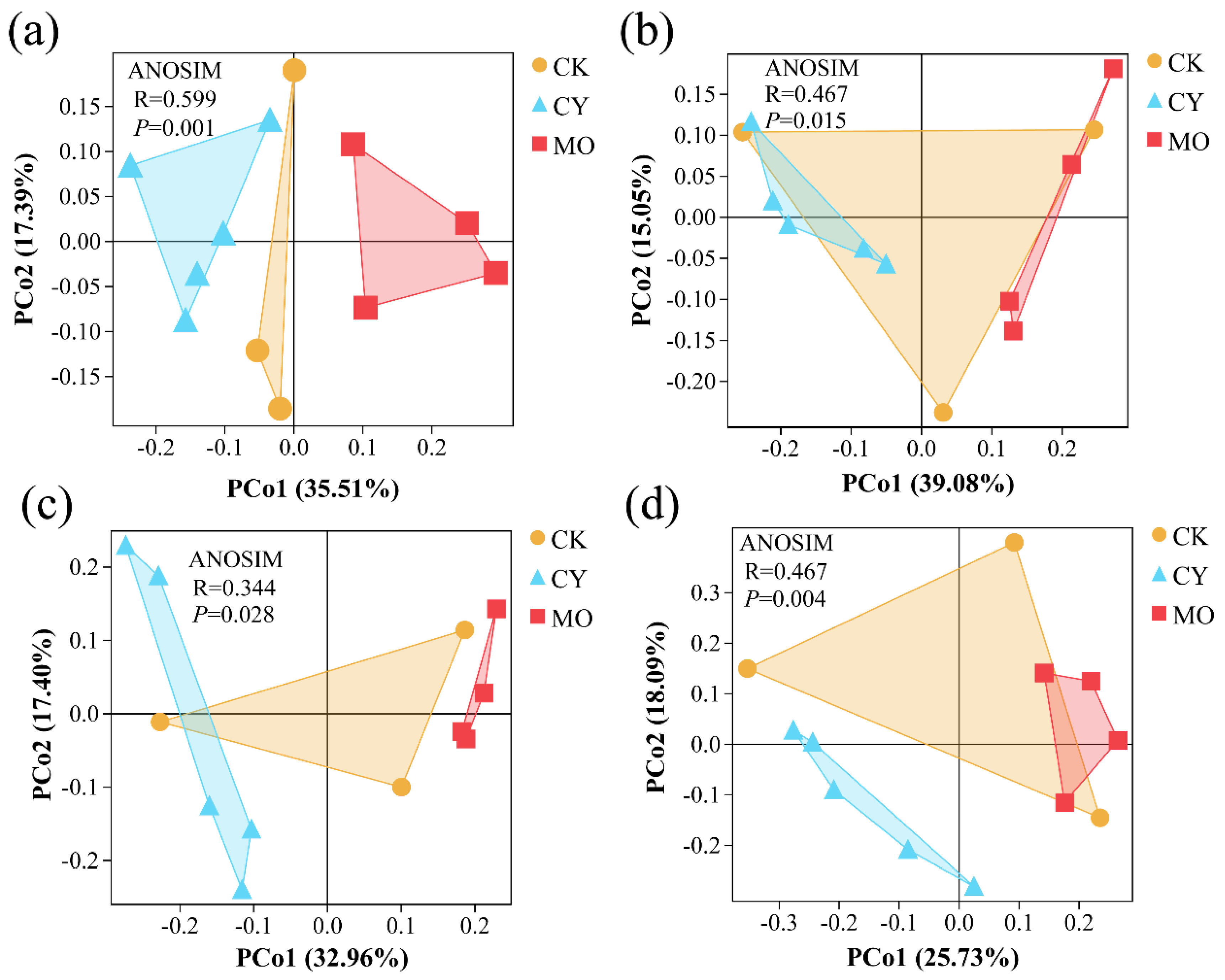
Principal coordinate analysis (PCoA) revealed that bacterial compositions differed significantly between the CY and MO groups, whereas the CK group tended to cluster together with the other two groups (Figure 4a). The same situation occurred in the compositions of phytoplankton, zooplankton, and fungi (Figure 4b–d).

3.6. Correlation Analysis between Species and Environmental Factors

According to our Mantel tests, the bacterial and zooplankton species distance was positively correlated with environmental factors (Figure S3), indicating a close relationship between water quality parameters and the species composition of bacteria and zooplankton. Furthermore, we used canonical correspondence analysis (CCA) and redundancy analysis (RDA) to display the relationship among species, groups, and environmental factors. In terms of bacterial species, the CY group was positively correlated with TN and TAN, and negatively correlated with the NPR (Figure 5a). In contrast, the MO group was positively correlated with NPR, and negatively correlated with TAN and TN (Figure 5a). Similar results were found in terms of phytoplankton and zooplankton species (Figure 5b,c). However, water quality parameters had little effect on the distribution of fungal species (Figure 5d).
Among the detected water quality parameters, TN and TAN made major contributions to the bacterial species composition (Figure S4a). Interestingly, TN also made major contributions to the zooplankton species composition (Figure S4c). In contrast, TP made major contributions to the phytoplankton and fungal species composition (Figure S4b,d).
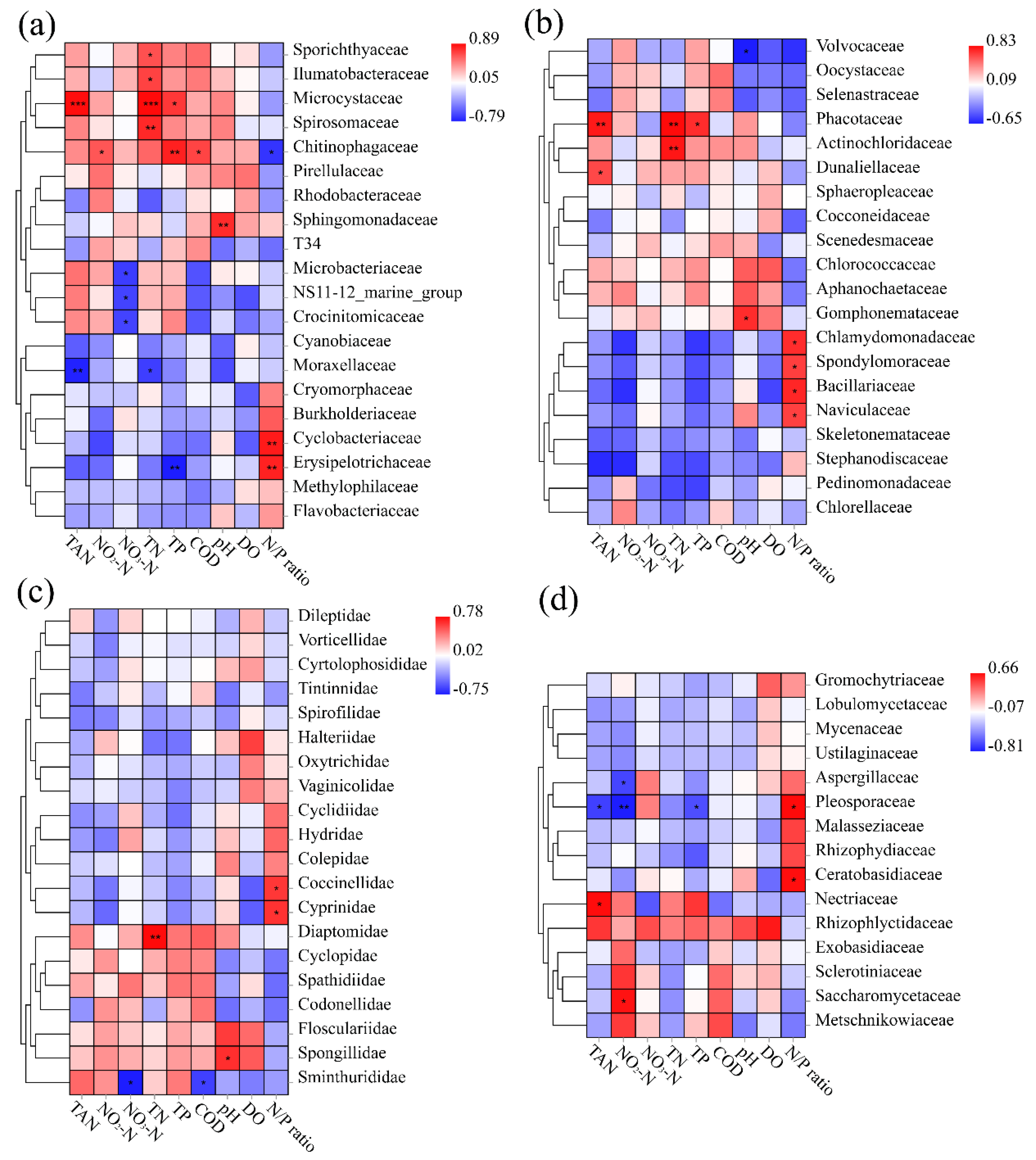
At the family level, Microcystaceae, Spirosomaceae, Ilumatobacteraceae, and Sporichthyacea were significantly positively correlated with TN, while Moraxellaceae was negatively correlated with TN (Figure 6a). Microcystaceae was also significantly positively correlated with TAN and TP. Interestingly, Chitinophagaceae and Erysipelotrichaceae were significantly positively and negatively (negatively and positively) correlated with TP (NPR), respectively (Figure 6a). In eukaryotes, Diaptomidae was significantly positively correlated with TN (Figure 6c), while Phacotaceae (Figure 6b) and Pleosporaceae (Figure 6d) were significantly positively and negatively correlated with TP, respectively. In phytoplankton, a significant positive correlation was also found between Phacotaceae and TAN/TN, Dunaliellaceae and TAN, Gomphonemataceae and pH, Chlamydomonadaceae and NPR, etc. (Figure 6b). In contrast, a significant negative correlation was only found between Volvocaceae and pH (Figure 6b).

4. Discussion

4.1. Limitations of Water Quality Indicators

In aquaculture, water quality parameters are commonly used indicators for monitoring the health of the pond ecosystem. The most used water quality indicators are dissolved oxygen, pH, temperature, nitrogen, and phosphorus nutrients. Their detection methods are simple, quick, and capable of indicating a variety of problems in ponds [31]. Similar to a previous study [32], all water quality parameters depicted good water conditions in our study; however, it is not possible to distinguish normal from cyanobacterial or “pond moss” blooms using water quality parameters. Water quality parameters provide limited information on the ongoing stress on the pond ecosystem, leaving risks to cultured species. To avoid these risks, we must screen biological indicators.

4.2. Relationship between NPR and Microalgae

Microalgae, bacteria, and fungi are central players in the cycling of nitrogen and phosphorus in the aquatic ecosystem. In return, NPR influences the function of bacteria and fungi [33], as well as the compositions of phytoplankton [34,35], which is supported by the present study. In line with previous studies [36,37], we found that cyanobacterial blooms correlated with low NPR in E. sinensis ponds, and “pond moss” occurrence correlated with high NPR. Thus, NPR manipulation seems to be an effective strategy to regulate the composition of microorganisms in aquaculture ponds. To clarify this finding, previous researchers have conducted numerous studies. To date, it is generally acknowledged that patterns of nutrient limitation in microalgae can be divided into nitrogen limitation, suitable range, and phosphorus limitation. NPR served as an easy indicator of nutrient limitation patterns.
Cyanobacteria blooms were more likely to occur under nitrogen limitation conditions, i.e., NPR below 16:1 by atoms or 7:1 by mass [38], due to their outstanding ability in acquiring nitrogen [39,40]. However, NPR seems less indicative for cyanobacteria blooms in highly eutrophic systems [41,42], raising doubts that low NPR is both a cause and result of the cyanobacteria blooms. In later studies, we awarded the concentration of phosphorus and nitrogen [43,44], as well as the type of nitrogen sources [45] that could influence the optimal NPR for microalgae. Therefore, we suggest that NPR, nitrogen, and phosphorus concentration, as well as nitrogen source types, are factors to consider in the risk assessment of algal blooms.

4.3. Biological Indicators for Algal Blooms

In this study, the bacterial species isolated from the CK group were E. indicum, a bacterium who has been reported to be suitable for Cr(VI) reduction from saline effluents [46]. In the CY group, two bacterial species and one fungal species were identified in the present study. Among these, Nitrospira sp. is a chemolithoautotrophic nitrite-oxidizing bacterium that is important in the biogeochemical nitrogen cycle and plays a number of important roles in the oxidation of the nitrite to nitrate in the nitrification process [47]. Flectobacillus sp. BAB-3569 is one of the unclassified Flectobacillus within the phylum Bacteroidetes. Fusarium oxysporum is a widely distributed large species complex of both plant and human pathogens [48]. In the MO group, one bacterial species and two fungal species were identified in the present study. The bacterial species—S. warneri, one of the most frequently found coagulase-negative staphylococci in natural waters—is an etiological agent of infections in humans and animals and can cause different types of infections ranging from superficial skin infections to severe systemic infections [49]. Interestingly, two indicator fungal species of the MO group were involved in the class Chytridiomycetes. Similar to the parasitic habits of G. mamkaevae [50], aquatic Rhizophydium sp. are mostly saprophytes and parasites [51]. In the present study, Nitrospira sp., Flectobacillus sp. BAB-3569, and S. warneri were screened as the indicator bacterial species; meanwhile, F. oxysporum, G. mamkaevae, and Rhizophydium sp. JEL317 were screened as the indicator fungal species in order to lay the foundation for the prevention of algal blooms at an early stage during E. sinensis culture.
In the present study, C. parallestriata was an indicator of phytoplankton species in the MO group, while L. monstruosa and C. raudensis were indicator phytoplankton species in the CY group, suggesting their strong ability to survive and proliferate under intense competition. This might be attributed to their efficient capacity for DNA excision repair and/or PSI-driven cyclic electron flow [52,53,54]. To clarify this finding, further studies are needed. Interestingly, these three indicator species are both green algae belonging to the order Chlamydomonadales. As evolutionary intermediates between cyanobacteria and higher plants [55], we suggested them as potential competitive inhibitors for controlling harmful algal blooms.

4.4. Strategies for Risk Prevention and Control in the Culture of E. sinensis

As reported in previous studies [56,57,58], we found a close relationship between water quality parameters and the species composition of bacteria and zooplankton in the present study. Specifically, TN and AN made major contributions to the bacterial species composition. In contrast, TP made major contributions to the phytoplankton species composition. According to the dominant families and their relationship with the water quality parameters, we suggest reducing TN, TAN, pH, and TP for the prevention and control of cyanobacteria blooms. For “pond moss” prevention and control, we suggest increasing TN slightly, while also decreasing pH and TAN.
To regulate the dominant microbial families that were less affected by water quality parameters, probiotics seem to be an effective solution [15,59,60]; for example, we might be able to use Lactogen 13 (Lactobacillus rhamnosus IMC 501) [61] and NanShuiLiSheng-03 [62] to reduce the abundance of Microbacteriaceae in cyanobacteria blooms or “pond moss” ponds. It is well documented that Chlorella vulgaris had a high removal efficiency of nutrients in aquaculture waste, such as nitrate and phosphate [63,64,65]. By effectively purifying the aquaculture water, C. vulgaris creates a good living environment for aquaculture species. In addition, dietary C. vulgaris had multiple beneficial effects on Pacific white shrimp, Litopenaeus vannamei, such as improved growth performance and tolerance of hypoxia and ammonia stress [66]. As a ubiquitous native species [67,68,69], C. vulgaris is suitable for the purification of aquaculture water and beneficial for the surrounding waters from both economic and ecological points of view. Given the interactions between microalgae and bacteria [70], it is an applicable way to regulate the pond ecosystem with a combination of probiotics and microalgae, e.g., C. vulgaris, in aquaculture [18,71].

5. Conclusions

Consistent with the macro appearance of ponds, the micro composition of normal ponds, cyanobacteria blooms ponds, and “pond moss” ponds are different, especially for the latter two groups. In addition to the normal ponds for E. sinensis cultivation, we identified the compositions of microbial and microalgae communities of ponds that erupted in harmful algal blooms. We screened Nitrospira sp., Flectobacillus sp. BAB-3569, S. warneri, F. oxysporum, G. mamkaevae, and Rhizophydium sp. JEL317 as bioindicators for harmful algal blooms. Based on the close relationships between water quality parameters and the species composition of bacteria and zooplankton, we suggested that NPR, nitrogen, and phosphorus concentration, as well as nitrogen sources types, are factors driving algal blooms, and we have indicated possible measures to prevent harmful algal blooms. We suggested regulating the pond ecosystem with a combination of probiotics and microalgae, e.g., C. vulgaris, to prevent and control potential risks in the culture of E. sinensis.

Supplementary Materials

The following supporting information can be downloaded at: https://www.mdpi.com/article/10.3390/fishes7040180/s1, Figure S1: Relative abundance (%) of dominant communities within (a) bacterial, (b) phytoplankton, (c) zooplankton, and (d) fungal phyla in each group; Figure S2: Dilution curve of each sample and OTU rank abundance curve of each group. (a) and (c) 16s rDNA, (b) and (d) 18S rDNA; Figure S3: Mantel tests on environmental distance and (a) bacterial, (b) phytoplankton, (c) zooplankton and (d) fungal species distance Bbray–Curtis distance); Figure S4: Variance partitioning analysis of the contribution of environmental factors to the distribution differences of (a) bacterial, (b) phytoplankton, (c) zooplankton and (d) fungal families; Table S1: Information for 12 sampling ponds. CK denoted normal group, CY denoted cyanobacteria bloom group, and MO denoted pond moss group; Table S2: The number of tags and OUTs of 12 procaryotic and 12 eukaryotic sequencing libraries; Table S3: Basic information of 12 procaryotic and 12 eukaryotic sequencing libraries; Table S4: The common and specific species of each group. (a) bacteria, (b) phytoplankton, (c) zooplankton, and (d) fungi.

Author Contributions

Conceptualization, Writing—Original draft preparation & Funding acquisition, J.G.; Visualization & Investigation, L.S.; Investigation, Z.N.; Formal analysis, H.Z.; Data curation, L.C.; Resources, J.D. and F.D.; Funding acquisition, Supervision &Writing—review & editing, G.X. All authors contributed to manuscript writing and approved the final version. All authors have read and agreed to the published version of the manuscript.

Funding

This work was supported by “JBGS” Project of Seed Industry Revitalization in Jiangsu Province [JBGS [2021]125]; and the Central Public-interest Scientific Institution Basal Research Fund, CAFS [NO. 2021XT0701].

Institutional Review Board Statement

Not applicable.

Informed Consent Statement

Not applicable.

Data Availability Statement

The datasets generated and/or analyzed for the current study are available from the corresponding author on reasonable request.

Conflicts of Interest

There is no conflict of interest in the present study.

References

  1. Fishery Administration Bureau of the Ministry of Agriculture and Villages; National Aquatic Products Technology Extension Station; China Society of Fisheries. 2021 China Fishery Statistical Yearbook; Chinese Agricultural Press: Beijing, China, 2021; Volume 34. [Google Scholar]
  2. Gao, J.; Tai, X.; Shao, N.; Sun, Y.; Nie, Z.; Wang, Y.; Li, Q.; Xu, P.; Xu, G. Effects of effective microorganisms on the growth performance, nutritional composition and flavour quality of the pond-cultured Eriocheir sinensis. Aquac. Res. 2021, 52, 871–880. [Google Scholar] [CrossRef]
  3. Dai, H.; Sun, Y.; Ren, N.; Lu, X. Investigation of Chinese hairy crab industry and analysis of development strategies. Jiangsu Agric. Sci. 2021, 49, 248–252. [Google Scholar] [CrossRef]
  4. Ma, X.; Ge, J.; Wang, Z. Study on changes and regulation of ammonia nitrogen and nitrite in Eriocheir sinensis culture. J. Aquac. 2020, 41, 43–45. [Google Scholar]
  5. Chi, C.; Yu, X.-W.; Zhang, C.-Y.; Liu, J.-D.; Ye, M.-W.; Zhang, D.-D.; Liu, W.-B. Acute exposure to microcystin-LR induces hepatopancreas toxicity in the Chinese mitten crab (Eriocheir sinensis). Arch. Toxicol. 2021, 95, 2551–2570. [Google Scholar] [CrossRef]
  6. Hasan, M.R.; Chakrabarti, R. Use of Algae and Aquatic Macrophytes as Feed in Small-Scale Aquaculture: A Review. In FAO Fisheries and Aquaculture Technical Paper No. 531; FAO: Rome, Italy, 2009; 123p. [Google Scholar]
  7. Cannell, R.J.; Farmer, P.; Walker, J.M. Purification and characterization of pentagalloylglucose, and alpha-glucosidase inhibitor/antibiotic from the freshwater green alga Spirogyra varians. Biochem. J. 1988, 255, 937–941. [Google Scholar] [CrossRef]
  8. Weber, J.; Schagerl, M. Strategies of Spirogyra against epiphytes. Algol. Stud. 2007, 123, 57–72. [Google Scholar] [CrossRef]
  9. Mao, G.; Tang, Y. Causes, hazards and prevention of pond moss in crab pond. Sci. Fish Farming 2016, 12, 61–62. [Google Scholar] [CrossRef]
  10. Huisman, J.; Codd, G.A.; Paerl, H.W.; Ibelings, B.W.; Verspagen, J.M.H.; Visser, P.M. Cyanobacterial blooms. Nat. Rev. Microbiol. 2018, 16, 471–483. [Google Scholar] [CrossRef]
  11. Zhang, Q.; Zhang, Z.; Lu, T.; Peijnenburg, W.J.G.M.; Gillings, M.; Yang, X.; Chen, J.; Penuelas, J.; Zhu, Y.-G.; Zhou, N.-Y.; et al. Cyanobacterial blooms contribute to the diversity of antibiotic-resistance genes in aquatic ecosystems. Commun. Biol. 2020, 3, 737. [Google Scholar] [CrossRef]
  12. Havens, K.E. Cyanobacteria blooms: Effects on aquatic ecosystems. Adv. Exp. Med. Biol. 2008, 619, 733–747. [Google Scholar] [CrossRef]
  13. Wu, J.X.; Huang, H.; Yang, L.; Zhang, X.F.; Zhang, S.S.; Liu, H.H.; Wang, Y.Q.; Yuan, L.; Cheng, X.M.; Zhuang, D.G.; et al. Gastrointestinal toxicity induced by microcystins. World J. Clin. Cases 2018, 6, 344–354. [Google Scholar] [CrossRef] [PubMed]
  14. Chen, X.; Pan, L.; Zhang, Y. Formation and countermeasures of harmful filamentous algae in water. South China Fish. Sci. 2011, 7, 77–81. [Google Scholar] [CrossRef]
  15. Martínez Cruz, P.; Ibáñez, A.L.; Monroy Hermosillo, O.A.; Ramírez Saad, H.C. Use of probiotics in aquaculture. ISRN Microbiol. 2012, 2012, 916845. [Google Scholar] [CrossRef] [PubMed] [Green Version]
  16. Wang, A.; Ran, C.; Wang, Y.; Zhang, Z.; Ding, Q.; Yang, Y.; Olsen, R.E.; Ringø, E.; Bindelle, J.; Zhou, Z. Use of probiotics in aquaculture of China-a review of the past decade. Fish Shellfish. Immunol. 2019, 86, 734–755. [Google Scholar] [CrossRef]
  17. Kuebutornye, F.K.A.; Abarike, E.D.; Lu, Y. A review on the application of Bacillus as probiotics in aquaculture. Fish Shellfish Immunol. 2019, 87, 820–828. [Google Scholar] [CrossRef]
  18. Ahmad, M.T.; Shariff, M.; Yusoff, F.M.; Goh, Y.M.; Banerjee, S. Applications of microalga Chlorella vulgaris in aquaculture. Rev. Aquac. 2020, 12, 328–346. [Google Scholar] [CrossRef]
  19. Sathasivam, R.; Radhakrishnan, R.; Hashem, A.; Abd_Allah, E.F. Microalgae metabolites: A rich source for food and medicine. Saudi J. Biol. Sci. 2019, 26, 709–722. [Google Scholar] [CrossRef]
  20. Chen, S.; Zhou, Y.; Chen, Y.; Gu, J. fastp: An ultra-fast all-in-one FASTQ preprocessor. Bioinformatics 2018, 34, i884–i890. [Google Scholar] [CrossRef]
  21. Magoč, T.; Salzberg, S.L. FLASH: Fast length adjustment of short reads to improve genome assemblies. Bioinformatics 2011, 27, 2957–2963. [Google Scholar] [CrossRef]
  22. Bokulich, N.A.; Subramanian, S.; Faith, J.J.; Gevers, D.; Gordon, J.I.; Knight, R.; Mills, D.A.; Caporaso, J.G. Quality-filtering vastly improves diversity estimates from Illumina amplicon sequencing. Nat. Methods 2013, 10, 57–59. [Google Scholar] [CrossRef]
  23. Edgar, R.C. UPARSE: Highly accurate OTU sequences from microbial amplicon reads. Nat. Methods 2013, 10, 996–998. [Google Scholar] [CrossRef] [PubMed]
  24. Edgar, R.C.; Haas, B.J.; Clemente, J.C.; Quince, C.; Knight, R. UCHIME improves sensitivity and speed of chimera detection. Bioinformatics 2011, 27, 2194–2200. [Google Scholar] [CrossRef] [PubMed] [Green Version]
  25. Wang, Q.; Garrity, G.M.; Tiedje, J.M.; Cole, J.R. Naive Bayesian classifier for rapid assignment of rRNA sequences into the new bacterial taxonomy. Appl. Environ. Microbiol. 2007, 73, 5261–5267. [Google Scholar] [CrossRef] [PubMed] [Green Version]
  26. Pruesse, E.; Quast, C.; Knittel, K.; Fuchs, B.M.; Ludwig, W.; Peplies, J.; Glöckner, F.O. SILVA: A comprehensive online resource for quality checked and aligned ribosomal RNA sequence data compatible with ARB. Nucleic Acids Res. 2007, 35, 7188–7196. [Google Scholar] [CrossRef] [Green Version]
  27. Edgar, R.C. MUSCLE: Multiple sequence alignment with high accuracy and high throughput. Nucleic Acids Res. 2004, 32, 1792–1797. [Google Scholar] [CrossRef] [PubMed] [Green Version]
  28. Price, M.N.; Dehal, P.S.; Arkin, A.P. FastTree 2—Approximately maximum-likelihood trees for large alignments. PLoS ONE 2010, 5, e9490. [Google Scholar] [CrossRef] [PubMed]
  29. Lozupone, C.; Knight, R. UniFrac: A new phylogenetic method for comparing microbial communities. Appl. Environ. Microbiol. 2005, 71, 8228–8235. [Google Scholar] [CrossRef] [PubMed] [Green Version]
  30. Revelle, W. psych: Procedures for Psychological, Psychometric, and Personality Research; Northwestern University: Evanston, IL, USA, 2021. [Google Scholar]
  31. Hu, Z.; Li, R.; Xia, X.; Yu, C.; Fan, X.; Zhao, Y. A method overview in smart aquaculture. Environ. Monit. Assess. 2020, 192, 493. [Google Scholar] [CrossRef]
  32. Cai, C.; Gu, X.; Huang, H.; Dai, X.; Ye, Y.; Shi, C. Water quality, nutrient budget, and pollutant loads in Chinese mitten crab (Eriocheir sinensis) farms around East Taihu Lake. Chin. J. Oceanol. Limnol. 2012, 30, 29–36. [Google Scholar] [CrossRef]
  33. Güsewell, S.; Gessner, M.O. N: P ratios influence litter decomposition and colonization by fungi and bacteria in microcosms. Funct. Ecol. 2009, 23, 211–219. [Google Scholar] [CrossRef]
  34. Rhee, G.-Y.; Gotham, I.J. Optimum N:P ratios and coexistence of planktonic algae. J. Phycol. 1980, 16, 486–489. [Google Scholar] [CrossRef]
  35. Rasdi, N.W.; Qin, J.G. Effect of N:P ratio on growth and chemical composition of Nannochloropsis oculata and Tisochrysis lutea. J. Appl. Phycol. 2015, 27, 2221–2230. [Google Scholar] [CrossRef]
  36. Palus, J. Effects of N:P Ratio on the Occurrence of Harmful Algal Blooms. BSc Thesis, The Ohio State University, Columbus, OH, USA, 2015. [Google Scholar]
  37. Kim, H.-S.; Hwang, S.-J.; Shin, J.-K.; An, K.-G.; Yoon, C.G. Effects of limiting nutrients and N:P ratios on the phytoplankton growth in a shallow hypertrophic reservoir. Hydrobiologia 2007, 581, 255–267. [Google Scholar] [CrossRef]
  38. Falkowski, P.G.; Davis, C.S. Natural proportions. Nature 2004, 431, 131. [Google Scholar] [CrossRef]
  39. Bergström, A.-K. The use of TN:TP and DIN:TP ratios as indicators for phytoplankton nutrient limitation in oligotrophic lakes affected by N deposition. Aquat. Sci. 2010, 72, 277–281. [Google Scholar] [CrossRef]
  40. Paerl, H.W.; Xu, H.; Hall, N.S.; Zhu, G.; Qin, B.; Wu, Y.; Rossignol, K.L.; Dong, L.; McCarthy, M.J.; Joyner, A.R. Controlling cyanobacterial blooms in hypertrophic Lake Taihu, China: Will nitrogen reductions cause replacement of non-N2 fixing by N2 fixing taxa? PLoS ONE 2014, 9, e113123. [Google Scholar] [CrossRef] [Green Version]
  41. Paerl, H.W.; Fulton, R.S., 3rd; Moisander, P.H.; Dyble, J. Harmful freshwater algal blooms, with an emphasis on cyanobacteria. Sci. J. 2001, 1, 76–113. [Google Scholar] [CrossRef]
  42. Xie, L.; Xie, P.; Li, S.; Tang, H.; Liu, H. The low TN:TP ratio, a cause or a result of Microcystis blooms? Water Res. 2003, 37, 2073–2080. [Google Scholar] [CrossRef]
  43. Liu, Y.; Li, L.; Jia, R. The Optimum Resource Ratio (N:P) for the Growth of Microcystis Aeruginosa with Abundant Nutrients. Procedia Environ. Sci. 2011, 10, 2134–2140. [Google Scholar] [CrossRef] [Green Version]
  44. Zhang, Q.; Hu, G. Effect of nitrogen to phosphorus ratios on cell proliferation in marine micro algae. Chin. J. Oceanol. Limnol. 2011, 29, 739–745. [Google Scholar] [CrossRef]
  45. Podevin, M.; De Francisci, D.; Holdt, S.L.; Angelidaki, I. Effect of nitrogen source and acclimatization on specific growth rates of microalgae determined by a high-throughput in vivo microplate autofluorescence method. J. Appl. Phycol. 2015, 27, 1415–1423. [Google Scholar] [CrossRef] [Green Version]
  46. Mohapatra, R.K.; Parhi, P.K.; Thatoi, H.; Panda, C.R. Bioreduction of hexavalent chromium by Exiguobacterium indicum strain MW1 isolated from marine water of Paradip Port, Odisha, India. Chem. Ecol. 2017, 33, 114–130. [Google Scholar] [CrossRef]
  47. Daims, H.; Wagner, M. Nitrospira. Trends Microbiol. 2018, 26, 462–463. [Google Scholar] [CrossRef] [PubMed]
  48. Zhang, Y.; Ma, L.-J. Chapter Five—Deciphering Pathogenicity of Fusarium oxysporum from a Phylogenomics Perspective. In Advances in Genetics; Townsend, J.P., Wang, Z., Eds.; Academic Press: Cambridge, MA, USA, 2017; Volume 100, pp. 179–209. [Google Scholar]
  49. Silva, V.; Caniça, M.; Capelo, J.L.; Igrejas, G.; Poeta, P. Diversity and genetic lineages of environmental staphylococci: A surface water overview. FEMS Microbiol. Ecol. 2020, 96, fiaa191. [Google Scholar] [CrossRef]
  50. Karpov, S.A.; Kobseva, A.A.; Mamkaeva, M.A.; Mamkaeva, K.A.; Mikhailov, K.V.; Mirzaeva, G.S.; Aleoshin, V.V. Gromochytrium mamkaevae gen. & sp. nov. and two new orders: Gromochytriales and Mesochytriales (Chytridiomycetes). Persoonia 2014, 32, 115–126. [Google Scholar] [CrossRef] [Green Version]
  51. Letcher, P.M.; Vélez, C.G.; Barrantes, M.E.; Powell, M.J.; Churchill, P.F.; Wakefield, W.S. Ultrastructural and molecular analyses of Rhizophydiales (Chytridiomycota) isolates from North America and Argentina. Mycol. Res. 2008, 112, 759–782. [Google Scholar] [CrossRef]
  52. Chaudhari, V.R.; Vyawahare, A.; Bhattacharjee, S.K.; Rao, B.J. Enhanced excision repair and lack of PSII activity contribute to higher UV survival of Chlamydomonas reinhardtii cells in dark. Plant Physiol. Biochem. 2015, 88, 60–69. [Google Scholar] [CrossRef]
  53. Dolhi, J.M.; Maxwell, D.P.; Morgan-Kiss, R.M. Review: The Antarctic Chlamydomonas raudensis: An emerging model for cold adaptation of photosynthesis. Extremophiles 2013, 17, 711–722. [Google Scholar] [CrossRef]
  54. Stahl-Rommel, S.; Kalra, I.; D’Silva, S.; Hahn, M.M.; Popson, D.; Cvetkovska, M.; Morgan-Kiss, R.M. Cyclic electron flow (CEF) and ascorbate pathway activity provide constitutive photoprotection for the photopsychrophile, Chlamydomonas sp. UWO 241 (renamed Chlamydomonas priscuii). Photosynth. Res. 2022, 151, 235–250. [Google Scholar] [CrossRef]
  55. Allahverdiyeva, Y.; Suorsa, M.; Tikkanen, M.; Aro, E.M. Photoprotection of photosystems in fluctuating light intensities. J. Exp. Bot. 2015, 66, 2427–2436. [Google Scholar] [CrossRef]
  56. Xu, Y.; Li, A.J.; Qin, J.; Li, Q.; Ho, J.G.; Li, H. Seasonal patterns of water quality and phytoplankton dynamics in surface waters in Guangzhou and Foshan, China. Sci. Total Environ. 2017, 590–591, 361–369. [Google Scholar] [CrossRef] [PubMed]
  57. Abirhire, O.; North, R.L.; Hunter, K.; Vandergucht, D.M.; Sereda, J.; Hudson, J.J. Environmental factors influencing phytoplankton communities in Lake Diefenbaker, Saskatchewan, Canada. J. Great Lakes Res. 2015, 41, 118–128. [Google Scholar] [CrossRef]
  58. Catherine, A.; Selma, M.; Mouillot, D.; Troussellier, M.; Bernard, C. Patterns and multi-scale drivers of phytoplankton species richness in temperate peri-urban lakes. Sci. Total Environ. 2016, 559, 74–83. [Google Scholar] [CrossRef] [PubMed]
  59. Romano, N. Chapter 5—Probiotics, prebiotics, biofloc systems, and other biocontrol regimens in fish and shellfish aquaculture. In Aquaculture Pharmacology; Kibenge, F.S.B., Baldisserotto, B., Chong, R.S.-M., Eds.; Academic Press: Cambridge, MA, USA, 2021; pp. 219–242. [Google Scholar]
  60. Sun, R.; Sun, P.; Zhang, J.; Esquivel-Elizondo, S.; Wu, Y. Microorganisms-based methods for harmful algal blooms control: A review. Bioresour. Technol. 2018, 248, 12–20. [Google Scholar] [CrossRef] [PubMed]
  61. Gioacchini, G.; Ciani, E.; Pessina, A.; Cecchini, C.; Silvi, S.; Rodiles, A.; Merrifield, D.L.; Olivotto, I.; Carnevali, O. Effects of Lactogen 13, a new probiotic preparation, on gut microbiota and endocrine signals controlling growth and appetite of Oreochromis niloticus Juveniles. Microb. Ecol. 2018, 76, 1063–1074. [Google Scholar] [CrossRef] [Green Version]
  62. Wang, R.; Guo, Z.; Tang, Y.; Kuang, J.; Duan, Y.; Lin, H.; Jiang, S.; Shu, H.; Huang, J. Effects on development and microbial community of shrimp Litopenaeus vannamei larvae with probiotics treatment. AMB Express 2020, 10, 109. [Google Scholar] [CrossRef]
  63. Hesni, M.A.; Hedayati, A.; Qadermarzi, A.; Pouladi, M.; Zangiabadi, S.; Naqshbandi, N. Using Chlorella vulgaris and iron oxide nanoparticles in a designed bioreactor for aquaculture effluents purification. Aquac. Eng. 2020, 90, 102069. [Google Scholar] [CrossRef]
  64. Tejido-Nuñez, Y.; Aymerich, E.; Sancho, L.; Refardt, D. Treatment of aquaculture effluent with Chlorella vulgaris and Tetradesmus obliquus: The effect of pretreatment on microalgae growth and nutrient removal efficiency. Ecol. Eng. 2019, 136, 1–9. [Google Scholar] [CrossRef]
  65. Daneshvar, E.; Antikainen, L.; Koutra, E.; Kornaros, M.; Bhatnagar, A. Investigation on the feasibility of Chlorella vulgaris cultivation in a mixture of pulp and aquaculture effluents: Treatment of wastewater and lipid extraction. Bioresour. Technol. 2018, 255, 104–110. [Google Scholar] [CrossRef]
  66. Pakravan, S.; Akbarzadeh, A.; Sajjadi, M.M.; Hajimoradloo, A.; Noori, F. Chlorella vulgaris meal improved growth performance, digestive enzyme activities, fatty acid composition and tolerance of hypoxia and ammonia stress in juvenile Pacific white shrimp Litopenaeus vannamei. Aquac. Nutr. 2018, 24, 594–604. [Google Scholar] [CrossRef]
  67. Hodač, L.; Hallmann, C.; Spitzer, K.; Elster, J.; Faßhauer, F.; Brinkmann, N.; Lepka, D.; Diwan, V.; Friedl, T. Widespread green algae Chlorella and Stichococcus exhibit polar-temperate and tropical-temperate biogeography. FEMS Microbiol. Ecol. 2016, 92, fiw122. [Google Scholar] [CrossRef] [PubMed] [Green Version]
  68. Huang, Y.; Shen, Y.; Zhang, S.; Li, Y.; Sun, Z.; Feng, M.; Li, R.; Zhang, J.; Tian, X.; Zhang, W. Characteristics of phytoplankton community structure and indication to water quality in the lake in agricultural areas. Front. Environ. Sci. 2022, 10, 833409. [Google Scholar] [CrossRef]
  69. Zhang, Y.; Gao, W.; Li, Y.; Jiang, Y.; Chen, X.; Yao, Y.; Messyasz, B.; Yin, K.; He, W.; Chen, Y. Characteristics of the phytoplankton community structure and water quality evaluation in autumn in the Huaihe river (China). Int. J. Environ. Res. Public Health 2021, 18, 12092. [Google Scholar] [CrossRef]
  70. Fuentes, J.L.; Garbayo, I.; Cuaresma, M.; Montero, Z.; González-Del-Valle, M.; Vílchez, C. Impact of microalgae-bacteria interactions on the production of algal biomass and associated compounds. Mar. Drugs 2016, 14, 100. [Google Scholar] [CrossRef] [PubMed] [Green Version]
  71. Hasan, K.N.; Banerjee, G. Recent studies on probiotics as beneficial mediator in aquaculture: A review. J. Basic Appl. Zool. 2020, 81, 53. [Google Scholar] [CrossRef]
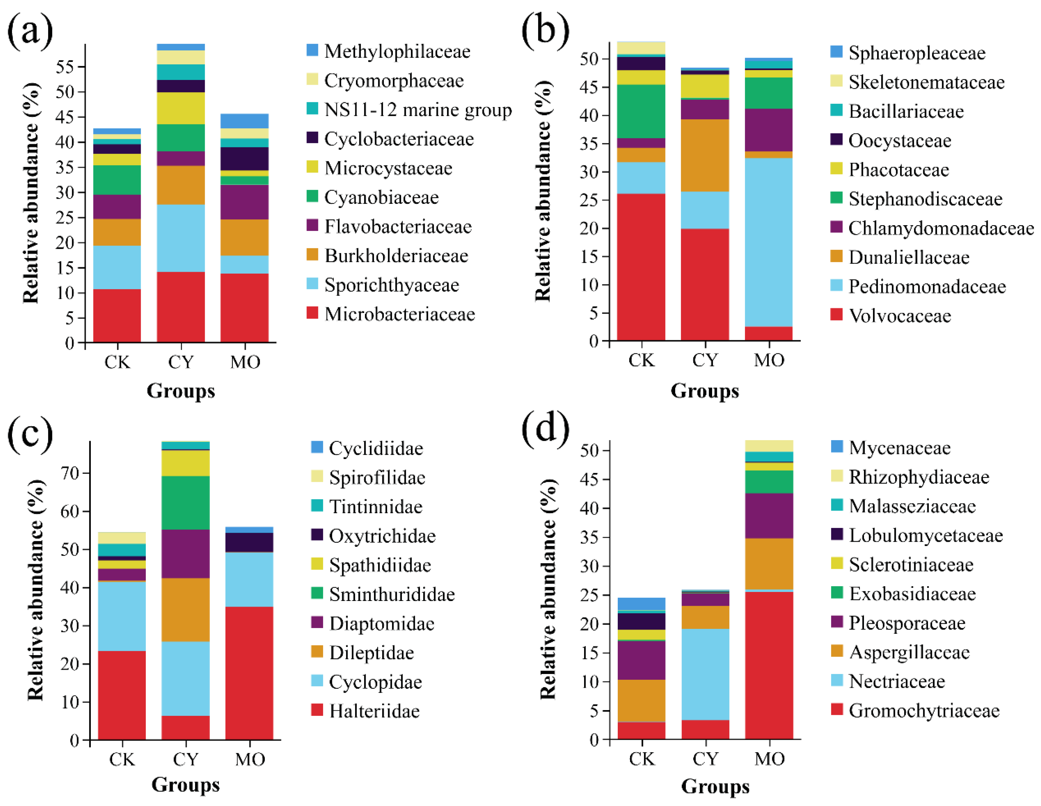
Figure 1. Relative abundance of top 10 dominant (a) bacterial, (b) phytoplankton, (c) zooplankton, and (d) fungal families in each group.
Fishes 07 00180 g001
Figure 2. Venn diagram analysis of the common and specific species of each group: (a) bacteria, (b) phytoplankton, (c) zooplankton, and (d) fungi.
Fishes 07 00180 g002
Figure 3. Indicator species of (a) bacteria, (b) phytoplankton, (c) zooplankton, and (d) fungi in each group.
Fishes 07 00180 g003
Figure 4. PCoA analysis and an ANOSIM test of the differences among (a) bacterial, (b) phytoplankton, (c) zooplankton, and (d) fungal communities in different groups based on OTU level. Distance between bacterial communities was calculated using the weighted-unifrac method, while distance between phytoplankton, zooplankton, and fungal communities was calculated using the unweighted-unifrac method.
Fishes 07 00180 g004
Figure 5. Correlation analysis of environmental factors and the relative abundance of top 20 abundant (a) bacterial, (b) phytoplankton, (c) zooplankton, and (d) fungal species.
Fishes 07 00180 g005
Figure 6. Correlation between environmental factors and families’ abundance of (a) bacteria, (b) phytoplankton, (c) zooplankton and (d) fungi. * represents p value < 0.05, ** represents p value < 0.01, and *** represents p value < 0.001.
Fishes 07 00180 g006
Table 1. Description statistic of water quality parameters and their variations across different groups. CK denoted normal group, CY denoted cyanobacteria bloom group, and MO denoted pond moss group. Values are presented as means ± SD. Statistically significant differences are indicated by different letters (p < 0.05).
GroupCKCYMO
Total ammonia nitrogen (TAN)0.10 ± 0.13 ab0.37 ± 0.04 a0.09 ± 0.04 b
Nitrite nitrogen (NO2-N)0.002 ± 0.0010.002 ± 0.0010.002 ± 0.001
Nitrate nitrogen (NO3-N)0.22 ± 0.020.21 ± 0.060.21 ± 0.02
Total nitrogen (TN)0.88 ± 0.41 b1.82 ± 0.58 a0.77 ± 0.20 b
Total phosphorus (TP)0.16 ± 0.08 ab0.22 ± 0.01 a0.06 ± 0.01 b
Chemical oxygen demand (COD)9.66 ± 0.759.32 ± 1.269.04 ± 0.29
pH9.37 ± 1.089.50 ± 0.669.40 ± 0.29
Dissolved oxygen (DO)12.29 ± 3.7010.19 ± 3.4710.70 ± 4.10
N/P ratio (NPR)6.41 ± 2.86 b8.31 ± 2.54 ab14.32 ± 4.62 a
Table 2. The distribution of annotated tags in different taxonomic classifications.
Sequencing TypeSample IDDomainPhylumClassOrderFamilyGenusSpecies
(a) 16S rDNACK167,24166,492 (98.89%)66,271 (98.56%)61,369 (91.27%)54,342 (80.82%)39,314 (58.47%)7641 (11.36%)
CK2115,727115,348 (99.67%)115,062 (99.43%)104,470 (90.27%)89,308 (77.17%)71,261 (61.58%)7902 (6.83%)
CK394,08393,649 (99.54%)93,369 (99.24%)88,040 (93.58%)71,858 (76.38%)44,265 (47.05%)5299 (5.63%)
CKmean92,350.3391,829.67 (99.44%)91,567.33 (99.15%)84,626.33 (91.64%)71,836.00 (77.79%)51,613.33 (55.89%)6947.33 (7.52%)
CY196,21695,995 (99.77%)94,320 (98.03%)88,079 (91.54%)82,684 (85.94%)53,475 (55.58%)9955 (10.35%)
CY264,23764,025 (99.67%)63,920 (99.51%)58,677 (91.34%)48,519 (75.53%)37,198 (57.91%)7498 (11.67%)
CY370,03969,689 (99.50%)69,554 (99.31%)66,061 (94.32%)63,490 (90.65%)47,345 (67.60%)10,991 (15.69%)
CY469,55869,290 (99.61%)69,258 (99.57%)66,936 (96.23%)60,453 (86.91%)43,103 (61.97%)1585 (2.28%)
CY558,45058,015 (99.26%)57,924 (99.10%)55,798 (95.46%)50,875 (87.04%)36,710 (62.81%)2370 (4.05%)
CYmean71,700.0071,402.80 (99.59%)70,995.20 (99.02%)67,110.20 (93.60%)61,204.20 (85.36%)43,566.20 (60.76%)6479.80 (9.04%)
MO1104,543104,253 (99.72%)103,999 (99.48%)95,491 (91.34%)76,683 (73.35%)50,391 (48.20%)3000 (2.87%)
MO279,86779,101 (99.04%)78,843 (98.72%)72,527 (90.81%)58,087 (72.73%)39,720 (49.73%)7101 (8.89%)
MO3101,091100,759 (99.67%)100,609 (99.52%)88,947 (87.99%)72,576 (71.79%)53,235 (52.66%)12,908 (12.77%)
MO4104,645104,255 (99.63%)103,888 (99.28%)94,707 (90.50%)75,193 (71.86%)51,555 (49.27%)4865 (4.65%)
MOmean97,536.5097,092.00 (99.54%)96,834.75 (99.28%)87,918.00 (90.14%)70,634.75 (72.42%)48,725.25 (49.96%)6968.50 (7.14%)
(b) 18S rDNACK181,12175,557 (93.14%)67,328 (83.00%)48,553 (59.85%)39,688 (48.92%)34,071 (42.00%)22,643 (27.91%)
CK298,33597,345 (98.99%)93,351 (94.93%)79,612 (80.96%)62,979 (64.05%)58,996 (59.99%)50,642 (51.50%)
CK3105,828104,404 (98.65%)100,742 (95.19%)87,007 (82.22%)77,651 (73.37%)66,507 (62.84%)57,388 (54.23%)
CKmean95,094.6792,435.33 (97.20%)87,140.33 (91.64%)71,724.00 (75.42%)60,106.00 (63.21%)53,191.33 (55.94%)43,557.67 (45.80%)
CY1108,732107,926 (99.26%)105,512 (97.04%)89,218 (82.05%)84,700 (77.90%)82,683 (76.04%)77,331 (71.12%)
CY260,59559,973 (98.97%)58,800 (97.04%)54,832 (90.49%)35,878 (59.21%)32,964 (54.40%)6093 (10.06%)
CY3108,869105,713 (97.10%)97,828 (89.86%)89,936 (82.61%)87,373 (80.26%)83,170 (76.39%)78,928 (72.50%)
CY4112,691111,388 (98.84%)99,026 (87.87%)83,779 (74.34%)55,383 (49.15%)50,548 (44.86%)25,269 (22.42%)
CY597,82896,991 (99.14%)61,628 (63.00%)58,405 (59.70%)51,231 (52.37%)47,761 (48.82%)42,149 (43.08%)
CYmean97,743.0096,398.20 (98.62%)84,558.80 (86.51%)75,234.00 (76.97%)62,913.00 (64.37%)59,425.20 (60.80%)45,954.00 (47.02%)
MO196,82895,199 (98.32%)78,525 (81.10%)69,429 (71.70%)62,588 (64.64%)43,185 (44.60%)20,838 (21.52%)
MO299,28897,175 (97.87%)91,076 (91.73%)71,515 (72.03%)69,638 (70.14%)64,130 (64.59%)14,049 (14.15%)
MO3108,951107,673 (98.83%)105,916 (97.21%)84,000 (77.10%)72,832 (66.85%)66,802 (61.31%)56,694 (52.04%)
MO487,86484,904 (96.63%)81,274 (92.50%)71,157 (80.99%)34,887 (39.71%)24,659 (28.06%)16,340 (18.60%)
MOmean98,232.7596,237.75 (97.97%)89,197.75 (90.80%)74,025.25 (75.36%)59,986.25 (61.07%)49,694.00 (50.59%)26,980.25 (27.47%)
Table 3. Descriptive analysis and difference significance test of alpha diversity for each group.
IndexCKCYMO
(a) bacteriaSpecies richness1514.00 ± 663.521269.00 ± 107.041439.00 ± 493.03
Shannon7.35 ± 0.166.68 ± 0.407.16 ± 0.53
Simpson (×10−2)97.79 ± 0.1197.07 ± 1.0897.56 ± 0.84
Chao11609.38 ± 626.191375.82 ± 112.651535.81 ± 459.37
Ace1635.49 ± 625.401414.71 ± 119.701539.02 ± 469.03
Good’s coverage (×10−2)99.79 ± 0.1199.67 ± 0.1099.84 ± 0.05
Pielou’s evenness (×10−2)70.33 ± 2.8264.85 ± 3.7368.60 ± 3.08
PD-whole tree212.72 ± 98.17184.27 ± 15.49196.83 ± 71.28
(b) phytoplanktonSpecies richness117.67 ± 10.97b146.80 ± 17.54a117.75 ± 19.75b
Shannon4.38 ± 0.704.10 ± 1.153.96 ± 0.97
Simpson (×10−2)89.18 ± 7.5684.63 ± 12.5180.68 ± 15.07
Chao1147.31 ± 6.60169.45 ± 15.95137.03 ± 22.86
Ace152.99 ± 8.15168.17 ± 17.39138.09 ± 21.90
Good’s coverage (×10−2)99.67 ± 0.0899.66 ± 0.0899.75 ± 0.06
Pielou’s evenness (×10−2)63.91 ± 11.3356.74 ± 15.0457.49 ± 12.62
PD-whole tree9.85 ± 1.1910.66 ± 0.8910.24 ± 1.10
(c) zooplanktonSpecies richness56.33 ± 5.1362.20 ± 12.5855.00 ± 6.00
Shannon3.52 ± 0.562.87 ± 0.772.81 ± 0.27
Simpson (×10−2)82.95 ± 7.5172.85 ± 13.7276.23 ± 5.55
Chao161.72 ± 6.4064.75 ± 13.0363.60 ± 7.22
Ace65.55 ± 8.0966.85 ± 11.5361.05 ± 5.88
Good’s coverage (×10−2)99.92 ± 0.0299.93 ± 0.0199.92 ± 0.01
Pielou’s evenness (×10−2)60.42 ± 8.1548.04 ± 11.5448.87 ± 6.04
PD-whole tree6.92 ± 0.677.68 ± 1.507.22 ± 0.62
(d) fungiSpecies richness43.67 ± 13.8050.60 ± 7.9646.00 ± 9.59
Shannon4.52 ± 0.703.71 ± 0.973.91 ± 0.88
Simpson (×10−2)93.89 ± 3.0382.97 ± 11.9984.57 ± 14.46
Chao151.53 ± 12.6161.51 ± 10.6953.83 ± 12.81
Ace54.04 ± 7.0659.89 ± 7.5255.33 ± 14.75
Good’s coverage (×10−2)99.02 ± 0.0798.61 ± 0.2598.84 ± 0.38
Pielou’s evenness (×10−2)83.55 ± 5.5065.34 ± 14.9670.66 ± 13.22
PD-whole tree5.42 ± 1.265.95 ± 0.665.61 ± 1.11
Publisher’s Note: MDPI stays neutral with regard to jurisdictional claims in published maps and institutional affiliations.