Article

Seasonal Changes in Plasma Hormones, Sex-Related Genes Transcription in Brain, Liver and Ovary during Gonadal Development in Female Rainbow Trout (Oncorhynchus mykiss)

1
Faculty of Animal Science and Technology, Plateau Aquacultural College, Yunnan Agricultural University, Kunming 650201, China
2
Key Laboratory of Plateau Fishery Resources Protection and Sustainable Utilization of Yunnan Province, Yunnan Agricultural University, Kunming 650201, China
3
College of Veterinary Medicine, Yunnan Agricultural University, Kunming 650201, China
*
Author to whom correspondence should be addressed.
Fishes 2021, 6(4), 62; https://doi.org/10.3390/fishes6040062
Submission received: 19 October 2021 / Revised: 4 November 2021 / Accepted: 10 November 2021 / Published: 12 November 2021
(This article belongs to the Section Physiology and Biochemistry)

Abstract

:
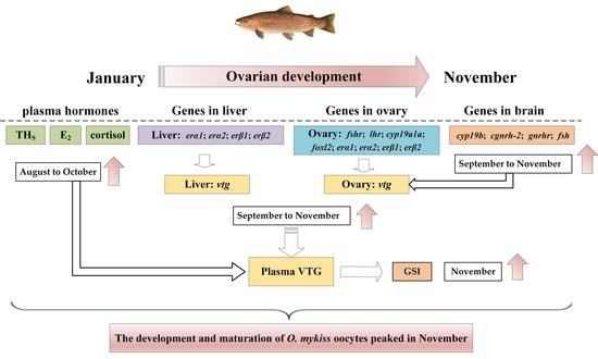
The purpose of this study was to investigate the periodic seasonal changes in endocrine activity and gonadal development of female rainbow trout (Oncorhynchus mykiss) in a high-altitude cold-water environment. The fish were sampled monthly from January to November and the levels of plasma hormones (estradiol (E2), cortisol and thyroid hormones (THS)) and vitellogenin (VTG) were measured by ELISA. Moreover, the transcriptions of sex-related genes in the ovary, brain, and liver were detected by qRT-PCR. The results showed a seasonal fluctuation of plasma hormones and VTG together with the development of the ovary, which reached a peak from August to October. Similarly, the transcription of hypothalamic gonadotropin-releasing hormone-2 (cgnrh-2), hypothalamic gonadotropin-releasing hormone receptors (gnrhr) and follicle-stimulating hormone (fsh) in the brain varied from January to September, but the highest level was detected in September to November. In addition, the transcription of sex-related genes located in the ovary and liver increased significantly during August to October, accompanied by a continuous increase in the gonadosomatic index (GSI) and a decrease in the hepatosomatic index (HSI). Therefore, plasma hormones and sex-related genes regulate the development and maturation of O. mykiss oocytes with the change in seasons and peaked in November. The results of this study provide a reference for improving the efficiency of the artificial reproduction of O. mykiss.

Graphical Abstract

1. Introduction

Reproduction is the basis of the whole ontogeny, which requires energy, ecology, anatomy, biochemistry and endocrine adaptations [1]. The obtainability of a high-quality fry and the capability to control fish breeding are the confining factors for fish culture [2]. Therefore, sufficient information related to reproductive constant, developmental biochemistry, and molecular regulation mechanism of gonadal development are very important in aquaculture.
Throughout the reproductive cycle, seasonal changes in tissue biochemical composition are related to gonadal weight, especially in females, in which the hepatic metabolism are stimulated during vitellogenesis [3]. Hence, monthly variations in the gonadosomatic index (GSI) helps to determine the breeding season of the fish, and study of the hepatosomatic index (HSI) is also important because the liver is a key organ in fish for production of vitellogenin which plays a significant role in the development of eggs [4,5]. Moreover, several studies have reported the seasonal endocrine and aromatase changes associated with reproductive activity, such as in O. mykiss [6], salmonids [7], rainbow trout (Salmo gairdneri) [8], and frog (Pelophylax esculentus) [9,10,11].
Increasing evidence demonstrates that neurosteroids might regulate neurogenesis in the developing or adult central nervous system. In particular, it can lead to permanent sexual differentiation of certain structures involved in sexual behavior and the neuroendocrine control of reproduction [12,13]. For instance, a gonadotropin-releasing hormone (GnRH) is a neuropeptide that participates in the reproductive regulation of all vertebrates. The primary function of GnRH is to regulate the synthesis and release of pituitary gonadotropins (GtHs), follicle-stimulating hormone (FSH), and luteinizing hormone (LH), and then stimulate steroidogenesis and gonadal development [14]. At least two GnRH forms have been characterized in O. mykiss, salmon GnRH (sgnrh-1), and chicken II GnRH (cgnrh-2) [15]. The sgnrh-1 appears to have a central role in eliciting the release of pituitary gonadotropins, whereas cgnrh-2 appears to act as a neuromodulator [16]. Furthermore, cytochrome P450 aromatase is the key enzyme that converts androgens to estrogens, and participates in the gonadal development and differentiation of fish. In teleost fish, two separate genes, cyp19a and cyp19b, that encode distinct aromatase isoforms, have been identified. The activity of cyp19a and cyp19b are predominantly associated with the ovary and brain, respectively [17]. Therefore, examination of the transcription of the gnrh, gnrhr, and brain aromatase genes in the hypothalamus and pituitary might help to understand the reproductive process of O. mykiss during the seasonal changes. In addition, gonadal steroids are also involved in the regulation of the hypothalamus-pituitary-thyroid (HPT) axis [18,19], affecting the level of thyroxine (T4), free thyroxine (FT4), triiodothyronine (T3), and free triiodothyronine (FT3), thus affecting the development, growth and reproduction of fish [20,21,22].
Environmental and genetic factors affect sex determination through a complex process [23]. During sex determination, the onset of a cascade of transcriptional or mRNA splicing factors are activated by a primary signal, allowing the final differentiation of the gonads into testis or ovary [24]. Foxl2, which is a putative winged helix/fork head transcription factor gene and a sexually dimorphic marker of ovarian differentiation, is involved in the ovarian development [25]. Previous studies suggest that ovarian hormones regulate the transcription of foxl2 thereby expanding the number of genes controlled by the hypothalamic-pituitary-gonadal (HPG) axis, e.g., gnrhr and fsh, that ultimately dictate reproductive fitness [26]. In addition, foxl2 also upregulates the transcription of cyp19a1a, which leads to the increase in cyp19a1a transcription [27]. Moreover, four nuclear estrogen receptor genes, erα1, erα2, erβ1, and erβ2, have been detected in rainbow trout [28]. Studies showed the positively correlated relationship between the transcription of ers and vtg [29,30], as the oocyte matures, the transcription of the vtg gene gradually increases. Therefore, detecting the transcription of sex-related genes during gonadal development may help to understand the reproductive process of O. mykiss during seasonal changes.
O. mykiss is a cold-water economic fish of the genus Oncorhynchus of the family Salmonidae. Yunnan is rich in cold-water resources and suitable climatic conditions, which are necessary conditions to promote the sustainable development of the O. mykiss industry. However, at present, the low egg quality, low fertilization rate, and low hatching rate of cultured female O. mykiss still restricts the development of cold-water fish culture in Yunnan. Therefore, to explore the characteristics of seasonal changes on plasma hormones and sex-related genes transcription during gonadal development of female O. mykiss, and to understand its endocrine regulation mechanism, it is of great significance to provide the data reference for O. mykiss reproduction, so as to further promote the healthy development of O. mykiss farming in Yunnan.

2. Materials and Methods

2.1. Fishes and Sample Collection

The O. mykiss used in this study were bought from Kunming Tanghao Aquaculture Company of China. In December 2017, the fish were domesticated in the indoor water tank of the circulatory system for a month in Yunnan Agricultural University and fed twice a day to satiety. Thirty-three female O. mykiss were sampled monthly from January to November 2018 (Table 1). After a fast for 2 d, three fish were anesthetized by 100 mg/L MS-222 (tricaine methane sulfonate, Sigma, St. Louis, MO, USA) before sampling every month. Plasma was separated from blood samples after centrifuging at 3000 rpms for 15 min, and then stored at −80 °C until assayed. The liver, brain and ovary were separated from the abdominal cavity and ventricle, then collected in an RNAase-free tube and stored at −80 °C for further testing. The experimental animals used in this experiment are strictly in accordance with the requirements of the guidelines for the use of Experimental Animals of Yunnan Agricultural University, and were approved by the Experimental Ethics Committee of Yunnan Agricultural University (YNAU2017llwyh131).

2.2. Determination of HSI and GSI

In order to calculate the values of HSI and GSI, the liver and ovary were weighed immediately after decapitation. HSI was calculated using the equation: HSI = [liver weight (g)/body weight (g)] × 100%. GSI was calculated as [ovary weight (g)/body weight (g)] × 100%.

2.3. Plasma Hormones and Vitellogenin (VTG) Analyses

Cortisol, E2, and VTG were determined by the ELISA kit, which was produced by Shanghai Enzymatic Biotechnology Co., Ltd. (Shanghai, China), according to the instructions of the kit. The Intra-assay CV(%) is less than 10% and Inter-assay CV(%) is less than 15%. The minimum detectable dose of cortisol, E2, and VTG are typically less than 10 pg/mL, 0.1 pmol/L and 1.0 ng/mL, respectively.
THS were also determined by ELISA kit, which was produced by Beijing North Institute of Biotechnology Co., Ltd., Beijing, China, according to the instructions of the kit. The Intra-assay CV(%) and Inter-assay CV(%) is less than 15%. The minimum detection concentration is less than 10 ng/mL, 0.4 ng/mL, 4.0 pmol/L, and 2.0 pmol/L for T4, T3, FT4, and FT3, respectively. All samples were measured at 450 nm wavelength using a 96 Flat Bottom Transparent Polystyrol (Greiner BioOne, Kremsmünster, Austria). The standard curve was established to calculate the concentration of cortisol, E2, VTG, and THS.

2.4. RNA Extraction and cDNA Synthesis

The total RNA of the brain, liver, and ovary of three female O. mykiss in each month were extracted using the TRIpure reagent (Aidlab Biotechnologies Co., Ltd., Beijing, China) following the manufacturer’s instruction. The quality and concentration of the total RNA were detected by NanoDrop 2000c apparatus (Thermo Fisher Scientific, Waltham, MA, USA), with an A260 nm/A280 nm ratio from 1.8 to 2.1. Then 1 μg total RNA was used for reverse transcription according to the manufacturer’s instruction of the TRUEscript 1st Strand cDNA Synthesis Kit user manual with gDNA Eraser (Aidlab Biotechnologies Co., Ltd., Beijing, China).

2.5. Gene Transcription

The transcription of sex-related genes during the oocyte development was detected using the quantitative real-time PCR. PCR reactions (20 μL) contained 1 μL of cDNA, 0.5 μL of each primer (10 μM), and 10 μL of 2 X TSINGKE Master qPCR Mix (SYBR Green I, Beijing TsingKe Biotech Co., Ltd., Beijing, China). The amplification procedure of these genes was: pre-heating at 95 °C for 3 min, followed by 40 cycles of 95 °C for 20 s, 55 °C for 20 s, and a final extension step at 72 °C for 20 s. The samples were analyzed in triplicate with a CFX96 Touch Real-Time PCR Detection System (Bio-Rad, Hercules, CA, USA). The elongation factor 1-alpha (ef1α) was used as the internal control to calculate the relative transcription of target gene by the 2△△Ct method [31]. The primers of genes were shown in Table 2.

2.6. Statistical Analysis

The Tukey method of a one-way analysis of variance (ANOVA) was used to evaluate the differences of value among the months through the SPSS 16.0 software (IBM Inc., Armonk, NY, USA). A value of p < 0.05 was considered statistically significant. All data were expressed as mean ± standard deviation (SD).

3. Results

3.1. Seasonal Changes in HSI and GSI

From January to July, no monthly significant differences in the HSI were observed. However, in August, HSI significantly reduced. After that, HSI returned to the normal levels from September to November (Figure 1A). Moreover, from January to October, no significant differences of GSI values were observed. The highest value of GSI of 4.11% was observed in November (Figure 1B).

3.2. Plasma Cortisol, E2 and VTG Levels of Female O. mykiss during Oocyte Development

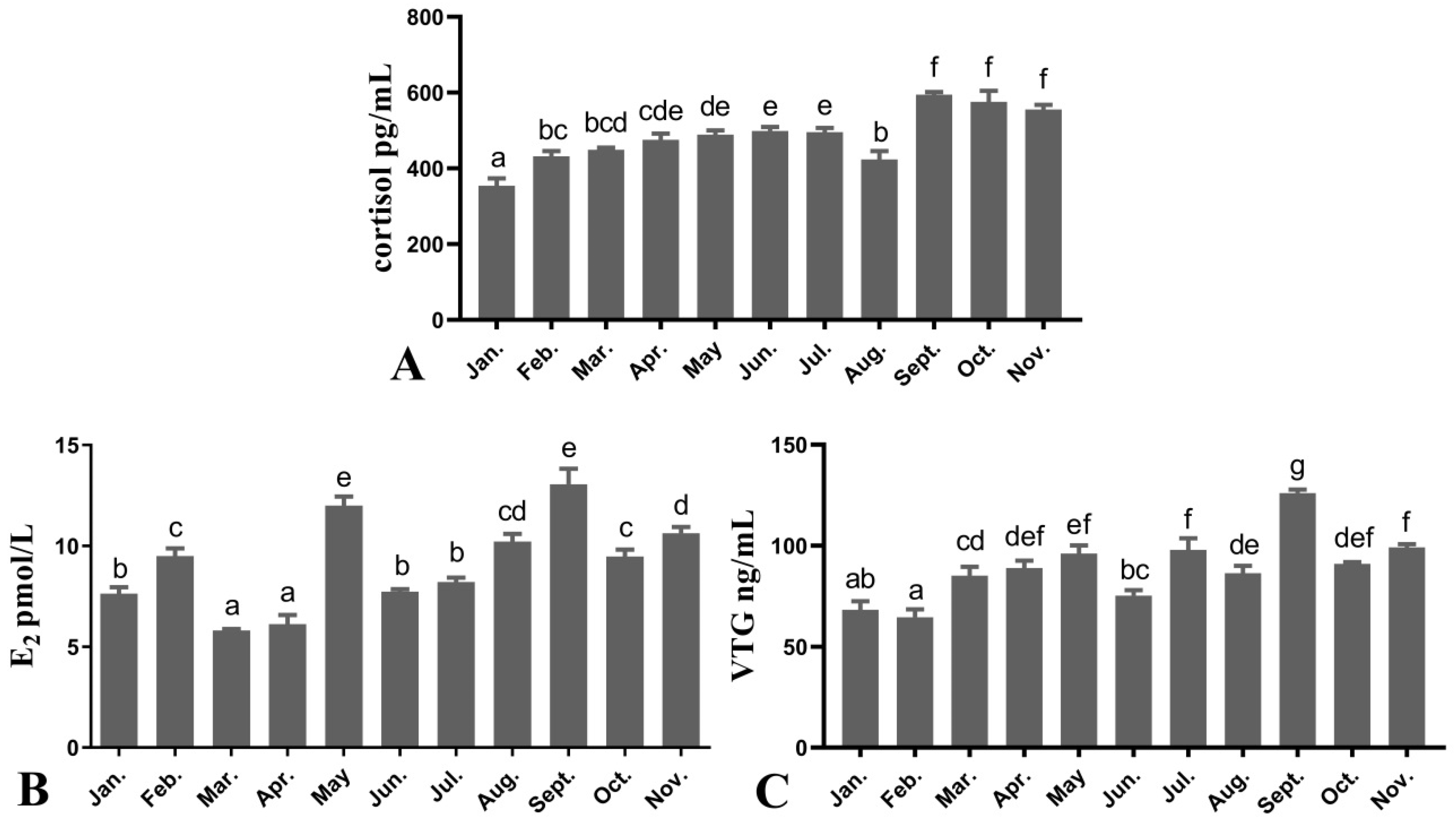
Plasma cortisol levels of females gradually increased from January to July (354.02 ± 19.60~485.85 ± 10.65 pg/mL). Then, cortisol levels decreased rapidly in August (423.58 ± 22.31 pg/mL) and increased again in September (594.65 ± 7.05 pg/mL) and remained at a high level until November (Figure 2A). Plasma E2 levels of females fluctuated from January to July (5.80 ± 0.07~12.00 ± 0.45 pmol/L) and peaked in May. E2 levels then increased rapidly in September (13.05 ± 0.77 pmol/L) and decreased again from October and November (Figure 2B). Plasma VTG levels of females fluctuated and were low in January to July (65.56 ± 3.95~97.96 ± 5.70 ng/mL), increased in September and reached the peak (126.09 ± 1.66 ng/mL). VTG levels then decreased rapidly in October and remained at a low level until November (Figure 2C).

3.3. Plasma THS Levels of Female O. mykiss during Oocyte Development

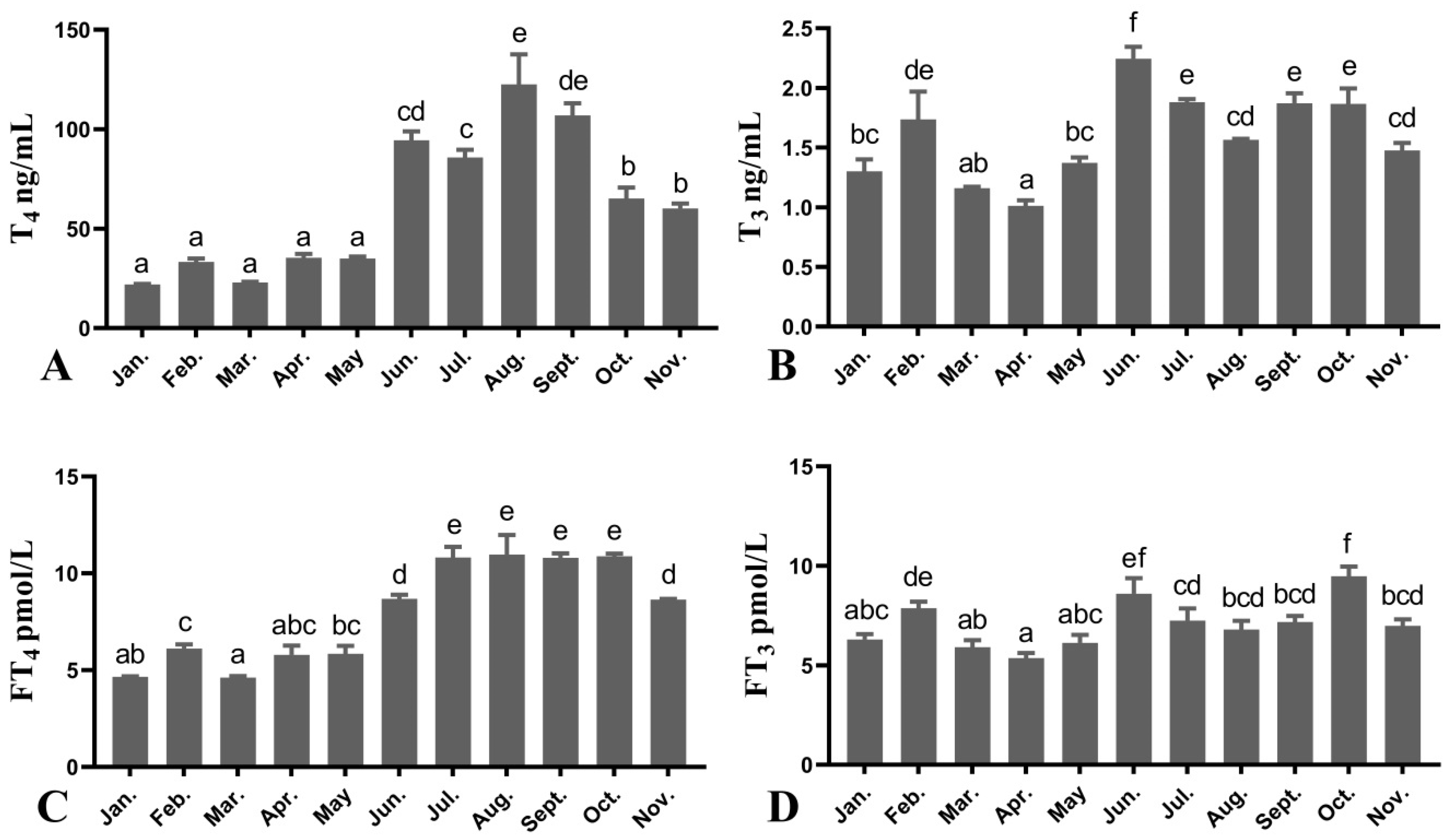
Plasma concentrations of both T4, FT4, T3, and FT3 were determined in female O. mykiss during the oocyte development (Figure 3). Plasma T4 levels remained at around 30 ng/mL from January to May, and then significantly increased in June. The highest plasma T4 levels were observed in August (122.59 ± 15.08 ng/mL). After that, T4 levels gradually declined from September to November (Figure 3A). Plasma T3 levels of females fluctuated throughout the year. From January to April, T3 levels increased first and then decreased (1.01 ± 0.05~1.74 ± 0.23 ng/mL). After that, plasma T3 remained elevated from May and reached the peak in June (2.24 ± 0.10 ng/mL). However, T3 concentrations continued to decrease from July to November (Figure 3B). The changes in FT4 levels in the plasma were similar to T4. Plasma FT4 increased in June and reached peak levels by the beginning of July (8.69 ± 0.21 pmol/L), and then maintained a high level until October (10.80 ± 0.22~10.97 ± 1.00 pmol/L) (Figure 3C). Then, a significant decrease was observed in November. Similar to T3, FT3 levels fluctuated irregularly throughout the year. The highest level of FT3 appeared in October (9.48 ± 0.48 pmol/L) (Figure 3D).

3.4. Seasonal Changes in the Levels of Female O. mykiss Brain Genes during Oocyte Development

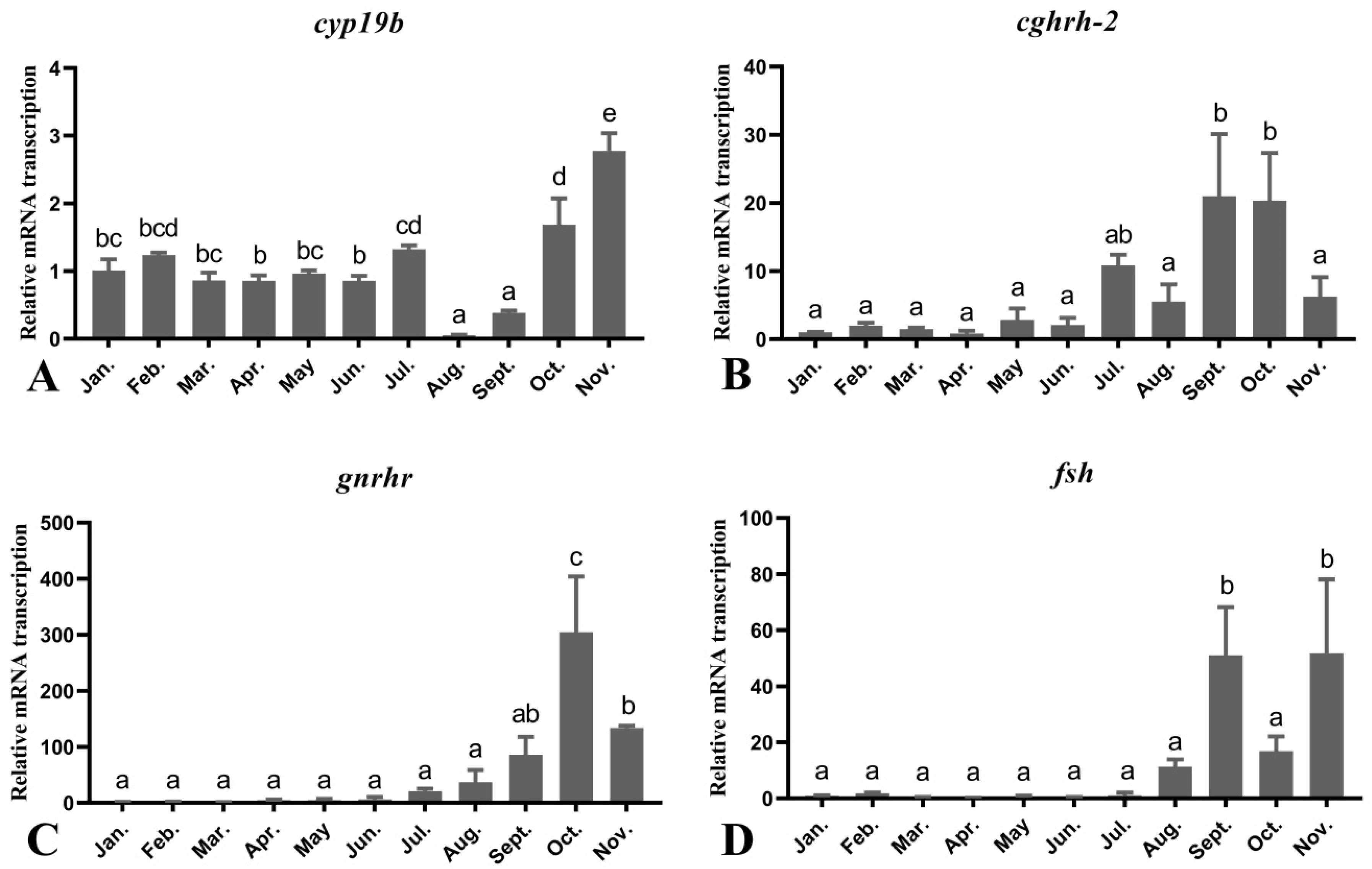
The transcription levels of genes in the brain were examined through an areal-time PCR assay (Figure 4). The transcription of cyp19b did not fluctuate significantly from January to July. However, a significant decrease was detected in August and September. From October to November, cyp19b levels gradually raised and then peaked in November (Figure 4A). The levels of cgnrh-2 were low in January to August, and increased significantly in September and October in association with active vitellogenesis. In November, the cgnrh-2 level decreased to the similar levels of January to August (Figure 4B). Moreover, similar to cgnrh-2, the levels of gnrhr were low from January to August. After that, the levels of gnrhr substantially increased in September and October, and the level in October was significantly higher than those in other months. The transcription of gnrhr significantly decreased in November (Figure 4C). The transcription levels of fsh were low from January to August. However, the levels of fsh fluctuated irregularly from September to November, showing a transcription trend that decreased first and then increased. The transcription peaks of fsh were observed in September and November (Figure 4D).

3.5. Seasonal Changes in the Levels of Female O. mykiss Gonadal Genes during Oocyte Development

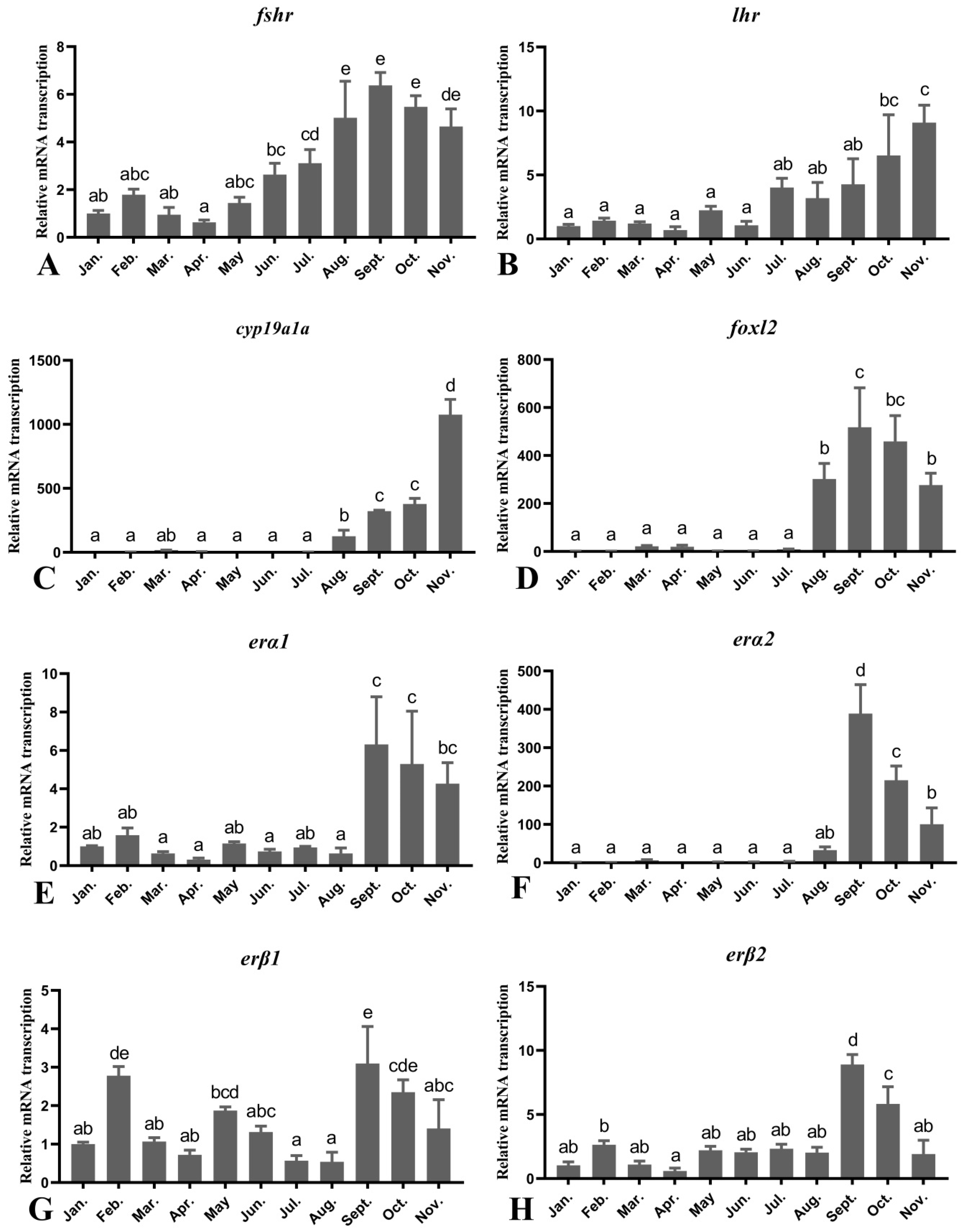
The real-time PCR was further used to validate the differentially expressed gonadal genes during seasonal changes (Figure 5). The transcription levels of fshr did not change significantly during the period from January to May, followed by a gradually increase from June to September, which reached the highest levels in September. In October and November, the transcription of fshr also remained at a high level (Figure 5A). Different from fshr, the levels of lhr transcription were low from January to September, and then gradually increased and reached its peak in November (Figure 5B). Transcripts of cyp19a1a remained at the same levels from January to July, and then significantly increased from August to November. The transcription peak of cyp19a1a was observed in November (Figure 5C). The levels of foxl2 transcription were low from January to July, and significantly increased from August and reached the maximum in September. After that, the transcription of foxl2 gradually decreased to the same level in August (Figure 5D).
According to the real-time PCR data, erα1 and erα2 were expressed at low levels from January to August and then became significantly high in September, followed by a gradually decrease from October to November (Figure 5E,F). However, irregular fluctuations of the erβ1 transcription were observed from January to August, which showed transcription peaks in September (Figure 5G). Similar to erα1 and erα2, the erβ2 transcription remained at the same levels from January to August, and then significantly increased in September. After that, the transcription of erβ2 gradually decreased to the same level in January to August (Figure 5H).

3.6. Seasonal Changes in the Levels of Female O. mykiss Liver Genes during Oocyte Development

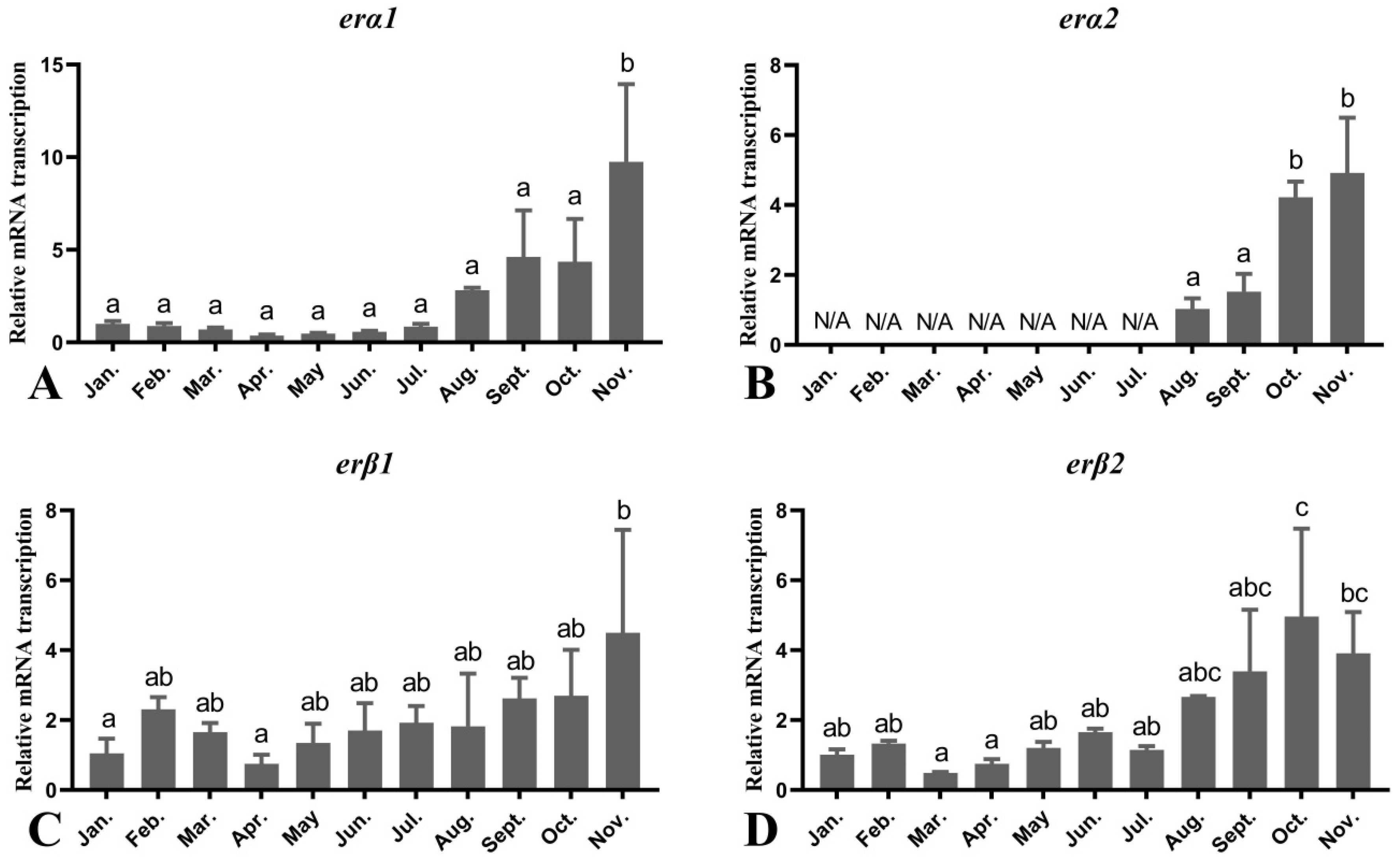
Both the erα1 and erα2 gene transcription profile showed the lowest transcription levels from January to July, then a significant elevation of erα1 transcription was observed in November, but erα2 transcription increased sharply between October and November (Figure 6A,B). There was no significant change in erβ1 transcription during the annual cycle. The erβ2 transcription was detected in the liver during the annual cycle with low levels of transcription, except for the October, in which the transcription level was significantly higher compared with January to July (Figure 6C,D).

3.7. Seasonal Changes in the Levels of Female O. mykiss vtg Gene during Oocyte Development

Transcription levels of vtg in the ovary were low from January to August, and its levels increased significantly from September to November when a peak was reached in November (Figure 7A). The vtg transcription levels in the liver increased significantly from August to November and reached a peak in November (Figure 7B).

4. Discussion

O. mykiss is an iterative, oviparous fish with a synchronized set of annual reproductive cycles. In the whole reproductive cycle, GSI and HSI may help to identify the breeding period or reproductive peak of fish, and further reflect the ovarian maturation time and spawning time of O. mykiss. In S. Gairdneri, GSI elevated gradually from June to August, increased significantly from September to December and reached a peak in December, and then decreased again from January to May. Similarly, HSI values increased gradually from March to December, and then decreased again in January and February [35]. Therefore, it showed that the ovarian maturation and spawning period of S. Gairdneri is in December. In this study, GSI increased gradually from August to November and peaked in November, while HSI was the lowest in August. When HSI decreases and GSI increases, it indicates that the liver loses weight during reproduction, and the VTG synthesized by the liver is transported to the gonads to promote gonadal maturation [36]. Therefore, the period of rapid gonadal development of O. mykiss is from August to November, and November is the most suitable breeding season for this species.
Steroid hormones such as testosterone, estradiol, progesterone, and corticosteroids play a key role in the reproductive process of fish. These hormones act on the reproductive process of fish directly or through feedback mechanisms. In female fish, E2 is necessary for ovarian development and controls egg maturation, ovulation, and egg laying. Meanwhile, circulating E2 regulates the transcription of vtg in hepatocytes, leading to the synthesis of several closely related VTG proteins [37]. In this study, the plasma levels of cortisol, E2 and VTG in female O. mykiss changed significantly during oocyte development. Especially in the pre-oviposition stage, the level of E2 in plasma increased, the yolk generation was active, and then the level of VTG in plasma increased subsequently. In the study of catfish (Silurus asotus), the levels of E2 and plasma cortisol increased during the period of yolk generation and spawning [38]. Female O. mykiss had a low gonad E2 synthesis January to August. However, the plasma E2 content increased significantly in September, indicating that the ovarian development of female O. mykiss was the fastest in September in this study. Similarly, the plasma VTG content of female O. mykiss also has a similar trend with E2. The content of VTG has a gradually increasing trend from January to June. In September, the plasma VTG content increased significantly, indicating that the synthesis of VTG is the largest in September, followed by the size of the oocyte, which increases rapidly. In addition, the trend of female O. mykiss plasma cortisol and VTG is consistent, reaching a peak in September, and then decreasing. Therefore, the trend of plasma cortisol is consistent with E2 and VTG, indicating that cortisol is also involved in the development of female O. mykiss oocytes in this study. During the reproductive cycle, plasma cortisol levels also increase from spawning or spawning periods, such as in the plaice (Pleuronectes platessa) [39], two species of trout, Salmo trutta L. and S. gairdneri [40], O. mykiss [41]. Therefore, in O. mykiss reproduction, the increase in the plasma cortisol level during reproductive periods may play a key role in ovarian growth and vitellogenesis in addition to stress [42,43].
In fish species, gonadal steroids can also affect circulating levels of thyroid hormone and/or thyroid activity [18,19]. THS include T3, T4, FT3, and FT4 [44]. Thyroids participate in the physiologic stress response envisaged chiefly because of the involvement in THS in almost all aspects of the physiologic processes [45,46,47]. The capability of the thyroid axis is to respond to the stimuli which arise from the other factors that engage in its cross talk [48]. Therefore, thyroid hormones have a wide range of effects on the development, growth, and reproduction of fish [20,21]. These effects are also passed on from generation, because THS play a pivotal role in the early development of offspring [21,49]. The plasma THS levels of many fishes show periodic changes. These hormones are related to sexual maturity [50,51] and reproductive cycles [52,53]. In this study, plasma concentrations of THS were determined in female O. mykiss during the year of oocyte development. Plasma THS levels increased in August and decreased in November before spawning. This is similar to the study of two strains of rainbow trout (O. mykiss Shasta and Kamloops), the levels of FT3 and FT4, T3 and T4 were the lowest during the spawning period, and the circulating THS showed similar seasonal changes [54]. These results indicated that THS participated in the regulation of yolk accumulation in rainbow trout.
With the change in seasons, the HPG axis plays an important role in oocyte development of O. mykiss. GnRH, synthesized and released by the hypothalamus, is a key hormone that regulates reproduction [55]. In salmonid fishes, two forms of GnRH, sgnrh-1 and cgnrh-2, have been detected [15]. GnRH is conveyed to the pituitary via the hypothalamo-hypophyseal portal vessels, and regulates synthesis and release of GtH [56]. In the process of signal transduction, ligands (cgnrh-2, fsh, lh) bind and interact with their receptors (gnrhr, fshr, lhr) distributed in target cells to achieve their physiological functions [57]. In this study, the transcription level of cgnrh-2 and gnrhr gradually increased from August to October. Moreover, a previous study showed that the transcription of fshr is associated predominantly with vitellogenesis, while the lhr is prevalent during oocyte maturation and ovulation [57]. In female greater amberjack (Seriola dumerili) [58], ovarian fshr and plasma E2 gradually increased during ovarian development, which suggested that fsh plays a role in ovarian development and during the post-spawning period [58]. In this study, the ovarian fsh, fshr of O. mykiss increased continuously from August to September and decreased from September to October, but fsh increased significantly again in November. Similarly, the ovarian lhr increased continuously from August to November, and the peak was in November. Therefore, the transcription of fsh and lh plays a crucial role in the oocyte development of O. mykiss and has seasonal correlation. In addition, GnRH can act as a neuromodulator, and administration of exogenous GnRH facilitates sexual behavior in many species [59]. Therefore, cgnrh-2 can affect the transcription of fsh and lh through gnrhr, and then participate in the regulation of E2 synthesis in O. mykiss.
Aromatase is a key enzyme in the synthesis of estrogen, also known as estrogen synthetase. It catalyzes the conversion of testosterone and androstendione to estrone and estradiol in animals, and converts androgen to estrogen. Therefore, aromatase shows its regulatory role in the early differentiation of female gonadal gland in nonmammalian vertebrates [60]. The cyp19 encodes cytochrome P450 aromatase. Two cyp19 genes, cyp19a and cyp19b, which belong to two independent CYP19 subfamilies, were identified. The cyp19a is transcribed in the ovary, while the cyp19b gene is transcribed in the brain [61]. The transcription of the O. mykiss cyp19b is consistent with the gonadal development period. From January to July, the transcription of the O. mykiss cyp19b is at a high level. The cyp19b promotes the release of estrogen and gonadotropin. Then, the transcription of the O. mykiss cyp19b decreased from August, and significantly increased until the gonad maturation process. These results indicate that cyp19b is based on the transcription of the brain, which regulates the final maturation and ovulation of O. mykiss in November.
The foxl2 is the fork transcription factor in the process of the ovarian development [62]. As shown in previous studies, the foxl2 transcription is a genetic factor that activates the transcription of aromatase [63]. In the O. mykiss gonadal development of this study, the transcription of foxl2 reached its peak in September, decreased gradually from October to November, and decreased the lowest in November. Therefore, foxl2 may have a regulatory relationship with cyp19a1a, and the transcription of cyp19a1a gradually increases from August to November and peaked in November. A previous study also revealed that cyp19a1a and cyp19a1b were co-expressed with foxl2, except in the early yolk stage of the ovary [24]. In summary, during fish gonadal development, the highest transcription level of foxl2 was earlier than cyp19a1a, suggesting that foxl2 is involved in fish gonadal differentiation and the maintenance of ovarian function [64].
In addition, estrogen regulates ovarian development, differentiation, and maintenance, as well as oogenesis, and stimulates liver synthesis of vtg and choriogenin [29,30]. These effects are principally mediated through ers which belong to the nuclear hormone receptor superfamily. After binding of a ligand to the ligand-binding domain (LBD) of ERS, this complex binds as homo dimer to estrogen response elements (EREs) in the promoter regions of estrogen responsive target genes and regulates their transcription [65]. Four nuclear estrogen receptor genes were identified in O. mykiss: erα1, erα2, erβ1, and erβ2 [28]. Recent knockout studies in goldfish (Carassius auratus Linnaeus) and zebrafish (Danio rerio) have shown that erβ1 and erβ2 are necessary for the estrogen-mediated up-regulation of erα and vtg transcription [66,67]. The vtg was also positively correlated with the ers subtype [29,30]. In this study, under the effect of estrogen in female O. mykiss, the transcription of ers in the ovary increased significantly in September, and decreased gradually from September to November. Similarly, the transcription of vtg in the ovary increased gradually in August to November and peaked in the November. As an important exogenous organ, the transcription of ers and vtg in the liver also increased with the change in season. These results showed that the increasing of estrogen level and its receptor genes transcription, as well as vtg, participated in the accumulation of vitelloprotein during female O. mykiss ovarian development with seasonal changes. Therefore, cortisol, E2, THS can interfere with the transcription of sex-related genes on the HPG axis, thereby promoting the transcription of vtg genes in the ovary and liver, increasing the plasma VTG levels, and finally leading to the reproduction of O. mykiss in November.

5. Conclusions

In conclusion, this experiment studied the periodic seasonal changes in plasma hormones, and sex-related genes transcription in the brain, liver, and ovary during the gonadal development of female O. mykiss under the regulation of HPG axis. The plasma hormones gradually increased from January to August, and peaked from August to October, which promotes the synthesis of VTG, and then accelerated the development and maturation of oocytes in O. mykiss. Similarly, the transcription level of sex-related genes in the HPG axis increased significantly since August and maintained a high level until November, which affected the synthesis of estrogen, and then participated in the regulation of gonadal development. In addition, estrogen can affect the transcription of vtg by increasing the ers transcription, thereby regulating the synthesis and accumulation of yolk in O. mykiss oocytes, and ultimately affects the value of GSI in November. Therefore, these sex-related hormones and genes can accelerate the synthesis and accumulation of yolk by affecting the HPG axis from August, and reach a peak in November to promote the spawning and reproduction of O. mykiss. These results strongly proved that November is the most suitable season for O. mykiss breeding in Yunnan.

Author Contributions

Conceptualization, H.C.; Data curation, H.C.; Funding acquisition, Q.H.; Methodology, H.C.; Project administration, Q.H.; Resources, B.B. and L.K.; Software, H.R. and Y.S.; Writing—original draft, H.C.; Writing—review and editing, Q.H. All authors have read and agreed to the published version of the manuscript.

Funding

This work was supported by the National Natural Science Foundation of China (grant number 32060825, 31602141), the Yunnan Fundamental Research Projects (grant number 2017FD077), and the Zhuangping expert workstation of Yunnan Province.

Institutional Review Board Statement

The experimental animals used in this experiment are strictly in accordance with the requirements of the guidelines for the use of Experimental Animals of Yunnan Agricultural University, and have been approved by the Experimental Ethics Committee of Yunnan Agricultural University (YNAU2017llwyh131).

Data Availability Statement

Data are contained within the article.

Acknowledgments

The author would like to thank the students majoring in Aquaculture in Yunnan Agricultural University for their help in this study.

Conflicts of Interest

The authors declare no conflict of interest.

References

  1. Rocha, M.J. Fish Reproduction; CRC Press: Boca Raton, FL, USA, 2008. [Google Scholar]
  2. Karimi, S.; Kochanian, P.; Salati, A.P.; Gooraninejad, S. Plasma sex steroids and gonadosomatic index variations during ovarian development of female wild yellowfin seabream (Acanthopagrus latus). Ichthyol. Res. 2013, 61, 68–75. [Google Scholar] [CrossRef]
  3. Tolussi, C.E.; Gomes, A.D.; Ribeiro, C.D.S.; Caneppele, D.; Moreira, R.G.; Honji, R.M. Mobilization of energetic substrates in the endangered catfish Steindachneridion parahybae (Siluriformes: Pimelodidae): Changes in annual reproductive cycle in captivity. Neotrop. Ichthyol. 2018, 16. [Google Scholar] [CrossRef]
  4. Jan, M.; Ahmed, I. Assessment of fecundity, gonadosomatic index and hepatosomatic index of snow trout, Schizothorax plagiostomus in river Lidder, from Kashmir Himalaya, India. Int. J. Fish. Aquat. Stud. Delhi 2016, 4, 370–375. [Google Scholar]
  5. Jan, M.; Jan, N. Studies on the fecundity (F), gonadosomatic index (GSI) and hepatosomatic index (HSI) of Salmo trutta fario (Brown trout) at Kokernag trout fish farm, Anantnag, Jammu and Kashmir. Int. J. Fish. Aquat. Stud. Delhi 2017, 5, 170–173. [Google Scholar]
  6. Estay, F.; Colihueque, N.; Araneda, C. Comparison of Oogenesis and Sex Steroid Profiles between Twice and Once Annually Spawning of Rainbow Trout Females (Oncorhynchus mykiss). Sci. World J. 2012, 2012, 1–7. [Google Scholar] [CrossRef] [Green Version]
  7. Teitsma, C.; Desdoits-Lethimonier, C.; Tujague, M.; Anglade, I.; Saligaut, D.; Bailhache, T.; Pakdel, F.; Kah, O.; Ducouret, B. Identification of potential sites of cortisol actions on the reproductive axis in rainbow trout. Comp. Biochem. Physiol. Part C Pharmacol. Toxicol. Endocrinol. 1998, 119, 243–249. [Google Scholar] [CrossRef]
  8. Cyr, D.G.; Eales, J.G. Influence of thyroidal status on ovarian function in rainbow trout, Salmo gairdneri. J. Exp. Zoöl. 1988, 248, 81–87. [Google Scholar] [CrossRef]
  9. Di Fiore, M.M.; Assisi, L.; Botte, V. Aromatase and testosterone receptor in the liver of the female green frog, Rana esculenta. Life Sci. 1998, 62, 1949–1958. [Google Scholar] [CrossRef]
  10. Burrone, L.; Santillo, A.; Pinelli, C.; Baccari, G.C.; Di Fiore, M.M. Induced synthesis of P450 Aromatase and 17β-estradiol by D-aspartate in frog brain. J. Exp. Biol. 2012, 215, 3559–3565. [Google Scholar] [CrossRef] [Green Version]
  11. Paolucci, M.; Di Fiore, M.M. Sex Steroid Binding Proteins in the Plasma of the Green Frog, Rana esculenta: Changes during the Reproductive Cycle and Dependence on Pituitary Gland and Gonads. Gen. Comp. Endocrinol. 1994, 96, 401–411. [Google Scholar] [CrossRef]
  12. Morris, J.A.; Jordan, C.L.; Breedlove, S.M. Sexual differentiation of the vertebrate nervous system. Nat. Neurosci. 2004, 7, 1034–1039. [Google Scholar] [CrossRef]
  13. Vizziano-Cantonnet, D.; Anglade, I.; Pellegrini, E.; Gueguen, M.-M.; Fostier, A.; Guiguen, Y.; Kah, O. Sexual dimorphism in the brain aromatase expression and activity, and in the central expression of other steroidogenic enzymes during the period of sex differentiation in monosex rainbow trout populations. Gen. Comp. Endocrinol. 2011, 170, 346–355. [Google Scholar] [CrossRef]
  14. Dickey, J.T.; Swanson, P. Effects of Salmon Gonadotropin-Releasing Hormone on Follicle Stimulating Hormone Secretion and Subunit Gene Expression in Coho Salmon (Oncorhynchus kisutch). Gen. Comp. Endocrinol. 2000, 118, 436–449. [Google Scholar] [CrossRef] [PubMed]
  15. Von Schalburg, K.R.; Warby, C.M.; Sherwood, N.M. Evidence for Gonadotropin-Releasing Hormone Peptides in the Ovary and Testis of Rainbow Trout1. Biol. Reprod. 1999, 60, 1338–1344. [Google Scholar] [CrossRef] [PubMed] [Green Version]
  16. Okuzawa, K.; Amano, M.; Kobayashi, M.; Aida, K.; Hanyu, I.; Hasegawa, Y.; Miyamoto, K. Differences in salmon GnRH and chicken GnRH-II contents in discrete brain areas of male and female rainbow trout according to age and stage of maturity. Gen. Comp. Endocrinol. 1990, 80, 116–126. [Google Scholar] [CrossRef]
  17. Chang, X.; Kobayashi, T.; Senthilkumaran, B.; Kobayashi-Kajura, H.; Sudhakumari, C.C.; Nagahama, Y. Two types of aromatase with different encoding genes, tissue distribution and developmental expression in Nile tilapia (Oreochromis niloticus). Gen. Comp. Endocrinol. 2005, 141, 101–115. [Google Scholar] [CrossRef]
  18. Cyr, D.; Bromage, N.; Duston, J.; Eales, J. Seasonal patterns in serum levels of thyroid hormones and sex steroids in relation to photoperiod-induced changes in spawning time in rainbow trout, Salmo gairdneri. Gen. Comp. Endocrinol. 1988, 69, 217–225. [Google Scholar] [CrossRef]
  19. Maclatchy, D.L.; Eales, J. Short-term treatment with testosterone increases plasma 3,5,3′-triiodo-l-thyronine and hepatic l-thyroxine 5′-monodeiodinase levels in arctic charr, Salvelinus alpinus. Gen. Comp. Endocrinol. 1988, 71, 10–16. [Google Scholar] [CrossRef]
  20. Blanton, M.L.; Specker, J.L. The Hypothalamic-Pituitary-Thyroid (HPT) Axis in Fish and Its Role in Fish Development and Reproduction. Crit. Rev. Toxicol. 2007, 37, 97–115. [Google Scholar] [CrossRef]
  21. Leatherland, J.F. Reflections on the thyroidology of fishes: From molecules to humankind. Guelph Ichthyol. Rev. 1994, 2, 1–67. [Google Scholar]
  22. Power, D.M.; Llewellyn, L.; Faustino, M.; Nowell, M.A.; Björnsson, B.T.; Einarsdóttir, I.E.; Canario, A.V.; Sweeney, G.E. Thyroid hormones in growth and development of fish. Comp. Biochem. Physiol. Part C Toxicol. Pharmacol. 2001, 130, 447–459. [Google Scholar] [CrossRef]
  23. Devlin, R.H.; Nagahama, Y. Sex determination and sex differentiation in fish: An overview of genetic, physiological, and environmental influences. Aquaculture 2002, 208, 191–364. [Google Scholar] [CrossRef]
  24. Mu, W.J.; Wen, H.S.; Li, J.F.; He, F. Cloning and expression analysis of Foxl2 during the reproductive cycle in Korean rockfish, Sebastes schlegeli. Fish Physiol. Biochem. 2013, 39, 1419–1430. [Google Scholar] [CrossRef] [PubMed]
  25. Wang, D.-D.; Zhang, G.-R.; Wei, K.-J.; Ji, W.; Gardner, J.P.A.; Yang, R.-B.; Chen, K.-C. Molecular identification and expression of the Foxl2 gene during gonadal sex differentiation in northern snakehead Channa argus. Fish Physiol. Biochem. 2015, 41, 1419–1433. [Google Scholar] [CrossRef]
  26. Herndon, M.K.; Nilson, J.H. Maximal Expression of Foxl2 in Pituitary Gonadotropes Requires Ovarian Hormones. PLoS ONE 2015, 10, e0126527. [Google Scholar] [CrossRef] [PubMed] [Green Version]
  27. Wang, D.S.; Kobayashi, T.; Zhou, L.Y.; Nagahama, Y. Molecular cloning and gene expression of Foxl2 in the Nile tilapia, Oreochromis niloticus. Biochem. Biophys. Res. Commun. 2004, 320, 83–89. [Google Scholar] [CrossRef]
  28. Nagler, J.J.; Cavileer, T.; Sullivan, J.; Cyr, D.G.; Rexroad, C., III. The complete nuclear estrogen receptor family in the rainbow trout: Discovery of the novel ERα2 and both ERβ isoforms. Gene 2007, 392, 164–173. [Google Scholar] [CrossRef] [Green Version]
  29. Guiguen, Y.; Fostier, A.; Piferrer, F.; Chang, C.-F. Ovarian aromatase and estrogens: A pivotal role for gonadal sex differentiation and sex change in fish. Gen. Comp. Endocrinol. 2010, 165, 352–366. [Google Scholar] [CrossRef]
  30. Lubzens, E.; Young, G.; Bobe, J.; Cerdà, J. Oogenesis in teleosts: How fish eggs are formed. Gen. Comp. Endocrinol. 2010, 165, 367–389. [Google Scholar] [CrossRef]
  31. Livak, K.J.; Schmittgen, T.D. Analysis of Relative Gene Expression Data Using Real-Time Quantitative PCR and the 2−ΔΔCT Method. Methods 2001, 25, 402–408. [Google Scholar] [CrossRef]
  32. Delalande, C.; Goupil, A.-S.; Lareyre, J.-J.; Le Gac, F. Differential expression patterns of three aromatase genes and of four estrogen receptors genes in the testes of trout (Oncorhynchus mykiss). Mol. Reprod. Dev. 2015, 82, 694–708. [Google Scholar] [CrossRef]
  33. Hou, Z.-S.; Wen, H.-S.; Li, J.-F.; He, F.; Li, Y.; Tao, Y.-X. Hypothalamus-pituitary-gonad axis of rainbow trout (Oncorhynchus mykiss) during early ovarian development and under dense rearing condition. Gen. Comp. Endocrinol. 2016, 236, 131–138. [Google Scholar] [CrossRef]
  34. Vizziano, D.; Randuineau, G.; Baron, D.; Cauty, C.; Guiguen, Y. Characterization of early molecular sex differentiation in rainbow trout, Oncorhynchus mykiss. Dev. Dyn. 2007, 236, 2198–2206. [Google Scholar] [CrossRef] [PubMed]
  35. van Bohemen, C.; Lambert, J.; Peute, J. Annual changes in plasma and liver in relation to vitellogenesis in the female rainbow trout, Salmo gairdneri. Gen. Comp. Endocrinol. 1981, 44, 94–107. [Google Scholar] [CrossRef]
  36. Zin, T.; Than, A.A.; Naing, T.T. Fecundity (F), gonadosomatic index (GSI), hepatosomatic index (HSI), condition factor (K) and length-weight relationship (LWR) in Channa orientalis Bloch & Schneider, 1801. Univ. Res. J. 2011, 4, 47–62. [Google Scholar]
  37. Soranganba, N.; Singh, I. Role of some steroidogenic hormones in fish reproduction. Chem. Sci. Rev. Lett. 2019, 8, 64–69. [Google Scholar]
  38. Lamba, V.J.; Goswami, S.V.; Sundararaj, B.I. Circannual and circadian variations in plasma levels of steroids (cortisol, estradiol-17β estrone, and testosterone) correlated with the annual gonadal cycle in the catfish, Heteropneustes fossilis (Bloch). Gen. Comp. Endocrinol. 1983, 50, 205–225. [Google Scholar] [CrossRef]
  39. Wingfield, J.; Grimm, A. Seasonal changes in plasma cortisol, testosterone and oestradiol-17β in the plaice, Pleuronectes platessa L. Gen. Comp. Endocrinol. 1977, 31, 1–11. [Google Scholar] [CrossRef]
  40. Pickering, A.D.; Christie, P. Changes in the concentrations of plasma cortisol and thyroxine during sexual maturation of the hatchery-reared brown trout, Salmo trutta L. Gen. Comp. Endocrinol. 1981, 44, 487–496. [Google Scholar] [CrossRef]
  41. Kusakabe, M.; Nakamura, I.; Young, G. 11β-Hydroxysteroid Dehydrogenase Complementary Deoxyribonucleic Acid in Rainbow Trout: Cloning, Sites of Expression, and Seasonal Changes in Gonads. Endocrinology 2003, 144, 2534–2545. [Google Scholar] [CrossRef] [Green Version]
  42. Shankar, D.S.; Kulkarni, R.S. Tissue cholesterol and serum cortisol level during different reproductive phases of the female freshwater fish Notopterus notopterus (Pallas). J. Environ. Biol. 2007, 28, 137–139. [Google Scholar] [PubMed]
  43. Hou, Y.Y.; Han, X.D.; Suzuki, Y. Annual changes in plasma levels of cortisol and sex steroid hormones in male rainbow trout, Oncorhynchus mykiss. Chin. J. Oceanol. Limnol. 2001, 19, 217–221. [Google Scholar]
  44. Abdollahpour, H.; Falahatkar, B.; Efatpanah, I.; Meknatkhah, B.; Van Der Kraak, G. Hormonal and physiological changes in Sterlet sturgeon Acipenser ruthenus treated with thyroxine. Aquaculture 2019, 507, 293–300. [Google Scholar] [CrossRef]
  45. Gorbman, A. Thyroid function and its control in fishes—ScienceDirect. Fish Physiol. 1969, 2, 241–274. [Google Scholar]
  46. Grau, E.G. Environmental Influences on Thyroid Function in Teleost Fish. Am. Zoöl. 1988, 28, 329–335. [Google Scholar] [CrossRef] [Green Version]
  47. Leatherland, J.F. Endocrine Factors Affecting Thyroid Economy of Teleost Fish. Am. Zoöl. 1988, 28, 319–328. [Google Scholar] [CrossRef]
  48. Peter, M.S. The role of thyroid hormones in stress response of fish. Gen. Comp. Endocrinol. 2011, 172, 198–210. [Google Scholar] [CrossRef]
  49. Kang, D.-Y.; Chang, Y.J. Effects of maternal injection of 3,5,3′-triiodo-l-thyronine (T3) on growth of newborn offspring of rockfish, Sebastes schlegeli. Aquaculture 2004, 234, 641–655. [Google Scholar] [CrossRef]
  50. Cyr, D.G.; Idler, D.R.; Audet, C.; McLeese, J.M.; Ealesd, J.G. Effects of Long-Term Temperature Acclimation on Thyroid Hormone Deiodinase Function, Plasma Thyroid Hormone Levels, Growth, and Reproductive Status of Male Atlantic Cod, Gadus morhua. Gen. Comp. Endocrinol. 1998, 109, 24–36. [Google Scholar] [CrossRef]
  51. Pavlidis, M.; Greenwood, L.; Mourot, B.; Kokkari, C.; Le Menn, F.; Divanach, P.; Scott, A. Seasonal Variations and Maturity Stages in Relation to Differences in Serum Levels of Gonadal Steroids, Vitellogenin, and Thyroid Hormones in the Common Dentex (Dentex dentex). Gen. Comp. Endocrinol. 2000, 118, 14–25. [Google Scholar] [CrossRef]
  52. Eales, J. Modes of action and physiological effects of thyroid hormones in fish. Fish Endocrinol. 2006, 2, 767–808. [Google Scholar]
  53. Supriya, A.; Raghuveer, K.; Swapna, I.; Rasheeda, M.K.; Kobayashi, T.; Nagahama, Y.; Gupta, A.D.; Majumdar, K.C.; Senthilkumaran, B. Thyroid hormone modulation of ovarian recrudescence of air-breathing catfish Clarias gariepinus. Fish Physiol. Biochem. 2005, 31, 267–270. [Google Scholar] [CrossRef] [PubMed]
  54. Pavlidis, M.; Dessypris, A.; Christofidis, I. Seasonal fluctuations in plasma thyroid hormones, in two strains of rainbow trout (Oncorhynchus mykiss), during the first and second reproductive cycle: Relation with their photoperiodically altered spawning time. Aquaculture 1991, 99, 365–385. [Google Scholar] [CrossRef]
  55. Millar, R.P. GnRHs and GnRH receptors. Anim. Reprod. Sci. 2005, 88, 5–28. [Google Scholar] [CrossRef]
  56. Sherwood, N.M.; Lovejoy, D.A.; Coe, I.R. Origin of mammalian gonadotropin-releasing hormones. Endocr. Rev. 1993, 14, 241–254. [Google Scholar] [CrossRef] [PubMed]
  57. Yaron, Z.; Levavi-Sivan, B. Endocrine regulation of fish reproduction. Encycl. Fish Physiol. Genome Environ. 2011, 2, 1500–1508. [Google Scholar]
  58. Nyuji, M.; Kazeto, Y.; Izumida, D.; Tani, K.; Suzuki, H.; Hamada, K.; Mekuchi, M.; Gen, K.; Soyano, K.; Okuzawa, K. Greater amberjack Fsh, Lh, and their receptors: Plasma and mRNA profiles during ovarian development. Gen. Comp. Endocrinol. 2016, 225, 224–234. [Google Scholar] [CrossRef]
  59. Pfaff, D.W.; Jorgenson, K.; Kow, L. Luteinizing Hormone-Releasing Hormone in Rat Brain: Gene Expression, Role as Neuromodulator, and Functional Effects. Ann. N. Y. Acad. Sci. 1987, 519, 323–333. [Google Scholar] [CrossRef]
  60. Tchoudakova, A.; Kishida, M.; Wood, E.; Callard, G.V. Promoter characteristics of two cyp19 genes differentially expressed in the brain and ovary of teleost fish. J. Steroid Biochem. Mol. Biol. 2001, 78, 427–439. [Google Scholar] [CrossRef]
  61. Chiang, E.F.L.; Yan, Y.L.; Tong, S.K.; Hsiao, P.H.; Guiguen, Y.; Postlethwait, J.; Chung, B.C. Characterization of duplicated zebrafish cyp19 genes. J. Exp. Zool. 2001, 290, 709–714. [Google Scholar] [CrossRef]
  62. Baron, D.; Cocquet, J.; Xia, X.; Fellous, M.; Guiguen, Y.; Veitia, R.A. An evolutionary and functional analysis of FoxL2 in rainbow trout gonad differentiation. J. Mol. Endocrinol. 2004, 33, 705–715. [Google Scholar] [CrossRef] [PubMed]
  63. Wang, D.-S.; Kobayashi, T.; Zhou, L.-Y.; Paul-Prasanth, B.; Ijiri, S.; Sakai, F.; Okubo, K.; Morohashi, K.-I.; Nagahama, Y. Foxl2 Up-Regulates Aromatase Gene Transcription in a Female-Specific Manner by Binding to the Promoter as Well as Interacting with Ad4 Binding Protein/Steroidogenic Factor 1. Mol. Endocrinol. 2007, 21, 712–725. [Google Scholar] [CrossRef] [PubMed] [Green Version]
  64. Liu, H.; Mu, X.; Gui, L.; Su, M.; Li, H.; Zhang, G.; Liu, Z.; Zhang, J. Characterization and gonadal expression of FOXL2 relative to Cyp19a genes in spotted scat Scatophagus argus. Gene 2015, 561, 6–14. [Google Scholar] [CrossRef] [PubMed]
  65. Tohyama, S.; Miyagawa, S.; Lange, A.; Ogino, Y.; Mizutani, T.; Tatarazako, N.; Katsu, Y.; Ihara, M.; Tanaka, H.; Ishibashi, H.; et al. Understanding the Molecular Basis for Differences in Responses of Fish Estrogen Receptor Subtypes to Environmental Estrogens. Environ. Sci. Technol. 2015, 49, 7439–7447. [Google Scholar] [CrossRef]
  66. Griffin, L.B.; January, K.E.; Ho, K.W.; Cotter, K.; Callard, G.V. Morpholino-Mediated Knockdown of ERα, ERβa, and ERβb mRNAs in Zebrafish (Danio rerio) Embryos Reveals Differential Regulation of Estrogen-Inducible Genes. Endocrinology 2013, 154, 4158–4169. [Google Scholar] [CrossRef] [Green Version]
  67. Nelson, E.; Habibi, H.R. Functional Significance of Nuclear Estrogen Receptor Subtypes in the Liver of Goldfish. Endocrinology 2010, 151, 1668–1676. [Google Scholar] [CrossRef] [Green Version]
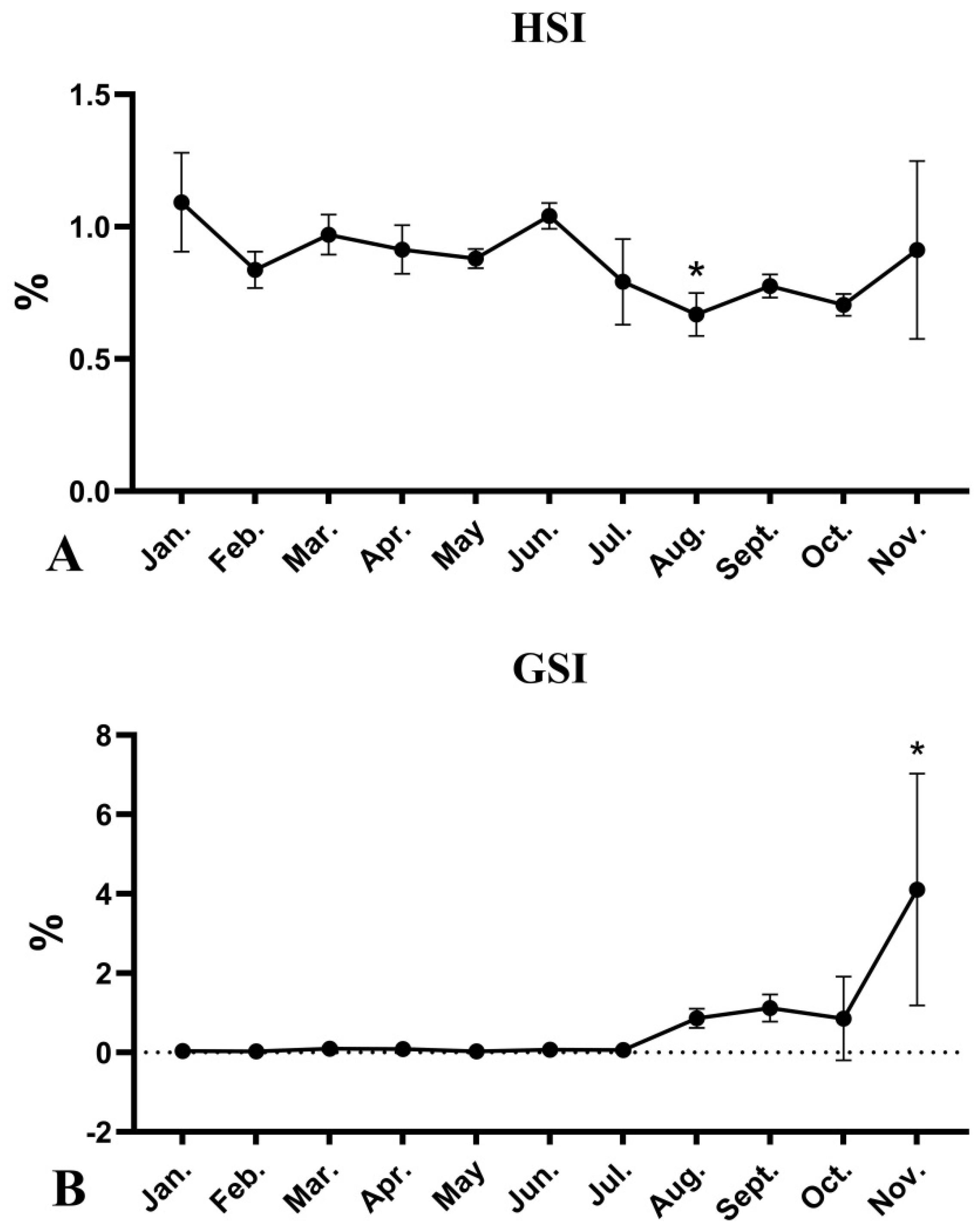
Figure 1. Monthly changes in HSI (A) and GSI (B) values of female O. mykiss during the oocyte development. *: Significant differences between the Jan and other month (Tukey method of the one-way ANOVA test, p < 0.05).
Fishes 06 00062 g001
Figure 2. Monthly changes in cortisol (A), E2 (B) and VTG (C) values of female O. mykiss during the oocyte development. Different letters denote statistically significant differences among the groups (Tukey method of the one-way ANOVA test, p < 0.05).
Fishes 06 00062 g002
Figure 3. Monthly changes in THS of female O. mykiss during the oocyte development. (A), T4; (B), T3; (C), FT4; (D), FT3. Different letters denote statistically significant differences among the groups (Tukey method of the one-way ANOVA test, p < 0.05).
Fishes 06 00062 g003
Figure 4. Monthly changes in genes transcription of female O. mykiss brain during the oocyte development. (A), cyp19b; (B), cgnrh-2; (C), gnrhr; (D), fsh. Different letters denote statistically significant differences among the groups (Tukey method of the one-way ANOVA test, p < 0.05).
Fishes 06 00062 g004
Figure 5. Monthly changes in genes transcription of female O. mykiss ovary during the oocyte development. (A), fshr; (B), lhr; (C), cyp19a1a; (D), foxl2; (E), erα1; (F), erα2; (G), erβ1; (H), erβ2. Different letters denote statistically significant differences among the groups (Tukey method of the one-way ANOVA test, p < 0.05).
Fishes 06 00062 g005
Figure 6. Monthly changes in genes transcription of female O. mykiss liver during the oocyte development. (A), erα1; (B), erα2; (C), erβ1; (D), erβ2. Different letters denote statistically significant differences among the groups (Tukey method of the one-way ANOVA test, p < 0.05).
Fishes 06 00062 g006
Figure 7. Monthly changes in vtg transcriptions of female O. mykiss during the oocyte development. (A), vtg gene transcription in ovary; (B), vtg gene transcription in liver. Different letters denote statistically significant differences among the groups (Tukey method of the one-way ANOVA test, p < 0.05).
Fishes 06 00062 g007
Table 1. Body weight, liver weight and ovary weight of female O. mykiss sampled monthly (n = 3).
MonthBody Weight (g)Liver Weight (g)Ovary Weight (g)
Jan.82.37 ± 11.190.91 ± 0.240.03 ± 0.02
Feb.154.93 ± 1.461.30 ± 0.120.04 ± 0.002
Mar.181.40 ± 57.931.75 ± 0.570.18 ± 0.13
Apr.308.87 ± 62.432.84 ± 0.720.28 ± 0.06
May291.47 ± 62.762.58 ± 0.640.09 ± 0.04
Jun.550.73 ± 149.845.77 ± 1.770.40 ± 0.13
Jul.661.73 ± 162.315.17 ± 1.340.34 ± 0.24
Aug.909.17 ± 49.996.08 ± 0.867.90 ± 2.54
Sept.1047.23 ± 212.678.07 ± 1.2611.76 ± 4.85
Oct.991.93 ± 60.766.70 ± 0.818.91 ± 11.29
Nov.1033.43 ± 286.979.98 ± 5.6247.86 ± 37.95
Table 2. Nucleotide primers used in real-time PCR.
GenePrimer NameSequence (5′–3′)GenBank No. or Article Source
erα1erα1-FCCCTGCTGGTGACAGAGAGAA[28]
erα1-RATCCTCCACCACCATTGAGACT
erα2erα2-FGTGGCACTGCTGGTGACAAC[28]
erα2-RACCACCGAAGCTGCTGTTCT
erβ1erβ1-FCCCAAGCGGGTCCTAGCT[28]
erβ1-RTCCTCATGTCCTTCTGGAGGAA
erβ2erβ2-FCTGACCCCAGAACAGCTGATC[28]
erβ2-RTCGGCCAGGTTGGTAAGTG
vtgvtg-FGTGGACTGGATGAAGGGACAAY049952.1
vtg-RAGAGCGGCTCAGGTTGGAAT
cyp19bcyp19b-FGAGGAAGGCACTGGAAGATGAC[32]
cyp19b-RGCTGGAAGAAACGACTGGGC
fshfsh-FGCGAAACAACGGACCTGAACTAT[33]
fsh-RGGACCACTCCTTGAAGTTACACA
cgnrh-IIcgnrh-II-FCTGTGAGGCAGGAGAATG[33]
cgnrh-II-RACGGTTGATAGGTTGTCTAA
gnrhrgnrhr-FGTCTTTTCCAACCCAGGATGTCAJ272116.1
gnrhr-RGGAAACTGGGACATGTTTGAGAG
fshrfshr-FTCAGTCACCTGACGATCTGCAA[33]
fshr-RTCCTGCAGGTCCAGCAGAAACG
lhrlhr-FCTTCTCAACCTCAATGAAATCTTC[33]
lhr-RGGATATACTCAGATAACGCAGCTT
cyp19a1acyp19a1a-FCTCTCCTCTCATACCTCAGGTT[34]
cyp19a1a-RAGAGGAACTGCTGAGTATGAAT
foxl2foxl2-FTGTGCTGGATTTGTTTTTTGTT[34]
foxl2-RGTGTCGTGGACCATCAGGGCCA
ef1αef1α-FAGGCCATCTGATCTACAAGTGCAF498320.1
ef1α-RGGTGATACCACGCTCCCTCT
Publisher’s Note: MDPI stays neutral with regard to jurisdictional claims in published maps and institutional affiliations.