Brief Report

Innate Immune Encapsulation Allows Gonadal Tolerance to Anisakis simplex (s.s.) Infection in Atlantic mackerel (Scomber scombrus)


1
Department of Life Sciences and Biotechnology, University of Ferrara, 44121 Ferrara, Italy
2
Center of Electron Microscopy, University of Ferrara, 44121 Ferrara, Italy
3
Department of Public Health and Infectious Diseases, Sapienza University of Rome, 00185 Rome, Italy
4
Department of Environmental and Prevention Sciences, University of Ferrara, 44121 Ferrara, Italy
*
Author to whom correspondence should be addressed.
Fishes 2026, 11(8), 453; https://doi.org/10.3390/fishes11080453
Submission received: 25 May 2026 / Revised: 16 June 2026 / Accepted: 30 July 2026 / Published: 1 August 2026
(This article belongs to the Section Fish Pathology and Parasitology)

Abstract

Most of the publications on Anisakis simplex (Rudolphi, 1809) focus upon systematic aspects and zoonotic risk but there is lack of detailed information on infection and cellular immune response of the gonads. The current investigation detected whether gonadal infection by A. simplex affects reproductive development and how Atlantic mackerel, Scomber scombrus (L.), responds immunologically. Thirty-four specimens out of 62 S. scombrus (55%) individuals had the larvae on or in the gonads, and live and calcified dead larvae often co-occurred. Nematode larvae were found in both mature and immature gonads. The intensity of infection varied from 1 to 31 nematodes per gonad (5.68 ± 5.53, mean ± standard deviation). In ovaries that harboured the nematode (inside the organ and in the mesenteries around it), in close vicinity to the larvae, mast cells (MCs), rodlet cells (RCs), macrophages, and macrophage aggregates (MAs) occurred. Fewer of the above cells were observed in the mesenteries of parasitized testes. In the gonads of infected fish, within the granulomas that surrounded the calcified and live larvae, putative epithelioid cells, fibroblasts, MCs, MAs and collagen fibres were present. The absence of a correlation between the intensity of infection and the host gonadosomatic index in female and male gonads might indicate that A. simplex larvae do not affect the production of gametes and gonad development in Atlantic mackerel. Our results suggest that localized encapsulation represents an effective tolerance strategy allowing parasite persistence while preserving host reproductive function.
Key Contribution: Gonads of male and female mackerel harboured larvae of Anisakis simplex; around most nematode larvae, the presence of granulomas was noticed; in granulomas, epithelioid cells, fibroblasts, MCs, MAs and collagen fibres occurred; and there was no correlation between intensity of infection and host gonadosomatic index.

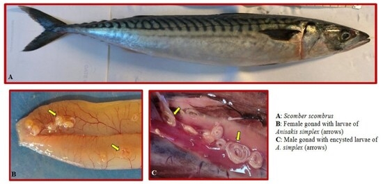
Graphical Abstract

1. Introduction

Anisakid nematodes of the genus Anisakis are widespread marine, brackish and freshwater parasites with an indirect life cycle involving crustaceans such as copepods or euphausiids as intermediate hosts, fish and squid as paratenic hosts and marine mammals (cetaceans and pinnipeds) as definitive hosts [1,2]. In paratenic hosts, L3 larvae of Anisakis species are often encysted in visceral organs and musculature. Their occurrence is of remarkable economic and public health significance because of their zoonotic risks to humans as agents of a gastrointestinal and/or allergic disease known as Anisakiosis [2,3].
Beside the public health concerns, there are consequences for fish health status, even if recent studies suggested that Anisakis spp. L3 larvae in paratenic hosts showed non-feeding behaviour [4]. The immune and reproductive systems are interconnected and are responsible for the defence of the gonads, and this might clarify the existence of sexual dimorphism in the immune response [5]. There are numerous published papers on the occurrence and identification of various nematode parasites of fish gonads from different parts of the world [6,7]. Conversely the effect of parasitic nematodes on fish reproduction has received less attention and available records mainly focus upon the nematodes of the family Philometridae [7]. In most records on the nematode–fish gonad system, authors cited generic terms of organ inflammation and hemorrhages with no micrographic documentation.
Fish generally react to extraintestinal helminth larvae with connectival encapsulation or granulomas [8,9,10]. Most of the investigations on the Atlantic mackerel–A. simplex system were focused on the occurrence of the nematode in fish flesh due to the potential zoonotic implications, and authors frequently ignored other infected organs of the host. A lack of histopathological data due to this nematode on other organs resulted in the publication of the first record of the effect of infection on gross pathology [11]. Afterwards, more S. scombrus individuals were examined in order to document the impact of A. simplex on host reproductive organs in both sexes. The current investigation was undertaken to establish whether infection by the nematode induces any detectable alteration in gonad development. Understanding immune responses within the gonads is essential for assessing the biological costs of infection. We hypothesized that gonadal infection induces a localized innate immune response while having limited effects on reproductive development; thus efforts have been made to ascertain such an assumption.

2. Materials and Methods

Sixty-two individuals of Atlantic mackerel, S. scombrus (36 females and 26 males), with total lengths varying from 29.00 to 44.50 cm (35.39 ± 3.53, mean ± SD) and body weights from 200 to 738 g (358.96 ± 106.13, mean ± SD), destined for food purposes were provided by local fishermen at different times during 2025 and 2026 from the Bay of Biscay (45°30′ N 4°24′ W, FAO Zone 27 VIIIB). Freshly caught fish were eviscerated onboard the vessel and visceral organs were examined. The gonads of each fish were isolated and weighed (g) and the gonadosomatic index (GSI) was calculated according to the following equation provided by Anderson et al. [12]: GSI = gonad weight /body weight × 100. The gonadal maturity was determined using Maier’s scale. Twenty-one mackerel were immature and 41 were mature.
From each Atlantic mackerel opened ventrally, the visceral organs were removed and put in a separate Petri dish in order to easily detect the presence of parasites on/in fish reproductive organs. The parasite load was defined by the prevalence (percentage of infected fish) and the mean intensity (mean number of parasites per infected fish), with deviation (± SD) and range of infection (min–max) determined according to Bush et al. [13].
Statistical analyses were performed in RStudio software (version 3.4.3) with the significance level set at p = 0.05. The normality and homogeneity of variances of the data were assessed through the Shapiro test and Levene test, respectively. When the data did not meet these two assumptions even after transformation, nonparametric statistics were used instead of parametric ones. The Kruskal–Wallis test was applied to verify if the intensity of infection was different between male and female fish. The numbers of live and calcified dead larvae in the gonads of each fish were compared using the Mann–Whitney–Wilcoxon test for paired data. Chi-square was applied to test for differences in the prevalence and intensity of infection in gonads between immature and mature fish. After square root transformation of the data, linear regression was employed to investigate the relationship between intensity of infection and mackerel size (total length and weight) and between intensity of infection and gonadosomatic index (GSI) in both males and females.
Larval nematodes of the Anisakis genus collected from the gonads of 5 Scomber scombrus and classified as live or calcified were washed in 0.9% saline solution and stored in 70% ethanol until undergoing molecular analyses. A subsample of 20 live and 15 calcified L3 larvae was characterized at species level using the diagnostic key based on RFLP of the nuclear ITS, according to D’Amelio et al. [14]. In particular, genomic DNA was isolated using Quick DNA Kit (Zymo Research, Irvine, CA, USA ) following the manufacturers’ instructions and material was used to amplify the regions of interest. For the ITS, the forward primer NC5 (5′-GTAGGTGAACCTGCGGAAGGATCATT-3′) and the reverse primer NC2 (5′-TTAGTTTCTTCCTCCGCT-3′) were used [15]. For each PCR, a negative control without genomic DNA was included and individual PCR products were detected on agarose gels (1%) and stained with SYBRSafe (Invitrogen, Waltham, MA, USA). Positive ITS PCRs were incubated with the endonuclease HinfI for 4 h at 37 °C and then resolved by electrophoresis in 2% agarose gels. The sizes of fragments were characterized by comparison to a 100 bp DNA ladder as a size marker (Promega, Amsterdam, The Netherlands) and to reference specific patterns according to D’Amelio et al. [14].
For transmission electron microscopy (TEM), several pieces, mostly up to 7 × 7 mm in size, of infected and uninfected gonads of both sexes were fixed in 2.5% glutaraldehyde in 0.1 M sodium cacodylate buffer for 3 h at 4 °C. The fixed tissues were post-fixed in 1% osmium tetroxide for 2 h, rinsed, and stored in 0.1 M sodium cacodylate buffer containing 6% sucrose for 48 h. Thereafter, the tissue pieces were dehydrated using a graded acetone series and embedded in epoxy resin (Durcupan ACM, Fluka, Milan, Italy). Semithin sections (1.5 µm) were cut using a Reichert Om U2 ultramicrotome (Reichert, Wien, Austria), stained with Toluidine Blue, examined and photographed using a Nikon Microscope ECLIPSE 80i (Nikon, Tokyo, Japan). Ultra-thin sections (90 nm) were stained with 4% uranyl acetate solution in 50% ethanol and Reynold’s lead citrate and then examined using a Talos L120C transmission electron microscope (Thermo Fisher Scientific Company, Waltham, MA, USA).

3. Results

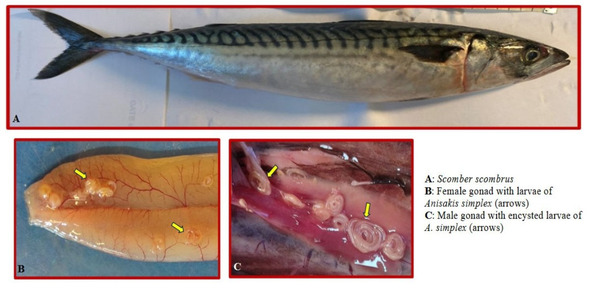
Sixty-one mackerel (prevalence 98%) harboured A. simplex L3 larvae in visceral organs with an intensity of infection ranging from 4 to 650 larvae per fish (58.56 ± 90.09, mean ± SD) and 34 of them had infected gonads (prevalence 55%). A total of 3572 A. simplex larvae were counted, of which 193 were in the gonads and 3379 in other organs. The intensity of infection in gonads ranged from 1 to 31 larvae (5.68 ± 5.53, mean ± SD) and did not differ significantly between male and female mackerel (W = 129, p > 0.05). Prevalence and intensity of infection were similar in immature and mature gonads (p > 0.05). There were no statistically significant differences in the number of calcified dead larvae (i.e., 93) compared to the number of live ones (i.e., 100) in gonads (V = 11, p > 0.05).
The number of nematode larvae found in gonads was not correlated to mackerel size (R2 = 0.038, p > 0.05 for total length; R2 = 0.032, p > 0.05 for weight). The relationships between the gonad development expressed by the GSI and the intensity of infection (general intensity, i.e., considering larvae in all fish organs, and gonadic intensity, i.e., considering only larvae in gonads) were analyzed and no significant correlations were found in both female (general intensity R2 = 0.011, p > 0.05; gonadic intensity R2 = 0.060, p > 0.05) and male (general intensity R2 = 0.052, p > 0.05; gonadic intensity R2 = 0.058, p > 0.05) mackerel.
Genomic DNA from a total of 20 live and 15 calcified larvae were isolated from gonads and the ITS was amplified at PCR. Positive amplicons were obtained for 95% of the live larvae analyzed (19/20) and for 13% of the calcified larvae (2/15), with an amplicon size of ~1000 bp for the ITS at gel visualization. All the 21 good amplicons digested with HinfI showed a pattern typical of Anisakis simplex (s.s.), with two main bands of around 620 bp and 250 bp and a smaller of around 80 bp.
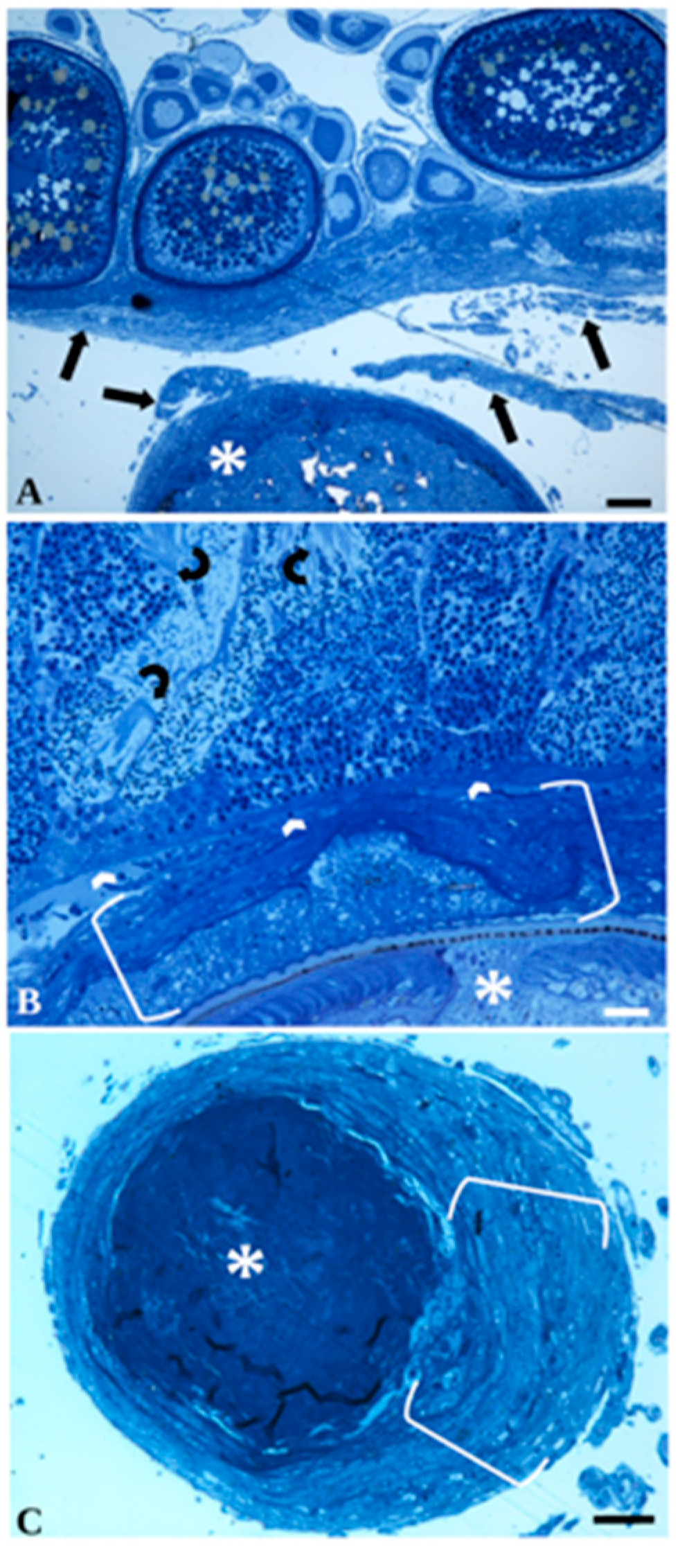
Observations of the semithin sections documented the interface region between A. simplex L3 larvae and female and male gonads (Figure 1A and Figure 1B respectively). A variable number of live and calcified nematode larvae co-occurred on the surface of gonads or were penetrated inside. The mesenteries covering the gonads were one of most infected sites. Inside the ovaries, the nematode larvae typically occupied the lumen of the organ and were not encircled by granulomas. Accordingly, when the nematode occurred inside the ovaries and testes, no intense host cellular reaction was induced and the worm’s body substituted germinal tissues. Interestingly, the nematode larvae located on the surface of the organ did not have an effect on the ovary germinal epithelium and oocytes in different stages of development appeared intact, with no sign of necrosis and with an absence of liquefaction of yolk globules (Figure 1A). Similarly, A. simplex L3 larvae over the surface of the testis did not induce any damage to the germinal cells and the occurrence of numerous spermatocytes and sperms with long flagella was documented (Figure 1B). When the A. simplex larva was observed on the external surface of the ovaries and testes, the nematode was surrounded by a granuloma consisting of three layers (Figure 1B,C).
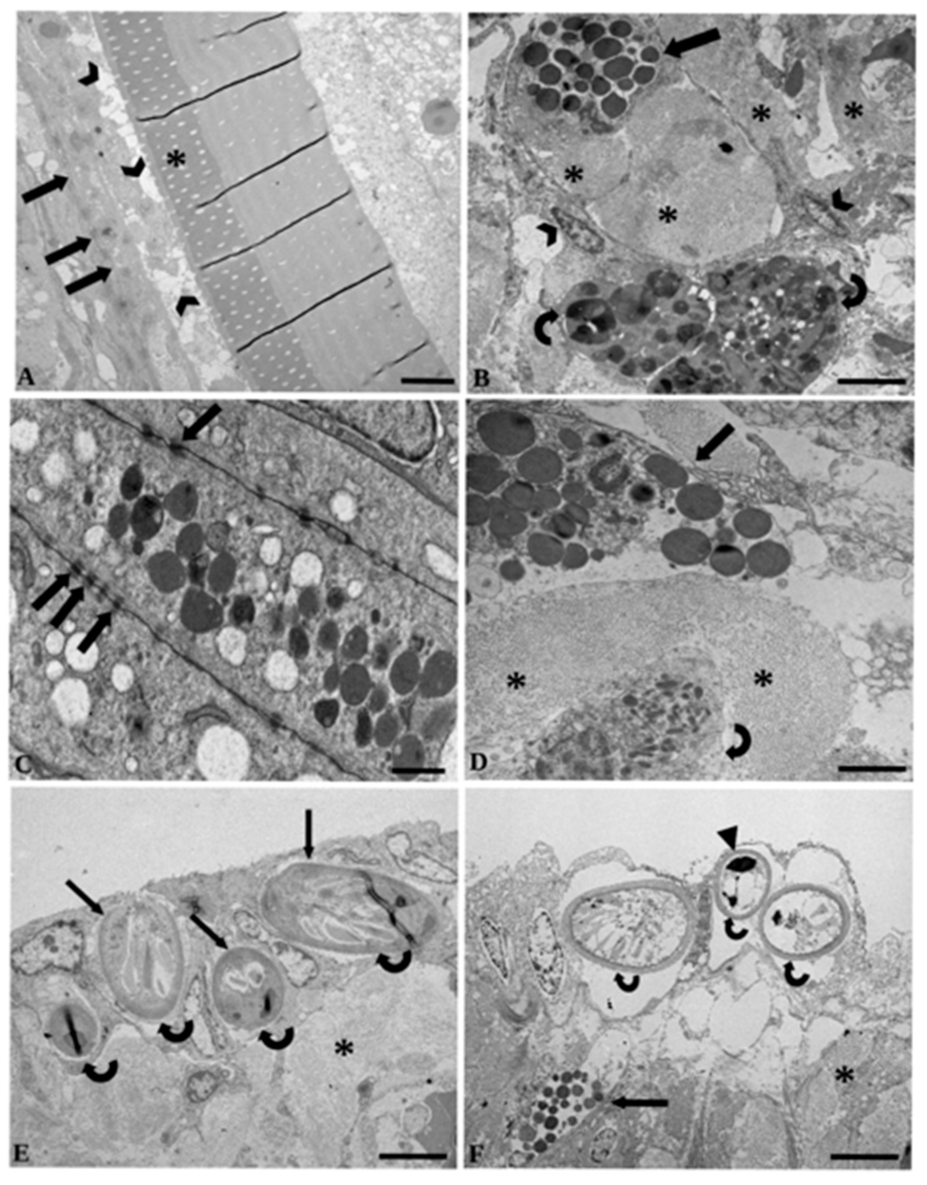
The most inner layer of the granuloma in close vicinity to the parasite’s cuticle was made up of elongated cells with numerous desmosomes between them which might suggest they were epithelioid cells (Figure 2A), and frequently with a rarefied cytoplasm. Macrophages and MCs were abundant in the middle layer of the granuloma. Macrophages were big round-to-oval cells with an irregular outline and many electron-dense and electron-lucent vesicles in the cytoplasm (Figure 2B). An MC appeared as an elongated cell (Figure 2C) or a round–oval-shaped cell, with an intact nucleus with peripheral heterochromatin and a cytoplasm containing numerous round electron-dense granules and seldom also contained some electron-lucent vesicles (Figure 2C,D). MCs presented numerous desmosomes with surrounded cells (Figure 2C). Within the middle layer, the co-occurrence of MCs and macrophages (Figure 2B) and MCs and neutrophils (Figure 2D) was frequent; indeed, collagen fibres were scattered among these types of cells (Figure 2D). The external layer of the granuloma was commonly constituted by macrophages and some fibroblasts, and seldom contained neutrophils and a mesh of collagen fibres among cells.
Within the mesenteries around the ovaries and testes, RCs were noticed (Figure 2E and Figure 2F respectively). These cells were characterized by a distinctive cell cortex and conspicuous inclusions, called rodlets (Figure 2E,F), which account for their name. In some instances, RCs showed an apoptotic nucleus with condensed electron-dense chromatin; indeed, the cytoplasm was vacuolated and rodlets were small in size and uncommon in aspect (Figure 2F). In mesenteries of several infected gonads of both sexes, MCs and RCs co-occurred (Figure 2F). A limited number of RCs were noticed in uninfected gonads.

4. Discussion

The reproduction and the immune system are interconnected and are responsible for immune defence in the gonads [5]. Fish react to extraintestinal helminths by producing granulomas or connectival encapsulation [9,16,17,18], a chronic inflammatory focal lesion that appears as a nodule in tissues/organs of the host [10,11,16,19]. Encapsulation is a mutual adaptation between the host’s immune response and the parasite, reaching a strategic compromise between the two to ensure the survival of both species [18,20]. It is stated that sequestration of a high number of live nematode larvae [20] and cestode larvae [18] is a host response itself, but might also be an advantage for the parasite that exploits it for survival. Granulomas can frequently also contain dead parasites, indicating that fibrosis itself is an adaptation to reduce infections rather than a mere side-effect of inflammation [18].
In the A. simplexS. scombrus system, larvae on the surface of male and female gonads were isolated inside the granuloma and this encapsulation of the nematode did not interfere with organ function. Concerning the nematode larvae inside the gonads, the mass of worms was very low, and likely uninfected portions of the organ preserved their integrity. Accordingly, the majority of infected Atlantic mackerel were sexually mature with a high quantity of gametes and our results showed that the intensity of infection was not correlated to GSI, suggesting that the larvae did not significantly alter the gonad development in male and female fish. This finding is consistent with that of Chou et al. [21] on A. simplex third-stage larvae in the spotted mackerel S. australasicus. Nevertheless, there is controversy among authors on the impact of nematodes on fish reproduction (see for details [6]) and host gonadosomatic index (GSI). Some accounts mentioned that A. pegreffii and Eustrongylides sp. altered the GSI of the host [22,23], reducing the effective volume of the gonad and leading to lower fecundity of the fish [23,24] or full castration [25,26]. A total of 1845 nematode specimens of the genus Philometra infect wild and cultured marine fish gonads with various consequences including a severe decrease in reproductive fitness [24,25,27]. One point needs to be emphasized: Philometra parasitizes the fish gonads at the adult stage, and they are hematophagous and larger in dimension in comparison to Anisakis spp., which in fish are at the larval stage. The results provided in the current study complement those listed above (summarized in [27]). It is reported that immature fish are not susceptible to infection with nematodes because the parasite is more likely to infect a host with developed gonads that provide a plentiful food supply to the worm [25,27]. However, in the present study, live larvae were also encysted on some immature gonads.
Only a few papers published on the effects of nematodes on fish gonads provided an adequate documentation of the type of immune cells involved in the organ response. Exceptionally, Bakenthaster et al. [28] in their study on the effects of the nematode Philometra floridensis in ovarian tissue of Sciaenops ocellatus listed the types of immune cells involved but no clear photographic documentation was provided to substantiate the authors’ results. In infected female gonads of S. scombrus, co-occurrence of some types of immune cells was frequent. Some insights on the epithelioid cells, macrophages, MAs, neutrophils, MCs, fibroblasts and RCs encountered in the granulomas around A. simplex L3 larva encysted on/in gonads of S. scombrus will be discussed below. The inner layer of the granuloma nearest to the pathogen/parasite is mainly formed by dark necrotic epithelioid cells and non-necrotic epithelioid cells. The occurrence of these cells, that are transformed macrophages [16], was mentioned in several granulomas around different nematode species in infected fish organs [11,29].
The presence of macrophages and MAs within the thickness of the granuloma around the A. simplex larvae in gonads of Atlantic mackerel tallies with similar findings mentioned in accounts on other organs (e.g., liver, intestine, and pyloric caeca) in different fish species that harboured nematode larvae [11,17,30]. Neutrophils have been shown to be the dominant cell type during the first immediate inflammatory response [31,32]. Within the thickness of granulomas in infected male and female gonads of S. scombrus, only some neutrophils were noticed. The lack or rare presence of neutrophils in granulomas around the larvae of nematodes residing in fish visceral organs was also reported in [10,29].
Mast cells (MCs) comprise a heterogeneous cell population and are important components of adaptive and innate immunity [33,34]. An impressive number of MCs were documented in infected gonads of male and female S. scombrus in close proximity to the A. simplex larvae. Similar findings were reported in different fish species that harbour metazoan parasites, and evidence for the role of MC tryptase in different fish–parasite systems reinforces its active role in tissue repair after worm damage [10]. The presence of layers of fibroblasts and fibrocytes in the granuloma tallies with known processes in the development of chronic inflammation [8,10,16,29]. Clusters of RCs were encountered in the ovaries and testes of S. scombrus that harboured A. simplex larvae. Rodlet cells (RCs) are exclusive to fish and have been associated most often with epithelia. Records concerning the role of RCs as immune effector cells have focused on their mobilization and recruitment in response to helminth parasites growing [10,19,35].
In vertebrates, sex plays an essential role in shaping the immune response [5]. Innate and adaptive immune responses develop against infections and commonly differ between females and males, and numerous accounts support the fact that hormones of the endocrine system are involved in the immunological dimorphism between two sexes [36,37]. In female fish, ovarian function is dependent on the establishment and continual remodelling of a complex vascular system which enables the follicle and/or corpus luteum to receive the required supply of nutrients, oxygen and hormonal support, and facilites the release of steroids [38]. The immune response of S. scomber documented herein was less intense in parasitized testes than in infected ovaries. More MCs, macrophages, MAs and RCs are encountered in infected female gonads and, since the above cell types move with the circulatory system, their efficient recruitment in ovaries could be related to the rich vascularization of this organ. The above results on sex differences in immune reactions are in agreement with the statement that both innate and adaptive immune responses are typically lower in males than in females, a phenomenon that has evolved in different animal species [37].

5. Conclusions

The results of the current investigation show that A. simplex did not induce very intense pathological alterations in Atlantic mackerel gonads. Gonads reacted to the parasite with the recruitment of different immune cell types, and mast cells were especially numerous. The inflammatory reaction seems to be more pronounced in the ovaries than in the testes and possible explanations could be hormonal regulation and differences in gonadal vascularization. Localized granuloma formation was shown to represent a tolerance strategy that balances parasite persistence with the preservation of the reproductive capacity of the host.

Author Contributions

Writing—original draft preparation, B.S.D.; Investigation, B.S.D., F.P., E.F., S.C. and S.D.; Conceptualization, B.S.D. and L.G.; Methodology, F.P., E.F., E.S., P.B., S.C. and S.D.; Visualization, F.P. and E.F.; Data curation, S.C. and S.D.; Funding acquisition, B.S.D.; Writing—review and editing, B.S.D. and L.G.; Validation, L.G. All authors have read and agreed to the published version of the manuscript.

Funding

This study was supported by local grants from the University of Ferrara to B. Sayyaf Dezfuli (grant number FAR 2025).

Institutional Review Board Statement

In Italy there is no need to have university ethics committee approval when we work on fish destined for food purposes. In Biscay Bay, total annual Atlantic mackerel catches range roughly between 10,000 and 15,000 tonnes, which means 27 tonnes per day are destined to the market, and we had 62 individuals for this investigation.

Data Availability Statement

The original contributions presented in this study are included in the article. Further inquiries can be directed to the corresponding author.

Acknowledgments

We are indebted to A. Gavioli from the University of Ferrara for helping with the statistical analyses of data. We also thank C. Holland from Trinity College Dublin for the English correction of the MS.

Conflicts of Interest

The authors declare no conflicts of interest.

References

  1. Cipriani, P.; Giulietti, L.; Palomba, M.; Fernandez, V.R.; Mattiucci, S.; Bjørge, A.; Levsen, A.; Bao, M. Anisakid biodiversity in two young harbour seals (Phoca vitulina L.) from coastal South-West Norway. Parasitol. Res. 2025, 124, 110. [Google Scholar] [CrossRef] [Scilit]
  2. Mattiucci, S.; Cipriani, P.; Levsen, A.; Paoletti, M.; Nascetti, G. Molecular epidemiology of Anisakis and anisakiasis: An ecological and evolutionary road map. Adv. Parasitol. 2018, 99, 93–263. [Google Scholar] [CrossRef] [Scilit] [PubMed]
  3. EFSA Panel on Biological Hazards (BIOHAZ); Allende, A.; Alvarez-Ordóñez, A.; Bortolaia, V.; Bover-Cid, S.; De Cesare, A.; Dohmen, W.; Guillier, L.; Herman, L.; Jacxsens, L.; et al. Re-evaluation of certain aspects of the EFSA scientific opinion of April 2010 on risk assessment of parasites in fishery products, based on new scientific data. Part 2. EFSA J. 2024, 22, e09090. [Google Scholar] [CrossRef] [Scilit] [PubMed]
  4. Sabadel, A.; Gay, M.; Lane, H.S.; Bourgau, O.; Bury, S.J.; Delgado, J.; Duflot, M. Just hitching a ride: Stable isotopes reveal non-feeding behaviour of Anisakis simplex within its host fish. J. Fish Dis. 2025, 48, e14043. [Google Scholar] [CrossRef] [Scilit] [PubMed]
  5. Caballero-Huertas, M.; Salazar-Moscoso, M.; Ribas, L. Sex is a crucial factor in the immune response: An ichthyological perspective. Rev. Fish. Sci. Aquacult. 2025, 33, 217–237. [Google Scholar] [CrossRef] [Scilit]
  6. Moravec, F.; de Buron, I. A synthesis of our current knowledge of philometrid nematodes, a group of increasingly important fish parasites. Folia Parasitol. 2013, 60, 81–101. [Google Scholar] [CrossRef] [Scilit] [PubMed]
  7. Moravec, F.; Fazaa, H.A.; Ali, A.H.; Saud, H.A. Two new and one newly recorded gonad-infecting species of Philometra Costa, 1845 (Nematoda: Philometridae) parasitising marine fishes in Iraq. Syst. Parasitol. 2023, 100, 439–453. [Google Scholar] [CrossRef] [Scilit] [PubMed]
  8. López-Verdejo, A.; Born-Torrijos, A.; Raga, J.A.; Canovas-Cabrera, H.; Montero, F.E. Validation of mollies and gilthead seabreams as new model organisms for the study of anisakids: Experimental infection and histological analysis of the infective process in sailfin molly. Aquaculture 2025, 599, 742120. [Google Scholar] [CrossRef] [Scilit]
  9. Weber, J.N.; Steinel, N.C.; Peng, F.; Shim, K.C.; Lohman, B.K.; Fuess, L.E.; Subramanian, S.; De Lisle, S.P.; Bolnick, D.I. Evolutionary gain and loss of a pathological immune response to parasitism. Science 2022, 377, 1206–1211. [Google Scholar] [CrossRef] [Scilit] [PubMed]
  10. Sayyaf Dezfuli, B.; Pironi, F.; Castaldelli, G.; Giari, L.; Lanzoni, M.; Buchmann, K.; Kania, P.W.; Bosi, G. Anguilla anguilla vs Contracaecum rudolphii: Granuloma allows host tolerance and parasite survival. Aquaculture 2024, 591, 741138. [Google Scholar] [CrossRef] [Scilit]
  11. Sayyaf Dezfuli, B.; Bernacchia, G.; De Bastiani, M.; Pinca, M.; Franchella, E.; Bellini, L.; Castaldelli, G.; Giari, L. Anisakid larvae in the organs of Atlantic mackerel, Scomber scombrus (L.): Host or parasite superiority? Food Waterborne Parasitol. 2025, 41, e00300. [Google Scholar] [CrossRef] [Scilit] [PubMed]
  12. Anderson, R.O.; Gutreuter, S.J. Length, weight and associated structural indices. In Fisheries Techniques; Nielsen, L.A., Johnson, D.L., Eds.; American Fisheries Society: Bethesda, MD, USA, 1983; pp. 283–300. [Google Scholar]
  13. Bush, A.O.; Lafferty, K.D.; Lotz, J.M.; Shostak, A.W. Parasitology meets ecology on its own terms: Margolis et al. revisited. J. Parasitol. 1997, 83, 575–583. Available online: https://www.jstor.org/stable/3284227 (accessed on 16 June 2026). [CrossRef] [Scilit]
  14. D’Amelio, S.; Mathiopoulos, K.D.; Santos, C.P.; Pugachev, O.N.; Webb, S.C.; Picanço, M.; Paggi, L. Genetic markers in ribosomal DNA for the identification of members of the genus Anisakis (Nematoda: Ascaridoidea) defined by polymerase-chain-reaction-based restriction fragment length polymorphism. Int. J. Parasitol. 2000, 30, 223–226. [Google Scholar] [CrossRef] [Scilit] [PubMed]
  15. Zhu, X.Q.; D’Amelio, S.; Paggi, L.; Gasser, R.B. Assessing sequence variation in the internal transcribed spacers of ribosomal DNA within and among members of the Contracaecum osculatum complex (Nematoda: Ascaridoidea: Anisakidae). Parasitol. Res. 2000, 86, 677–683. [Google Scholar] [CrossRef] [Scilit] [PubMed]
  16. Ferguson, H.W. Systemic Pathology of Fish: A Text and Atlas of Normal Tissues in Teleosts and their Responses in Disease; Scotian Press: London, UK, 2006. [Google Scholar]
  17. Kumas, K.; Sayyaf Dezfuli, B.; Franchella, E.; Duan, Y.; Kania, P.W.; Buchmann, K. Cellular responses in rainbow trout Oncorhynchus mykiss to experimental Anisakis simplex infection. Parasitol. Res. 2025, 124, 109. [Google Scholar] [CrossRef] [Scilit] [PubMed]
  18. De Lisle, S.P.; Bolnick, D.I. Male and female reproductive fitness costs of an immune response in natural populations. Evolution 2021, 75, 2509–2523. [Google Scholar] [CrossRef] [Scilit] [PubMed]
  19. Sayyaf Dezfuli, B.; Lorenzoni, M.; Carosi, A.; Giari, L.; Bosi, G. Teleost innate immunity, an intricate game between immune cells and parasites of fish organs: Who wins, who loses. Front. Immunol. 2023, 14, 1250835. [Google Scholar] [CrossRef] [Scilit] [PubMed]
  20. Buchmann, K. Fish immune responses against endoparasitic nematodes—Experimental models. J. Fish. Dis. 2012, 35, 623–635. [Google Scholar] [CrossRef] [Scilit] [PubMed]
  21. Chou, Y.Y.; Wang, C.S.; Chen, H.G.; Chen, H.Y.; Chen, S.N.; Shih, H.H. Parasitism between Anisakis simplex (Nematoda: Anisakidae) third-stage larvae and the spotted mackerel Scomber australasicus with regard to the application of stock identification. Vet. Parasitol. 2011, 177, 324–331. [Google Scholar] [CrossRef] [Scilit] [PubMed]
  22. Eissa, A.E.; Korany, R.M.; El Zlitne, R.A.; Magdy, A.; Sharaf, M.S.; Abdelbaky, A.A.; Mahmoud, A.E.; Abd ElMaged, R.; Qorany, R.A.; Mohamed, A.M.K.; et al. Impact of Anisakis pegreffi infection on gonadal health and gonadosomatic index of European hake (Merluccius merluccius). JAVS 2023, 8, 67–74. [Google Scholar] [CrossRef] [Scilit]
  23. Kaur, P.; Shrivastav, R.; Dar, B.A.; Jha, G.N.; Qureshi, T.A. Suppression of ovarian development in freshwater fishes due to endo-helminthic infection. Indian. J. Fish. 2015, 62, 128–132. [Google Scholar]
  24. Moravec, F.; Bakenhaster, M.D.; Seyoum, S.; Tringali, M.D. Morphological and genetic description of two new species of philometrid nematodes (Philometridae) parasitic in needlefishes (Belonidae) from estuaries of Florida, USA. Folia Parasitol. 2021, 68, 008. [Google Scholar] [CrossRef] [Scilit] [PubMed]
  25. Chávez, R.A.; Oliva, M.E. Philometra chilensis (Nematoda, Philometridae) affects the fecundity of the red cusk-eel, Genypterus chilensis (Guichenot) (Pisces, Ophidiidae) in Chile. Acta Parasit. 2011, 56, 236–237. [Google Scholar] [CrossRef] [Scilit]
  26. Moravec, F.; Salgado-Maldonado, G. A new species of Philometra (Nematoda, Philometridae) from the gonads of the rock hind Epinephelus adscensionis (Osteichthyes) from the southern Gulf of Mexico. Acta Parasitol. 2007, 52, 376–381. [Google Scholar] [CrossRef] [Scilit]
  27. Hesp, S.A.; Hobbs, R.P.; Potter, I.C. Infection of the gonads of Glaucosoma hebvaicum by the nematode Philometva lateolabracis: Occurrence and host response. J. Fish Biol. 2002, 60, 663–673. [Google Scholar] [CrossRef] [Scilit]
  28. Bakenhaster, M.D.; Lowerre-Barbieri, S.; Kiryu, Y.; Walters, S.; Fajer-Avila, E.J. Philometra floridensis (Nematoda: Philometridae) damages ovarian tissue without reducing host (Sciaenops ocellatus) fecundity. Dis. Aquat. Org. 2014, 108, 227–239. [Google Scholar] [CrossRef] [Scilit] [PubMed]
  29. Molnár, K.; Székely, C.; Baska, F.; Müller, T.; Zuo, S.; Kania, P.W.; Nowak, B.; Buchmann, K. Differential survival of 3rd stage larvae of Contracaecum rudolphii type B infecting common bream (Abramis brama) and common carp (Cyprinus carpio). Parasitol. Res. 2019, 118, 2811–2817. [Google Scholar] [CrossRef] [Scilit] [PubMed]
  30. Speirs, Z.C.; Loynes, C.A.; Mathiessen, H.; Elks, P.M.; Renshaw, S.A.; Jørgensen, L.V.G. What can we learn about fish neutrophil and macrophage response to immune challenge from studies in zebrafish. Fish Shellfish Immunol. 2024, 148, 109490. [Google Scholar] [CrossRef] [Scilit] [PubMed]
  31. Bader, A.; Gao, J.; Rivière, T.; Schmid, B.; Walzog, B.; Maier-Begandt, D. Molecular insights into neutrophil biology from the zebrafish perspective: Lessons from CD18 deficiency. Front. Immunol. 2021, 12, 677994. [Google Scholar] [CrossRef] [Scilit] [PubMed]
  32. Ying, C.; Fang, X.; Wang, H.; Yang, Y.; Xu, P.; Liu, K.; Yin, G. Anisakidae parasitism activated immune response and induced liver fibrosis in wild anadromous Coilia nasus. J. Fish Biol. 2022, 100, 958–969. [Google Scholar] [CrossRef] [Scilit] [PubMed]
  33. Wang, X.; Zhang, P.; Tang, Y.; Chen, Y.; Zhou, E.; Gao, K. Mast cells: A double-edged sword in inflammation and fibrosis. Front. Cell Dev. Biol. 2024, 12, 1466491. [Google Scholar] [CrossRef] [Scilit] [PubMed]
  34. Da Silva, W.F.; Simões, M.J.; Gutierre, R.C.; Egami, M.I.; Santos, A.A.; Antoniazzi, M.M.; Ranzani-Paiva, M.J.T. Special dyeing, histochemistry, immunohistochemistry and ultrastructure: A study of mast cells/eosinophilic granules cells (MCs/EGC) from Centropomus parallelus intestine. Fish Shellfish Immunol. 2017, 60, 502–508. [Google Scholar] [CrossRef] [Scilit] [PubMed]
  35. Sayyaf Dezfuli, B.; Pironi, F.; Maynard, B.; Simoni, E.; Bosi, G. Rodlet cells, fish immune cells and a sentinel of parasitic harm in teleost organs. Fish Shellfish Immunol. 2022, 121, 516–534. [Google Scholar] [CrossRef] [Scilit] [PubMed]
  36. Simkova, A.; Jarkovský, J.; Koubková, B.; Barus, V.; Prokes, M. Associations between fish reproductive cycle and the dynamics of metazoan parasite infection. Parasitol. Res. 2005, 95, 65–72. [Google Scholar] [CrossRef] [Scilit] [PubMed]
  37. Klein, S.L.; Flanagan, K.L. Sex differences in immune responses. Nat. Rev. Immunol. 2016, 16, 626–638. [Google Scholar] [CrossRef] [Scilit] [PubMed]
  38. Robinson, R.S.; Woad, K.J.; Hammond, A.J.; Laird, M.; Hunter, M.G.; Mann, G.E. Angiogenesis and vascular function in the ovary. Reproduction 2009, 138, 869–881. [Google Scholar] [CrossRef] [Scilit] [PubMed]
Figure 1. Semithin sections of Atlantic mackerel gonads infected with nematode Anisakis simplex larvae. (A) Infected ovary: A. simplex larva (asterisk) in close proximity to the surface of the organ is encircled by the mesentery (arrows). Within the ovary, numerous oocytes in different developmental stages with a lack of necrosis and liquefaction of the yolk globules are evident. Stain: Toluidine Blue. Scale bar: 100 μm. (B) Nematode larva (asterisk) lying on external surface of the mesenteries (arrow heads). In the periphery of the testis is the occurrence of sperm flagella (curved arrows) and numerous gametes in different developmental stages; note the thickness of the granuloma (parenthesis). Stain: Toluidine Blue. Scale bar: 25 μm. (C) A calcified larva (asterisk), where different layers of the granuloma are evident (parentheses). Stain: Toluidine Blue. Scale bar: 25 μm.
Fishes 11 00453 g001
Figure 2. Transmission electron micrographs of Atlantic mackerel gonads infected with the nematode Anisakis simplex larvae. (A) Interface region between A. simplex larval cuticle (asterisk) and inner part of the granuloma. Residues of host cells (arrow heads) and several desmosomes (arrows) between epithelioid cells can be observed. Scale bar: 5 μm. (B) Encysted larva on the surface of a female gonad. Among the elements of the granuloma, MCs (arrow), macrophages (curved arrows), collagen fibres (asterisks) and fibroblasts (arrow heads) are visible. Scale bar: 5 μm. (C) High magnification of an MC in a granuloma on a male testis. Electron-dense granules are scattered in the cytoplasm; note several desmosomes (arrows) between the MC and surrounding cells. Scale bar: 0.5 μm. (D) Co-occurrence of an MC in degranulation (arrow) and a neutrophil (curved arrow) separated by collagen fibres (asterisks). Scale bar: 2 μm. (E) Upper part of the mesentery around an infected ovary, with four RCs (curved arrows). Note the presence of the cell cortex (arrows) of each RC and that the cytoplasm is filled with small rodlets. Collagen fibres (asterisk) are also present. Scale bar: 5 μm. (F) Upper region of the mesentery around a parasitized testis; the co-presence of RCs (curved arrows) and an MC (thick arrow) is visible. Vacuolisation of RC cytoplasms, small-size rodlets with an unusual aspect and an apoptotic nucleus (arrow head) are evident; collagen fibres (asterisk) are also present. Scale bar: 5 μm.
Fishes 11 00453 g002
Disclaimer/Publisher’s Note: The statements, opinions and data contained in all publications are solely those of the individual author(s) and contributor(s) and not of MDPI and/or the editor(s). MDPI and/or the editor(s) disclaim responsibility for any injury to people or property resulting from any ideas, methods, instructions or products referred to in the content.