Article

LncRNA-Mediated Transcriptional Responses to Piscirickettsia salmonis Infection in Rainbow Trout Skeletal Muscle and Primary Myotubes


by
Rodrigo Zuloaga
1,2,3,†,
Luciano Ahumada-Langer
1,†,
Phillip Dettleff
4,
Alfredo Molina
2,3 and
Juan Antonio Valdés
1,2,3,*
1
Laboratorio de Biotecnología Molecular, Facultad de Ciencias de la Vida, Universidad Andrés Bello, Santiago 8370186, Chile
2
Interdisciplinary Center for Aquaculture Research—Applied Research (INCAR2), Universidad Andres Bello, Santiago 8370146, Chile
3
Centro de Investigacion Marina Quintay (CIMARQ), Universidad Andres Bello, Quintay 2340000, Chile
4
Escuela de Medicina Veterinaria, Facultad de Agronomia y Sistemas Naturales, Facultad de Ciencias Biologicas y Facultad de Medicina, Pontificia Universidad Catolica de Chile, Santiago 7820436, Chile
*
Author to whom correspondence should be addressed.
These authors contributed equally to this work.
Fishes 2026, 11(7), 398; https://doi.org/10.3390/fishes11070398 (registering DOI)
Submission received: 5 May 2026 / Revised: 21 June 2026 / Accepted: 2 July 2026 / Published: 6 July 2026
(This article belongs to the Special Issue Aquaculture Omics: Current Status and Future Perspectives)

Abstract

Piscirickettsia salmonis is one of the most significant pathogens affecting salmon farming. Besides liver, head kidney and spleen, skeletal muscle has shown transcriptional immune responses to these bacteria, but the contribution of non-coding RNAs remains poorly understood. This study investigates the role of long non-coding RNAs (lncRNAs) in the immune response of rainbow trout skeletal muscle and primary myotube cultures infected with P. salmonis. Using RNA-seq data from both in vivo and in vitro muscle under control and infected conditions, the analysis identified 4263 candidate lncRNAs through a stringent bioinformatics pipeline. These lncRNAs were mostly classified as exonic and intergenic, showing distinct genomic distributions and structural differences depending on the source. Expression analyses revealed that cell type had a stronger effect on lncRNA profiles than infection status. From 764 differentially expressed lncRNAs, 191 were uniquely associated with infected and 180 with control conditions, mainly unannotated. Functional predictions based on co-expression and proximity to coding genes suggest that lncRNAs are primarily involved in downregulation of structural-cellular maintenance under control conditions, whereas during infection, they are related to immunity, signaling, and apoptosis. Overall, the findings indicate that lncRNAs exhibit origin-specific regulatory roles and are modulated by P. salmonis infection, highlighting their potential importance in fish immune responses.
Key Contribution: Identification of 4263 novel lncRNAs in rainbow trout skeletal muscle and myotube cultures reveals strong tissue-specific expression patterns, with cell type exerting a greater influence than Piscirickettsia salmonis infection. Differential expression and co-expression analyses suggest that lncRNAs play key regulatory roles in immune response, cell signaling, and apoptotic processes during P. salmonis infection in fish skeletal muscle cells.

1. Introduction

Skeletal muscle is the most abundant tissue in teleost fishes, accounting for 50–60% of their body weight [1]. This tissue is important for motor activities and plays key roles in energy and protein metabolism; previous research has even demonstrated its immunomodulatory capacity against viruses and bacteria [2,3,4]. The immune response of skeletal muscle to pathogens has been demonstrated using in vitro and in vivo methods. Primary culture infection studies with cytokines, pathogen-associated molecular patterns (PAMPs) or bacteria have been conducted on Atlantic salmon (Salmo salar) and rainbow trout (Oncorhynchus mykiss), which have demonstrated the induction of expression of genes related to innate and adaptive immune responses [5,6,7]. On the other hand, infection studies using pathogens in fine flounder (Paralichthys adspersus), Nile tilapia (Oreochromis niloticus) or rainbow trout, among others, revealed variations in the expression of immune-related molecules in skeletal muscle, such as toll-like receptors, pro-inflammatory cytokines, and antimicrobial peptides [2,8,9,10].
Piscirickettsia salmonis is one of the most significant pathogens affecting salmon farming [11]. This pathogen is a Gram-negative, nonmotile, facultatively intracellular bacterium and has been described as the causative agent of salmonid rickettsial septicemia (SRS) or piscirickettsiosis [12,13]. This septicemic disease affects various species of salmonids, such as Atlantic salmon, coho salmon (Oncorhynchus kisutch), and rainbow trout [14]. Also, P. salmonis can affect several organs, including liver, spleen, head kidney, and, more recently described, skeletal muscle [11,15,16]. To delve into the effects of pathogen infection on skeletal muscle and the molecular mechanisms associated with its immunocompetence, next-generation sequencing (NGS) appears as an excellent alternative to research this concern [17]. RNA-Seq analysis of rainbow trout skeletal muscle infected with P. salmonis exhibited a strong transcriptional inflammatory response that included activation of IL-8-related pathways, and showed that many genes involved in the production, organization, or function of the contractile apparatus were downregulated [18]. Similarly, transcriptomic response of trout muscle cells with cortisol and/or P. salmonis showed differentially expressed transcripts related to apoptosis, inhibition of cell proliferation and innate immune enriched biological processes [19]. Despite this work, previous reports do not describe or characterize additional (non-protein-coding) RNAs, including non-coding RNAs (ncRNAs), which have become more recognized as relevant as critical regulators of gene expression [20].
The ncRNAs are generally grouped into short ncRNAs and long ncRNAs (lncRNAs) based on their length [21]. LncRNAs are typically over 200 nucleotides in length and have been classified into long intronic ncRNAs, long intergenic ncRNAs, natural antisense transcripts, and a new group of ncRNAs known as circular RNAs (circRNAs) [22,23]. In general, it has been reported that lncRNAs can modulate gene expression, and they have gained recognition in recent years as key regulators of several biological processes [24,25]. In teleost fishes, the lncRNAs have been found to be related to development and differentiation, and emerging evidence suggests that they could act as potential regulators of the immune response [26,27,28]. Analysis of the colocalization and co-expression of lncRNAs and genes in the brain, spleen, and head kidney of salmon infected with P. salmonis shows that they regulate processes associated with endocytosis and iron homeostasis [29]. Additionally, a study of these effects in combination with other pathogens of major importance in the salmon industry, such as the infectious salmon anemia virus (ISA) or ectoparasite copepod Caligus rogercresseyi, showed that the modulation is pathogen-specific and primarily correlated with genes associated with the innate immune response [30]. Although there is existing evidence regarding the regulation of lncRNAs by P. salmonis [29,30], no studies have examined these effects in the skeletal muscle of teleost fishes or determined how they exert their effects in an integrated manner across isolated and systemic systems.
The aim of this study was to identify and characterize lncRNAs in rainbow trout RNA-seq libraries derived from skeletal muscle and a primary myotube culture under normal and infected conditions, and to determine how they interact with one another. We found that lncRNAs exhibited distinct characteristics and functions depending on the cell type from which they originate, modulated by P. salmonis infection related to immune response, apoptosis, and cell-signaling processes.

2. Materials and Methods

2.1. In Vivo Rainbow Trout Challenge and Sampling

Briefly, sixteen juvenile rainbow trout (15.2 ± 2.8 g) from a commercial farm in La Araucanía, Chile, were acclimated for two weeks in recirculating systems at 15 °C and fed a commercial diet. Water quality (oxygen and temperature) was monitored daily. After confirming health status, 48 fish were randomly assigned to four groups: two infected and two controls. Infected fish received an intraperitoneal injection of P. salmonis (LF-89 strain; 1 × 10⁶ bacteria), while controls received sterile PBS. No mortality occurred. At 12 h post-infection (selected based on immune response kinetics), fish were euthanized by benzocaine solution (300 mg/L), and white skeletal muscle samples were collected and preserved in liquid nitrogen. All procedures were approved by the Bioethics Committee of Andrés Bello University, approval code: 012/2020. Further information is detailed in [18].

2.2. In Vitro Rainbow Trout Myotube Challenge and Sampling

Skeletal muscle cells obtained from four juvenile rainbow trout (5–8 g; weight 9.4 ± 1.8 g; length 10 ± 2 cm) free of P. salmonis were isolated from dorsal white muscle using mechanical dissociation and enzymatic digestion with collagenase type II (0.2%) and trypsin–EDTA (0.1%) diluted in DMEM (plus 10% donor horse serum). Cells were then seeded in 12-well plates (cell density of 8 × 10⁵ per well), previously treated with poly-L-lysine (2 g/cm2) and laminin (20 g/mL), and held at 18 °C for 7 days with DMEM plus 10% donor horse serum. After the culture period, the donor horse serum was decreased from 10% to 2% for 7 days. The procedure to obtain rainbow trout myotubes was repeated three times independently (n = 3).
The P. salmonis strain LF-89 (ATCC VR-1361) was cultured as previously described [19], in a defined basal medium, and growth was monitored by measuring optical density at 600 nm to ensure conditions in the logarithmic phase (between 0.3 and 0.6). Bacterial cells were harvested by centrifugation and resuspended in culture medium prior to infection assays.
Briefly, the experimental design included four groups: (i) control and (ii) infected with P. salmonis LF-89. Myotubes were preincubated with vehicle (DMSO) for 3 h at 18 °C, followed by infection with P. salmonis LF-89 (in the logarithmic phase) at a multiplicity of infection (MOI) of 50 for 8 h under controlled temperature conditions (18 °C). After 8 h, the RNA from myotubes was extracted using an E.Z.N.A® Total RNA kit (R6834, Omega Bio-Tek, Norcross, GA, USA) following the manufacturer’s recommendations. Further information is detailed in Zuloaga et al. [19].

2.3. Data Collection

A total of 10 paired-end cDNA libraries were obtained from primary myotube cultures and skeletal muscle tissue under control and P. salmonis-infected conditions using the TruSeq RNA Sample Preparation kit v2 (Illumina®, San Diego, CA, USA). The libraries were sequenced on a HiSeq 4000 platform with 100 bp paired-end reads (Macrogen, Seoul, Republic of Korea) [18,19]. These data are available at NCBI under accession numbers PRJNA732666 for myotubes and PRJNA676020 for skeletal muscle.

2.4. Quality Control, Mapping and Assembly

The quality control of raw reads was measured by FastQC v0.11.9 [31]. Short reads, low-quality bases (Phred quality value < 30) and adapter contamination were removed using Fastp v0.22.0 [32]. The clean reads were separately aligned to the reference genome OmykA_1.1 (Genbank assembly accession: GCA_013265735.3) with HISAT2 v2.2.1 [33], and the mapped reads were sorted and deduplicated using Samtools v1.6 [34]. The resulting reads were independently assembled with Stringtie v2.2.1 [35], and the “merge” option of Stringtie was used to make a reference transcriptome for this study.

2.5. Identification of Candidate lncRNAs

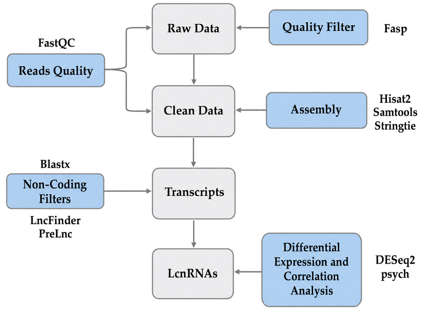
The lncRNA identification pipeline is shown in Figure 1. First, the sequences of the transcripts were queried against the non-redundant protein (nr) and Uniprot databases with Diamond v0.9.14.115 [36] using E-value cut-offs of 10−6 and 10−3, respectively. Transcripts that matched a coding gene were discarded. Second, transcripts with a length < 200 bp were also discarded. Third, the remaining sequences were filtered by PreLnc [37] and LncFinder [38], running with models constructed with the reference coding (cds), messenger RNA (mRNA) and lncRNA sequences obtained from Ensembl (https://www.ensembl.org/Oncorhynchus_mykiss/, accessed on 19 June 2025). For LncFinder, the “SecondaryStructure” feature was used, and only transcripts with a non-coding potential > = 0.9 and < = 0.1 were regarded as candidate lncRNAs. Finally, to classify these candidates according to their genomic characteristics, gffcompare software [39] was used.

2.6. Differential Expression, Co-Expression and Functional Enrichment Analysis

To compare the expression levels in pairwise fashion between conditions or cell types, a read count was performed by prepDE.py, a Python 3.2.6 script provided by Stringtie. The count data were normalized by the median of ratios by DESeq2 [40], a differential gene expression analysis was performed, and the transcripts with a p-adjusted value (FDR) < 0.05 and absolute Log2(FC) > 1 were considered as significantly differentially expressed. To construct a co-expression network, we predicted the target protein-coding genes of candidate lncRNAs using the trans acting role. The trans role indicates the effect on the expression level of any gene; therefore, Pearson’s correlation coefficient (PCC) was calculated between expression levels of differentially expressed (DE) lncRNAs and DE mRNAs using a threshold of |PCC| ≥ 0.9 to determine strong correlations. The resulting correlations were then ranked using GENIE3 [41], and only the top 10% of predicted interactions were retained for network construction. Functional enrichment analysis of the resulting gene sets was performed using DAVID [42] to identify overrepresented biological processes, cellular components, molecular function and KEGG pathways.
Thresholds for differential expressions were selected following criteria commonly applied in lncRNA studies in teleost fishes [43,44]. For co-expression network construction, a Pearson correlation coefficient threshold of |PCC| ≥ 0.9 was applied to retain only strong linear associations between DE lncRNAs and DE mRNAs, consistent with previously used stringency criteria in lncRNA regulatory network analyses in fish [26]. The top 10% of GENIE3-ranked interactions were retained to reduce network complexity while preserving the most likely regulatory relationships, following approaches used in lncRNA-mRNA network inference.

3. Results

3.1. Transcriptome Assembly and Identification of lncRNAs

A total of 609,882,820 raw reads were obtained from the RNA sequencing. Following the quality filter, 570,147,465 clean reads remained, with a minimum length of 50 bp. From these, on average 93.6% were successfully mapped to the reference genome OmykA_1.1. The reference assembly generated 175,595 transcripts with an N50 and an average length of 4697 and 3026, respectively. The transcripts were aligned against a non-redundant database (nr, NCBI) and the Uniprot database, obtaining an 80.5% annotation rate with a high level of homology to rainbow trout. We developed a process based on deep learning software, and from the transcripts analyzed by these algorithms, 4263 lncRNAs were predicted. Of the total of lncRNAs, 3725 (87.4%) were identified as new and 538 (12.6%) had already been previously annotated; also, 1008 were exonic (23.6%), 497 intronic (11.7%), 1390 intergenic (32.6%) and 1368 ambiguous (32.1%).

3.2. Characterization of lncRNAs

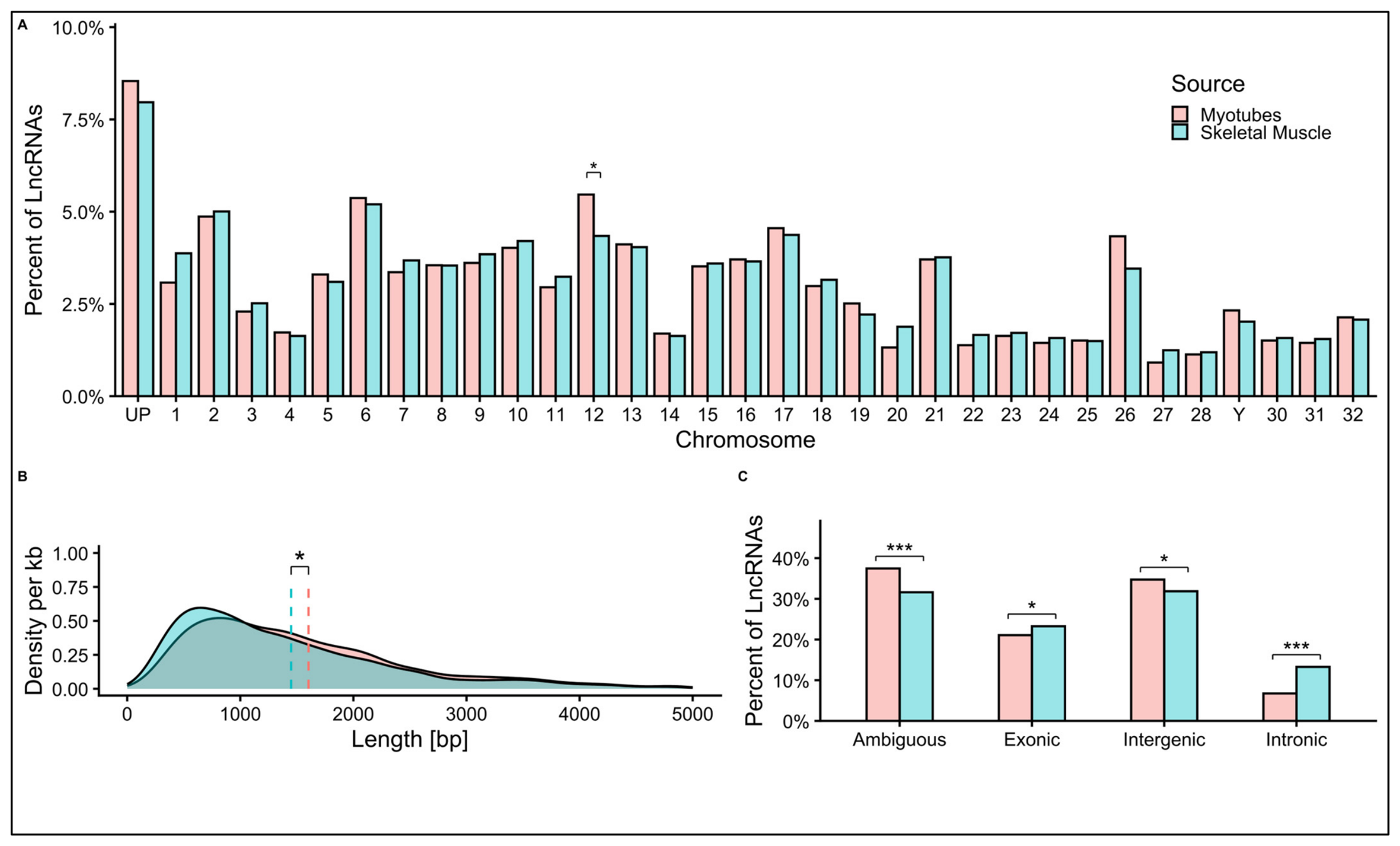
The results can characterize and make different comparisons of lncRNAs that come from myotubes or skeletal muscle. Of the total number of lncRNAs identified, 2913 (91.5%) were mapped to different chromosomal locations from myotubes and 3328 (92.0%) from skeletal muscle; however, 272 (8.5%) were unmapped (unplaced) in myotubes and 288 in muscle (7.96%) (Figure 2A). The genomic distribution of lncRNAs differed significantly between those from myotubes and those from skeletal muscle. Among the chromosomes with the greatest difference is chromosome 12, one of the largest chromosomes in the rainbow trout genome, with 102,853,256 base pairs (Figure 2A). Chromosome 12 was the only one that showed a statistically significant difference in lncRNA representation, with 174 lncRNAs (5.5% of total myotube lncRNAs) compared to 157 (4.3% of total skeletal muscle lncRNAs) (Figure 2A). This difference may reflect lncRNA expression specialization associated with the developmental state or origin of each cell type. The length distribution of the identified lncRNAs ranged from 201 to 8465 bp from both sources. When compared, the average length of lncRNAs in myotubes was 1603 bp, while the average length of lncRNAs in skeletal muscle was 1449 bp. A significant difference was observed between the two groups, with lncRNAs from myotubes being 154 bp longer than those from skeletal muscle (Figure 2B). Regarding gene localization, we can observe that most of the lncRNAs were in exonic and intergenic zones, totaling 21.1% and 23.3% in myotubes and skeletal muscle, respectively (Figure 2C). Only 6.8% of the lncRNAs in the myotube group were located in introns, compared with 13.3% in the skeletal muscle group.
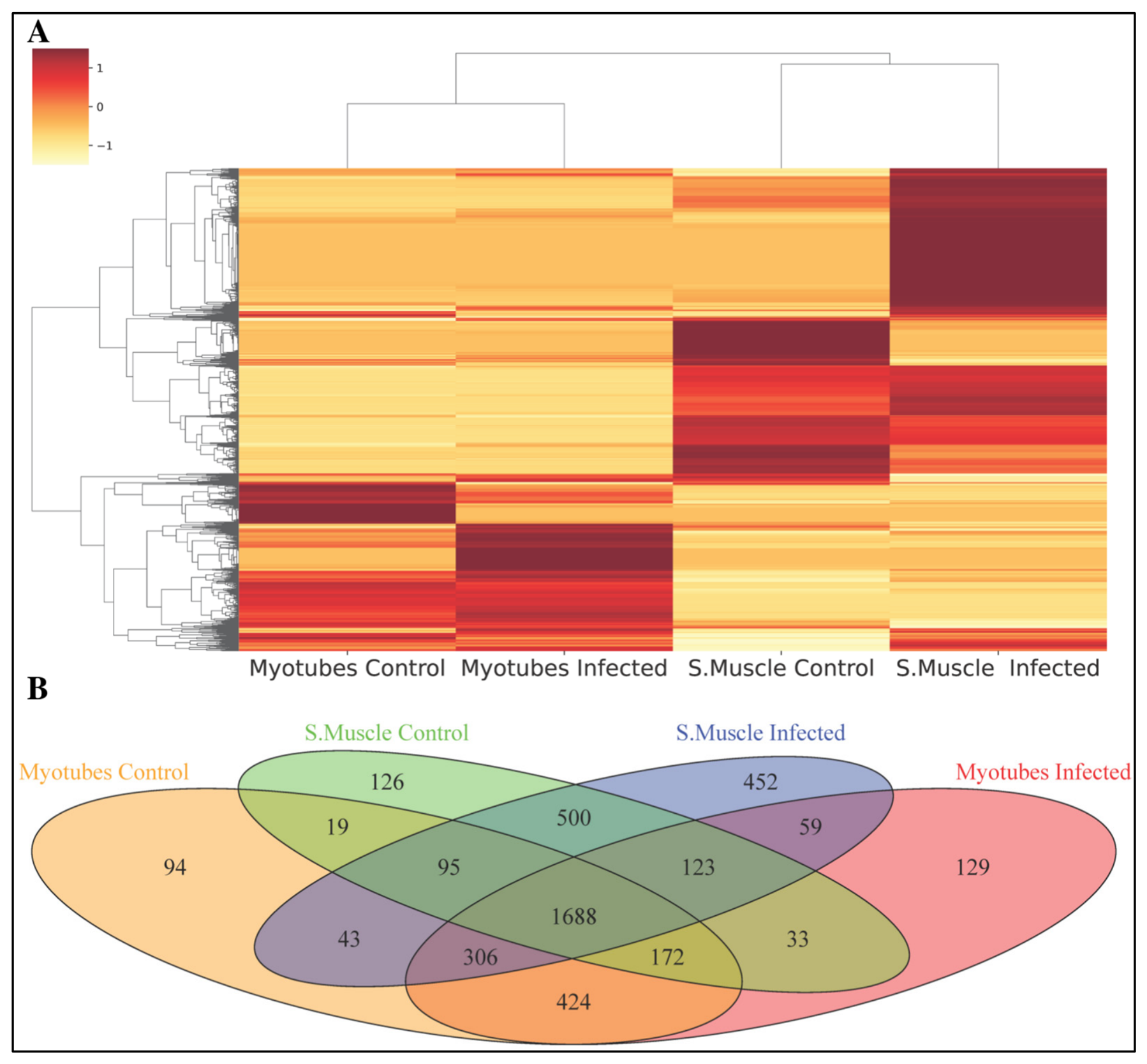
The 4263 predicted lncRNA candidates were evaluated based on their expression profiles in isolated cells (myotubes) and in skeletal muscle, comparing between the control group and the infected group to identify group-specific expression patterns. Comparing the samples, we observe that the differences in the expression levels due to the infection condition are fewer than those observed for the origin of the samples from the myotube and skeletal muscle groups (Figure 3A). In relation to this, we show these results in a Venn diagram (Figure 3B). There are 223 lncRNAs uniquely belonging to the myotube group (94 control and 129 infected), and 578 to the skeletal muscle group (126 control and 452 infected). Also, there were 78 lncRNAs shared between the myotube group and skeletal muscle (19 control and 59 infected); further, the number of lncRNAs shared among all groups was 1648.

3.3. Differential Expression and Correlation Analysis

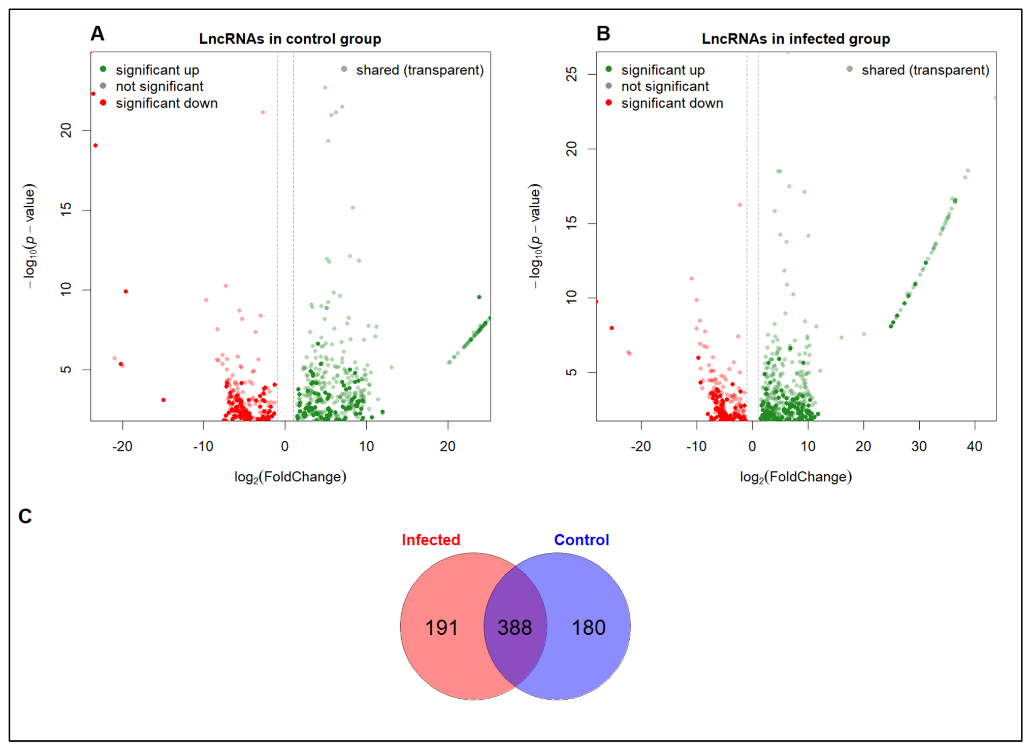
At a global level, the response to infection revealed a total of 764 differentially expressed (DE) lncRNAs, with 266 (34.82%) and 498 (65.18%) downregulated and upregulated, respectively. In the control group (568 DE lncRNAs), 191 (33.63%) were downregulated and 377 (66.37%) lncRNAs were upregulated (Figure 4A). Among 568 DE lncRNAs, 467 were classified as new and 101 as known. A high proportion of DE lncRNAs were observed in the control comparison, where the presence of transparent dots (green or lighter red in Figure 4A) indicates that a significant fraction of DE signals in the control group also appear in the infected group. The solid dots represent components that are specifically regulated under the control condition. Meanwhile, in the infected group (579 DE lncRNAs), 182 (31.44%) were downregulated and 397 (68.56%) lncRNAs were upregulated (Figure 4B). Among 579 DE lncRNAs, 479 were classified as new and 100 as known. These results show up- and downregulated lncRNAs, with a clear trend toward upregulation (positive log2FC values). The transparent dots indicate a core set of lncRNAs shared with the control group. The solid dots suggest an additional regulatory signature associated with infection. The complete list of DE lncRNAs under the control condition is shown in Supplementary Table S1 and those under the infected condition in Supplementary Table S2. Figure 4C compares the sets of significantly DE lncRNAs from the analyses shown in Figure 4A,B. The results show that there is a major group of 388 shared DE lncRNAs. Unique subsets are also observed under each condition (191 in the infected group and 180 in the control group), consistent with source-specific responses (Figure 4C).

3.4. Distribution of Enriched Biological Processes of Candidate lncRNAs

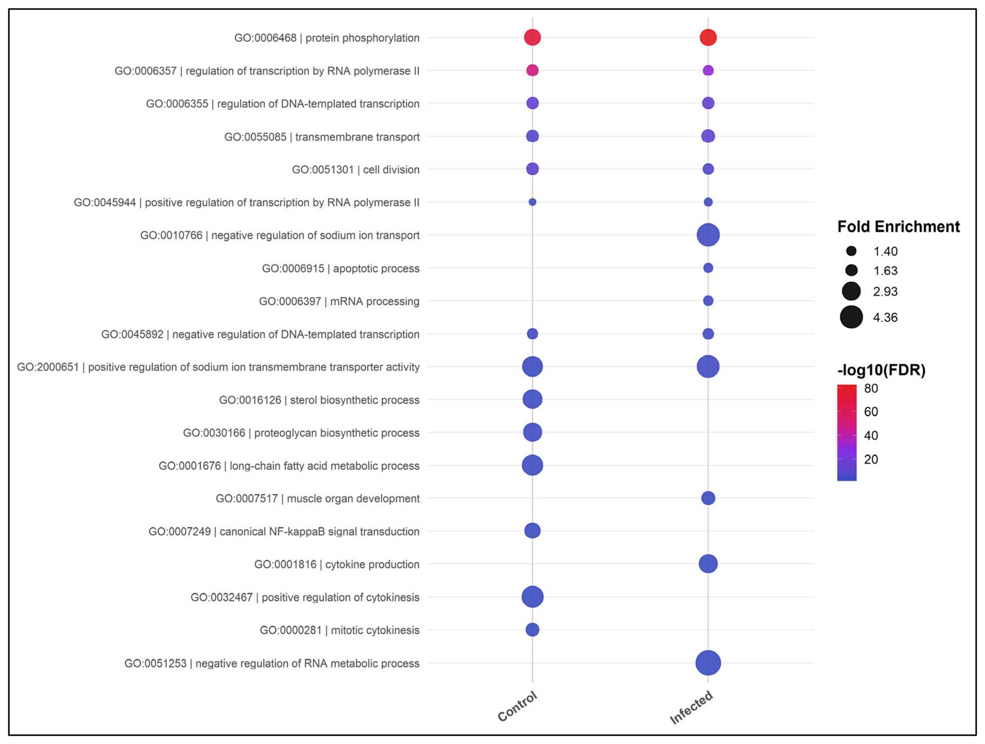
Candidate target genes for the lncRNAs were predicted based on their proximity to protein-encoding genes. Gene ontology (GO) analysis of the protein-encoding target genes revealed the modulation and enrichment of biological processes (BPs) with high significance related to protein phosphorylation (GO:0006468), regulation of transcription by RNA polymerase II (GO:0006357), regulation of DNA-templated transcription (GO:0006355), transmembrane transport (GO:0055085), and cell division (GO:0051301), which were shared between both comparisons (Figure 5). The most enriched BPs in the control group were related to biosynthesis/metabolism and cellular organization, such as the sterol biosynthetic process (GO:0016126), proteoglycan biosynthetic process (GO:0030166), long-chain fatty acid metabolic process (GO:0001676), and positive regulation of cytokinesis (GO:0032467). In contrast, in the infected group, the most common BPs were response to damage/stress and immune regulation; for example: the apoptotic process (GO:0006915), cytokine production (GO:0001816), negative regulation of sodium ion transport (GO:0010766), and negative regulation of RNA metabolism (GO:0051253). The complete lists of biological processes, cellular components, molecular functions, and KEGG pathways are shown in Supplementary Table S3 for the control condition and Supplementary Table S4 for the infected condition.

3.5. Expression Network of the Differentially Expressed lncRNAs and mRNAs in the Rainbow Trout Skeletal Muscle

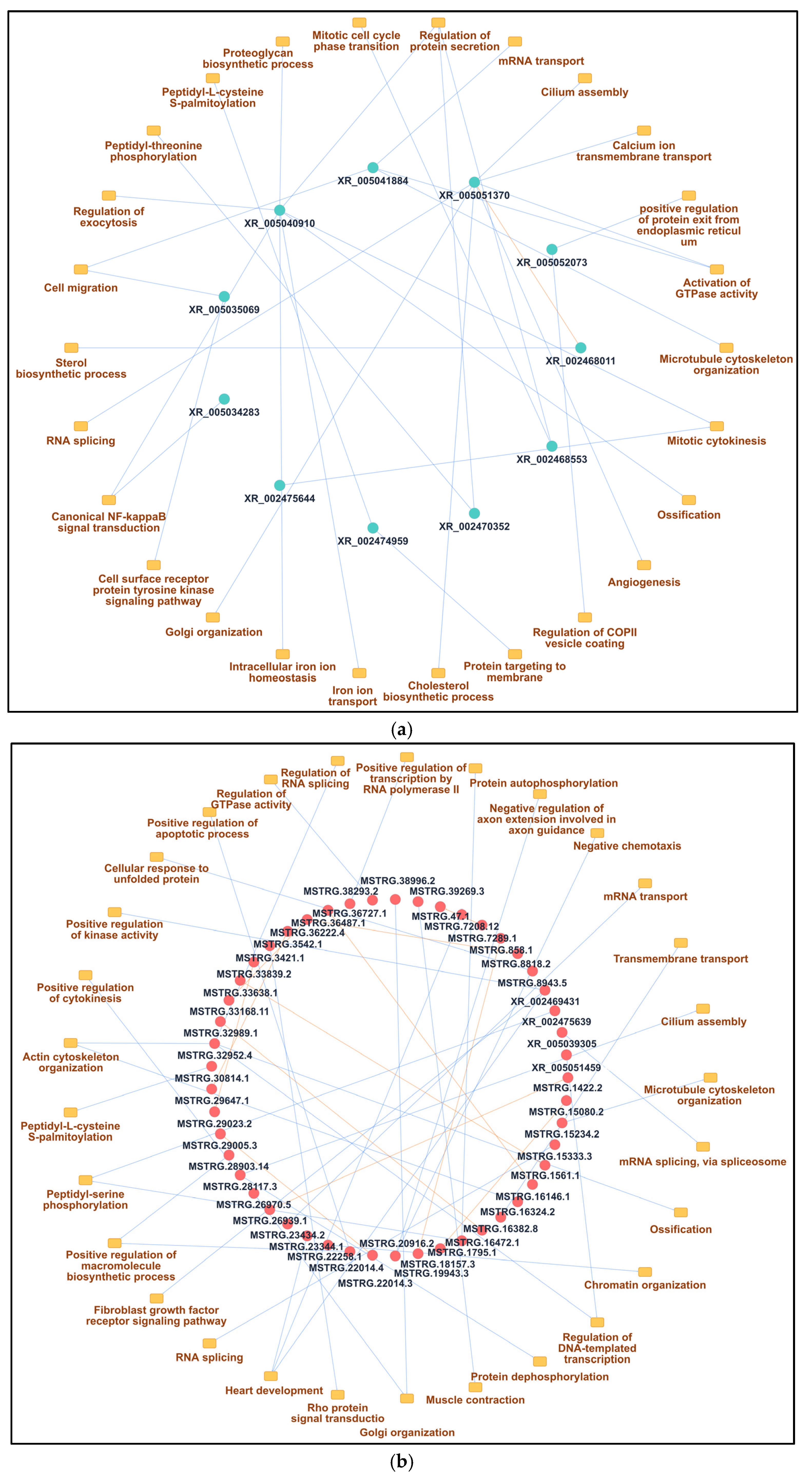
Analysis of DE lncRNA and DE mRNA co-expression networks in skeletal muscle and cell culture (myotubes) revealed that 33.51% and 32.71% of the genes were grouped into four main networks under the control and infected with P. salmonis conditions, respectively (Figure 6 and Figure 7, respectively). The networks shown in Figure 6 and Figure 7 were filtered using the criteria described in their respective captions. The first network primarily included 11 downregulated DE lncRNAs more enriched in the control group (Figure 6a). The three lncRNAs associated with the most biological processes were XR_005040910 (7 BP), XR_005051370 (7 BP), and XR_005041884 (4 BP). The BPs associated with the XR_005040910 lncRNA were mainly related to the proteoglycan biosynthetic process, regulation of exocytosis, mitotic cytokinesis, ossification, iron ion transport, intracellular iron ion homeostasis, and canonical NF-kappaB signal transduction. This lncRNA also interacted with the XR_002468011 lncRNA. The BPs associated with the XR_005051370 lncRNA were mainly related to cilium assembly, calcium ion transmembrane transport, activation of GTPase activity, angiogenesis, the cholesterol biosynthetic process, Golgi organization and RNA splicing. The BPs associated with the XR_005041884 lncRNA were mainly related to mRNA transport, microtubule cytoskeleton organization, activation of GTPase activity and cell migration. Under the control condition, only annotated lncRNAs (those beginning with the letters XR) were observed (Figure 6a). The second network primarily included 48 upregulated DE lncRNAs more enriched in the control group (Figure 6b). The three lncRNAs associated with the most biological processes were MSTRG.32952.4 (4 BP), MSTRG.28117.3 (3 BP), and XR_002469431 (3 BP). The BPs associated with the MSTRG.32952.4 lncRNA were mainly related to regulation of DNA-templated transcription, actin cytoskeleton organization, ossification, and regulation of RNA splicing. The BPs associated with the MSTRG.28117.3 lncRNA were mainly related to regulation of protein dephosphorylation, cilium assembly, and positive regulation of cytokinesis. The BPs associated with the XR_002469431 lncRNA were mainly related to positive regulation of kinase activity, the fibroblast growth factor receptor signaling pathway, and heart development. In the control condition, both annotated and unannotated lncRNAs (those beginning with the letters MRSTG) were observed, with the majority being unannotated lncRNAs (Figure 6b). It should be noted that some of the enriched biological processes (e.g., cilium assembly, ossification, heart development) may appear incongruent in a muscle context; however, these reflect the functional annotation of target genes in other biological contexts and do not necessarily imply active regulation of those processes in skeletal muscle or myotubes.
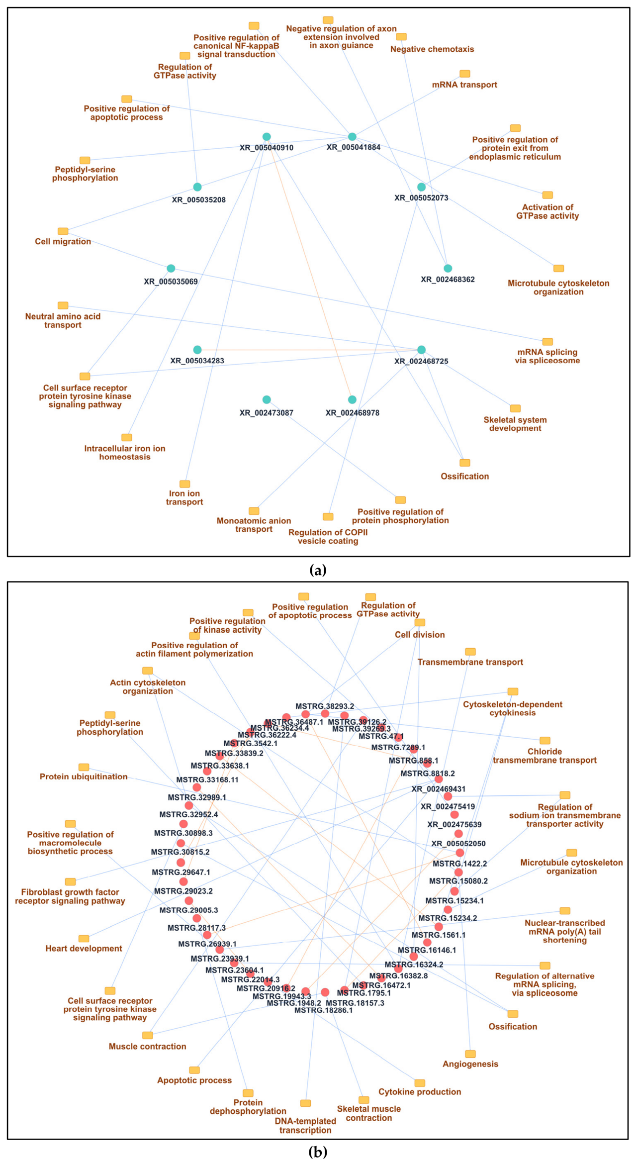
The third network primarily included 10 downregulated DE lncRNAs more enriched in the infected group (Figure 7a). The three lncRNAs associated with the most biological processes (BPs) were XR_005041884 (7 BP), XR_002468725 (5 BP), and XR_005040910 (3 BP). In the infected condition, two lncRNAs (XR_002468978 and XR_005034283) were observed that do not interact with BPs but rather interacted with other lncRNAs. The BPs associated with the XR_005041884 lncRNA were mainly related to microtubule cytoskeleton organization, activation of GTPase activity, mRNA transport, positive regulation of canonical NK-kappaB signal transduction, positive regulation of the apoptotic process, cell migration and peptidyl–serine phosphorylation. The BPs associated with the XR_002468725 lncRNA were mainly related to skeletal system development, ossification, monoatomic anion transport, the cell-surface receptor protein tyrosine kinase signaling pathway, and neutral aminoacidic transport. This lncRNA was also interacting with the XR_005034283 lncRNA. The BPs associated with the XR_005040910 lncRNA were mainly related to intracellular iron ion homeostasis, iron ion transport, and ossification. This lncRNA also interacted with the lncRNA XR_002468978. Similarly, as observed in downregulated DE lncRNAs with the control condition (Figure 6a), only annotated lncRNAs were observed in the infected condition (Figure 7a).
The fourth network primarily included 48 upregulated DE lncRNAs more enriched in the infected group (Figure 7b). The three lncRNAs associated with the most biological processes were XR_002469431 (4 BP), MSTRG.15080.2 (3 BP), and MSTRG.18286.1 (2 BP). The BPs associated with the XR_002469431 lncRNA were mainly related to positive regulation of kinase activity, the apoptotic process, and the fibroblast growth factor receptor signaling pathway. The BPs associated with the MSTRG.15080.2 lncRNA were mainly related to actin cytoskeleton organization, and angiogenesis. This lncRNA also interacted with the MSTRG.18157.3 lncRNA. The BPs associated with the MSTRG.18286.1 lncRNA were mainly related to skeletal muscle contraction. In the infected condition, both annotated and unannotated lncRNAs were observed, with the majority being unannotated lncRNAs (Figure 7b).

4. Discussion

Our research group had previously described the regulation of the overall immune response through RNA sequencing in skeletal muscle and primary myotube cultures of rainbow trout infected with P. salmonis [15,18,19]. To understand the role of lncRNAs in regulating P. salmonis infection in skeletal muscle tissue and cultured cells of rainbow trout, we used RNA-seq data to identify and assess the differential expression of lncRNAs between the control and infected groups.
The thorough description of lncRNAs and their co-expression with mRNAs within the skeletal muscle (in vivo and in vitro) during a bacterial infection provides a valuable contribution to the growing evidence showing that non-coding RNAs play a regulatory role in the immune response of fish. As in other RNA-seq studies on teleost fishes, the number of lncRNAs discovered and their classification into intergenic, antisense, and intronic groups do not differ from results previously reported for other species, including coho salmon and zebrafish (Danio rerio) [26,43,44]. The transcriptomic profiles of lncRNAs have been documented as the most abundant class of lncRNA discovered, with antisense and intronic lncRNAs constituting the second- and third-most abundant classes, respectively [22]. These findings are consistent across previous studies, supporting the suggestion that the structural lncRNA repertoire is evolutionarily conserved across vertebrates and lending further credence to the integrity of our identification pipeline. The genomic positions for lncRNAs found here reflect the patterns of vertebrate systems described above, where intergenic areas seem to be an important source for lncRNA transcription. The positional context of lncRNAs relative to protein-encoding genes is associated with their regulatory role in cis orientation affecting the level of expression of nearby genes [24]. Skeletal muscle, a notably plastic tissue, has a transcriptional landscape that is tightly coupled with physiological and cellular states such as growth, repair, and immunoactivity in fishes [45]. Therefore, lncRNA distributions among different areas of the genome may also be related to whether the cells are in a developmentally or functionally different phase.
Interestingly, we showed differences in transcription length across different experimental conditions, suggesting possible changes in the overall expressed lncRNA subclasses. It has been shown that smaller lncRNAs that have fewer exons could be correlated with quicker, more temporary transcriptional events and that the larger, more complex transcripts could be involved in long-term “regulating” events (chromatin remodeling and transcriptional scaffolding) [46,47]. It is also possible that changes in chromatin accessibility or transcriptional dynamics, considering a particular state within cells (such as immune activation), may influence the initiation and elongation of lncRNA transcripts, leading to the observed differences in transcriptional length [48,49,50]. We also found a difference between muscle tissue and cell culture in terms of length, with myotubes having longer lncRNAs than the skeletal muscle. In addition, only one chromosome (chromosome 12) shows a substantial amount of difference, which could represent a unique regulatory role that needs further analysis. Further research should explore whether the same chromosome-specific patterns are present in other teleosts, and what role these chromosomes might play in host–pathogen interactions and muscle physiology [51].
The global expression pattern for lncRNAs and mRNAs indicates that there are substantial differences in how these genes respond to pathogen exposure. The non-coding part of the transcriptome is therefore quite responsive to bacterial infection. The expression profile for lncRNAs that we observed agrees with previous findings from fishes and other vertebrates, where they have been shown to have much lower levels of expression than mRNAs, greater variability in expression than mRNAs, and specific tissue distributions [52,53]. This pattern lends support to the notion of lncRNAs being fine-tuners of gene regulation by producing context-dependent effects that may not always indicate large relative changes, but that are nonetheless biologically relevant [54]. The results suggest that the DE lncRNAs dynamics between in vivo and in vitro conditions comprise a shared, robust regulatory component across both comparisons, while also retaining a substantial fraction of condition-specific lncRNAs associated with either control or infected states. This dual pattern highlights the complexity of lncRNA-mediated regulation, where a conserved set of transcripts may underpin core regulatory mechanisms of the system, whereas condition-specific lncRNAs could act as mediators of context-dependent processes [55,56]. In this regard, lncRNAs uniquely expressed under specific conditions represent promising candidates for driving distinct biological responses, such as host–pathogen interactions or cellular adaptation, while the shared lncRNAs may reflect fundamental nuclear regulatory mechanisms operating across experimental contexts [57].
Common biological processes (such as protein phosphorylation and transcriptional regulation by RNA polymerase) support the interpretation that the two comparisons share a common core of DE lncRNAs. These data suggest that there is a basal adaptive program associated with cell signaling, regulation, transport, and dynamic proliferation in both in vivo and in vitro systems, and may represent a highly conserved intrinsic response by cells to changing experimental contexts. Nonetheless, additional regulatory mechanisms likely will be detected within individual condition-specific patterns of DE lncRNAs. The biologically enriched processes in the control condition are primarily related to biosynthetic and structural functions such as sterol and proteoglycan synthesis, long-chain fatty acid metabolism, and cytokinesis, implying that there is a stronger metabolic and structural basis for the differences between isolated cells and muscle tissue. By comparison, the infected group was found to be enriched for pathways related to stress and immune processes, including apoptosis, cytokine production, and the negative regulation of ion transport and RNA metabolism. This enrichment supports an alteration in the transcriptional environment to be highly reactive and immunologically active [58,59]. Hence, although there remains a highly conserved regulatory framework within each condition, infection could produce a functional reprogramming of the major stress- and immune-related signaling pathways, likely reflecting both host defense mechanisms and potential programming of cellular responses via infections [2,4].
The differences observed between the in vivo and in vitro models likely reflect not only intrinsic cellular responses but also the influence of the complex tissue microenvironment, including interactions with immune and stromal cells, endocrine signaling, and extracellular matrix components that are absent under in vitro conditions. Consequently, while myotube cultures provide a valuable system to investigate muscle-specific responses, the in vivo model captures additional regulatory mechanisms arising from multicellular and systemic interactions. The co-expression network analysis revealed several lncRNAs associated with structural-cellular maintenance, immune response, cell signaling and apoptosis. The lncRNA most connected to the networks was XR_005041884, which was predicted to participate in negatively regulating microtubule cytoskeleton organization, promoting GTPase-activating signals, and cell migration when under uninfected conditions. When infected with P. salmonis, this lncRNA was associated with negatively regulating the positive regulation of canonical NF-κB signaling and apoptotic processes. This might reflect a context-dependent regulatory function whereby lncRNAs may modulate the cytoskeletal organization dynamics and cellular homeostasis through their regulatory activity under normal conditions while also shaping immune and survival pathways through their active role during infection [60]. One important point about the repressive effects of NF-κB signaling and apoptosis is that they could occur through two mechanisms, the first being that the host is driving a regulatory mechanism to prevent excessive inflammation and tissue damage, and the second possibly resulting from the pathogenic microbes’ ability to suppress host immune responses and thus increase their chance of survival intracellularly [61,62]. This last hypothesis is supported by previous studies demonstrating how P. salmonis can alter host cell survival by causing macrophage death through the caspase-dependent apoptosis pathway, allowing evasion of the immune system and establishment of infection [62]. Additionally, P. salmonis can establish a productive infection in macrophages without causing early indicators of significant cytopathic effects, indicating that bacteria use immune modulation to persist within host cells [63]. Similarly, there is evidence for a temporally regulated apoptotic response in macrophages during initial phases of P. salmonis infection, where apoptosis of those cells is inhibited, aiding in bacterial survival and replication, and later apoptosis is promoted, aiding in their spread [64]. Thus, P. salmonis‘s ability to survive and replicate within macrophages supports its capacity to inhibit immune pathway function occurring in hosts [62,65]. Nevertheless, while supporting evidence as to the effect of P. salmonis on NF-kappaB modulation in fish is limited, it is possible that the observed repression of NF-kappaB in our dataset is indicative of a general immune signaling modulation strategy. Skeletal muscle is increasingly recognized as an immunocompetent tissue capable of producing cytokines, chemokines, and antimicrobial peptides that modulate both local and systemic immune responses, a role that is becoming especially evident in teleost fishes under conditions of stress and infection [2,3,4]. As such, the lncRNA–mRNA interactions discovered here are likely to provide an additional regulatory mechanism through which muscle regulates its immune response to P. salmonis infection.
Although the number of DE lncRNAs between control and infected groups parallels the results of other published RNA-seq studies, the overall amount of lncRNA expression is largely dictated by both the type of tissue being studied and the nature of the experiment being performed. For instance, the number of DE lncRNAs present in red cusk-eel (Genypterus chilensis) skeletal muscle was relatively low when animals were subjected to handling stress in comparison to those found in the more immunologically active tissues [66]. As such, the data presented here are consistent with the idea that the relatively moderate number of DE lncRNAs identified in skeletal muscle will be significantly lower than those found in studies that are typically done examining the head kidney or spleen, where various immune responses are generally associated with much larger numbers of lncRNAs [29,30]. Thus, there does appear to be a greater degree of transcriptional complexity exhibited by the skeletal muscle compared to other tissues; however, this degree of transcriptional complexity may still have important functional implications.
Several biotic or abiotic stimuli can determine how lncRNAs are regulated. In this sense, P. salmonis infection could induce a cascade of immunological events (innate immune activation, inflammation, and metabolic reprogramming) that may require different sets of regulatory lncRNAs than those triggered by stress conditions such as handling or confinement [67]. Thus, by using co-expression network analyses to characterize lncRNAs related to genes associated with immune-related functions, researchers can provide important evidence for the potential functional involvement of lncRNAs even if they exhibit moderate levels of differential expression. Our analysis, which conducted functional enrichment in a network-based manner, revealed an asymmetric distribution between DE lncRNAs, with predominantly annotated lncRNAs in the downregulated co-expression modules in both control and infected fish, and upregulated lncRNAs demonstrating the presence of both annotated and unannotated lncRNAs, with the majority unannotated. This finding suggests that a substantial portion of the transcriptional response (especially under conditions of activation) may involve uncharacterized or novel lncRNAs and illustrates the significant limitations of current genome annotation [26,28]. This result indicates that unannotated or novel lncRNAs may have important regulatory roles in specific biological contexts.
There are some limitations to the discussion of these findings that should be considered. The functional predictions of lncRNAs that were derived from co-expression and enrichment analyses are mostly based on correlation data. Further studies should include more comprehensive functional analysis by using other functional databases for enrichment, integrating identified differentially expressed coding genes to validate regulatory actions of lncRNAs, and carrying out expression analysis on the functional effects of lncRNAs on host–pathogen interaction in skeletal muscle. Also, functional validation using targeted molecular approaches, such as loss- or gain-of-function or reporter assays, will be required to confirm the regulatory roles of the candidate lncRNAs identified in this study. This would enhance our knowledge and validate the biological relevance of the identified patterns of lncRNAs, characterizing how they contribute to how lncRNAs interact with pathogens in skeletal muscle.
The fact that ncRNA expression is highly tissue-specific, and developmentally and physiologically dependent, implies that the biological context in which lncRNAs are studied is important for understanding their function [68]. The expression of lncRNAs specific to skeletal muscle likely reflects the interplay between metabolic, structural, and immune functions, especially during stress or infection. For this reason, the lncRNA expression profiles described in this study increase our understanding of muscle biology in teleosts and provide a basis for future studies to elucidate the molecular mechanisms that underline host–pathogen interactions in aquacultured animals.

5. Conclusions

These findings indicate that the lncRNA repertoire in teleost skeletal muscle is structurally conserved yet dynamically regulated in response to P. salmonis infection. The observed co-expression patterns with mRNAs suggest that lncRNAs may contribute to the modulation of immune response, apoptosis, and signaling pathways, reinforcing their emerging role as key origin-specific regulatory elements in fish physiology and disease.

Supplementary Materials

The following supporting information can be downloaded at https://www.mdpi.com/article/10.3390/fishes11070398/s1: Table S1: Complete list of DE lncRNAs in control condition. Table S2: Complete list of DE lncRNAs in infected with P. salmonis condition. Table S3: Complete list of biological processes, cellular components, molecular functions, and KEEG pathways in the control condition between skeletal muscle and myotubes of rainbow trout. Table S4: Complete list of biological processes, cellular components, molecular functions, and KEEG pathways in the infected condition between skeletal muscle and myotubes of rainbow trout.

Author Contributions

Conceptualization, J.A.V., A.M., P.D.;methodology, R.Z. and J.A.V.; software, L.A.-L.; formal analysis, R.Z., and L.A.-L.; investigation, J.A.V.; resources, J.A.V.; data curation, R.Z. and L.A.-L.; writing—original draft preparation, R.Z. and L.A.-L.; writing—review and editing, R.Z. and J.A.V.; supervision, J.A.V.; project administration J.A.V.; funding acquisition, J.A.V. All authors have read and agreed to the published version of the manuscript.

Funding

This work was supported by Agencia Nacional de Investigación y Desarrollo, ANID INCAR2 CIA250009 (to Juan Antonio Valdés); the Fondo Nacional de Desarrollo Científico y Tecnológico (FONDECYT) under grant numbers 1201498 and 1230794 (to Juan Antonio Valdés); and ANID Vinculación Internacional FOVI250249.

Institutional Review Board Statement

This research was approved by the Bioethics Committee of Andrés Bello University, approval code: 012/2020; and date: 14 April 2020.

Data Availability Statement

These data are available at NCBI under the codes PRJNA732666 for myotubes and PRJNA676020 for skeletal muscle. The datasets generated and analyzed during the current study are not publicly available due to privacy or ethical restrictions but are available from the corresponding author upon reasonable request.

Conflicts of Interest

The authors declare no conflicts of interest.

Abbreviations

The following abbreviations are used in this manuscript:
bpbase pair
BPbiological process 
circRNAscircular RNAs 
DEdifferentially expressed 
DMSOdimethyl sulfoxide
GOgene ontology 
ISAinfectious salmon anemia 
lncRNAlong non-coding RNA
MOImultiplicity of infection
BHmultiple comparisons 
NGSnext-generation sequencing 
ncRNAnon-coding RNA
PAMPspathogen-associated molecular patterns 
PCCPearson’s correlation coefficient 
FDRp-adjusted value 
SRSsalmonid rickettsial septicemia 
RNA-SeqRNA sequencing

References

  1. Grunow, B.; Stange, K.; Bochert, R.; Tönißen, K. Histological and biochemical evaluation of skeletal muscle in the two salmonid species Coregonus maraena and Oncorhynchus mykiss. PLoS ONE 2021, 16, e0255062. [Google Scholar] [CrossRef]
  2. Valenzuela, C.A.; Zuloaga, R.; Poblete-Morales, M.; Vera-Tobar, T.; Mercado, L.; Avendaño-Herrera, R.; Valdés, J.A.; Molina, A. Fish skeletal muscle tissue is an important focus of immune reactions during pathogen infection. Dev. Comp. Immunol. 2017, 73, 1–9. [Google Scholar] [CrossRef]
  3. Chatterjee, A.; Roy, D.; Patnaik, E.; Nongthomba, U. Muscles provide protection during microbial infection by activating innate immune response pathways in Drosophila and zebrafish. Dis. Model. Mech. 2016, 9, 697–705. [Google Scholar] [CrossRef]
  4. Debbarma, S.; Narsale, S.A.; Acharya, A.; Singh, S.K.; Mocherla, B.P.; Debbarma, R.; Yirang, Y. Drawing immune-capacity of fish-derived antimicrobial peptides for aquaculture industry: A comprehensive review. Comp. Immunol. Rep. 2024, 6, 200150. [Google Scholar] [CrossRef]
  5. Pooley, N.J.; Tacchi, L.; Secombes, C.J.; Martin, S.A. Inflammatory responses in primary muscle cell cultures in Atlantic salmon (Salmo salar). BMC Genom. 2013, 14, 747. [Google Scholar] [CrossRef]
  6. Aedo, J.E.; Reyes, A.E.; Avendaño-Herrera, R.; Molina, A.; Valdés, J.A. Bacterial lipopolysaccharide induces rainbow trout myotube atrophy via Akt/FoxO1/Atrogin-1 signaling pathway. Acta Biochim. Biophys. Sin. 2015, 47, 932–937. [Google Scholar] [CrossRef]
  7. Iturriaga, M.; Espinoza, M.B.; Poblete-Morales, M.; Feijoo, C.G.; Reyes, A.E.; Molina, A.; Avendaño-Herrera, R.; Valdés, J.A. Cytotoxic activity of Flavobacterium psychrophilum in skeletal muscle cells of rainbow trout (Oncorhynchus mykiss). Vet. Microbiol. 2017, 210, 101–106. [Google Scholar] [CrossRef]
  8. Rivas-Aravena, A.; Fuentes-Valenzuela, M.; Escobar-Aguirre, S.; Gallardo-Escarate, C.; Molina, A.; Valdés, J.A. Transcriptomic response of rainbow trout (Oncorhynchus mykiss) skeletal muscle to Flavobacterium psychrophilum. Comp. Biochem. Physiol. Part D Genom. Proteom. 2019, 100, 596. [Google Scholar] [CrossRef]
  9. ELbialy, Z.I.; Atef, E.; Al-Hawary, I.I.; Salah, A.S.; Aboshosha, A.A.; Abualreesh, M.H.; Assar, D.H. Myostatin-mediated regulation of skeletal muscle damage post-acute Aeromonas hydrophila infection in Nile tilapia (Oreochromis niloticus L.). Fish Physiol. Biochem. 2023, 49, 1–17. [Google Scholar] [CrossRef]
  10. Aedo, J.; Aravena-Canales, D.; Dettleff, P.; Fuentes-Valenzuela, M.; Zuloaga, R.; Rivas-Aravena, A.; Molina, A.; Valdés, J.A. RNA-seq analysis reveals the dynamic regulation of proteasomal and autophagic degradation systems of rainbow trout (Oncorhynchus mykiss) skeletal muscle challenged with infectious pancreatic necrosis virus (IPNV). Aquaculture 2022, 552, 738000. [Google Scholar] [CrossRef]
  11. Rozas-Serri, M. Why Does Piscirickettsia salmonis Break the Immunological Paradigm in Farmed Salmon? Biological Context to Understand the Relative Control of Piscirickettsiosis. Front. Immunol. 2022, 13, 856896. [Google Scholar] [CrossRef]
  12. Ramírez, C.; Romero, J. Know Your Enemy: Piscirickettsia salmonis and Phage Interactions Using an In Silico Perspective. Antibiotics 2025, 14, 558. [Google Scholar] [CrossRef]
  13. Islam, S.I.; Shahed, K.; Ahamed, M.I.; Khang, L.T.P.; Jung, W.-K.; Sangsawad, P.; Dinh-Hung, N.; Permpoonpattana, P.; Linh, N.V. Pathogenomic Insights into Piscirickettsia salmonis with a Focus on Virulence Factors, Single-Nucleotide Polymorphism Identification, and Resistance Dynamics. Animals 2025, 15, 1176. [Google Scholar] [CrossRef]
  14. Figueroa, J.; Cárcamo, J.; Yañez, A.; Olavarria, V.; Ruiz, P.; Manríquez, R.; Muñoz, C.; Romero, A.; Avendaño-Herrera, R. Addressing viral and bacterial threats to salmon farming in Chile: Historical contexts and perspectives for management and control. Rev. Aquac. 2019, 11, 299–324. [Google Scholar] [CrossRef]
  15. Carrizo, V.; Valenzuela, C.A.; Zuloaga, R.; Aros, C.; Altamirano, C.; Valdés, J.A.; Molina, A. Effect of cortisol on the immune-like response of rainbow trout (Oncorhynchus mykiss) myotubes challenged with Piscirickettsia salmonis. Vet. Immunol. Immunopathol. 2021, 237, 110240. [Google Scholar] [CrossRef]
  16. Valenzuela, C.A.; Azúa, M.; Álvarez, C.A.; Schmitt, P.; Ojeda, N.; Mercado, L. Evidence of the Autophagic Process during the Fish Immune Response of Skeletal Muscle Cells against Piscirickettsia salmonis. Animals 2023, 13, 880. [Google Scholar] [CrossRef]
  17. Garg, M. Chapter 5—RNA sequencing: A revolutionary tool for transcriptomics. In Advances in Animal Genomics; Mondal, S., Singh, R.L., Eds.; Academic Press: Cambridge, MA, USA, 2021; pp. 61–73. [Google Scholar] [CrossRef]
  18. Carrizo, V.; Valenzuela, C.A.; Aros, C.; Dettleff, P.; Valenzuela-Muñoz, V.; Gallardo-Escarate, C.; Altamirano, C.; Molina, A.; Valdés, J.A. Transcriptomic analysis reveals a Piscirickettsia salmonis-induced early inflammatory response in rainbow trout skeletal muscle. Comp. Biochem. Physiol. Part D Genom. Proteom. 2021, 39, 100859. [Google Scholar] [CrossRef]
  19. Zuloaga, R.; Dettleff, P.; Bastias-Molina, M.; Meneses, C.; Altamirano, C.; Valdés, J.A.; Molina, A. RNA-Seq-Based Analysis of Cortisol-Induced Differential Gene Expression Associated with Piscirickettsia salmonis Infection in Rainbow Trout (Oncorhynchus mykiss) Myotubes. Animals 2021, 11, 2399. [Google Scholar] [CrossRef]
  20. Antonazzo, G.; Gaudet, P.; Lovering, R.C.; Attrill, H. Representation of non-coding RNA-mediated regulation of gene expression using the Gene Ontology. RNA Biol. 2024, 21, 981–993. [Google Scholar] [CrossRef]
  21. Diamantopoulos, M.A.; Boti, M.A.; Sarri, T.; Scorilas, A. Non-Coding RNAs in Health and Disease: From Biomarkers to Therapeutic Targets. LabMed 2025, 2, 17. [Google Scholar] [CrossRef]
  22. Mattick, J.S.; Amaral, P.P.; Carninci, P.; Carpenter, S.; Chang, H.Y.; Chen, L.L.; Chen, R.; Dean, C.; Dinger, M.E.; Fitzgerald, K.A.; et al. Long non-coding RNAs: Definitions, functions, challenges and recommendations. Nat. Rev. Mol. Cell Biol. 2023, 24, 430–447. [Google Scholar] [CrossRef]
  23. Chen, L.; Kim, V.N. Small and long non-coding RNAs: Past, present, and future. Cell 2024, 187, 6451–6485. [Google Scholar] [CrossRef]
  24. Statello, L.; Guo, C.J.; Chen, L.L.; Huarte, M. Gene regulation by long non-coding RNAs and its biological functions. Nat. Rev. Mol. Cell Biol. 2021, 22, 96–118. [Google Scholar] [CrossRef]
  25. Kang, J.; Chung, A.; Suresh, S.; Bonzi, L.C.; Sourisse, J.M.; Ramirez-Calero, S.; Romeo, D.; Petit-Marty, N.; Pegueroles, C.; Schunter, C. Long non-coding RNAs mediate fish gene expression in response to ocean acidification. Evol. Appl. 2024, 17, e13655. [Google Scholar] [CrossRef]
  26. Zhou, Z.; Leng, C.; Wang, Z.; Long, L.; Lv, Y.; Gao, Z.; Wang, Y.; Wang, S.; Li, P. The potential regulatory role of the lncRNA-miRNA-mRNA axis in teleost fish. Front. Immunol. 2023, 21, 1065357. [Google Scholar] [CrossRef]
  27. Wang, J.; Fu, L.; Koganti, P.P.; Wang, L.; Hand, J.M.; Ma, H.; Jao, J. Identification and functional prediction of large intergenic noncoding RNAs (lincRNAs) in rainbow trout (Oncorhynchus mykiss). Mar. Biotechnol. 2016, 18, 271–282. [Google Scholar] [CrossRef]
  28. Cao, Y.; He, X.; Zheng, W.; Luo, Q.; Xu, T.; Sun, Y. The conserved long noncoding RNA LTCONS12135 positively regulates innate immunity in teleost. Comp. Biochem. Physiol. B Biochem. Mol. Biol. 2026, 284, 111220. [Google Scholar] [CrossRef]
  29. Valenzuela-Miranda, D.; Gallardo-Escárate, C. Novel insights into the response of Atlantic salmon (Salmo salar) to Piscirickettsia salmonis: Interplay of coding genes and lncRNAs during bacterial infection. Fish Shellfish Immunol. 2016, 59, 427–438. [Google Scholar] [CrossRef]
  30. Tarifeño-Saldivia, E.; Valenzuela-Miranda, D.; Gallardo-Escárate, C. In the shadow: The emerging role of long non-coding RNAs in the immune response of Atlantic salmon. Dev. Comp. Immunol. 2017, 73, 193–205. [Google Scholar] [CrossRef]
  31. Andrews, S. FastQC: A Quality Control Tool for High Throughput Sequence Data. 2010. Available online: https://www.bioinformatics.babraham.ac.uk/projects/fastqc/ (accessed on 16 July 2025).
  32. Chen, S.; Zhou, Y.; Chen, Y.; Gu, J. fastp: An ultra-fast all-in-one FASTQ preprocessor. Bioinformatics 2018, 34, i884–i890. [Google Scholar] [CrossRef]
  33. Kim, D.; Paggi, J.M.; Park, C.; Bennett, C.; Salzberg, S.L. Graph-based genome alignment and genotyping with HISAT2 and HISAT-genotype. Nat. Biotechnol. 2019, 37, 907–915. [Google Scholar] [CrossRef]
  34. Danecek, P.; Bonfield, J.K.; Liddle, J.; Marshall, J.; Ohan, V.; Pollard, M.O.; Whitwham, A.; Keane, T.; McCarthy, S.A.; Davies, R.M.; et al. Twelve years of SAMtools and BCFtools. GigaScience 2021, 10, giab008. [Google Scholar] [CrossRef]
  35. Kovaka, S.; Zimin, A.V.; Pertea, G.M.; Razaghi, R.; Salzberg, S.L.; Pertea, M. Transcriptome assembly from long-read RNA-seq alignments with StringTie2. Genome Biol. 2019, 20, 278. [Google Scholar] [CrossRef]
  36. Buchfink, B.R. Sensitive protein alignments at tree-of-life scale using DIAMOND. Nat. Methods 2021, 18, 366–368. [Google Scholar] [CrossRef]
  37. Cao, L.W. PreLnc: An Accurate Tool for Predicting lncRNAs Based on Multiple Features. Genes 2020, 11, 981. [Google Scholar] [CrossRef]
  38. Han, S.L. LncFinder: An integrated platform for long non-coding RNA identification utilizing sequence intrinsic composition, structural information and physicochemical property. Brief. Bioinform. 2019, 20, 2009–2027. [Google Scholar] [CrossRef]
  39. Pertea, G.; Pertea, M. GFF Utilities: GFFRead and GFFCompare. F1000Research 2020, 9, 304. [Google Scholar] [CrossRef]
  40. Love, M.I.; Huber, W.; Anders, S. Moderated estimation of fold change and dispersion for RNA-seq data with DESeq2. Genome Biol. 2014, 15, 550. [Google Scholar] [CrossRef]
  41. Huynh-Thu, V.A.; Irrthum, A.; Wehenkel, L.; Geurts, P. Inferring regulatory networks from expression data using tree-based methods. PLoS ONE 2010, 5, e12776. [Google Scholar] [CrossRef]
  42. Sherman, B.T.; Hao, M.; Qiu, J.; Jiao, X.; Baseler, M.W.; Lane, H.C.; Imamichi, T.; Chang, W. DAVID: A web server for functional enrichment analysis and functional annotation of gene lists (2021 update). Nucleic Acids Res. 2022, 50, W216–W221. [Google Scholar] [CrossRef]
  43. Pauli, A.; Valen, E.; Lin, M.F.; Garber, M.; Vastenhouw, N.L.; Levin, J.Z.; Fan, L.; Sandelin, A.; Rinn, J.L.; Regev, A.; et al. Systematic identification of long noncoding RNAs expressed during zebrafish embryogenesis. Genome Res. 2012, 22, 577–591. [Google Scholar] [CrossRef]
  44. Leiva, F.; Rojas-Herrera, M.; Reyes, D.; Bravo, S.; Garcia, K.K.; Moya, J.; Vidal, R. Identification and characterization of miRNAs and lncRNAs of coho salmon (Oncorhynchus kisutch) in normal immune organs. Genomics 2019, 112, 45–54. [Google Scholar] [CrossRef]
  45. Oudhoff, H.; Hisler, V.; Baumgartner, F.; Rees, L.; Grepper, D.; Jazwinska, A. Skeletal muscle regeneration after extensive cryoinjury of caudal myomeres in adult zebrafish. npj Regen. Med. 2024, 9, 8. [Google Scholar] [CrossRef]
  46. Graf, J.; Kretz, M. From structure to function: Route to understanding lncRNA mechanism. BioEssays 2020, 42, e2000027. [Google Scholar] [CrossRef]
  47. Ali, T.; Grote, P. Beyond the RNA-dependent function of LncRNA genes. eLife 2020, 9, e60583. [Google Scholar] [CrossRef]
  48. Heward, J.A.; Lindsay, M.A. Long non-coding RNAs in the regulation of the immune response. Trends Immunol. 2014, 35, 408–419. [Google Scholar] [CrossRef]
  49. Li, P.; Leonard, W.J. Chromatin Accessibility and Interactions in the Transcriptional Regulation of T Cells. Front. Immunol. 2018, 9, 2738. [Google Scholar] [CrossRef]
  50. Zhang, Y.; Cao, X. Long noncoding RNAs in innate immunity. Cell Mol. Immunol. 2016, 13, 138–147. [Google Scholar] [CrossRef]
  51. Duran, B.O.S.; Garcia de la Serrana, D.; Zanella, B.T.T.; Perez, E.S.; Mareco, E.A.; Santos, V.B.; Carvalho, R.F.; Dal-Pai-Silva, M. An insight on the impact of teleost whole genome duplication on the regulation of the molecular networks controlling skeletal muscle growth. PLoS ONE 2021, 16, e0255006. [Google Scholar] [CrossRef]
  52. Al-Tobasei, R.; Paneru, B.; Salem, M. Genome-Wide Discovery of Long Non-Coding RNAs in Rainbow Trout. PLoS ONE 2016, 11, e0148940. [Google Scholar] [CrossRef]
  53. Chen, L.; Zhang, Y.H.; Pan, X.; Liu, M.; Wang, S.; Huang, T.; Cai, Y.D. Tissue Expression Difference between mRNAs and lncRNAs. Int. J. Mol. Sci. 2018, 19, 3416. [Google Scholar] [CrossRef]
  54. Dahl, M.; Kristensen, L.S.; Grønbæk, K. Long Non-Coding RNAs Guide the Fine-Tuning of Gene Regulation in B-Cell Development and Malignancy. Int. J. Mol. Sci. 2018, 19, 2475. [Google Scholar] [CrossRef]
  55. Kyung, J.; Kim, M.; Shin, H.R.; Kim, E.; Oh, H.J. Multi-layered gene regulation by long non-coding RNAs: From chromatin to genome architecture. BMB Rep. 2026, 59, 112–123. [Google Scholar] [CrossRef]
  56. Herman, A.B.; Tsitsipatis, D.; Gorospe, M. Integrated lncRNA function upon genomic and epigenomic regulation. Mol. Cell 2022, 82, 2252–2266. [Google Scholar] [CrossRef]
  57. Agliano, F.; Rathinam, V.A.; Medvedev, A.E.; Vanaja, S.K.; Vella, A.T. Long Noncoding RNAs in Host-Pathogen Interactions. Trends Immunol. 2019, 40, 492–510. [Google Scholar] [CrossRef]
  58. Guo, H.; Dixon, B. Understanding acute stress-mediated immunity in teleost fish. Fish Shellfish Immunol. Rep. 2021, 2, 100010. [Google Scholar] [CrossRef]
  59. Özcan, F.; Arserim, N.B. Antibacterial immunity in teleost fish: Integrating innate and adaptive responses for sustainable aquaculture. Vet. Immunol. Immunopathol. 2026, 297, 111113. [Google Scholar] [CrossRef]
  60. Arunima, A.; van Schaik, E.J.; Samuel, J.E. The emerging roles of long non-coding RNA in host immune response and intracellular bacterial infections. Front. Cell. Infect. Microbiol. 2023, 13, 1160198. [Google Scholar] [CrossRef]
  61. Rozas-Serri, M.; Peña, A.; Maldonado, L. Transcriptomic profiles of post-smolt Atlantic salmon challenged with Piscirickettsia salmonis reveal a strategy to evade the adaptive immune response and modify cell-autonomous immunity. Dev. Comp. Immunol. 2017, 81, 348–362. [Google Scholar] [CrossRef]
  62. Rojas, V.; Galanti, N.; Bols, N.C.; Jiménez, V.; Paredes, R.; Marshall, S.H. Piscirickettsia salmonis induces apoptosis in macrophages and monocyte-like cells from rainbow trout. J. Cell. Biochem. 2010, 110, 468–476. [Google Scholar] [CrossRef]
  63. Rojas, V.; Galanti, N.; Bols, N.C.; Marshall, S.H. Productive infection of Piscirickettsia salmonis in macrophages and monocyte-like cells from rainbow trout, a possible survival strategy. J. Cell. Biochem. 2009, 108, 631–637. [Google Scholar] [CrossRef]
  64. Díaz, S.; Rojas, M.E.; Galleguillos, M.; Maturana, C.; Smith, P.I.; Cifuentes, F.; Contreras, I.; Smith, P.A. Apoptosis inhibition of Atlantic salmon (Salmo salar) peritoneal macrophages by Piscirickettsia salmonis. J. Fish Dis. 2017, 40, 1895–1902. [Google Scholar] [CrossRef]
  65. Mccarthy, U.; Bron, J.; Brown, L.; Pourahmad, F.; Bricknell, I.; Thompson, K.; Adams, A.; Ellis, A. Survival and replication of Piscirickettsia salmonis in rainbow trout head kidney macrophages. Fish Shellfish Immunol. 2008, 25, 477–484. [Google Scholar] [CrossRef]
  66. Dettleff, P.; Hormazabal, E.; Aedo, J.; Fuentes, M.; Meneses, C.; Molina, A.; Valdes, J.A. Identification and evaluation of long noncoding RNAs in response to handling stress in red cusk-eel (Genypterus chilensis) via RNA-seq. Mar. Biotechnol. 2018, 40, 25–32. [Google Scholar] [CrossRef]
  67. Martínez, D.P.; Oliver, C.; Santibañez, N.; Coronado, J.L.; Oyarzún-Salazar, R.; Enriquez, R.; Vargas-Chacoff, L.; Romero, A. PAMPs of Piscirickettsia salmonis Trigger the Transcription of Genes Involved in Nutritional Immunity in a Salmon Macrophage-Like Cell Line. Front. Immunol. 2022, 13, 849752. [Google Scholar] [CrossRef]
  68. Mouhou, E.; Genty, F.; El M’selmi, W.; Chouali, H.; Zagury, J.F.; Le Clerc, S.; Proudhon, C.; Noirel, J. High tissue specificity of lncRNAs maximises the prediction of tissue of origin of circulating DNA. Sci. Rep. 2025, 15, 12941. [Google Scholar] [CrossRef]
Figure 1. Bioinformatic pipeline for the assembly and identification of lncRNAs in the O. mykiss transcriptome.
Fishes 11 00398 g001
Figure 2. Characterization of lncRNAs identified in O. mykiss myotubes and skeletal muscle. (A) Distribution of the identified lncRNAs on chromosomes. Using χ2 analysis, the asterisk denotes significant differences in length. UP: Unplaced lncRNAs in chromosomes. (B) Length–density graph per kilobase (kb) of the identified lncRNAs by length in base pairs (bp), using the Welch t-test for statistical analysis. (C) Genomic location of the identified lncRNAs. The data show the proportion per chromosome for each source, rather than absolute counts. Ambiguous: everything that is not exonic, intergenic or intronic. The statistical analysis used was a category-based comparison with multiple comparisons Benjamini–Hochberg (BH) correction, where each individual set of brackets summarizes the significance of a category with BH adjustment. The significance values are * p ≤ 0.05, *** p ≤ 0.001 between myotubes and skeletal muscle.
Fishes 11 00398 g002
Figure 3. Expression profiles of the lncRNAs identified in O. mykiss myotubes and skeletal muscle for samples exposed or not exposed to P. salmonis. (A) Heatmap and hierarchical clustering of the expression levels of the lncRNAs identified present among the respective conditions. (B) Venn diagram of the lncRNAs identified present among the respective conditions. S. Muscle Control: skeletal muscle, control; S. Muscle Infected: skeletal muscle, infected.
Fishes 11 00398 g003
Figure 4. Differentially expressed (DE) lncRNAs in response to the source of the cells under the same conditions. (A) Volcano plot of the DE lncRNAs under the infected condition (skeletal muscle vs. myotubes); of a total of 568 DE lncRNAs, 377 were upregulated and 191 downregulated. (B) Volcano plot of the DE lncRNAs in the control condition (skeletal muscle vs. myotubes); of a total of 579 DE lncRNAs, 397 were upregulated and 182 downregulated. Each point in graphs A and B represents a lncRNA. The X-axis is plotted as log2(FoldChange), where positive values indicate higher expression and negative values indicate lower expression of the lncRNAs in the skeletal muscle control group compared to the myotube control. The Y-axis is plotted as log10(p-value), where higher values indicate greater statistical evidence of a difference. Additionally, significantly upregulated lncRNAs are shown in green, significantly downregulated ones in red, and non-significant ones in gray; lighter green or red points represent DE lncRNAs shared between both comparisons; and solid-colored points represent lncRNAs that are uniquely DE in each comparison. (C) Venn diagram of the DE lncRNAs in the infected and control conditions (skeletal muscle vs. myotubes); we use FDR < 0.05 and |FoldChange| > 1 as thresholds for DE.
Fishes 11 00398 g004
Figure 5. Distribution of enriched biological processes with gene ontology (GO) terms for candidate lncRNA-associated protein-encoding genes in rainbow trout. Comparison of GO terms for genes involved in enriched biological processes and interacting with lncRNAs: (i) skeletal muscle control condition compared to myotubes, and (ii) P. salmonis infection in skeletal muscle versus in myotubes. Each row is a GO term, and each column represents a group (control or infected). The size of the bubble represents the fold enrichment, where a larger bubble indicates greater overrepresentation of the GO term. The color of the bubble is expressed in −log10(false discovery rate or FDR), where a more intense color indicates greater statistical significance (p < 0.05).
Fishes 11 00398 g005
Figure 6. Gene expression network between differentially expressed (DE) lncRNAs and biological processes (BPs) of DE mRNAs in the skeletal muscle and myotubes of rainbow trout with the control condition. (a) Downregulated DE lncRNA enrichments associated with BPs of DE mRNAs. The criteria used to generate the figures were a Pearson correlation coefficient of at least 0.9, a GENIE3 score of 90, and an indegree of 20. (b) Upregulated DE lncRNA enrichments associated with BPs of DE mRNAs. The criteria used to generate the figure were a Pearson correlation coefficient of at least 0.95, a GENIE3 score of 99, and an indegree of 10. The yellow squares indicate a BP (of DE mRNAs) composed of genes that are regulated by lncRNAs. The blue bars indicate DE lncRNAs that interact with the BP, and the orange bars show the interaction between different DE lncRNAs. The red circles indicate upregulated DE lncRNAs, while the green circles show downregulated DE lncRNAs.
Fishes 11 00398 g006
Figure 7. Gene expression network between differentially expressed (DE) lncRNAs and biological processes (BPs) of DE mRNAs in the skeletal muscle and myotubes of rainbow trout under the infected condition. (a) Down-regulated DE lncRNA enrichments associated with BPs of DE mRNAs. The criteria used to generate the figure were a Pearson correlation coefficient of at least 0.9, a GENIE3 score of 90, and an indegree of 20. (b) Upregulated DE lncRNAs associated with BPs of DE mRNAs. The criteria used to generate the figure were a Pearson correlation coefficient of at least 0.95, a GENIE3 score of 99, and an indegree of 10. The yellow squares indicate BPs (of DE mRNAs) composed of genes that are regulated by lncRNAs. The blue bars indicate DE lncRNAs that interact with the BP, and the orange bars show the interaction between different DE lncRNAs. The red circles indicate upregulated DE lncRNAs, while the green circles show downregulated DE lncRNAs.
Fishes 11 00398 g007
Disclaimer/Publisher’s Note: The statements, opinions and data contained in all publications are solely those of the individual author(s) and contributor(s) and not of MDPI and/or the editor(s). MDPI and/or the editor(s) disclaim responsibility for any injury to people or property resulting from any ideas, methods, instructions or products referred to in the content.