Article

Effect of Dandelion Root Extract on Growth, Biochemical Indices, Antioxidant Capacity, Intestinal Histomorphology and Microbiota in Micropterus salmoides

1
State Key Laboratory of Mariculture Breeding, Fisheries College, Jimei University, Xiamen 361021, China
2
Xiamen Key Laboratory for Feed Quality Testing and Safety Evaluation, Fisheries College, Jimei University, Xiamen 361021, China
3
The Key Laboratory of Healthy Mariculture for the East China Sea, Ministry of Agriculture, Fisheries College, Jimei University, Xiamen 361021, China
*
Authors to whom correspondence should be addressed.
Fishes 2025, 10(9), 446; https://doi.org/10.3390/fishes10090446
Submission received: 23 June 2025 / Revised: 14 August 2025 / Accepted: 26 August 2025 / Published: 3 September 2025
(This article belongs to the Special Issue Intestinal Health of Aquatic Organisms)

Abstract

Intensive aquaculture systems face challenges including compromised immune function and hepatointestinal damage in cultured species. Therefore, it is urgent to seek effective feed additives to strengthen health and immunity in intensive aquaculture. This study evaluated the potential of dandelion root extract (DRE) for improving growth performance, immune response, and hepatointestinal health in juvenile largemouth bass (Micropterus salmoides). Fish were fed diets supplemented with 0.05% (DRE1), 0.1% (DRE2), and 0.15% (DRE3) DRE for 42 days. The results showed that DRE supplementation had no significant effect on growth performance indicators (p > 0.05). However, compared to the control group, the DRE2 and DRE3 groups exhibited significantly reduced AST and ALT activities (p < 0.05). Lysozyme (LZM) activity increased significantly in all DRE groups, while alkaline phosphatase (AKP) activity was significantly elevated in the DRE2 and DRE3 groups (p < 0.05). In the liver, catalase (CAT) activity was significantly higher in the DRE2 group compared to the control (p < 0.05), and total antioxidant capacity (T-AOC) was significantly enhanced in DRE2 (p < 0.05). DRE also improved intestinal morphology, with significantly greater muscularis thickness in DRE2 and villus height in DRE3 compared to the control (p < 0.05). Also, serum D-lactate content was significantly decreased in all DRE-supplemented groups. Regarding intestinal microbiota, DRE2 supplementation resulted in an increased relative abundance of beneficial bacteria (Firmicutes) and a decreased relative abundance of potentially pathogenic bacteria (Proteobacteria), indicating favorable restructuring of the gut microbiota by DRE. In conclusion, dietary DRE supplementation, notably at 0.10%, enhanced antioxidant capacity and immunity while improving hepatointestinal health in largemouth bass, demonstrating potential as a functional feed additive in aquaculture.
Key Contribution: In this study, we found that while dandelion root extract (DRE) did not affect the growth performance of largemouth bass, it significantly enhanced hepatic antioxidant capacity and humoral immunity. Furthermore, DRE improved the structure of the gut microbial community and strengthened gut barrier function, thereby promoting intestinal health. This research provides theoretical groundwork for the application of DRE as a functional feed additive in aquaculture.

1. Introduction

As the global aquaculture industry rapidly transitions from extensive to intensive production models, the inherent limitations of high-density farming practices and their management technologies have become increasingly apparent. These shortcomings directly lead to a systemic decline in the immune function of farmed animals and a persistent intensification of various environmental stress responses, consequently triggering frequent outbreaks of large-scale diseases [1,2]. This poses a serious constraint to the healthy and sustainable development of the entire industry. To address the escalating challenges of disease control, the widespread abuse of antibiotics has become common in industry practices. However, this approach has triggered a sharp increase in the risk of multi-drug resistance among pathogenic microorganisms and raised widespread concern over its far-reaching potential food safety implications [3,4]. Therefore, identifying safe and effective feed additives to strengthen fish health and enhance immunity has become a major research focus.
Dandelion is a traditional Chinese medicinal plant; its roots are rich in diverse bioactive compounds such as triterpenes, essential oils, flavonoids, polysaccharides, and phenolic derivatives [5]. These constituents exhibit a wide range of pharmacological activities, such as antimicrobial [6], anticancer [7], antioxidant [8], anti-inflammatory [9], and immunomodulatory [10] effects. Studies have demonstrated that dandelion root extract alleviates hepatic inflammation in mice by modulating the Nrf2-Keap1 signaling pathway [11]. Furthermore, dandelion extract mitigates LPS-induced inflammatory responses in RAW264.7 macrophages through regulation of polarization and apoptosis [12]. Notably, dandelion polysaccharides enhance intestinal barrier function, suppress pro-inflammatory cytokines, promote anti-inflammatory mediators, and restore gut microbial homeostasis, thereby ameliorating intestinal inflammation [9]. In aquaculture, 0.3% dandelion extract enhanced non-specific immunity in rainbow trout (Oncorhynchus mykiss) [13] and 5.42% dandelion extract improved antioxidant capacity in common carp (Cyprinus carpio) [14]. Collectively, these findings highlight dandelion’s potential as a functional feed additive in aquaculture, offering a strategy to enhance disease resistance in cultured species.
Largemouth bass (Micropterus salmoides) is a commercial fish species valued worldwide for its rapid growth, palatable flesh, and high nutritional content. Since its introduction to China in the 1980s, largemouth bass farming has expanded rapidly, the species becoming key in freshwater aquaculture [15]. While dandelion demonstrates potential as a natural feed additive, its dose-dependent effects and underlying mechanisms in largemouth bass remain insufficiently characterized, necessitating further investigation. This study aimed to evaluate the impacts of dietary supplementation with graded concentrations of dandelion root extract (DRE) on growth performance, immune function, and hepatic and intestinal health in largemouth bass, thereby providing essential evidence for developing targeted functional feed additives to mitigate associated pathologies in intensive aquaculture systems.

2. Materials and Methods

2.1. Feed Preparation

Largemouth bass were sourced from a commercial aquaculture facility in Zhangzhou, Fujian, China. Experimental diets contained fish meal and soybean meal as primary protein sources, supplemented with soybean oil and fish oil as lipid sources (complete formulation and proximate composition detailed in Table 1). The basal diet (DRE0) served as control, while experimental diets contained graded concentrations of dandelion root extract (DRE; ≥5 g/kg flavonoids, Warman Biotech, Guangzhou): 0.05% (DRE1), 0.1% (DRE2), and 0.15% (DRE3) [16]. Dry ingredients were hammer-milled, sieved (60 mesh), and proportionally blended. Following distilled water addition, mixtures were homogenized, pelleted (2.5 mm), oven-dried (55 °C), vacuum-sealed, and stored at −20 °C until feeding.

2.2. Fish Rearing and Sample Collection

All animal procedures received approval from the Animal Care and Use Committee of Jimei University, Xiamen, China (approval number: 2011–58). This experiment was conducted at the Aquaculture Research Institute of Jimei University, Xiamen, China. Fish with an initial body weight of 5.46 ± 0.3 g were distributed into 12 tanks with a density of 30 fish per tank after acclimating for one month. The feeding trial lasted 42 days [17]. Fish were fed twice daily (8:00 am and 5:30 pm) to apparent satiation. Residual waste was removed and water replaced 30 min post-feeding. During the experiment, 40% of the water was exchanged daily to remove dissolved waste, and water temperature was maintained at 25–27 °C.
At the conclusion of the feeding trial, fish were subjected to 24 h fasting and subsequently anesthetized with diluted eugenol (1:1000). Following the recording of body length and weight for all fish, blood samples were collected from the caudal vein of 20 individuals using 1 mL syringes and centrifuged for serum separation. Liver and intestine samples from three fish per replicate were collected, placed in sterile 5 mL cryopreservation tubes, immediately frozen in liquid nitrogen, and stored at −80 °C for subsequent analysis. Additionally, intestines from two fish per replicate were collected and fixed in Bouin’s solution (PH0976; Phygene Life Sciences Company, Fuzhou, China) for intestinal histomorphological assessment.

2.3. Growth Performance

Initial body weight (IBW) and final body weight (FBW) were recorded for every fish across all treatment groups at the commencement and termination of the trial. Growth performance and morphometric parameters were calculated as follows:
WGR (%) = (Wt − W0)/W0 × 100
SGR (%/d) = (LnWt − LnW0)/T × 100
FCR = Wf/(Wt − W0)
CF (g/cm3) = Ws/I3 × 100
HSI (%) = Wh/Ws × 100
SR (%) = Nf/Ni × 100
Note: W0 = Initial mean body weight (g); Wt = Final mean body weight (g); Wf = Individual feed intake (g); Ws = Whole-body mass (g); Wh = Liver weight (g); T = Feeding duration (days); I = Body length (cm); Nf = Final fish number; Ni = Initial fish number.

2.4. Biochemical Indicators

Serum parameters were quantified using commercial kits following manufacturers’ protocols: acid phosphatase (ACP) activity, lysozyme (LYZ) activity, alanine aminotransferase (ALT) activity, aspartate aminotransferase (AST) activity, total protein (TP) content, immunoglobulin M (IgM) levels, and complement 3 (C3) levels using kits from Nanjing Jiancheng Bioengineering Institute (Nanjing, China); alkaline phosphatase (AKP) activity and D-lactate content with kits from Abbkine Scientific Co., Ltd. (Wuhan, China).

2.5. Antioxidant Capacity

Hepatic antioxidant activities were quantified using commercial assay kits following manufacturers’ protocols: glutathione peroxidase (GSH-Px) with a kit from Nanjing Jiancheng Bioengineering Institute (Nanjing, China); malondialdehyde (MDA), superoxide dismutase (SOD), and total antioxidant capacity (T-AOC) with kits from Abbkine Scientific Co., Ltd. (Wuhan, China); and catalase (CAT) with a kit from Sino Best Biological Technology Co., Ltd. (Beijing, China).

2.6. Intestinal Histology

For intestinal morphometric analysis, two fish per tank were euthanized and dissected to collect 1 cm hindgut segments from standardized anatomical locations [18]. Tissue samples underwent 24 h fixation in Bouin’s solution. Paraffin embedding, sectioning, and hematoxylin-eosin (HE) staining were performed by Wuhan Powerful Biotechnology Co., Ltd. (Wuhan, China). Histological imaging was conducted using a DM5000B fluorescence microscope (Leica, Wetzlar, Germany) at the Core Facility of Aquatic Sciences, Jimei University.

2.7. Intestinal Microbiota

Total DNA from intestinal samples was extracted using a commercial DNA extraction kit [19]. The V3–V4 region of the 16S rRNA gene was subsequently amplified via polymerase chain reaction (PCR) with forward primer 338F (5′-ACTCCTACGGGGAGGCAGCA-3′) and reverse primer 806R (5′-GGACTACHVGGGGTWTCTAAT-3′). High-throughput sequencing was performed on the Illumina HiSeq platform (Beijing Biomarker Biotechnology Co., Ltd., Beijing, China). Gut microbiota analysis followed established protocols on the BMKCloud Platform (BioMarker Cloud Platform), comprising the following workflow: After chimeric sequence identification and removal, high-quality sequences were clustered into operational taxonomic units (OTUs) at ≥97% similarity threshold using Usearch V10. Cluster analysis was conducted in QIIME (v1.7.0) with results visualized in R software (v2.15.3). Final analyses included alpha diversity indices (Shannon, Simpson, Chao1, ACE), beta diversity (PCA), and microbial abundance at the phylum and genus levels.

2.8. Statistical Analysis

The experimental results were expressed as the mean ± standard error (SEM) (n = 3). As data from fish belonging to the same tank were not available, each fish was considered as an individual experimental unit. All data were analyzed by one-way ANOVA with Duncan’s multiple-range test for post hoc comparisons using SPSS version 26.0 (IBM Corporation, Chicago, IL, USA), with statistical significance set at p < 0.05.

3. Results

3.1. Effect of DRE on Growth Performance of Largemouth Bass

Dietary DRE supplementation did not significantly affect growth performance (FBW, WGR, SGR), feed utilization (FCR), physiological indices (CF, HSI), or survival rate (SR) compared to the control (p > 0.05, Table 2).

3.2. Effect of DRE on Serum Biochemical Parameters of Largemouth Bass

As shown in Figure 1, dietary supplementation groups DRE2 and DRE3 showed significantly reduced serum activities of aspartate aminotransferase (AST) and alanine aminotransferase (ALT) compared with the control (p < 0.05), whereas DRE1 showed no significant difference (p > 0.05). D-lactate content decreased significantly across all DRE-supplemented groups (lowest in DRE2, p < 0.05), while lysozyme (LZM) activity increased significantly in all DRE groups. Alkaline phosphatase (AKP) activity showed significant elevation, specifically in DRE2 (peak) and DRE3 (p < 0.05), whereas the acid phosphatase (ACP) activity and immunoglobulin M (IgM) content showed non-significant upward trends in treated groups compared to controls (p > 0.05). Complement 3 (C3) levels remained comparable across all dietary treatments (p > 0.05).

3.3. Effect of DRE on Antioxidant Indexes in the Liver of Largemouth Bass

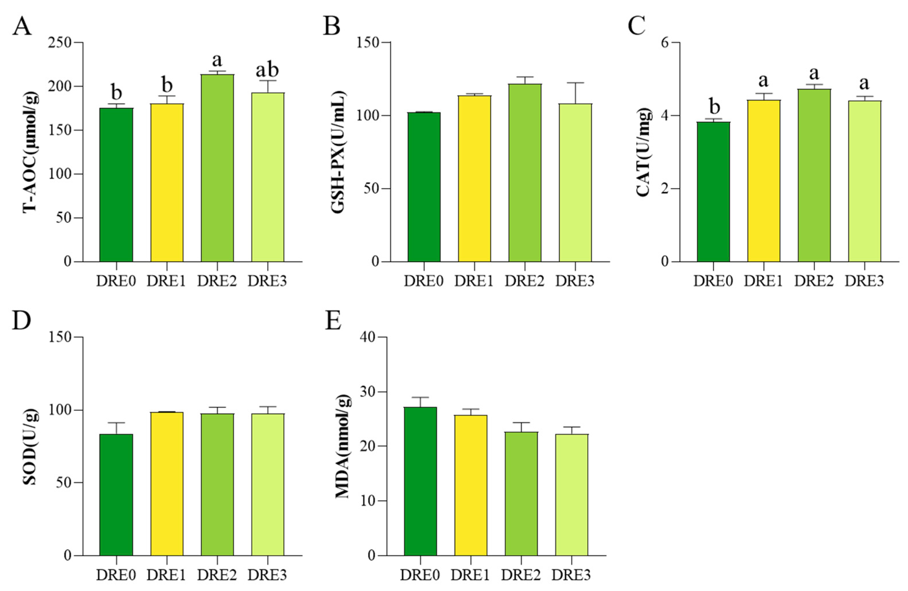
CAT activity was significantly higher in DRE1, DRE2, and DRE3 compared with the control, with the highest activity in DRE2 (p < 0.05, Figure 2). T-AOC was significantly enhanced in DRE2 compared with the control (p < 0.05, Figure 2). Dietary DRE supplementation elevated superoxide dismutase (SOD) and glutathione peroxidase (GSH-Px) activities while reducing malondialdehyde (MDA) content, albeit non-significantly (p > 0.05, Figure 2).

3.4. Effect of DRE on the Intestinal Morphology of Largemouth Bass

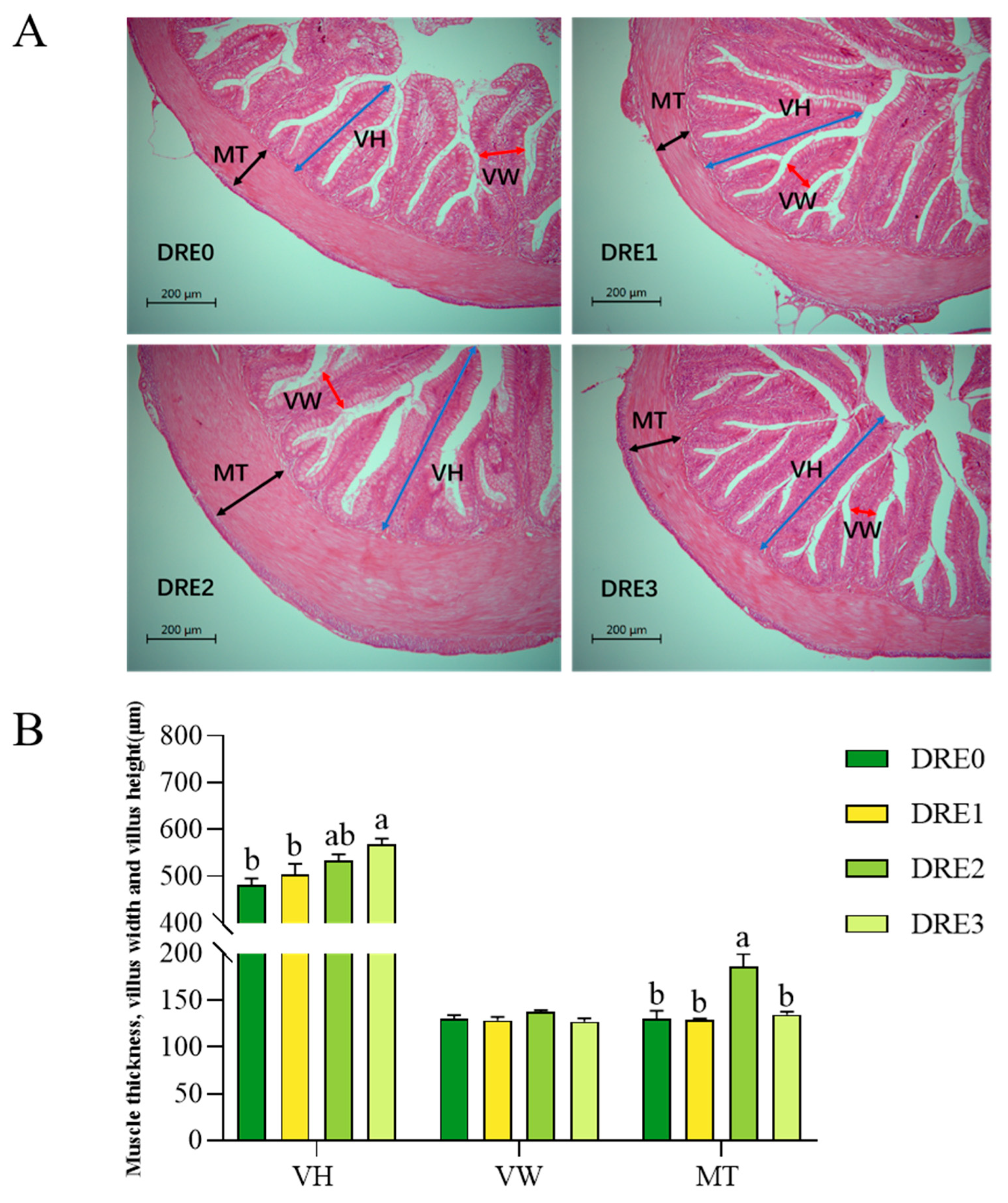
The control group exhibited atrophied and shortened intestinal villi, whereas the DRE-supplemented groups displayed a well-structured, intact intestinal architecture with abundant, well-developed villi arranged in orderly alignment and projecting perpendicularly into the intestinal lumen. Significantly higher muscular thickness was observed in DRE2 compared to the control (p < 0.05, Figure 3). Villus height was significantly elevated in DRE3 compared to the control (p < 0.05, Figure 3) and increased (but not significantly) in DRE2 (p > 0.05, Figure 3).

3.5. Effect of DRE on the Intestinal Microbiota of Largemouth Bass

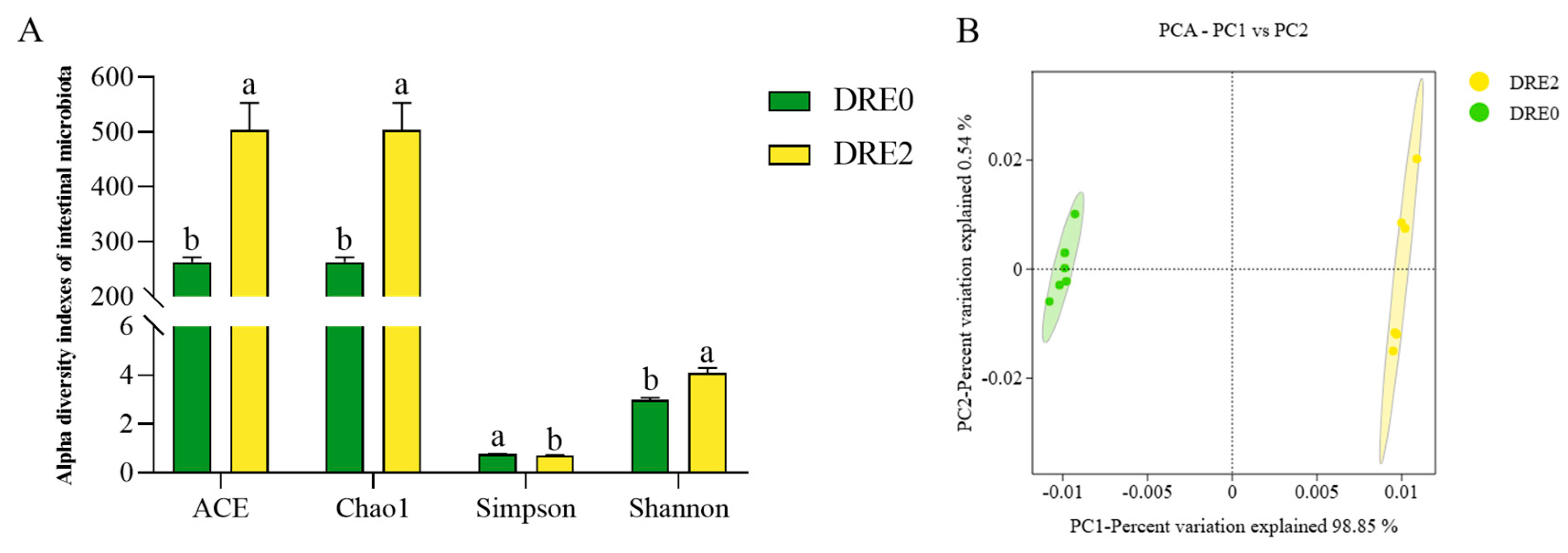
Based on the mentioned experimental data, group DRE2 demonstrated optimal overall effects and was selected for intestinal microbiota assessment. Altered microbial diversity was observed in DRE2 compared to the control. Alpha diversity indices (ACE, Chao1, Simpson, and Shannon) differed significantly between DRE2 and the control (p < 0.05, Figure 4). Principal component analysis revealed distinct clustering patterns, with samples from the DRE2 group distributed in quadrants I and IV, while control samples occupied quadrants II and III, indicating pronounced taxonomic divergence between the two groups (Figure 4).
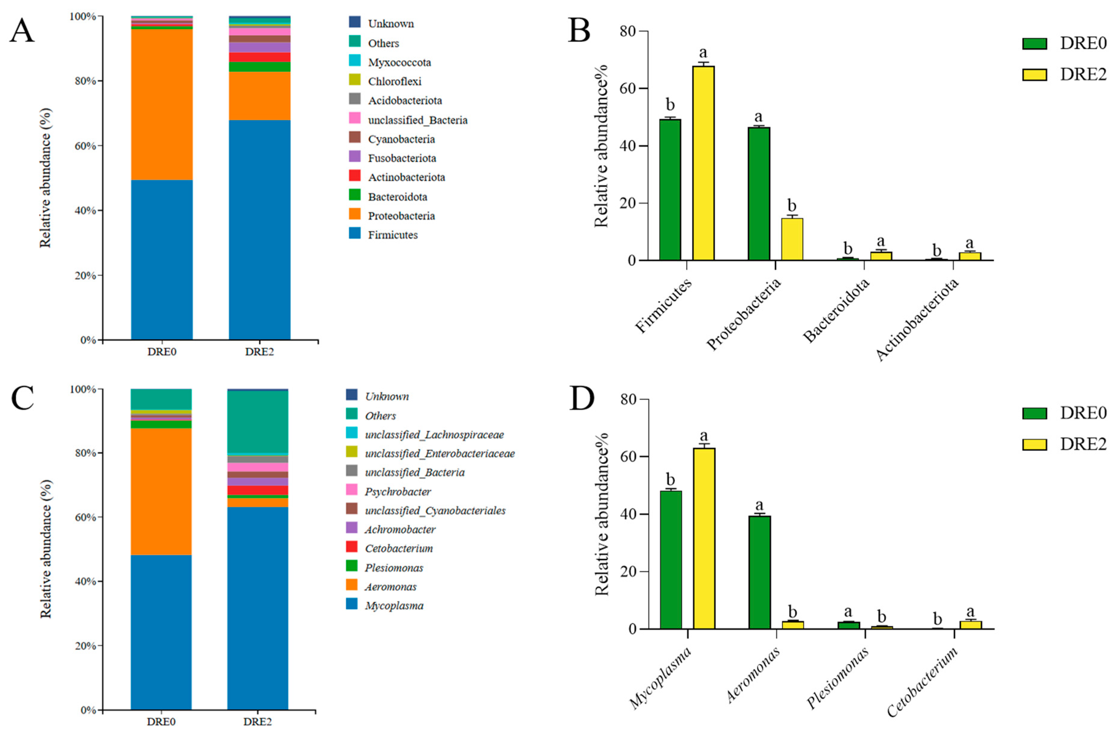
At the phylum level, Firmicutes, Proteobacteria, Bacteroidetes, and Actinobacteria were identified as the dominant bacterial phyla. Compared to the control, DRE2 exhibited increased relative abundance trends for Firmicutes, Bacteroidetes, and Actinobacteria alongside decreased Proteobacteria abundance, though these differences were not statistically significant (p < 0.05; Figure 5). At the genus level, Mycoplasma, Aeromonas, Plesiomonas, and Cetobacterium constituted the predominant genera. Significantly higher abundances of Cetobacterium and Mycoplasma, with correspondingly reduced relative abundances of Aeromonas and Plesiomonas, were observed in DRE2 versus the control (p < 0.05; Figure 5).

4. Discussion

Dandelion, a medicinal and edible herb rich in bioactive compounds, exerts beneficial regulatory effects on animal physiology [5]. This study systematically investigated the impacts of dandelion root extract (DRE) on growth performance, hepatointestinal health, and immune function in largemouth bass. Although dietary supplementation with graded DRE levels showed no significant effects on growth parameters such as final body weight (FBW) and weight gain rate (WGR), it notably enhanced hepatointestinal protection, immune modulation, and antioxidant responses. These findings underscore the potential of DRE as a functional feed additive in aquaculture.
Studies have indicated that incorporating dandelion extract into feed can enhance the growth performance of various animals, including finishing pigs [20], broilers [21], rainbow trout [22], and golden pompano (Trachinotus ovatus) [23]. In aquatic animals, dietary supplementation with 0.25% dandelion extract for 56 days increased the weight gain rate and specific growth rate of common carp [24], while supplementation with 0.1% dandelion extract for 56 days significantly improved the weight gain rate and feed intake of golden pompano [23]. However, in the present study, supplementing different concentrations of dandelion root extract (DRE) did not significantly affect the growth performance indicators of largemouth bass. This observation differs from the growth-promoting effects of dandelion extract reported in other fish species. These differences may stem from variations in feeding habits, metabolism, and developmental stages among fish species. As a carnivorous species, largemouth bass may exhibit resistance to plant-derived additives; this could potentially reduce their feed intake and consequently affect growth performance. Similarly, supplementing hybrid grouper (Epinephelus lanceolatus ♂ × Epinephelus fuscoguttatus ♀) feed with 0.1–0.8% dandelion extract for 56 days did not improve growth performance but did enhance liver health [25]. Furthermore, the relatively short experimental duration in the current study may have limited the translation of DRE-induced physiological health improvements into measurable growth performance enhancements. This is analogous to findings where supplementing juvenile largemouth bass feed with 0.5–1.5% kelp laminarin additive for 28 days resulted in no significant difference in growth performance, yet enhanced antioxidant capacity and suppressed the abundance of harmful intestinal microbiota [26]. Given our hypothesis that DRE effectively reduces energy loss during fish development while enhancing immune function, we further conduct an in-depth investigation in the following section [27].
Serum biochemical parameters serve as critical physiological and pathological indicators for assessing fish health, with ACP, AKP, IgM, and LZM directly reflecting immune defense capabilities [28,29]. Dandelion extract, which is enriched with bioactive components such as flavonoids and polysaccharides, has demonstrated immunomodulatory potential [10,13]. For instance, dietary supplementation with DRE significantly enhanced LZM and AKP activities in the serum of common carp [30]. Similarly, supplementation with dandelion flavonoids in snakehead (Channa argus) feed improved serum immune markers, including AKP, ACP, and IgM [31]. Dietary supplementation with DRE significantly enhanced the activities of AKP and LZM in largemouth bass. While ACP and IgM showed a similar trend, the increases were not statistically significant. These results suggest an enhancement of the non-specific immune defense capacity in largemouth bass. It is noteworthy that this immune-enhancing effect occurred without any change in growth performance, which suggests that the active components in DRE may exert their physiological regulatory effects primarily by acting on the immune system, rather than directly influencing growth metabolism.
The liver functions as the principal detoxification organ in vertebrates, where AST and ALT activities serve as biomarkers for hepatic injury, while T-AOC capacity, along with CAT, SOD, and GSH-PX activities, collectively indicate antioxidant potential [32,33]. MDA content directly reflects lipid-peroxidation-induced membrane damage [34]. Our findings indicate that dietary DRE supplementation resulted in a decreasing trend in the hepatosomatic index. This may reflect improved metabolic efficiency leading to a modest reduction in liver size, consistent with observations in largemouth bass supplemented with Artemisia annua extract [17]. In contrast, pathological conditions involving hepatic injury can induce liver enlargement and consequently elevate hepatosomatic index [35]. Notably, 0.1–0.15% DRE supplementation significantly decreased AST and ALT activities, demonstrating enhanced hepatic function in largemouth bass. This observation parallels findings in hybrid grouper, where 0.2–0.4% DRE supplementation similarly reduced hepatosomatic index and aminotransferase activities while improving hepatic structural integrity [25]. Notably, although in this study SOD and GSH-Px activities only showed an upward trend and MDA content exhibited a downward trend, these directional changes closely align with the significantly enhanced T-AOC and CAT activities. This consistency indicates an optimization of hepatic antioxidant capacity that warrants further investigation through long-term feeding trials or higher supplementation levels. This protective effect is probably attributed to the polysaccharides in DRE functioning as natural antioxidants that reinforce hepatic protection through antioxidant system potentiation [11].
The intestine, serving as the central organ for digestion and absorption in fish, directly influences nutrient metabolic efficiency. Additionally, intestinal structural integrity is essential for both nutrient assimilation and primary immunological barrier function [36,37,38]. Villus height, muscular thickness, and mucosal integrity constitute critical morphological indicators for evaluating intestinal health [36]. Previous studies have demonstrated that dietary DRE supplementation significantly improves intestinal villus morphology and muscular thickness in rainbow trout [39]. In this study, dietary DRE supplementation significantly enhanced villus height and muscular thickness in largemouth bass, concurrently reducing serum D-lactate levels. As a sensitive biomarker of intestinal mucosal integrity, the decreased D-lactate concentration suggests enhanced intestinal barrier function, likely attributable to DRE-induced upregulation of tight junction protein genes (ZO-1 and Occludin), which promotes the reconstruction of interepithelial connections and reduces D-lactate leakage [16,23]. These findings collectively elucidate the multifaceted protective mechanisms of DRE in maintaining intestinal structural integrity and functional stability.
The gut microbiota plays crucial role in maintaining fish health, particularly in enhancing pathogen resistance and regulating intestinal immune homeostasis [40]. Previous research has shown that bioactive compounds in dandelion can modulate gut microbiota balance through antimicrobial effects [9,41,42]. In this study, supplementation with 0.1% DRE significantly increased microbial species richness and altered community composition. Notably, DRE treatment elevated the relative abundance of Firmicutes while decreasing the proportion of Proteobacteria. Firmicutes are commonly associated with beneficial metabolic functions, whereas Proteobacteria often include opportunistic pathogens [43]. At the genus level, DRE markedly decreased the abundance of pathogenic Aeromonas and Plesiomonas, while promoting the proliferation of beneficial Cetobacterium. These findings collectively suggest that DRE may optimize microbial structure by suppressing niche colonization of potential pathogens and creating competitive advantages for beneficial taxa. Notably, the enrichment of beneficial microbiota could maintain immune equilibrium by suppressing excessive inflammatory responses [44], thereby indirectly enhancing the host’s systemic disease resistance.

5. Conclusions

In conclusion, dietary supplementation with 0.10% DRE enhances antioxidant capacity, humoral immunity, and hepatic health in largemouth bass. Furthermore, DRE administration shaped intestinal microbiota, optimized intestinal morphology, and strengthened barrier function, collectively promoting intestinal health. These findings provide a theoretical foundation for developing functional feed additives based on DRE in aquaculture practices.

Author Contributions

P.-A.L.: Investigation, Writing—original draft. H.-L.Y.: Data analysis, Writing—review and editing. J.H.: Investigation, Writing. F.L.: Investigation. K.-Y.H.: Data analysis. Z.-X.L.: Investigation, Writing. G.-H.C.: Formal analysis, Writing—review and editing. Y.-Z.S.: Conceptualization, Supervision, Project administration, Funding acquisition, Review and editing. All authors have read and agreed to the published version of the manuscript.

Funding

This study was supported by Xiamen Science and Technology Bureau Project (2024CXY0307) and Fujian Province Agricultural Guidance (Key) Project (2023N0012).

Institutional Review Board Statement

The study was approved by The Animal Care and Use Committee at the Fisheries College of Jimei University No. 1067 2024-01-03.

Data Availability Statement

The data that support the findings of this study are available from the corresponding author upon reasonable request.

Acknowledgments

We sincerely thank the reviewers for their insightful comments to improve the manuscript.

Conflicts of Interest

The authors declare no conflicts of interest.

References

  1. Flores-Méndez, L.C.; Lizárraga-Velázquez, C.E.; Sánchez-Gutiérrez, E.Y.; Arrizon, J.; Leyva-López, N.; Hernández, C. Study of the Effect of Dietary Agavin Supplementation in Blood Parameters and Antioxidant Enzymes of Juvenile Nile Tilapia (Oreochromis niloticus) under Stress Conditions. Fishes 2022, 7, 340. [Google Scholar] [CrossRef]
  2. Dawood, M.A.O.; Metwally, A.E.-S.; El-Sharawy, M.E.; Atta, A.M.; Elbialy, Z.I.; Abdel-Latif, H.M.R.; Paray, B.A. The Role of β-Glucan in the Growth, Intestinal Morphometry, and Immune-Related Gene and Heat Shock Protein Expressions of Nile Tilapia (Oreochromis niloticus) under Different Stocking Densities. Aquaculture 2020, 523, 735205. [Google Scholar] [CrossRef]
  3. Cabello, F.C.; Godfrey, H.P.; Buschmann, A.H.; Dölz, H.J. Aquaculture as yet Another Environmental Gateway to the Development and Globalisation of Antimicrobial Resistance. Lancet 2016, 16, e127–e133. [Google Scholar] [CrossRef] [PubMed]
  4. Yuan, X.; Lv, Z.; Zhang, Z.; Han, Y.; Liu, Z.; Zhang, H. A Review of Antibiotics, Antibiotic Resistant Bacteria, and Resistance Genes in Aquaculture: Occurrence, Contamination, and Transmission. Toxics 2023, 11, 420. [Google Scholar] [CrossRef]
  5. Fan, M.; Zhang, X.; Song, H.; Zhang, Y. Dandelion (Taraxacum Genus): A Review of Chemical Constituents and Pharmacological Effects. Molecules 2023, 28, 5022. [Google Scholar] [CrossRef]
  6. Sharifi-Rad, M.; Roberts, T.H.; Matthews, K.R.; Bezerra, C.F.; Morais-Braga, M.F.B.; Coutinho, H.D.M.; Sharopov, F.; Salehi, B.; Yousaf, Z.; Sharifi-Rad, M.; et al. Ethnobotany of the Genus Taraxacum-Phytochemicals and Antimicrobial Activity. Phytother. Res. 2018, 32, 2131–2145. [Google Scholar] [CrossRef] [PubMed]
  7. Kang, L.; Miao, M.; Song, Y.; Fang, X.; Zhang, J.; Zhang, Y.; Miao, J. Total Flavonoids of Taraxacum Mongolicum Inhibit Non-Small Cell Lung Cancer by Regulating Immune Function. J. Ethnopharmacol. 2021, 281, 114514. [Google Scholar] [CrossRef]
  8. Lis, B.; Rolnik, A.; Jedrejek, D.; Soluch, A.; Stochmal, A.; Olas, B. Dandelion (Taraxacum officinale L.) Root Components Exhibit Anti-Oxidative and Antiplatelet Action in an in Vitro Study. J. Funct. Foods 2019, 59, 16–24. [Google Scholar] [CrossRef]
  9. Li, Z.; Li, X.; Shi, P.; Li, P.; Fu, Y.; Tan, G.; Zhou, J.; Zeng, J.; Huang, P. Modulation of Acute Intestinal Inflammation by Dandelion Polysaccharides: An In-Depth Analysis of Antioxidative, Anti-Inflammatory Effects and Gut Microbiota Regulation. Int. J. Mol. Sci. 2024, 25, 1429. [Google Scholar] [CrossRef]
  10. Qiao, Q.; Song, X.; Zhang, C.; Jiang, C.; Jiang, R. Structure and Immunostimulating Activity of Polysaccharides Derived from the Roots and Leaves of Dandelion. Chem. Biol. Technol. Agric. 2024, 11, 51. [Google Scholar] [CrossRef]
  11. Cai, L.; Wan, D.; Yi, F.; Luan, L. Purification, Preliminary Characterization and Hepatoprotective Effects of Polysaccharides from Dandelion Root. Molecules 2017, 22, 1409. [Google Scholar] [CrossRef]
  12. Li, W.; Luo, F.; Wu, X.; Fan, B.; Yang, M.; Zhong, W.; Guan, D.; Wang, F.; Wang, Q. Anti-Inflammatory Effects and Mechanisms of Dandelion in RAW264.7 Macrophages and Zebrafish Larvae. Front. Pharmacol. 2022, 13, 906927. [Google Scholar] [CrossRef] [PubMed]
  13. Hosseini Shekarabi, S.P.; Mostafavi, Z.S.; Mehrgan, M.S.; Islami, H.R. Dietary Supplementation with Dandelion (Taraxacum officinale) Flower Extract Provides Immunostimulation and Resistance against Streptococcus Iniae Infection in Rainbow Trout (Oncorhynchus mykiss). Fish Shellfish Immunol. 2021, 118, 180–187. [Google Scholar] [CrossRef]
  14. Xue, S.; Xia, B.; Zou, Y.; Li, L.; Zhang, B.; Shen, Z.; Xiang, Y.; Han, Y.; Chen, W. Dandelion Extract on Growth Performance, Immunity, Stress and Infection Resistance in Common Carp. Aquac. Rep. 2022, 26, 101330. [Google Scholar] [CrossRef]
  15. He, G.; Sun, H.; Liao, R.; Wei, Y.; Zhang, T.; Chen, Y.; Lin, S. Effects of Herbal Extracts (Foeniculum vulgare and Artemisia annua) on Growth, Liver Antioxidant Capacity, Intestinal Morphology and Microorganism of Juvenile Largemouth Bass, Micropterus salmoides. Aquac. Rep. 2022, 23, 101081. [Google Scholar] [CrossRef]
  16. Tan, X.; Sun, Z. Dietary Dandelion Extract Improved Growth Performance, Immunity, Intestinal Morphology and Microbiota Composition of Golden Pompano Trachinotus ovatus. Aquac. Rep. 2020, 18, 100491. [Google Scholar] [CrossRef]
  17. Salem, M.O.A.; Salem, T.A.; Yürüten Özdemir, K.; Sönmez, A.Y.; Bilen, S.; Güney, K. Antioxidant Enzyme Activities and Immune Responses in Rainbow Trout (Onchorhynchus mykiss) Juveniles Fed Diets Supplemented with Dandelion (Taraxacum officinalis) and Lichen (Usnea barbata) Extracts. Fish Physiol. Biochem. 2021, 47, 1053–1062. [Google Scholar] [CrossRef]
  18. Li, Q.; Fu, M.; Zhu, S.; Liu, J.; Li, Y.; Xue, Z.; Zhou, Z.; Yu, L. Effects of Dietary Hydroxy-Cinnamic Acid Derivatives on Growth, Muscle, and Intestinal Parameters of Tilapia (Oreochromis niloticus). Fish Physiol. Biochem. 2025, 51, 4. [Google Scholar] [CrossRef]
  19. Meng, X.; Cai, H.; Li, H.; You, F.; Jiang, A.; Hu, W.; Li, K.; Zhang, X.; Zhang, Y.; Chang, X.; et al. Clostridium Butyricum-Fermented Chinese Herbal Medicine Enhances the Immunity by Modulating the Intestinal Microflora of Largemouth Bass (Micropterus salmoides). Aquaculture 2023, 562, 738768. [Google Scholar] [CrossRef]
  20. Samolińska, W.; Grela, E.R.; Kowalczuk-Vasilev, E.; Kiczorowska, B.; Klebaniuk, R.; Hanczakowska, E. Evaluation of Garlic and Dandelion Supplementation on the Growth Performance, Carcass Traits, and Fatty Acid Composition of Growing-Finishing Pigs. Anim. Feed Sci. Technol. 2020, 259, 114316. [Google Scholar] [CrossRef]
  21. Mao, J.; Wang, Y.; Wang, W.; Duan, T.; Yin, N.; Guo, T.; Guo, H.; Liu, N.; An, X.; Qi, J. Effects of Taraxacum mongolicum Hand.-Mazz. (dandelion) on growth performance, expression of genes coding for tight junction protein and mucin, microbiota composition and short chain fatty acids in ileum of broiler chickens. BMC Vet. Res. 2022, 18, 180. [Google Scholar] [CrossRef]
  22. Mostafavi, Z.S.; Shekarabi, S.P.H.; Mehrgan, M.S.; Islami, H.R. Amelioration of Growth Performance, Physio-Metabolic Responses, and Antioxidant Defense System in Rainbow Trout, Oncorhynchus mykiss, Using Dietary Dandelion, Taraxacum officinale, Flower Extract. Aquaculture 2022, 546, 737296. [Google Scholar] [CrossRef]
  23. Tan, X.; Sun, Z.; Zhou, C.; Huang, Z.; Tan, L.; Xun, P.; Huang, Q.; Lin, H.; Ye, C.; Wang, A. Effects of Dietary Dandelion Extract on Intestinal Morphology, Antioxidant Status, Immune Function and Physical Barrier Function of Juvenile Golden Pompano Trachinotus ovatus. Fish Shellfish Immunol. 2018, 73, 197–206. [Google Scholar] [CrossRef]
  24. Giri, S.S.; Kim, S.G.; Woo, K.J.; Jung, W.J.; Lee, S.B.; Lee, Y.M.; Jo, S.J.; Kim, J.H.; Park, S.C. Impact of Dandelion Polysaccharides on Growth and Immunity Response in Common Carp Cyprinus carpio. Fish Shellfish Immunol. 2022, 128, 371–379. [Google Scholar] [CrossRef] [PubMed]
  25. Sun, Z.; Tan, X.; Xu, M.; Liu, Q.; Ye, H.; Zou, C.; Zhou, Y.; Su, N.; Chen, L.; Wang, A.; et al. Effects of Dietary Dandelion Extracts on Growth Performance, Liver Histology, Immune-Related Gene Expression and CCl4 Resistance of Hybrid Grouper (Epinephelus lanceolatus♂ × Epinephelus fuscoguttatus♀). Fish Shellfish Immunol. 2019, 88, 126–134. [Google Scholar] [CrossRef] [PubMed]
  26. Wu, Y.; Cheng, Y.; Qian, S.; Zhang, W.; Huang, M.; Yang, S.; Fei, H. An Evaluation of Laminarin Additive in the Diets of Juvenile Largemouth Bass (Micropterus salmoides): Growth, Antioxidant Capacity, Immune Response and Intestinal Microbiota. Animals 2023, 13, 459. [Google Scholar] [CrossRef]
  27. Zhang, X.; Huang, K.; Zhong, H.; Ma, Y.; Guo, Z.; Tang, Z.; Liang, J.; Luo, Y.; Su, Z.; Wang, L. Effects of Lycium Barbarum Polysaccharides on Immunological Parameters, Apoptosis, and Growth Performance of Nile Tilapia (Oreochromis niloticus). Fish Shellfish Immunol. 2020, 97, 509–514. [Google Scholar] [CrossRef]
  28. Mashoof, S.; Criscitiello, M.F. Fish Immunoglobulins. Biology 2016, 5, 45. [Google Scholar] [CrossRef]
  29. Lulijwa, R.; Young, T.; Symonds, J.E.; Walker, S.P.; Delorme, N.J.; Alfaro, A.C. Uncoupling Thermotolerance and Growth Performance in Chinook Salmon: Blood Biochemistry and Immune Capacity. Metabolites 2021, 11, 547. [Google Scholar] [CrossRef]
  30. Chen, W.; Xia, B.; Zou, Y.; Xiang, Y.; Shen, Z.; Xue, S. Protective Effect of Dietary Dandelion (Taraxacum officinale) Extract on Common Carp under Acute Ammonia Stress. Aquac. Rep. 2023, 30, 101609. [Google Scholar] [CrossRef]
  31. Du, J.-H.; Xu, M.-Y.; Wang, Y.; Lei, Z.; Yu, Z.; Li, M.-Y. Evaluation of Taraxacum mongolicum Flavonoids in Diets for Channa Argus Based on Growth Performance, Immune Responses, Apoptosis and Antioxidant Defense System under Lipopolysaccharide Stress. Fish Shellfish Immunol. 2022, 131, 1224–1233. [Google Scholar] [CrossRef]
  32. Madeira, D.; Narciso, L.; Cabral, H.N.; Vinagre, C.; Diniz, M.S. Influence of Temperature in Thermal and Oxidative Stress Responses in Estuarine Fish. Comp. Biochem. Physiol. A Mol. Integr. Physiol. 2013, 166, 237–243. [Google Scholar] [CrossRef] [PubMed]
  33. Shi, A.; Ma, H.; Shi, X.; Zhou, W.; Pan, W.; Song, Y.; Chen, Q.; Yu, X.; Niu, C.; Yang, Y.; et al. Effects of Microbe-Derived Antioxidants on Growth, Digestive and Aminotransferase Activities, and Antioxidant Capacities in the Hepatopancreas of Eriocheir Sinensis under Ammonia Nitrogen Stress. Aquac. Fish. 2024, 9, 957–966. [Google Scholar] [CrossRef]
  34. Storey, K.B. Oxidative Stress: Animal Adaptations in Nature. Braz. J. Med. Biol. Res. 1996, 29, 1715–1733. [Google Scholar] [PubMed]
  35. Chen, P.; Zhu, Y.; Wu, X.; Gu, X.; Xue, M.; Liang, X. Metabolic Adaptation to High-Starch Diet in Largemouth Bass (Micropterus salmoides) Was Associated with the Restoration of Metabolic Functions via Inflammation, Bile Acid Synthesis and Energy Metabolism. Br. J. Nutr. 2022, 129, 381–394. [Google Scholar] [CrossRef]
  36. Kocot, A.M.; Jarocka-Cyrta, E.; Drabińska, N. Overview of the Importance of Biotics in Gut Barrier Integrity. Int. J. Mol. Sci. 2022, 23, 2896. [Google Scholar] [CrossRef]
  37. Nadal, A.L.; Ikeda-Ohtsubo, W.; Sipkema, D.; Peggs, D.; McGurk, C.; Forlenza, M.; Wiegertjes, G.F.; Brugman, S. Feed, Microbiota, and Gut Immunity: Using the Zebrafish Model to Understand Fish Health. Front. Immunol. 2020, 11, 114. [Google Scholar] [CrossRef] [PubMed]
  38. Takiishi, T.; Fenero, C.I.M.; Câmara, N.O.S. Intestinal Barrier and Gut Microbiota: Shaping Our Immune Responses throughout Life. Tissue Barriers 2017, 5, e1373208. [Google Scholar] [CrossRef]
  39. Köse, Ö.; Karabulut, H.A.; Er, A. Dandelion Root Extract in Trout Feed and Its Effects on the Physiological Performance of Oncorhynchus mykiss and Resistance to Lactococcus garvieae Infection. Ann. Anim. Sci. 2024, 24, 161–177. [Google Scholar] [CrossRef]
  40. He, M.; Li, X.; Poolsawat, L.; Guo, Z.; Yao, W.; Zhang, C.; Leng, X. Effects of Fish Meal Replaced by Fermented Soybean Meal on Growth Performance, Intestinal Histology and Microbiota of Largemouth Bass (Micropterus salmoides). Aquac. Nutr. 2020, 26, 1058–1071. [Google Scholar] [CrossRef]
  41. Liang, Y.; Duan, H.; Zhang, P.; Han, H.; Gao, F.; Li, Y.; Xu, Z. Extraction and Isolation of the Active Ingredients of Dandelion and Its Antifungal Activity against Candida Albicans. Mol. Med. Rep. 2019, 21, 229–239. [Google Scholar] [CrossRef] [PubMed]
  42. Ye, D.; Sun, J.; Li, Y. Evaluation of Toxicity, Bacteriostatic, Analgesic, Anti-Inflammatory, and Antipyretic Activities of Huangqin-Honghua-Pugongying-Jinyinhua Extract. Vet. Sci. 2021, 8, 330. [Google Scholar] [CrossRef] [PubMed]
  43. Xu, J.-M.; Gao, W.-R.; Liang, P.; Cai, G.-H.; Yang, H.-L.; Lin, J.-B.; Sun, Y.-Z. Pleurotus Eryngii Root Waste and Soybean Meal Co-Fermented Protein Improved the Growth, Immunity, Liver and Intestinal Health of Largemouth Bass (Micropterus salmoides). Fish Shellfish Immunol. 2024, 149, 109551. [Google Scholar] [CrossRef] [PubMed]
  44. Round, J.L.; Mazmanian, S.K. The Gut Microbiota Shapes Intestinal Immune Responses during Health and Disease. Nat. Rev. Immunol. 2009, 9, 313–323. [Google Scholar] [CrossRef]
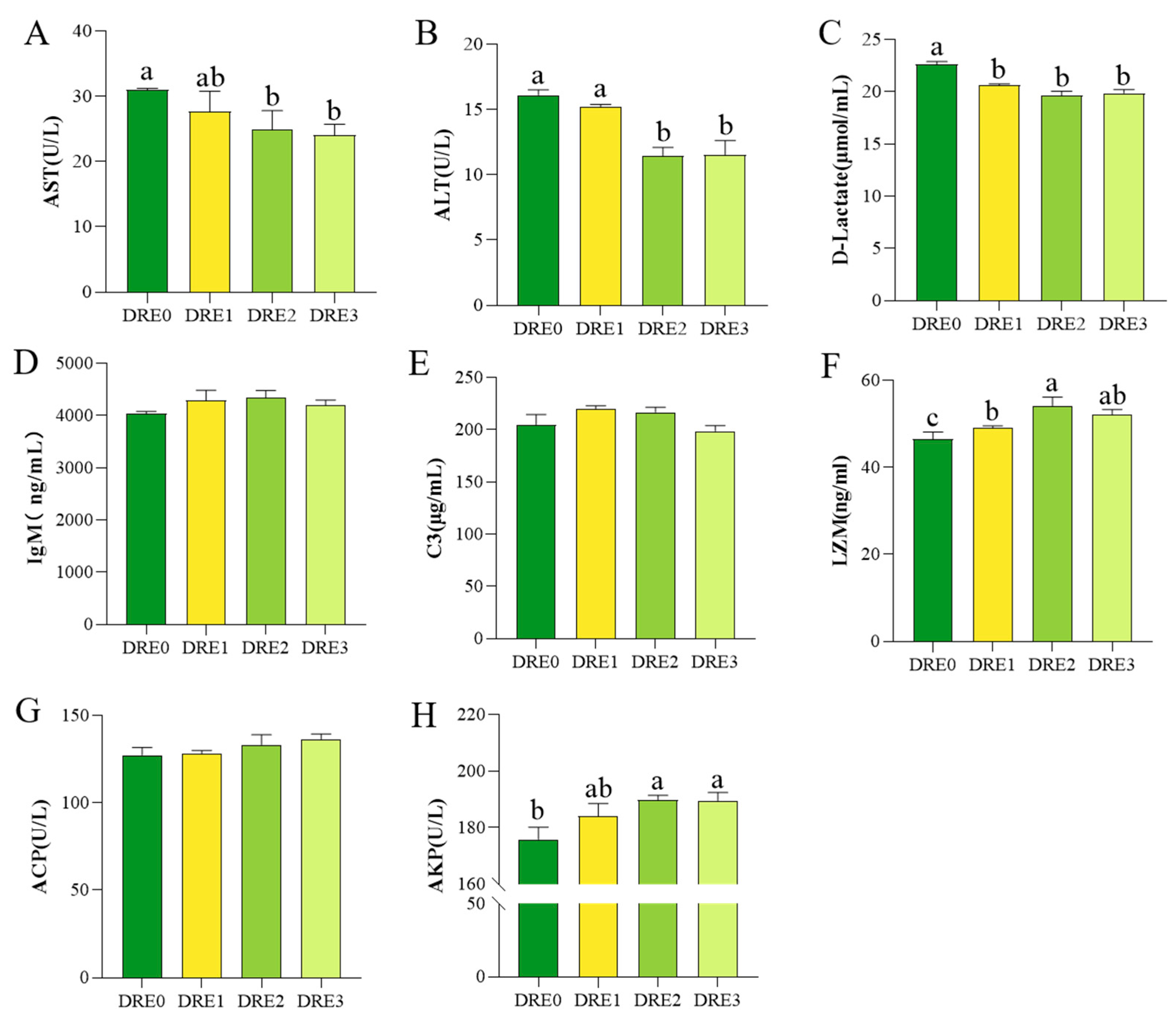
Figure 1. Effects of dietary DRE supplementation on serum biochemical parameters in Micropterus salmoides. (A) Aspartate aminotransferase (AST); (B) Alanine aminotransferase (ALT); (C) D-lactate; (D) Immunoglobulin M (IgM); (E) Complement 3 (C3); (F) Lysozyme (LZM); (G) Acid phosphatase (ACP); (H) Alkaline phosphatase (AKP). Different lowercase letters denote significant differences at p < 0.05.
Fishes 10 00446 g001
Figure 2. Effects of dietary DRE supplementation on hepatic antioxidant indices in Micropterus salmoides. (A) Total antioxidant capacity (T-AOC); (B) Glutathione peroxidase (GSH-Px); (C) Catalase (CAT); (D) Superoxide dismutase (SOD); (E) Malondialdehyde (MDA). Different lowercase letters denote significant differences at p < 0.05.
Fishes 10 00446 g002
Figure 3. Effects of dietary DRE supplementation on intestinal histomorphology in Micropterus salmoides. (A) Blue bidirectional arrow VH: Villus height; red bidirectional arrow VW: Villous width; black bidirectional arrow MT: Muscular thickness. (B) VH: Villus height; VW: Villous width; MT: Muscular thickness. Different lowercase letters denote significant differences at p < 0.05.
Fishes 10 00446 g003
Figure 4. Effects of dietary DRE supplementation on intestinal microbiota diversity in Micropterus salmoides. (A) α-Diversity indices (ACE, Chao1, Simpson, Shannon); (B) β-Diversity: Principal Coordinate Analysis (PCoA). Different lowercase letters denote significant differences at p < 0.05.
Fishes 10 00446 g004
Figure 5. Taxonomic composition of intestinal microbiota based on 16S rRNA gene (V3–V4 regions) sequencing. (A) Phylum-level taxonomy (intestinal samples); (B) Phylum-level taxonomy; (C) Genus-level taxonomy (intestinal samples); (D) Genus-level taxonomy. Only the top 10 most abundant phyla and genera by relative abundance are displayed. Different lowercase letters denote significant differences at p < 0.05.
Fishes 10 00446 g005
Table 1. Formulation and proximate composition of the experimental diets (g/100 g dry matter).
IngredientContent
Domestic fish meal 126
Steam fish meal 120
Soybean meal 120
Wheat flour 114
Soybean protein concentrate 16
Corn protein 15
Soybean oil3
Fish oil2
Lecithin0.6
Ca(H2PO4)21.8
Choline chloride0.5
Vitamin C0.1
Mineral premix 20.6
Vitamin premix 30.4
Proximate composition (g/100 g dry matter basis)
Crude protein47.4
Crude fat10.4
1 Supplied by Xiamen Jiakang Aquatic Feed Co., Ltd. (Fujian, China). Domestic fish meal: crude protein content 65%, crude fat content 10%. Steam fish meal: crude protein content 68%, crude fat content 7.8%. Soybean meal: crude protein content 43%, crude fat content 1.5%. Wheat flour: crude protein content 10%, crude fat content 1.2%. Soybean protein concentrate: crude protein content 65%, crude fat content 0.6%. Corn protein: crude protein content 60%, crude fat content 2.2%. 2 Vitamin premix containing the following (mg/kg diet): thiamin, 10; riboflavin, 8; pyridoxine HCl, 10; vitamin B12, 0.2, vitamin K3, 10; inositol, 100; pantothenic acid, 20; niacin acid, 50; folic acid, 2; biotin, 2; retinol acetate, 400; cholecalciferol, 5; alpha-tocopherol, 100; ethoxyquin, 150; wheat middling 132.8. 3 Mineral premix containing the following (mg/kg diet): KCI, 200; KI, 60; CoSO4, 100; CuSO4·5H2O, 24; FeSO4·H2O, 400; ZnSO4·H2O, 174; MnSO4·H2O, 78; MgSO4·7H2O, 800; Na2SeO3, 50; Zeolite, 311.4.
Table 2. Effect of DRE on growth performance indices of largemouth bass.
DRE0DRE1DRE2DRE3
FBW (g/fish)42.80 ± 0.9041.66 ± 0.5041.63 ± 0.3441.65 ± 1.04
WGR (%)684.79 ± 16.24662.26 ± 8.70662.69 ± 6.23663.55 ± 18.97
SGR (%/d)4.90 ± 0.054.84 ± 0.034.84 ± 0.024.84 ± 0.06
FCR0.83 ± 0.020.80 ± 0.010.83 ± 0.010.81 ± 0.01
CF (g/cm3)1.74 ± 0.041.73 ± 0.011.68 ± 0.011.73 ± 0.04
HSI (%)2.01 ± 0.061.95 ± 0.071.96 ± 0.091.89 ± 0.03
SR (%)100100100100
FBW = Final body weight; WGR = Weight gain rate; SGR = Specific growth rate; FCR = Feed conversion rate; CF = Condition factor; HSI = Hepatosomatic index; SR = Survival rate. Values are means ± SEM (n = 3).
Disclaimer/Publisher’s Note: The statements, opinions and data contained in all publications are solely those of the individual author(s) and contributor(s) and not of MDPI and/or the editor(s). MDPI and/or the editor(s) disclaim responsibility for any injury to people or property resulting from any ideas, methods, instructions or products referred to in the content.