Article

WSSV Infection in the Gut Microbiota of the Black Tiger Shrimp Penaeus monodon

1
Marine Science Research Institute of Shandong Province, Qingdao 266104, China
2
Wudi County Marine and Fishery Development Research Center, Binzhou 251900, China
3
Shandong Youfa Aquaculture Co., Ltd., Binzhou 251900, China
4
College of Fisheries and Life Science, Shanghai Ocean University, Shanghai 201306, China
*
Authors to whom correspondence should be addressed.
These authors contributed equally to this work.
Fishes 2025, 10(9), 440; https://doi.org/10.3390/fishes10090440
Submission received: 11 July 2025 / Revised: 16 August 2025 / Accepted: 28 August 2025 / Published: 3 September 2025
(This article belongs to the Section Welfare, Health and Disease)

Abstract

This study investigated the impacts of white spot syndrome virus (WSSV) on the gut microbiota of Penaeus monodon through a comparative microbiota analysis of infected and healthy shrimp using 16S rDNA sequencing. The WSSV-infected shrimp exhibited characteristic white spots, reduced feeding activity, and behavioral lethargy preceding 100% mortality. The comparative microbiota analysis revealed a significantly diminished α-diversity in the infected specimens, marked by phylum-level dominance shifts from Proteobacteria (72.68%) and Firmicutes (11.27%) in the controls to Cyanobacteria (75.51%) and Proteobacteria (15.63%) in the WSSV-infected shrimp. An LEfSe analysis identified Arthrospira_PCC-7345 and Halochromatium as significantly enriched taxa during infection, contrasting with depleted populations of Ruegeria, Marivita, Bacillus, and seven other genera. The distinct dysbiosis pattern characterized by the pathogen-favored taxa proliferation and commensal species suppression demonstrates WSSV-associated microbiota restructuring, potentially contributing to disease progression in farmed P. monodon. These findings establish intestinal microbial biomarkers for early WSSV detection in aquaculture systems.
Key Contribution: This study demonstrates that WSSV infection in P. monodon induces significant behavioral changes and gut microbiota dysbiosis, which may contribute to disease progression. These behavioral and microbial biomarkers provide a foundation for early WSSV detection in aquaculture. Future research should explore whether microbiota modulation could mitigate WSSV impacts, improving shrimp health and farm productivity.

1. Introduction

The global aquaculture landscape is undergoing a critical transformation, with Penaeus vannamei cultivation entering a phase of diminishing returns while China witnesses a remarkable expansion in P. monodon farming [1]. This industrial shift underscores the urgent need to understand the biological factors influencing shrimp health, particularly the underappreciated role of the intestinal microbiota. Emerging evidence reveals that symbiotic gut microorganisms constitute a fundamental biological barrier, providing a crucial immune defense during crustacean development [2,3] and serving as frontline protectors against pathogenic invasions [4,5,6]. However, this delicate microbial ecosystem remains vulnerable to environmental stressors and pathogen attacks, which can trigger structural dysbiosis, biodiversity loss, and consequent immunosuppression—ultimately predisposing the hosts to infection [7,8,9].
Contemporary advances in microbial ecology have been propelled by 16S ribosomal RNA (rRNA) gene amplicon sequencing, a gold-standard approach for profiling complex microbial communities. By targeting the hypervariable regions (e.g., V3–V4) of this phylogenetic marker gene, researchers can achieve genus-level taxonomic resolution while capturing > 90% of prokaryotic diversity [10]. While primer selection fundamentally dictates the scope and accuracy of microbial profiling, computational tools such as MultiPrime have revolutionized this process through intelligent multiplex PCR primer design, maximizing coverage while minimizing off-target amplification [11]. Compared to traditional culture-based methods, this high-throughput technique overcomes the “great plate count anomaly” by detecting both culturable and unculturable species. Building upon these primer optimization strategies, recent methodological breakthroughs, particularly full-length 16S sequencing via third-generation platforms (e.g., PacBio SMRT), further enhance classification accuracy through complete coverage of the 1500 bp gene [12]. These synergistically integrated advances—combining precision primer design with long-read sequencing—make amplicon sequencing indispensable for identifying subtle microbiota shifts during disease progression—a critical advantage when studying pathogen–microbiome interactions in non-model organisms such as shrimp.
Recent investigations into shrimp–pathogen interactions have illuminated specific microbial dynamics during disease progression. Studies of white spot syndrome virus (WSSV) infection demonstrate the proliferation of Proteobacteria (notably Photobacterium) and Fusobacteria in the affected shrimp populations, accompanied by the enrichment of the pathogenic Arcobacter and Flavobacterium genera that exacerbate intestinal microbiota heterogeneity [13]. Complementary findings reveal that WSSV infection heightens the susceptibility to secondary Photobacterium campbelli infections, accelerating mortality rates in the affected populations [14]. Parallel studies of other viral infections show similar microbial disruption patterns, including the depletion of beneficial Bacteroidetes and Firmicutes coupled with pathogenic Vibrio and Photobacterium colonization during Decapod iridescent virus 1 (DIV1) outbreaks [15]. Furthermore, research on acute hepatopancreatic necrosis disease (AHPND) identifies α-diversity collapse and community destabilization, manifested through elevated variation coefficients and dysbiosis indices compared to healthy specimens [16].
WSSV stands as the most devastating shrimp pathogen, capable of triggering 100% mortality within 7–9 days and causing catastrophic economic losses [17,18,19]. Current research has characterized WSSV-induced gut microbiota alterations in multiple crustacean species, such as the Pacific white shrimp (Litopenaeus vannamei) [13] and the red swamp crayfish (Procambarus clarkii) [20]. We aimed to identify WSSV-induced dysbiosis patterns and characterize species-specific microbial responses in P. monodon—a species of growing agricultural importance. This study investigated the WSSV–microbiota interactions in pond-reared P. monodon. Through a comparative analysis of gut microbial communities in healthy versus WSSV-infected specimens, we aimed to identify pathogen-induced dysbiosis patterns and characterize species-specific microbial responses. The findings may provide insights for developing WSSV prevention strategies in commercial P. monodon aquaculture.

2. Materials and Methods

2.1. Sample Collection and Preparation

Following the methodology described by Cornejo-Granados et al. [7], the moribund diseased shrimp (n = 90, body length: 6–7 cm) in the late stages of WSSV infection [21] were obtained from a pond with stocking density of 30,000 individuals, salinity of 27, and water temperature of 27~28 °C located at an aquaculture farm in Shandong Province, China. The healthy cultured shrimp (n = 90, body length: 6–7 cm) without disease signs of WSSV infection were obtained from another pond from the same aquaculture farm with the same stocking density, salinity, and water temperature. Both healthy and WSSV-infected shrimp surfaces were sterilized with 75% ethanol followed by aseptic collection of complete intestinal tissues, which were dissected and divided into 3 biological replicates, randomly labeled as Control 1–3 (healthy, n = 30) and WSSV 1–3 (infected, n = 30), and preserved in sterile tubes. All samples were flash-frozen in liquid nitrogen and stored at −80 °C within 24 h for downstream analysis. Gills and hepatopancreas were aseptically collected and labeled as 1–3 (healthy shrimp) and 4–6 (WSSV-infected) for WSSV detection.

2.2. WSSV Detection

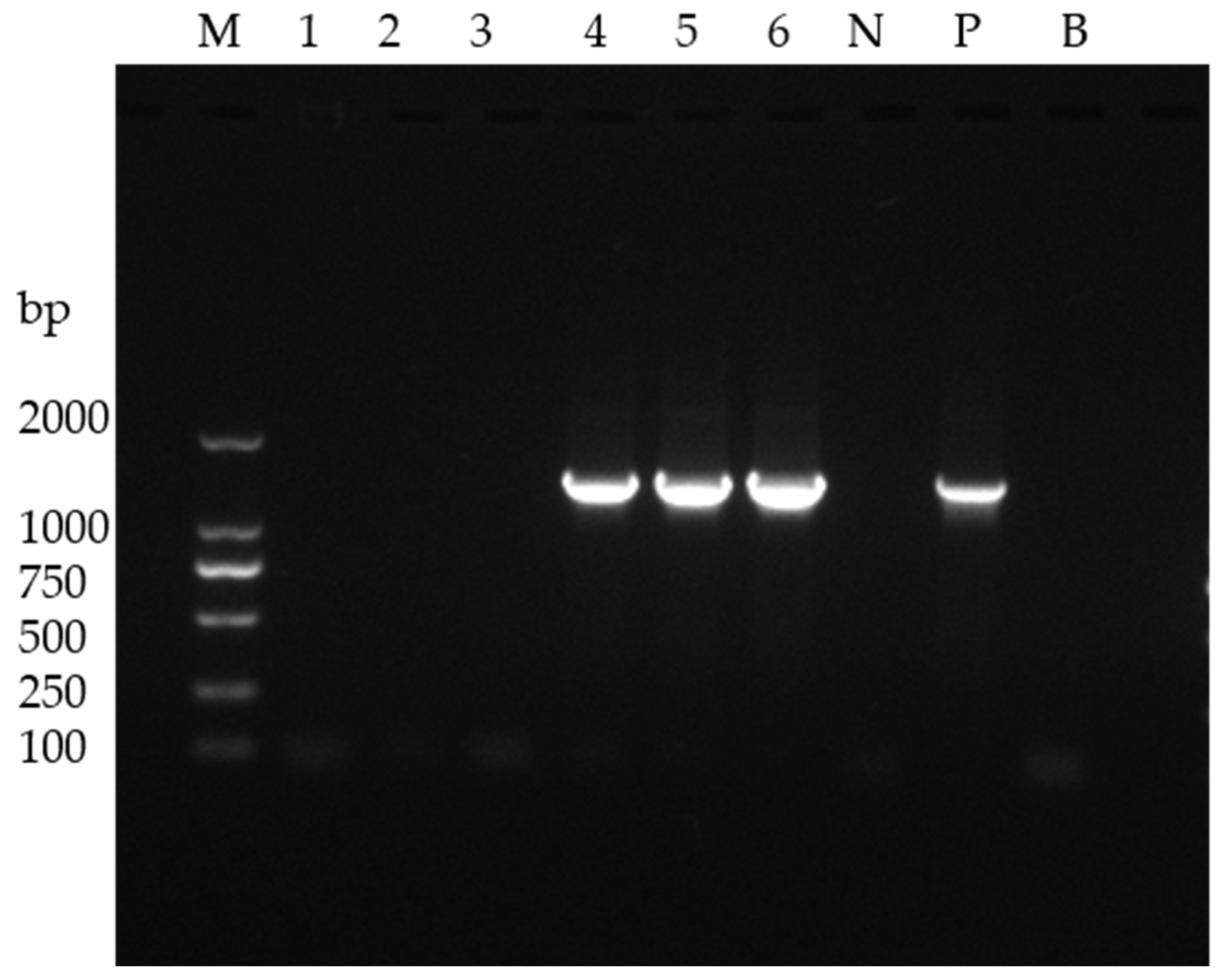
The diseased and healthy shrimp were examined by using nested PCR [22] to confirm WSSV presence. DNA extracted from gills and hepatopancreas of both groups underwent amplification with WSSV-specific primers, followed by agarose gel electrophoresis for validation.

2.3. Intestinal DNA Extraction and 16S rDNA Amplification

Total genomic DNA was isolated from intestinal tissues using CTAB/SDS methods. DNA purity and concentration were verified via 1% agarose gel electrophoresis, then normalized to 1 ng/μL. The 16S rDNA V1–V9 region was amplified using primers (F: AGAGTTTGATCCTGGCTCAG; R: GNTACCTTGTTACGACTT) under the following amplification conditions: an initial single cycle at 94 °C for 5 min, followed by 27 cycles of denaturation at 94 °C for 5 s, annealing at 55 °C for 30 s, extension at 72 °C for 30 s, and final a final hold at 4 °C.

2.4. High-Throughput Sequencing and Data Analysis

2.4.1. Quality Control

The 16S rDNA amplification products of intestinal microbiota were used for library construction according to the protocols and methods provided by Novogene Co., Ltd. (Beijing, China). The PCR products were purified and subsequently used to construct libraries, followed by third-generation full-length amplicon sequencing on the PacBio platform. Raw sequences were initially processed through the PacBio SMRT portal. Sequences were filtered for a minimum of 3 passes and a minimum predicted accuracy of 90% (minfullpass = 3, minPredicted accuracy = 0.9), which is defined as the threshold below which a CCS is considered noise. The files generated by the PacBio platform were then used for amplicon size trimming to remove sequences outside the expected amplicon size (min. length 1340 bp, max. length 1640 bp). The reads were assigned to samples based on their unique barcodes and truncated by cutting off the barcode and primer sequence. The reads were compared with the reference database using the UCHIME algorithm to detect chimera sequences, the chimera sequences were removed, and the clean reads were finally obtained.

2.4.2. OTU Cluster and Species Annotation

Sequence analysis was performed using Uparse software (Uparsev7.0.1001). Sequences with ≥97% similarity were assigned to the same OTU. A representative sequence for each OTU was screened for further annotation. For each representative sequence, the SSUrRNA Database of Silva Database was used, based on the Mothur (1.25.0) algorithm, to annotate taxonomic information. In order to study the phylogenetic relationships of different OTUs, and the difference in the dominant species in different samples (groups), multiple sequence alignment was conducted using the MUSCLE software (Version 3.8.31). OTU abundance information was normalized using a standard of sequence number corresponding to the sample with the most sequences. Subsequent analyses of alpha diversity and beta diversity were all performed based on this normalized output data.

2.4.3. Alpha Diversity

Alpha diversity is applied in analyzing the complexity of species diversity for a sample through indices, including Chao1, Shannon, Simpson, ACE, and Good-coverage. All these indices for our samples were calculated with QIIME (Version 1.9.1) and displayed with R software (Version 2.15.3). Chao1 and ACE were selected to identify community richness. Shannon and Simpson were used to identify community diversity. Good coverage was chosen to characterize sequencing depth.

2.4.4. Beta Diversity

UniFrac distance was computed using QIIME software (version 1.9.1). Principal coordinate analysis (PCoA) was performed and visualized in R software (version 2.15.3) with the WGCNA, stats, and ggplot2 packages. Beta diversity differences between groups were assessed in R using both parametric (Student’s t-test) and non-parametric tests.
LEfSe analysis was performed using the LEfSe software (LEfSe-1.0.8.post1-1) with the default LDA score threshold set at 4. Metastats analysis was conducted in R software, performing permutation tests between groups at different taxonomic levels to obtain p-values. The p-values were then adjusted using the Benjamini and Hochberg False Discovery Rate (FDR) method to derive q-values.

3. Results

3.1. WSSV Detection of Diseased and Healthy Shrimp

The diseased shrimp exhibited the characteristic disease signs of a WSSV infection, including lethargy, markedly reduced motility, and the presence of white spots (Figure 1). The nested PCR analysis validated this clinical progression timeline, with viral amplicons being exclusively detectable in the moribund specimens (Figure 2), thereby establishing a precise diagnostic concordance between the observed behavioral pathology and the virological confirmation.

3.2. Analysis of Intestinal Microbiota Diversity

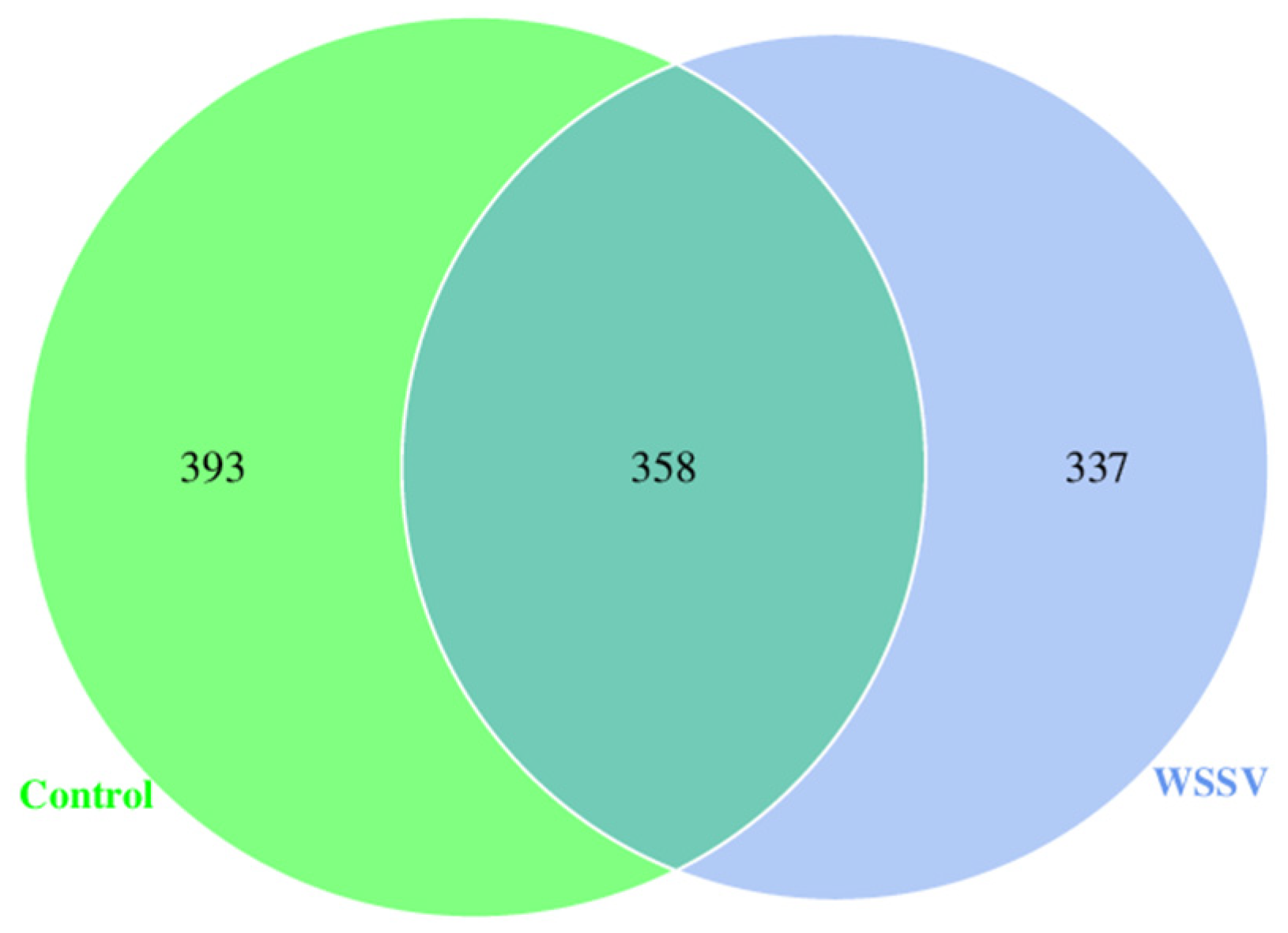
A comparative analysis of the OTUs revealed distinct microbial community profiles between the experimental groups. A systematic quantification demonstrated that while 291 OTUs were conserved across both the WSSV-infected and control specimens, the WSSV-infected group harbored 337 unique OTUs compared to 393 signature OTUs in the control group. The data (Figure 3) indicate that WSSV infection leads to a decreased diversity of gut microbial communities in P. monodon. The observed 14.2% reduction in unique OTUs (393 vs. 337) in the WSSV-challenged shrimp correlates with viral-induced dysbiosis, suggesting a pathogen-mediated suppression of commensal microbiota. These findings collectively demonstrate that WSSV infection not only alters microbial community architecture but also reduces niche-specific taxonomic diversity, potentially compromising the intestinal homeostasis critical for pathogen resistance.
A comprehensive α-diversity assessment at 97% sequence similarity revealed distinct microbial community structures between the experimental groups (Table 1, Table 2 and Table 3). The control group exhibited significantly elevated Shannon (5.357 vs. 2.523) and Simpson indices (0.939 vs. 0.445) compared to the WSSV-infected specimens (Shannon p = 0.0075, Simpson p = 0.0072), demonstrating superior species richness and evenness in the non-infected shrimp. Although not statistically significant (p > 0.05), higher Chao1 (507.52 vs. 485.55, p = 0.46) and ACE indices (506.70 vs. 490.03, p = 0.63) in the control group further suggested a microbial community contraction following the viral challenge, with both groups maintaining exceptional sequencing depth (Good’s coverage > 0.998).
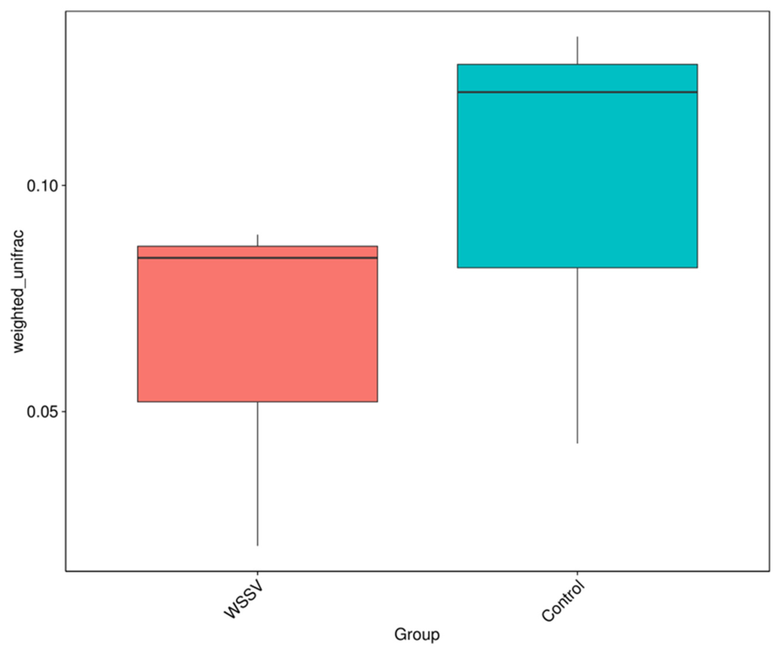
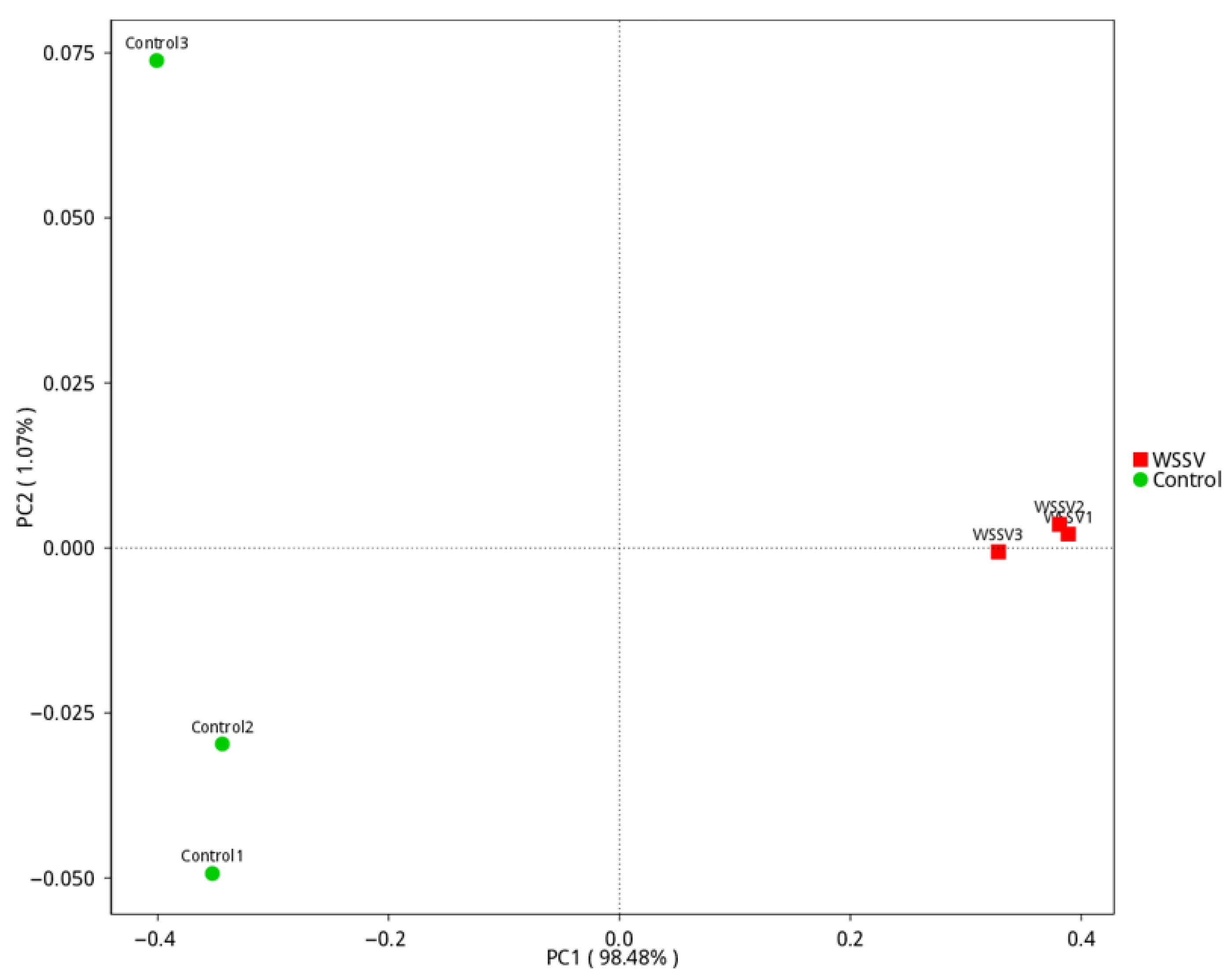
Complementing these α-diversity metrics, a β-diversity analysis through weighted UniFrac distances revealed enhanced microbial heterogeneity in the controls (Figure 4), though the intergroup differences lacked statistical significance (p > 0.05). The PCoA demonstrated a striking compositional divergence, with 98.48% variance explained along PCo1 (Figure 5). The pronounced dispersion between the groups in multivariate space confirms a fundamental restructuring of the microbial consortia, substantiating WSSV-induced dysbiosis. Collectively, these multidimensional analyses establish that healthy P. monodon maintain not only a greater microbial richness (α-diversity) but also more complex community architectures (β-diversity) compared to their infected counterparts, highlighting the destabilizing effects of WSSV infection on intestinal ecosystem integrity.

3.3. Composition and Differential Analysis of Intestinal Microbiota in P. monodon

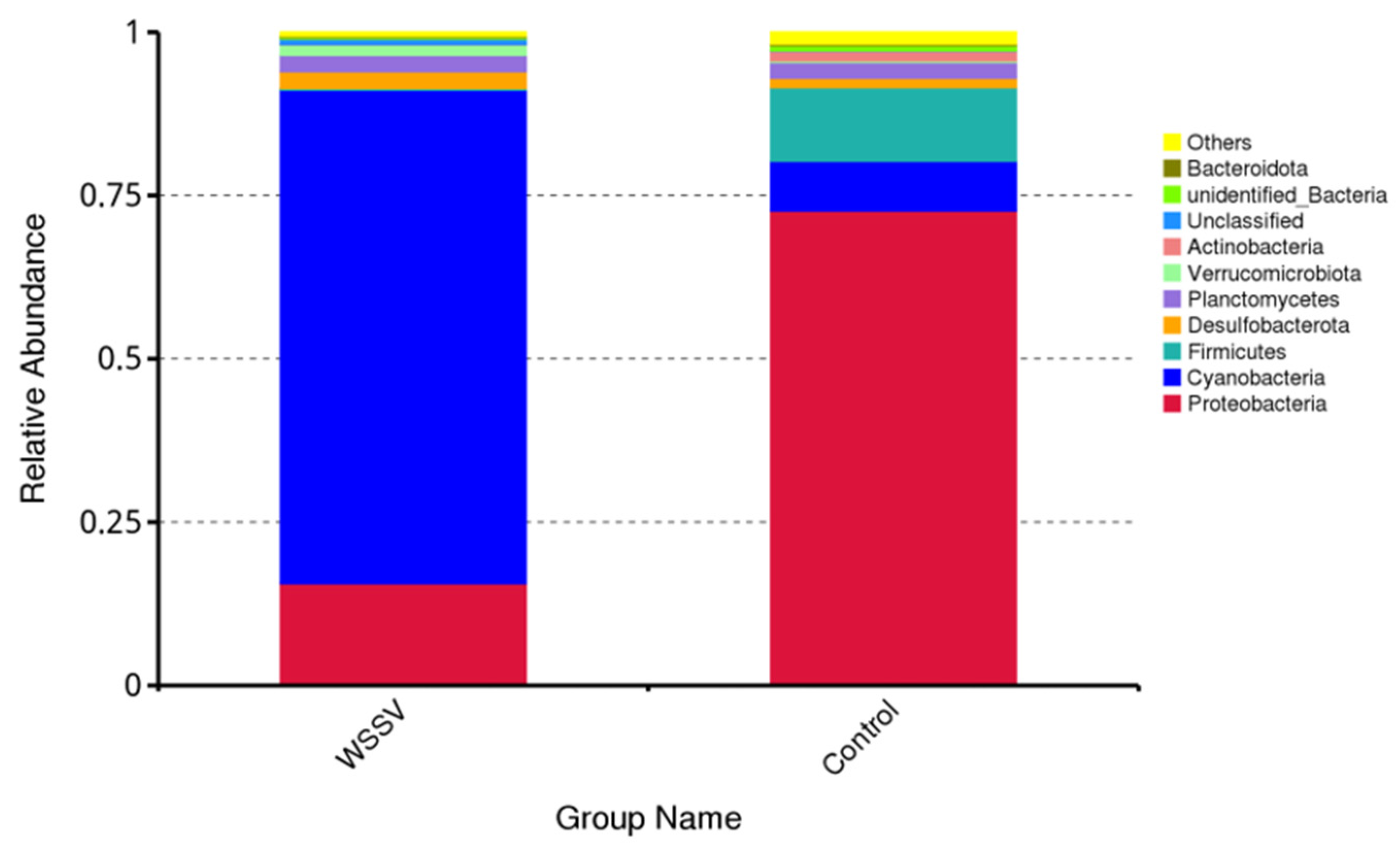
The species abundance of the intestinal microbiota in the WSSV-infected and control groups was annotated and analyzed at the phylum level (Figure 6, Table 4). More than 10 phyla were identified in both groups, including Cyanobacteria, Proteobacteria, Firmicutes, Desulfobacterota, Planctomycetes, Verrucomicrobiota, and Actinobacteria. A stacked bar chart illustrates the relative abundance of these phyla. In the control group, the dominant phyla were Proteobacteria (72.68%) and Firmicutes (11.27%). Conversely, the WSSV-infected group was dominated by Cyanobacteria (75.51%) and Proteobacteria (15.63%). Notably, the abundance of Cyanobacteria increased significantly (p < 0.05), while the abundance of Proteobacteria decreased significantly (p < 0.05) in the WSSV-infected group compared to the control group (Figure 7, Table 5).
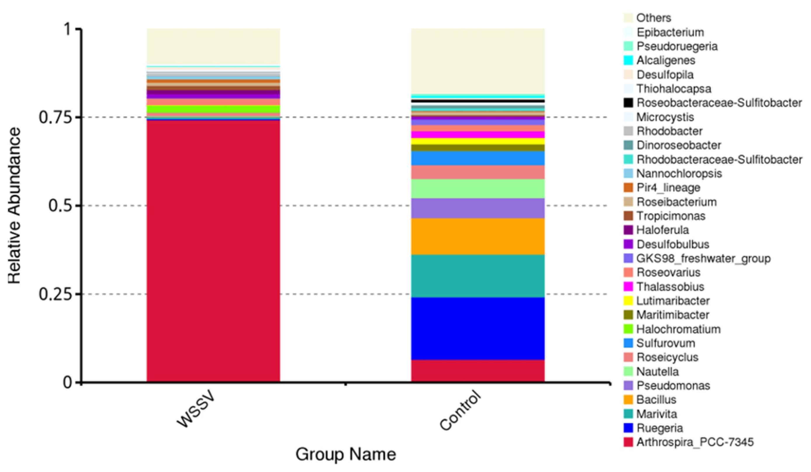
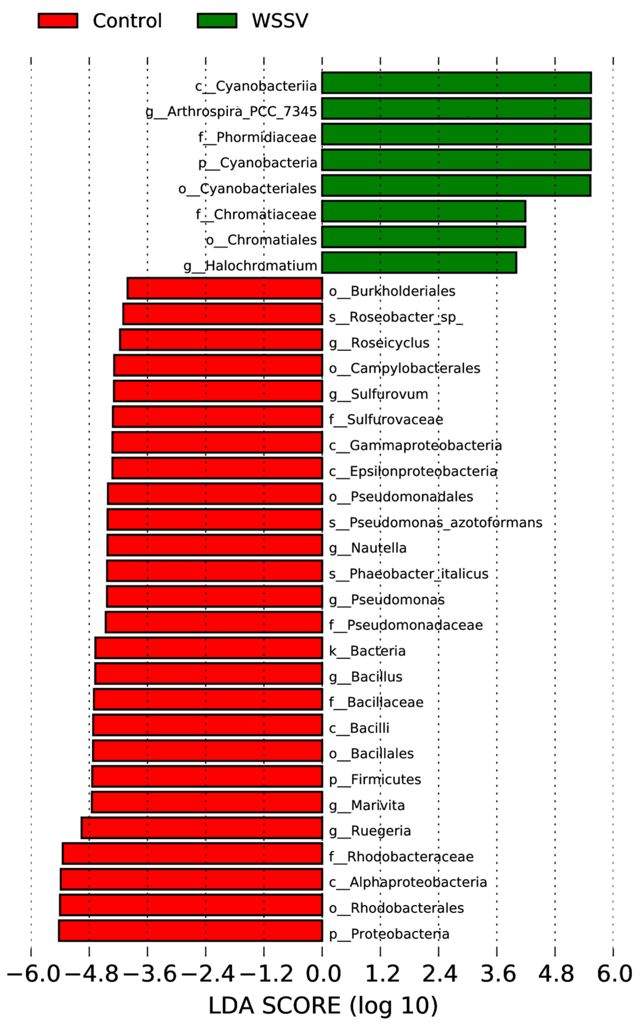
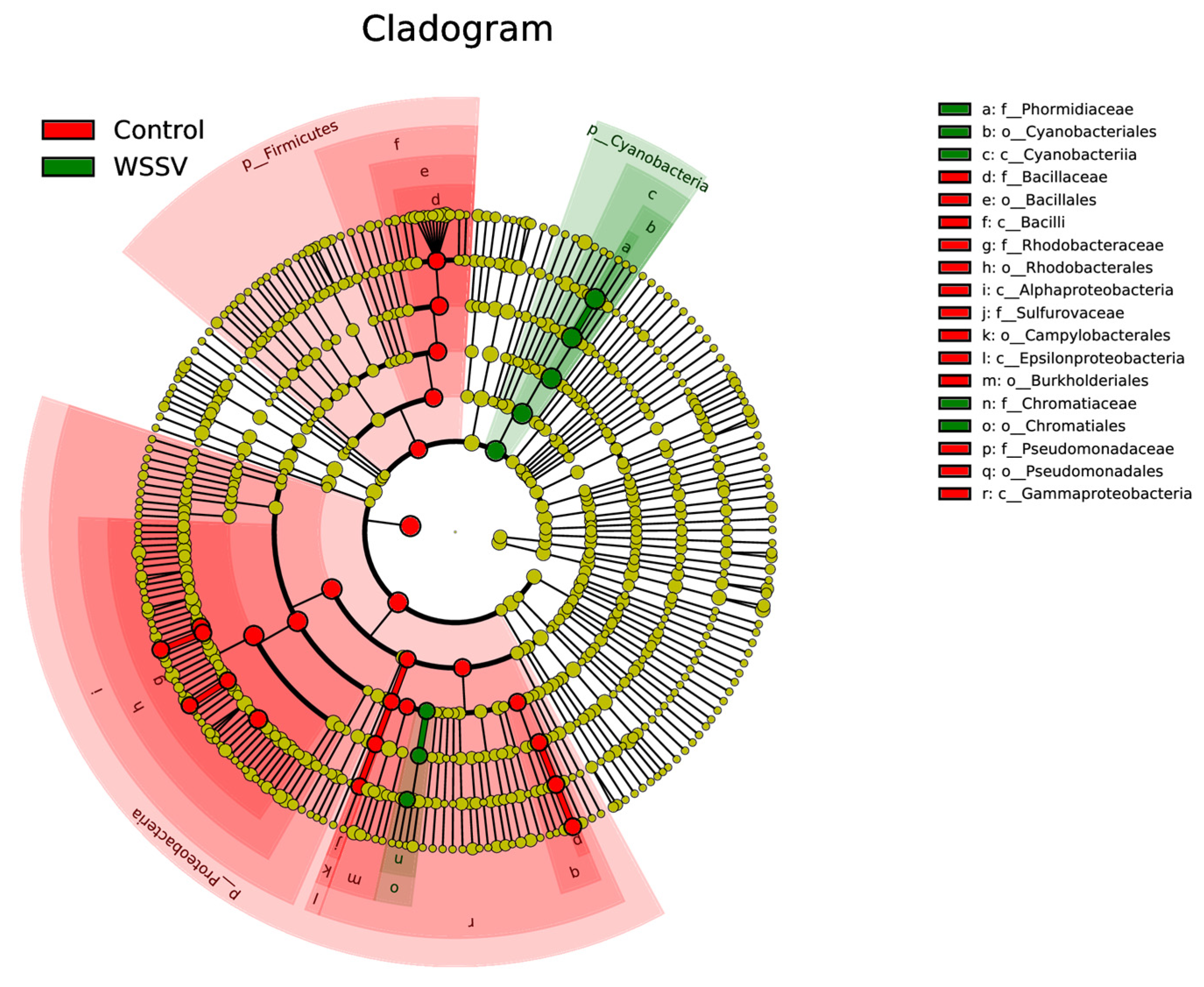
At the genus level, the composition and abundance of the dominant bacteria differed markedly between the two groups. A total of 306 bacterial genera were identified (Figure 7). In the WSSV-infected group, the dominant genera were Arthrospira_PCC-7345 (74.32%), Halochromatium (1.98%), and Roseovarius (1.56%). In contrast, the control group was dominated by Ruegeria (17.67%), Marivita (12.08%), and Bacillus (10.30%). To further explore the intestinal microbiota associated with WSSV infection in P. monodon, an LEfSe (Linear Discriminant Analysis Effect Size) analysis was conducted to compare the relative abundance of microbial taxa between the WSSV-infected and control groups. This analysis identified the taxa that exhibited significant differences. A linear discriminant analysis (LDA) and cladograms revealed that two generas (Arthrospira and Halochromatium), two families (Chromatiaceae and Phormidiaceae), two orders (Cyanobacteriales and Chromatiales), and one phylum (Cyanobacteria) were significantly higher in the WSSV-infected group (p < 0.05) (Figure 8 and Figure 9).
In contrast, the control group exhibited a significantly greater relative abundance of microbial taxa across multiple taxonomic levels compared to the WSSV-infected group (p < 0.05). Notably, the control group showed a greater abundance in seven genera, four families, five orders, and two phyla. These results highlight the pronounced alterations in the intestinal microbiota composition associated with WSSV infection, underscoring the potential involvement of specific microbial taxa in disease progression and the host’s immune response.

4. Discussion

The intestinal microbiota plays a crucial role in maintaining shrimp health by forming a complex micro-ecosystem with the host’s internal environment [23,24,25]. This dynamic system regulates various physiological functions and maintains ecological balance through mutual constraints and dependencies. The disruption of this balance may allow harmful microorganisms to invade, potentially leading to disease outbreaks in shrimp [26,27]. Identifying the differences in the intestinal microbial structures between healthy and diseased shrimp can thus provide valuable insights for early disease detection [28,29,30]. Previous studies on species such as Rimicaris exoculata and Litopenaeus vannamei have linked an intestinal microbiota imbalance to WSSV outbreaks [31,32,33]. Consistent with these findings, our results revealed significant differences in the intestinal microbiota composition between the WSSV-infected and control groups in P. monodon. In the control group, the dominant phyla were Proteobacteria and Firmicutes, which are commonly found in healthy shrimp intestines and are closely associated with host health. Within these phyla, the genera Ruegeria, Marivita (family Rhodobacteraceae), and Bacillus were particularly abundant [34,35]. Rhodobacteraceae species are recognized as core colonizers of the shrimp intestine and are known to improve aquaculture water quality by reducing COD levels, promoting the heterotrophic metabolism of carbon sources and regulating intestinal microbiota structure [36,37]. These beneficial effects contribute to enhanced disease resistance and stress tolerance, making Rhodobacteraceae an emerging probiotic candidate for shrimp disease prevention [38]. Bacillus as probiotics can enhance host immunity, decrease disease susceptibility, enhance immune responses, and improve resistance to WSSV infection [39,40]. However, there were several limitations in our study, such as the absence of stage-specific WSSV analysis during the sampling and the restriction of the 16S sequencing to relative quantification, which will be prioritized in future studies.
In contrast, the WSSV-infected group exhibited a markedly different microbiota profile, with Cyanobacteria dominating the intestinal microbial community. The genus Arthrospira showed a significantly higher abundance, diverging from the patterns observed in other crustaceans infected with WSSV [13,20,31]. This discrepancy may be attributed to aquaculture environmental factors influencing the shrimp’s microbiota. Notably, Cyanobacteria are not native to shrimp intestines but are introduced through feed and water sources [41]. This suggests that environmental deterioration may have contributed to the WSSV outbreak observed in this study.
Microbial diversity is widely regarded as a strong indicator of host health [42,43], playing a key role in maintaining ecosystem stability and functionality [44,45]. A reduced intestinal microbiota diversity can make shrimp more susceptible to environmental stress and microbial pathogens [46], as disease outbreaks are often linked to decreased microbial diversity [7,47,48]. In this study, the alpha and beta diversity analyses demonstrated that the intestinal microbiota diversity in the WSSV-infected shrimp was significantly lower than in the healthy individuals. This aligns with previous research on WSSV-infected Procambarus clarkii [20], Enterocytozoon hepatopenaei-infected Exopalaemon carinicauda [49], and Vibrio-infected Litopenaeus vannamei [50], all of which showed higher microbial diversity in the healthy shrimp compared to the diseased individuals. Proteobacteria are often considered a health indicator in aquaculture organisms. In this study, their higher abundance in the control group compared to the WSSV group further supports this view [51]. Following WSSV infection, Proteobacteria abundance significantly decreased, while Cyanobacteria, particularly Arthrospira, became dominant. This shift likely contributed to the reduced microbial diversity in the WSSV-infected group, further disrupting the intestinal microbiota structure and facilitating the proliferation of potentially pathogenic bacteria. This imbalance may be a critical factor in WSSV pathogenesis.
Probiotics are widely recognized as effective tools in enhancing shrimp health by promoting beneficial bacteria and suppressing pathogenic colonization. Composite probiotics have shown promising results in combating shrimp pathogens, including WFS-associated bacteria. For instance, Streptomyces has demonstrated protective effects in Artemia, P. monodon, and Litopenaeus vannamei [52,53,54]. Furthermore, adding Streptomyces sp. RL8 alone or in combination with Bacillus sp. has been shown to enhance intestinal microbial diversity and promote bacteria that produce antimicrobial agents [44]. Based on these findings, the strategic addition of probiotics to both aquaculture environments and feed may offer an effective approach to preventing WSSV outbreaks, ultimately improving shrimp health and disease resilience.

5. Conclusions

This study demonstrates that WSSV infection in P. monodon induces significant gut microbiota dysbiosis, which may contribute to disease progression. WSSV infection drastically reduced α-diversity, with Arthrospira_PCC-7345 and Halochromatium significantly enriched in the infected shrimp, suggesting their potential role in WSSV progression. Beneficial genera such as Ruegeria, Marivita, Bacillus, and others were suppressed, indicating a breakdown in gut homeostasis. These microbial biomarkers provide a foundation for early WSSV detection in aquaculture. Future research should explore whether microbiota modulation (e.g., probiotics or prebiotics) could mitigate WSSV impacts, improving shrimp health and farm productivity.

Author Contributions

Conceptualization, Y.W.; methodology, Y.W., X.W., C.G. and Y.L.; formal analysis, S.W., L.X. and L.L.; resources, R.F.; data curation, Y.W. and L.X.; writing—original draft preparation, Y.W.; writing—review and editing, H.C. and Y.F.; supervision, J.D., H.Y. and X.Y.; project administration, Y.W., Y.F., X.W., C.G. and Y.L.; funding acquisition, X.W. All authors have read and agreed to the published version of the manuscript.

Funding

This research was funded by the Earmarked Fund for Shandong Agriculture Innovation Team (SDAIT-13) and the Earmarked Fund for China Agriculture Research System (CARS-48).

Institutional Review Board Statement

Ethical review and approval were waived for this study due to the following reasons: Penaeus monodon is an invertebrate and is not subject to mandatory review under GB/T 35892-2018; healthy prawns were released back into the aquaculture ponds after the experiment.

Informed Consent Statement

Not applicable.

Data Availability Statement

The dataset is available on request from the authors.

Acknowledgments

We thank Ying Fan and Haipeng Cao for providing valuable insights during the manuscript preparation and the staff at Novogene Co., Ltd. Laboratory for their technical assistance. Finally, we acknowledge the support of Youfa Aquaculture Co., Ltd. for providing access to research facilities.

Conflicts of Interest

Author Ranghui Fu was employed by the Shandong Youfa Aquaculture company. The remaining authors declare that the research was conducted in the absence of any commercial or financial relationships that could be construed as a potential conflict of interest.

References

  1. Wang, Y.H.; Wang, X.L.; Yu, X.Q.; Xu, L.; Gai, C.L.; Wang, S.X.; Liu, H.J.; Diao, J. Effects of Chronic Nitrite Stress on Behavior, Digestive Enzyme Activities and Non-Specific Immunity of an African Population of Penaeus monodon. Period. Ocean. Univ. China 2023, 53, 43–49. [Google Scholar]
  2. Zhu, J.; Dai, W.; Qiu, Q.; Dong, C.; Zhang, J.; Xiong, J. Contrasting Ecological Processes and Functional Compositions between Intestinal Bacterial Community in Healthy and Diseased Shrimp. Microb. Ecol. 2016, 72, 975–985. [Google Scholar] [CrossRef]
  3. Fan, L.L.Q.X. Characteristics of Intestinal Microbiota in the Pacific White Shrimp Litopenaeus vannamei Differing Growth Performances in the Marine Cultured Environment. Aquaculture 2019, 505, 450–461. [Google Scholar] [CrossRef]
  4. Biesebeke, R.T. Balancing Microbial Ecosystems within Humans and Animals to Prevent Medical Conditions. J. Nutr. Food Res. Technol. 2018, 1, 40. [Google Scholar] [CrossRef]
  5. Butto, L.F.; Haller, D. Dysbiosis in Intestinal Inflammation: Cause or Consequence. Int. J. Med. 2016, 306, 302–309. [Google Scholar] [CrossRef] [PubMed]
  6. Pickard, J.M.; Zeng, M.Y.; Caruso, R.; Núñez, G. Gut Microbiota: Role in Pathogen Colonization, Immune Responses, and Inflammatory Disease. Immunol. Rev. 2017, 279, 70–89. [Google Scholar] [CrossRef] [PubMed]
  7. Cornejo-Granados, F.; Lopez-Zavala, A.A.; Gallardo-Becerra, L.; Mendoza-Vargas, A.; Sánchez, F.; Vichido, R.; Brieb, L.G.; Viana, M.; Sotelo-Mundo, R.R.; Ochoa-Leyva, A. Microbiome of Pacific White Leg Shrimp Reveals Differential Bacterial Community Composition between Wild, Aquacultured and AHPND/EMS Outbreak Conditions. Sci. Rep. 2017, 7, 11783. [Google Scholar] [CrossRef]
  8. Liu, W.C.; Zhou, S.H.; Balasubramanian, B.; Zeng, F.Y.; Sun, C.B.; Pang, H.Y. Dietary Seaweed (Enteromorpha) Polysaccharides Improves Growth Performance Involved in Regulation of Immune Responses, Intestinal Morphology and Microbial Community in Banana Shrimp Fenneropenaeus merguiensis. Fish Shellfish Immunol. 2020, 104, 202–212. [Google Scholar] [CrossRef]
  9. Shi, C.; Xia, M.; Li, R.; Mu, C.; Zhang, L.; Liu, L.; Ye, Y.; Wang, C. Vibrio alginolyticus Infection Induces Coupled Changes of Bacterial Community and Metabolic Phenotype in the Gut of Swimming Crab. Aquaculture 2019, 499, 251–259. [Google Scholar] [CrossRef]
  10. Sanschagrin, S.; Yergeau, E. Next-generation sequencing of 16S ribosomal RNA gene amplicons. J. Vis. Exp. 2014, 90, e51709. [Google Scholar] [CrossRef]
  11. Xia, H.; Zhang, Z.; Luo, C.; Wei, K.; Li, X.; Mu, X.; Duan, M.; Zhu, C.; Jin, L.; He, X.; et al. MultiPrime: A Reliable and Efficient Tool for Targeted Next-Generation Sequencing. iMeta 2023, 2, e143. [Google Scholar] [CrossRef] [PubMed]
  12. Curry, K.D.; Wang, Q.; Nute, M.G.; Tyshaieva, A.; Reeves, E.; Soriano, S.; Wu, Q.; Graeber, E.; Finzer, P.; Mendling, W.; et al. Emu: Species-level microbial community profiling of full-length 16S rRNA Oxford Nanopore sequencing data. Nat. Methods 2022, 19, 845–853. [Google Scholar] [CrossRef]
  13. Wang, J.; Huang, Y.; Xu, K.; Zhang, X.; Sun, H.; Fan, L.; Yan, M. White Spot Syndrome Virus (WSSV) Infection Impacts Intestinal Microbiota Composition and Function in Litopenaeus vannamei. Fish Shellfish Immunol. 2019, 84, 130–137. [Google Scholar] [CrossRef]
  14. Phuoc, L.H.; Corteel, M.; Nauwynck, H.J.; Pensaert, M.B.; Alday-Sanz, V.; Van den Broeck, W.; Sorgeloos, P.; Bossier, P. Increased Susceptibility of White Spot Syndrome Virus-Infected Litopenaeus vannamei to Vibrio campbellii. Environ. Microbiol. 2008, 10, 2718–2727. [Google Scholar] [CrossRef]
  15. Liao, M.; Liao, X.; Long, X.; Zhao, J.; He, Z.; Zhang, J.; Wu, T.; Sun, C. Host-Microbiota Interactions and Responses of Metapenaeus ensis Infected with Decapod Iridescent Virus 1. Front. Microbiol. 2023, 13, 1097931. [Google Scholar] [CrossRef]
  16. Dong, P.; Guo, H.; Wang, Y.; Wang, R.; Zhang, D. Gastrointestinal Microbiota Imbalance is Triggered by the Enrichment of Vibrio in Subadult Litopenaeus vannamei with Acute Hepatopancreatic Necrosis Disease. Aquaculture 2021, 533, 736199. [Google Scholar] [CrossRef]
  17. Zhai, Y.F.; He, P.M.; Jia, R. iTRAQ-Based Quantitative Proteomic Analysis of Differentially Expressed Proteins in the Hepatopancreas of Litopenaeus vannamei after WSSV Infection. Dis. Aquat. Org. 2021, 145, 51–61. [Google Scholar] [CrossRef] [PubMed]
  18. He, Y.; Li, X.; Luan, S.; Kong, J.; Cao, B.; Luo, K.; Tan, J.; Cao, J.; Cao, J.; Chen, B.; et al. Evaluation of Genetic Parameters for Growth and Survival Traits of Penaeus vannamei During White Spot Syndrome Virus Infection. Prog. Fish. Sci. 2024, 45, 155–164. [Google Scholar]
  19. Lo, C.F.; Hsu, H.C.; Tsai, M.F.; Ch, H.; Peng, S.; Kou, G. Specific Genomic DNA Fragment Analysis of Different Geographical Clinical Samples of Shrimp White Spot Syndrome Virus. Dis. Aquat. Org. 1999, 35, 175–185. [Google Scholar] [CrossRef]
  20. Chen, Y.; Gu, Z.; Li, L.; Chen, S.; Yuan, J. Effects of White Spot Syndrome Virus (WSSV) Infection on Intestinal Flora Composition of Procambarus clarkii. J. Huazhong Agric. Univ. 2020, 30, 40–46. [Google Scholar]
  21. Cheng, W.; Zhang, H.; Wang, P.; Wei, Y.; Li, T.; Zhou, Y.; Mao, Y. Clinical signs of naturally white spot syndrome virus (WSSV)-infected kuruma shrimp Marsupenaeus japonicas, based on their physiological and behavioural states. Aquaculture 2021, 533, 736104. [Google Scholar] [CrossRef]
  22. GB/T 28630.2-2012; Diagnostic Protocols for White Spot Disease—Part 2: Nested PCR Method. Ministry of Agriculture and Rural Affairs: Beijing, China, 2012.
  23. Sha, H.; Lu, J.; Chen, J.; Xiong, J. Rationally Designed Probiotics Prevent Shrimp White Feces Syndrome via the Probiotics Gut Microbiome Immunity Axis. npj Biofilms Microbiomes 2024, 10, 40. [Google Scholar] [CrossRef] [PubMed]
  24. Sha, H.; Song, T.; Zhan, P.; Zhang, X.; Chen, J.; Xiong, J. Sarafloxacin Hydrochloride Exposure Disrupts Gut Microbiota and Increases Shrimp Susceptibility to Vibrio anguillarum Infection. Aquaculture 2024, 586, 740810. [Google Scholar] [CrossRef]
  25. Zhang, S.; Sun, X. Core Gut Microbiota of Shrimp Function as a Regulator to Maintain Immune Homeostasis in Response to WSSV Infection. Microbiol. Spectr. 2022, 10, e0246521. [Google Scholar] [CrossRef]
  26. Xiong, J.B.; Sha, H.N.; Chen, J. Updated Roles of the Gut Microbiota in Exploring Shrimp Etiology, Polymicrobial Pathogens, and Disease Incidence. Zool. Res. 2024, 45, 910–923. [Google Scholar] [CrossRef]
  27. Holt, C.C.; Bass, D.; Stentiford, G.D.; van der Giezen, M. Understanding the Role of the Shrimp Gut Microbiome in Health and Disease. J. Invertebr. Pathol. 2021, 186, 107387. [Google Scholar] [CrossRef]
  28. Yu, X.; Wang, Y.; Bi, J.; Wang, X.; Wang, S.; Gai, C.; Xu, L.; Ye, H.; Li, L.; Diao, J.; et al. Distribution Characteristics of Microorganisms in Hepatopancreas and Intestine of Penaeus monodon under Industrial and High-Pond Culture Modes. Fish. Sci. Technol. Inf. 2024, 51, 362–372. [Google Scholar]
  29. Mao, J.N.; Lu, J.Q.; Chen, J.; Xiong, J. Consistent Features of the Gut Microbiota in Response to Diverse Shrimp Litopenaeus vannamei Diseases: A Meta-Analysis. Fish Fish. 2023, 24, 1103–1117. [Google Scholar] [CrossRef]
  30. Sha, H.N.; Lu, J.Q.; Chen, J. A Meta-Analysis Study of the Robustness and Universality of Gut Microbiota–Shrimp Diseases Relationship. Environ. Microbiol. 2022, 24, 3924–3938. [Google Scholar] [CrossRef]
  31. Huang, Z.; Zeng, S.; Chen, J.; Xiong, J. Microecological Koch’s Postulates Reveal that Intestinal Microbiota Dysbiosis Contributes to Shrimp White Feces Syndrome. Microbiome 2020, 8, 32. [Google Scholar] [CrossRef]
  32. Guri, M.; Durand, L.; Cueff-Gauchard, V.; Zbinden, M.; Crassous, P.; Shillito, B.; Cambon-Bonavita, M.A. Acquisition of Epibiotic Bacteria along the Life Cycle of the Hydrothermal Shrimp Rimicaris exoculata. ISME J. 2012, 6, 597–609. [Google Scholar] [CrossRef] [PubMed]
  33. Wang, Y.; Wang, K.; Huang, L.; Dong, P.; Wang, S.; Chen, H.; Lu, Z.; Hou, D.; Zhang, D. Fine-Scale Succession Patterns and Assembly Mechanisms of Bacterial Community of Litopenaeus vannamei Larvae across the Developmental Cycle. Microbiome 2020, 8, 106. [Google Scholar] [CrossRef]
  34. Landsman, A.; St-Pierre, B.; Rosales-Leija, M.; Brown, M.; Gibbons, W. Impact of Aquaculture Practices on Intestinal Bacterial Profiles of Pacific Whiteleg Shrimp Litopenaeus vannamei. Microorganisms 2019, 7, 93. [Google Scholar] [CrossRef]
  35. Zeng, S.; Khoruamkid, S.; Kongpakdee, W.; Wei, D.; Yu, L.; Wang, H.; Deng, Z.; Weng, S.; Huang, Z.; He, J.; et al. Dissimilarity of Microbial Diversity of Pond Water, Shrimp Intestine and Sediment in Aquamimicry System. AMB Express 2020, 1, 180. [Google Scholar] [CrossRef]
  36. Wang, W.; Cui, X.; Cai, Z.; Huang, Y. Analysis of Changes in the Waterflora and Intestinal Microflora of Penaeus monodon in Different Cultural Models. J. Xichang Univ. Nat. Sci. Ed. 2023, 37, 7–14. [Google Scholar]
  37. Hong, J.E. Succession of Intestinal Microbiota of Litopenaeus vannamei Larvae and the Impacts of Several Probiotics on Larviculture. Master’s Thesis, Guangdong Ocean University, Zhanjiang, China, 2020. [Google Scholar]
  38. Gao, F.; Yu, E.L.; Liu, W.; Gao, H.; Wang, Y.; Wang, R.; Yan, M.; Dong, P.; Zhang, D. Screening of Carbon Sources for Enrichment and Directional Isolation of Rhodobacteraceae from the Gut of Litopenaeus vannamei. Acta Microbiol. Sin. 2022, 65, 1805–1818. [Google Scholar]
  39. Seabkongseng, T.; Limkul, S.; Sriphuttha, C.; Phiwthong, T.; Aunkam, P.; Suwannathit, R.; Jaree, P.; Somboonwiwat, K.; Tittabutr, P.; Teaumroong, N. Supplementation of Bacillus velezensis S141 in feed as a probiotic enhances growth performance, pathogenic tolerances, and immune system in shrimp. Aquaculture 2025, 604, 742448. [Google Scholar] [CrossRef]
  40. Chai, P.C.; Song, X.L.; Chen, G.F.; Xu, H.; Huang, J. Dietary supplementation of probiotic Bacillus PC465 isolated from the gut of Fenneropenaeus chinensis improves the health status and resistance of Litopenaeus vannamei against white spot syndrome virus. Fish Shellfish Immunol. 2016, 54, 602–611. [Google Scholar] [CrossRef] [PubMed]
  41. Fan, Y.; Ma, D.; Li, L.; Jiang, J.; Wang, Y.; Wang, S.; Hou, F.; Ye, H.; Liu, H.; Yue, X. Effects of Lactobacillus plantarum on Growth, Immunity, Digestase Activities and Gut Microbiota Composition of Sebastes schlegelii. J. Guangdong Ocean. Univ. 2024, 44, 42–54. [Google Scholar]
  42. Li, T.; Li, H.; Gatesoupe, F.J.; She, R.; Lin, Q.; Yan, X.; Li, J.; Li, X. Bacterial Signatures of “Red-Operculum” Disease in the Gut of Crucian Carp (Carassius auratus). Microb. Ecol. 2017, 74, 510–521. [Google Scholar] [CrossRef]
  43. Li, E.; Wang, X.; Chen, K.; Xu, C.; Qin, J.G.; Chen, L. Physiological Change and Nutritional Requirement of Pacific White Shrimp Litopenaeus vannamei at Low Salinity. Rev. Aquac. 2017, 9, 57–75. [Google Scholar] [CrossRef]
  44. Turnbaugh, P.J.; Hamady, M.; Yatsunenko, T.; Cantarel, B.L.; Duncan, A.; Ley, R.E.; Sogin, M.L.; Jones, W.J.; Roe, B.A.; Affourtit, J.P.; et al. A Core Gut Microbiome in Obese and Lean Twins. Nature 2009, 457, 480–484. [Google Scholar] [CrossRef]
  45. Le Chatelier, E.; Nielsen, T.; Qin, J.; Prifti, E.; Hildebrand, F.; Falony, G.; Almeida, M.; Arumugam, M.; Batto, J.M.; Kennedy, S.; et al. Richness of Human Gut Microbiome Correlates with Metabolic Markers. Nature 2013, 500, 541–546. [Google Scholar] [CrossRef]
  46. Bass, D.; Stentiford, G.D.; Wang, H.C.; Koskella, B.; Tyler, C.R. The Pathobiome in Animal and Plant Diseases. Trends Ecol. Evol. 2019, 34, 996–1008. [Google Scholar] [CrossRef]
  47. Xiong, J.; Wang, K.; Wu, J.; Qiuqian, L.; Yang, K.; Qian, Y.; Zhang, D. Changes in Intestinal Bacterial Communities are Closely Associated with Shrimp Disease Severity. Appl. Microbiol. Biotechnol. 2015, 99, 6911–6919. [Google Scholar] [CrossRef]
  48. Xiong, J.; Zhu, J.; Dai, W.; Dong, C.; Qiu, Q.; Li, C. Integrating Gut Microbiota Immaturity and Disease-Discriminatory Taxa to Diagnose the Initiation and Severity of Shrimp Disease. Environ. Microbiol. 2017, 19, 1490–1501. [Google Scholar]
  49. Duan, J.; Hu, J.; Shen, Y.; Deng, G.; Gao, W.; Mou, H.; Zhang, Q.; Gao, H. Effect of Enterocytozoon hepatopenaei Infection on the Intestinal Microflora of Exopalaemon carinicauda. Prog. Fish. Sci. 2022, 43, 75–83. [Google Scholar]
  50. Chang, Y.T.; Ko, H.T.; Wu, P.L.; Kumar, R.; Wang, H.C.; Lu, H.P. Gut Microbiota of Pacific White Shrimp (Litopenaeus vannamei) Exhibits Distinct Responses to Pathogenic and Non-Pathogenic Vibrio parahaemolyticus. Microbiol. Spectr. 2023, 11, e0118023. [Google Scholar] [CrossRef]
  51. Blumberg, R.; Powrie, F. Microbiota, Disease, and Back to Health: A Metastable Journey. Pediatr. Neurol. 2016, 52, 566–584. [Google Scholar] [CrossRef] [PubMed]
  52. Bernal, M.G.; Marrero, R.M.; Campa-Córdova, Á.I.; Mazón-Suástegui, J.M. Probiotic Effect of Streptomyces Strains Alone or in Combination with Bacillus and Lactobacillus in Juveniles of the White Shrimp Litopenaeus vannamei. Aquac. Int. 2017, 25, 927–939. [Google Scholar] [CrossRef]
  53. Mazón-Suástegui, J.M.; Salas-Leiva, J.S.; Medina-Marrero, R.; Medina-García, R.; García-Bernal, M. Effect of Streptomyces Probiotics on the Gut Microbiota of Litopenaeus vannamei Challenged with Vibrio parahaemolyticus. Microbiol. Open 2019, 9, e967. [Google Scholar] [CrossRef] [PubMed]
  54. Augustine, D.; Jacob, J.C.; Philip, R. Exclusion of Vibrio spp. by an Antagonistic Marine Actinomycete Streptomyces rubrolavendulae M56. Aquac. Res. 2016, 47, 2951–2960. [Google Scholar] [CrossRef]
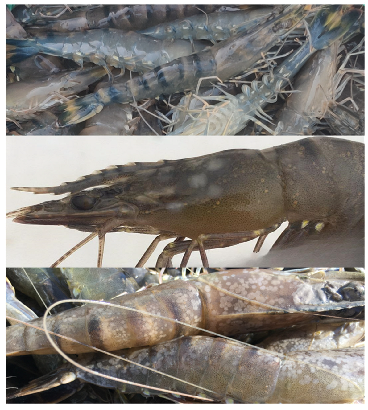
Figure 1. P. monodon infected with WSSV.
Fishes 10 00440 g001
Figure 2. WSSV detection in samples. M: marker; 1–3: healthy shrimp; 4–6: diseased shrimp; N: negative control; P: positive control.
Fishes 10 00440 g002
Figure 3. Venn digram of OUTs number between WSSV-infected and control groups.
Fishes 10 00440 g003
Figure 4. Beta diversity boxplot of intestinal samples from WSSV-infected and control P. monodon.
Fishes 10 00440 g004
Figure 5. Beta diversity principal coordinate analysis (PCoA) of intestinal samples from WSSV-infected and control P. monodon.
Fishes 10 00440 g005
Figure 6. Relative abundance of intestinal microbiota in WSSV-infected and control P. monodon at the phylum level.
Fishes 10 00440 g006
Figure 7. Relative abundance of intestinal microbiota in WSSV-infected and control P. monodon at the genus level.
Fishes 10 00440 g007
Figure 8. Analysis of LDA differences between WSSV-infected shrimp and the control group.
Fishes 10 00440 g008
Figure 9. The abundance of significant differences between WSSV-infected shrimp and the control group.
Fishes 10 00440 g009
Table 1. α-Diversity of WSSV-infected and control groups.
GroupObserved
Otus
ShannonSimpsonChao1ACEGoods
Coverage
Control4805.3570.939 507.516506.6980.999
WSSV4512.5230.445 485.552490.0270.998
Table 2. T-test for WSSVinfected–control.
IndicesWSSV-Infected–Control (p-Value)
Observed_otus0.4984
Shannon0.007528
Simpson0.00724
Chao10.4661
ACE0.6297
Goods_coverage0.5185
Table 3. Shapiro–Wilk and Levene tests for α-diversity.
IndicesKolmogorov–Smirnov Test (V) aShapiro–Wilk Test
StatsdfPStatsdfP
Observed_otus0.18460.200 *0.91360.0454
Shannon0.31360.0670.75660.230
Simpson0.31260.0700.75660.230
Chao10.21160.200 *0.94560.697
ACE0.23460.200 *0.85860.183
Goods_coverage0.31960.0560.68360.040
* This is a lower bound of the true significance. a Lilliefors correction. p > 0.05, the null hypothesis (H0) of normality cannot be rejected, suggesting that the data are consistent with a normal distribution.
Table 4. Relative abundance of intestinal microbiota in WSSV-infected and control P. monodon at the phylum level.
PhylumAbundance
WSSVControl
Proteobacteria15.63%72.68%
Cyanobacteria75.51%7.59%
Firmicutes0.28%11.27%
Desulfobacterota2.59%1.47%
Planctomycetes2.48%2.39%
Verrucomicrobiota1.61%0.24%
Actinobacteria0.07%1.47%
Unclassified0.84%0.08%
unidentified_Bacteria0.24%0.80%
Bacteroidota0.19%0.24%
Others0.56%1.77%
Table 5. Relative abundance of intestinal microbiota in WSSV-infected and control P. monodon at the genus level.
GenusAbundance
WSSVControl
Arthrospira_PCC-734574.32%6.57%
Ruegeria0.30%17.67%
Marivita0.55%12.08%
Bacillus0.09%10.30%
Pseudomonas0.13%5.67%
Nautella0.15%5.41%
Roseicyclus0.92%3.90%
Sulfurovum0.00%4.01%
Halochromatium1.98%0.01%
Maritimibacter0.04%1.89%
Others21.51%32.48%
Disclaimer/Publisher’s Note: The statements, opinions and data contained in all publications are solely those of the individual author(s) and contributor(s) and not of MDPI and/or the editor(s). MDPI and/or the editor(s) disclaim responsibility for any injury to people or property resulting from any ideas, methods, instructions or products referred to in the content.